Article

Molecular Detection and Antibiogram of Bacteria and Fungi in Table Eggs Under Different Storage Durations with Organoleptic Properties

by
Md Shahab Uddin
1,
Md Ahosanul Haque Shahid
1,2,*,
Saiduzzaman
1,
Marzia Rahman
1 and
K. H. M. Nazmul Hussain Nazir
1,*
1
Department of Microbiology and Hygiene, Bangladesh Agricultural University, Mymensingh 2202, Bangladesh
2
Department of Human Nutrition, Food, and Animal Sciences, University of Hawaii at Manoa, Honolulu, HI 96822, USA
*
Authors to whom correspondence should be addressed.
Bacteria 2025, 4(3), 40; https://doi.org/10.3390/bacteria4030040
Submission received: 1 June 2025 / Revised: 19 July 2025 / Accepted: 30 July 2025 / Published: 4 August 2025

Abstract

This study was undertaken to identify foodborne bacteria and fungi from different parts of eggs depending on their storage duration, organoleptic properties, total viable count, and antibiotic resistance profile. Thirty-two samples were randomly collected from commercial layer farms in Mymensingh. Following the protocol of sample preparation, outer-surface and inner-content samples were streaked onto various selective media. Isolation and identification were carried out by observing Gram staining and biochemical properties. Molecular detection was confirmed through a PCR assay using specific primers for Salmonella spp., E. coli, Staphylococcus spp., and fungus (Simplicillium spp. and Saccharomyces spp.). To determine the antibiotic resistance profile, the disk diffusion method was followed against nine antibiotic disks. The isolation rate of E. coli, Salmonella spp., and Staphylococcus spp. was 53.13%, 40.63%, and 40.63%, respectively, in the outer eggshell and 15.63%, 25%, and 15.63%, respectively, in the inner content of the eggs. Regarding the fungus content (yeast and mold), 100% was obtained in the outer eggshell, whereas there was an absence of fungus in the inner content. It was observed that all the isolates of E. coli, Salmonella spp., and Staphylococcus spp. were highly sensitive to either Ciprofloxacin or Levofloxacin and extremely resistant to Amoxicillin or Azithromycin drug disks or both. The data also shows that storage duration had a proportional relationship with TVC and an inversely proportional relationship with organoleptic properties. This study indicates that eggs harbor multidrug-resistant foodborne bacteria, which might constitute a public health hazard if these antibiotic-resistant bacteria are transferred to humans.

1. Introduction

Food safety remains a critical public health priority as foodborne infections continue to rise globally [1]. According to the World Health Organization (WHO) [2], foodborne diseases occur when pathogenic or toxic agents are ingested through contaminated foods. Foodborne illnesses attributable to microorganisms constitute a significant and escalating public health concern [3]. Eggs and the poultry meat industry contribute affordable, high-quality protein sources throughout the world and provide roughly one-quarter (22–27%) of the animal protein supply in Bangladesh. Eggs are versatile and healthy and may be stored in the fridge for weeks. They offer unique, well-balanced nutrients for all ages and are rich in choline, selenium, vitamin B12, phosphorus, and riboflavin. Folic acid, zinc, and vitamins A, D, E, and K are also abundant in the yolk [4].
Egg quality can be compromised by microbes via shell pores and transovarian routes, causing spoiling and disease transmission. Due to their valuable nutrient availability, high humidity, and temperature, germs can enter eggs, which then deteriorate, generating economic losses or public health hazards. Before now, from different studies in the literature, eggshells and an egg’s contents have been found to contain harmful bacteria like Listeria monocytogenes, Yersinia enterocolitica, Escherichia coli O157: H7, Salmonella spp., and Campylobacter spp. [5,6,7]. One study suggests that Salmonella can survive or slowly multiply in egg albumen, whereas rapid multiplication can occur in egg yolk [8]. Moreover, Salmonella infections are a big concern in Bangladesh’s chicken sector and have significant public health implications [4].
Again, eggs may potentially be contaminated by fungi before consumption [9]. Some yeasts can cause nail affections, skin lesions, vaginitis, and gastrointestinal disturbances, and some molds produce mycotoxins that are linked to food poisoning and neoplastic diseases like leukemia and liver cancer. Several fungus species create aflatoxins that harm food and agriculture. Such mycotoxins threaten food safety in many countries, particularly in tropical and subtropical climates, where molds and toxins thrive in the warmth and humidity. The transfer of hazardous toxins to food can harm human health [10].
Antibiotic resistance is a global issue, and Bangladesh is a key contributor due to its low healthcare standards, as well as the incorrect treatment or overuse of antibiotics. Antibiotic resistance has grown at a quicker pace in poultry E. coli and Salmonella spp. isolates than in human clinical isolates during the last few decades [11]. Initially, resistant bacteria can induce an asymptomatic carrier state, which may subsequently result in foodborne infections from foods that are not identified as contaminated [12]. Antimicrobial resistance genes have been discovered in commensal Gram-negative bacteria, which could extend to pathogenic strains [13,14]. Unhygienic conditions in farming areas, as well as transport and handling conditions, might cause contamination of eggs, which may play a potential role in the spread of antibiotic-resistant organisms. Additionally, storage duration might change the organoleptic properties of table eggs. Consumer preferences and purchase patterns are greatly influenced by organoleptic properties, such as albumen clarity, viscosity, yolk coloration, shape, color, freshness, and cleanliness of the shell. One of the main indicators of egg spoilage brought on by extended storage combined with bacterial or fungal contamination is organoleptic deterioration [15]. Bacterial invasion may manifest as sulfurous smells, watery albumen, enlarged air cells, or visible spots [16]. Sometimes the color of the yolk determines whether a customer decides to buy an egg. For instance, although the hen’s nutrition influences yolk coloration more, customers prefer to buy bright-yellow- or orange-colored yolk-containing eggs, assuming that they are fresher and more nutritious [17]. Maintaining excellent organoleptic standards can ultimately increase customer satisfaction and foster the perception that eggs that are microbiologically safe are trustworthy. It is necessary to close the gap between consumer perception and scientific confidence to maintain business success and protect public health.
Therefore, the present research was undertaken to isolate and molecularly identify the bacterial and fungal strains present in the outer eggshell and inner content of table eggs under different storage durations with antibiogram and organoleptic profiles.

2. Materials and Methods

2.1. Collection of Egg Samples

A total of 64 fresh egg samples were randomly collected from a nearby small-scale commercial layer farm in Mymensingh. Eggs with cracked shells, abnormal shapes (e.g., too small or round), double yolks, spoilage, dirt, or blood spots were excluded from the study. All eggs were transported to the laboratory within 1–2 h using sterile zip-lock bags wiped with 70% ethanol. Of the 64 eggs, 32 were used for microbiological analysis, while the remaining 32 eggs from the same batch were stored for organoleptic profile evaluation. Sensory evaluation and sample collection were conducted on the same day after respective storage periods of 5, 10, 15, 20, or 25 days. Throughout the experiment, eggs were stored in the microbiology laboratory refrigerator under hygienic conditions at a consistent temperature of 4 °C.

2.2. Processing of Egg Samples

2.2.1. External Surface

Without any treatment, the external surfaces of eggs were washed with 5 mL of PBS by pipetting, and 10-fold serial dilutions were made for TVC count. Next, 10 µL diluted samples were dropped on a plate count agar. Samples of 50 µL were spread on potato dextrose agar (antibiotic-treated) and incubated for 5–7 days for fungal analysis. A total of 1 mL of washed sample was collected and enriched in a test tube containing nutrient broth and incubated at 37 °C overnight.

2.2.2. Internal Contents (Albumin, Yolk Membrane, Interior Content of Yolk)

Individual eggs were dipped into alcohol solution to prevent contamination of egg albumin and yolk. The egg contents (egg yolk, albumin, yolk with membrane) were emptied into sterile large-sized Petri dishes. These were then mixed by using sterile glass spreaders. Further procedures were carried out following the same protocol as external surface examination.

2.3. Organoleptic Evaluation

For organoleptic evaluation, we randomly selected 10 judges (teacher or students from the Microbiology Department). Previously stored (at 4 °C) egg samples (5, 10, 15, 20, and 25 days) were boiled at equal temperature for equal time and divided into 10 equal portions. Before starting, judges were asked not to eat or smoke for 3 h before testing. The panelists used the Score Card technique to rate the general acceptability of the eggs as well as their color, texture, flavor, and taste [18]. The total score mark was 60, with each property (appearance, flavor, color, taste, odor, and overall acceptance) being up to 10 marks. Marks were collected from the judges individually. The average score was calculated for each of the organoleptic categories in an Excel file.

2.4. Total Viable Count (TVC)

TVC was calculated according to a previous researcher [19]. The total bacterial count was measured in colony-forming units (CFU) per milliliter of sample. Data was collected in Microsoft Excel and used for further analysis.

2.5. Isolation and Identification of E. coli, Salmonella, Staphylococcus, and Proteus

Initially, egg washing and content enriched in nutrient broth were used in this step. Cultures were streaked onto several agars, such as Salmonella–Shigella, Eosin Methylene Blue, and Mannitol Salt, and incubated aerobically at 37 °C for 24 h. Bacterial identification relied on colony characteristics, Gram staining, and biochemical assays [1].

2.6. Isolation and Identification of Fungus (Yeast and Mold)

Prepared samples from the egg washing and contents were directly streaked on potato dextrose agar media. Then, the petri dishes were incubated at a temperature of 28 °C for 7 days. Isolation was confirmed based on the cultural and morphological characteristics. Morphological characterization of fungus was conducted by the 1% Methylene Blue staining method [20].

2.7. Molecular Identification of E. coli, Salmonella spp., Staphylococcus spp., and Fungus (Yeast and Mold) by PCR

The genomic DNA of the isolated bacteria and fungus was extracted via conventional boiling as reported by earlier researchers [1,14]. Table 1 lists this study’s PCR primers and predicted product sizes. A 25 μL PCR reaction mixture was created using 12.5 μL of 2X Master Mix (Promega, San Luis Obispo, CA, USA), 1.0 μL of forward and reverse primer (10 pmol/μL), 5.0 μL of DNA template, and 5.5 μL of deionized water. Separation of PCR products was achieved using gel electrophoresis with 1.5% agarose gel in 1X TAE buffer. A UV transilluminator (Biometra, Göttingen, Germany) was used for visualization following ethidium bromide (0.5 μg/mL) staining [21,22].

2.8. Antibiotic Sensitivity Testing

Eleven commonly prescribed antibiotics (HiMedia, Maharashtra, India) were used for antimicrobial susceptibility testing by the disk diffusion, method where a McFarland 0.5 standard was maintained. Amoxycillin (AMX, 30 mg), Azithromycin (30 mg), Ciprofloxacin (CIP, 5 mg), Ceftriaxone (CTR, 30 mg), Cefixime (CFM, 5 mg), Gentamycin (GEN, 10 mg), Levofloxacin (LE, 5 mg), Oxacillin (OX, 1 mg), Meropenem (MEM, 10 mg), Tetracycline (TE, 30 mg), and Vancomycin (VA, 30 mg) are the drug discs used for this study. Results were recorded in an Excel file in accordance with the recommendations of CLSI [1,24]. Data was collected in Microsoft Excel and used for further analysis.

3. Results

3.1. Summary of Total Viable Count and Prevalence of Bacteria After 5 Days, 10 Days, 15 Days, 20 Days, and 25 Days (Eggshell and Content)

For better understanding, the average TVC is shown in per lot, not per sample. The TVC of each egg is presented in Supplementary File S1. The mean TVC of four eggs after 5 days of storage was 1.31 × 10¹³ on the eggshell (Figure 1). No TVC was found in the egg contents at day 5. Salmonella, E. coli, and Proteus were absent in both the eggshell and egg contents (Table 2). Only Staphylococcus spp. and fungus were found on the eggshell. The mean TVC of four eggs after 10 days of storage was 2.73 × 10¹³ on the eggshell. No TVC was found in the egg contents. Salmonella, E. coli, Staphylococcus, and Proteus were absent from both eggshell and contents. Only fungus was present on the eggshell. The mean TVC of eight eggs on the eggshell after 15 days of storage was 2.32 × 10¹³, and in the egg contents, it was 1.19 × 10¹³. Salmonella, E. coli, Staphylococcus, and Proteus were present on the eggshells, but only Salmonella and Staphylococcus were present in the egg contents. Fungus was present on the eggshells. The mean TVC of eight eggs after 20 days of storage was 3.25 × 10¹³, and in the egg contents, it was 7.6 × 10¹³. Salmonella, E. coli, and Staphylococcus were present on both the eggshell and in the egg contents, but only fungus was present on the eggshell. The mean TVC of eight eggs after 25 days of storage was 2.34 × 10¹³, and in the egg contents, it was 1.56 × 10¹³. Salmonella, E. coli, and Staphylococcus were present on the eggshells. Only E. coli was present in the egg contents. Fungus was only present on the eggshells.

3.2. Molecular Detection and Prevalence of Bacteria and Fungus

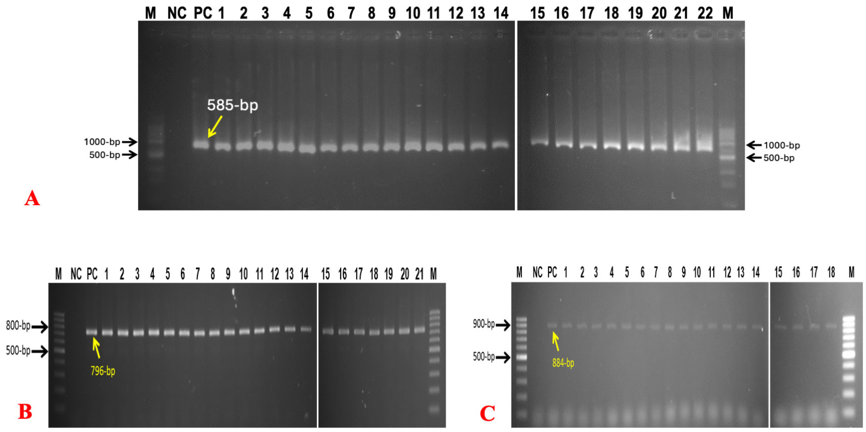
A total of 22 (34.38%) isolates were confirmed as E. coli by amplifying genus-specific 16S rRNA primers. A total of 21 (32.81%) isolates were confirmed as Salmonella spp. by amplifying genus-specific Inv-A primers. A total of 18 (28.13%) isolates were confirmed as Staphylococcus spp by amplifying the Tuf gene (Figure 2). Twenty-nine (45.31%) Simplicillium spp. were finally confirmed by PCR using genus-specific primers, with amplification at 606 bp, and ITS-1 and ITS-4 primers identified two (3.13%) as positive for Saccharomyces spp., showing amplification at 550 bp (Figure 3).
The numbers of isolated bacteria and fungus are listed in Table 3, with their prevalence for the 32 samples consisting of eggshell and egg contents. The p value of Salmonella spp. was calculated as 0.183 (p > 0.05), meaning that the result was not significant. For E. coli, the p value was calculated as 0.0016 (p < 0.05), meaning that the result was highly significant. For Staphylococcus spp., the p value was calculated as 0.026 (p < 0.05), meaning that the result was significant. For Proteus spp., the p value was calculated as 0.313 (p < 0.05), meaning that the result was not significant. For fungal species, the p value was calculated as 0.00 (p < 0.05), meaning that the result was not significant.

3.3. Results of Antibiotic Sensitivity Tests

Positive E. coli, Salmonella spp., and Staphylococcus spp. isolates were tested against nine different antibiotics. For E. coli, Ciprofloxacin showed the highest susceptibility pattern, followed by Levofloxacin. The highest resistance pattern was showed by Amoxycillin, Azithromycin, and Meropenem (Figure 4). Positive Salmonella spp. isolates showed the highest susceptibility pattern to Ceftriaxone, Ciprofloxacin, and Levofloxacin, whereas the highest resistance pattern was shown for Amoxycillin, Tetracycline, and Azithromycin (Figure 5). Against Staphylococcus spp. Isolates, Ciprofloxacin and Vancomycin were found to have the highest sensitivity in this study. The highest resistance pattern was shown by Amoxycillin, Gentamycin, and Oxacillin (Figure 6).

3.4. Result of Organoleptic Evaluation or Sensory Characters of Different Egg Samples Depending on Storage Duration

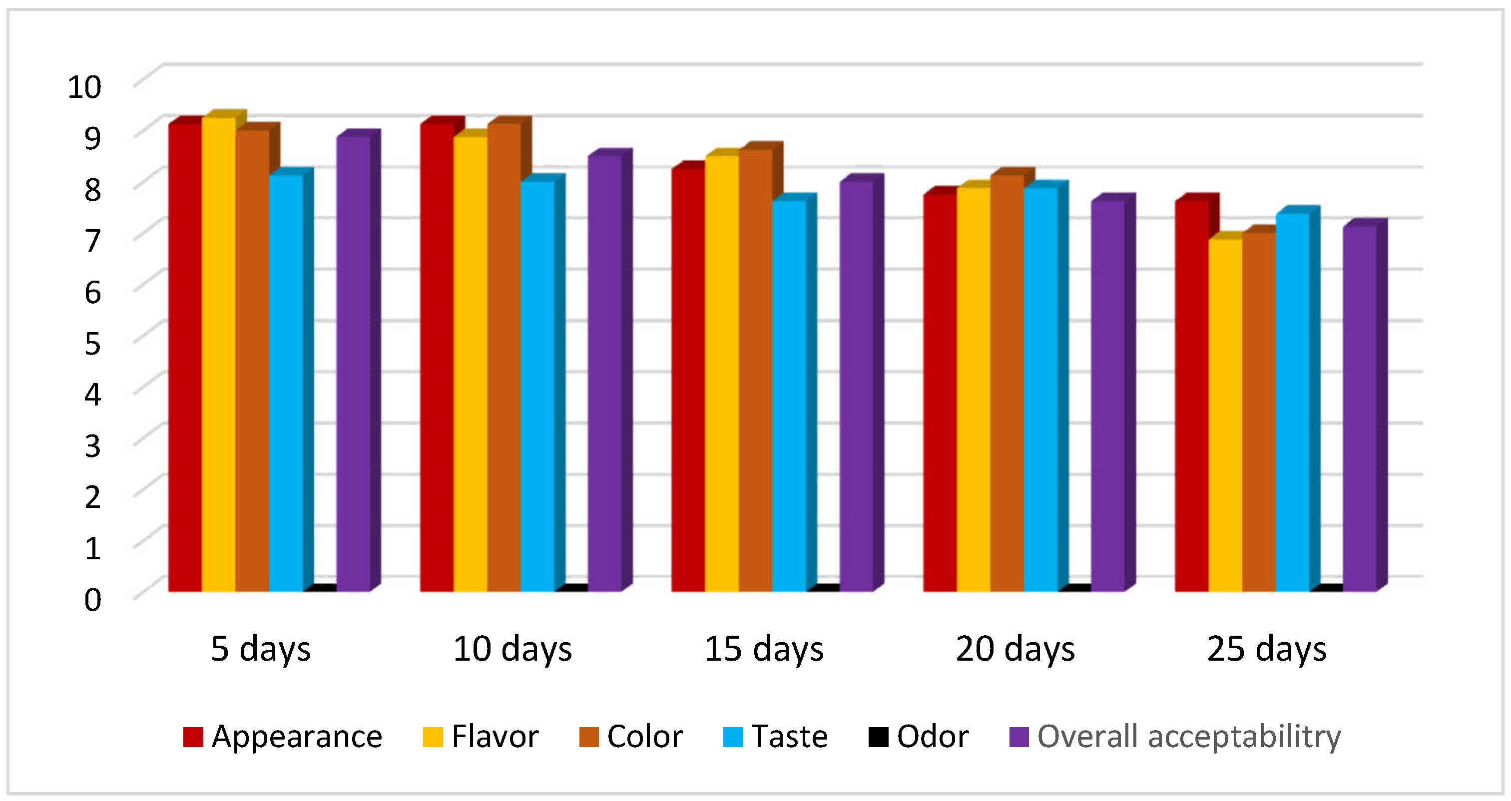
The total panel score marks were 60. The egg sample stored for 5 days scored 44.375, the sample stored for 10 days scored 43.63, the sample stored for 15 days scored 43.632, the sample stored for 20 days scored 39.25, and finally, the sample stored for 25 days scored 36.00, showing a gradually decreasing trend. For all the time intervals, there was no change in the odor parameter (Figure 7).

4. Discussion

Eggs are one of the most nutritious foods, and they often make up a healthy diet. They are sold at most food retailers in Bangladesh, although hygiene standards are not always properly maintained by sellers in developing countries. Thus, eggs can undergo rapid microbial deterioration, endangering human health through a variety of diseases. Many outbreaks of foodborne diseases, particularly those that are gastro-enteric (vomiting, diarrhea, gastro-intestinal upset) in nature, have been reported due to the consumption of undercooked and contaminated eggs. Eggs are also susceptible to fungal contamination at different stages until consumption, with some species of yeast constituting a public health hazard [9]. In addition, salmonella infections are among the most common foodborne illnesses directly linked to egg intake. The presence of Enterobacteriaceae in table eggs has been reported by several investigators [6,25]. The most frequently isolated bacteria were Enterobacter, Citrobacter, E. coli, and Klebsiella. In this study, four genera of bacteria and two genera of fungus were isolated and identified. The bacteria were E. coli, Salmonella spp., Staphylococcus spp., and Proteus spp. The fungi were Simplicillium spp. and Saccharomyces spp. The media used in this study were selected by considering the experience of a previous researcher who worked in various fields relevant to the present study [26,27].
The isolation rate of Salmonella was 40.63% in the outer eggshell and 25% in the inner egg contents. Stepien-pysniak et al. [28] found a prevalence of Salmonella at 88.3% on the eggshell surface. Musgrove et al. [29] showed that the prevalence of Salmonella ranges from 57% to 94%. The isolation rate of E. coli in the outer eggshell was 53.13%, and that in the inner egg content was 15.63%. Stepien-pysniak et al. [28] found a prevalence of E. coli at 58.7% in the outer shell and 4.3% in the inner contents, a result which is similar to our findings. However, the above result does not agree with the results reported by Adesiyun et al. [7] and Verma et al. [30], who observed a prevalence of E. coli at 88.3% and 84% in the outer eggshell and at 7.2% and 21% in the inner content of the eggs, respectively. Staphylococcus spp. was found in the outer eggshell at a rate of 40.63% and at a rate of 15.63% in the inner contents of eggs. This is lower than the findings from Jones et al. [31] and Verma et al. [30], where they reported prevalences of 78% and 77% for Staphylococcus in the outer eggshell and 4.4% and 15% in the inner contents of the egg. For fungus (yeast and mold), a rate of 100% was found in the outer eggshell, and there was an absence of fungus (yeast and mold) in the contents of the eggs. This disagrees with Higenyi and Kabasa [32], who stated a prevalence of fungi on the outer shell surface of eggs at 3%. The isolation rate of Proteus spp. in the outer eggshell was 3.13%. The difference in prevalence of the organisms may be due to a variation in the pre-enrichment technique or a change in the pattern of infection in layers or contamination of table eggs in the specific country or location. It was also found that the housing system can affect the risk of contamination. We did not categorize the prevalence of bacteria based on Gram staining properties or the location from where it was isolated, but it is noted that Gram-negative bacteria were found more often in yolks than in albumens [28].
The temperature and humidity at which eggs are kept are also key factors determining quantitative bacterial infection. The safety of eggs is determined by the number of bacterial cells on the eggshells and within the egg, as well as the pace at which they proliferate within it. From this study, it was revealed that the storage duration may have an influence on the quality of eggs. Bacterial contamination and proliferation mainly depend on the storage area and temperature. In this study, from TVC, we found that at day zero, the bacterial load was absent in the egg contents. However, after ten days, the presence of bacteria was found in the egg contents, which indicates that bacteria transferred through the pores on the eggshell. Salmonella spp. trans-ovarian transmission has been extensively reported in the literature [5,8]. Moreover, in the case of highly nutritious contents like egg albumin and egg yolk, bacteria were able to transfer through the route in order to find a balanced diet for their proliferation and growth. The aforementioned situation might be the cause for the tremendous increase in TVC/mL in the egg contents between days ten and twenty. However, the variation in TVC on the eggshell during the whole storage time did not show much fluctuation, except for the 20th day. Surprisingly, after the 20th day, the number of viable bacteria once again decreased. This could be due to a lack of required micronutrients or proper environmental conditions (pH, temperature) for microbes. Further research is needed in order to test this hypothesis. A numerical decrease in TVC was observed on the eggshells between day 20 and day 25; however, this reduction was 0.14 log units, which is within the expected measurement uncertainty (0.3–0.5 log units) for microbial analyses. Hence, the decrease is unlikely to be statistically or biologically meaningful. Furthermore, since the eggs were taken immediately after hatching without any treatment, we did find a higher level of TVC/mL than other researchers [16,33,34]. Most small layer farms in Bangladesh pick eggs immediately after they are laid and sell them without any further treatment. This serves as a possible pathway for the transfer of AMR bacteria in addition to being a means of introducing pathogenic germs into the human population. From the organoleptic evaluation, it was found that as time passed, the total taste and acceptability rates decreased. There was no odor change during this total period. However, the color, appearance, taste, and flavor quality also decreased day-wise. This could indicate that with a longer storage time, there is a further decrease in the quality of eggs. The changes in the internal color and appearance of the yolk also depends inversely on the storage period. Eggs kept in storage lose CO2 and water via their shell, raising the albumen’s pH and reducing its freshness [35]. Physicochemical evaluation is foundational in the food industry. It not only ensures that foods are safe and nutritious, but it also aligns them with consumer expectations and regulatory demands. Due to lack of funding availability, physicochemical analysis, which is more robust for evaluating any food, was not attempted and is a drawback of this study.
In Bangladesh, the prevalent usage of broad-spectrum antibiotics for various medical diseases signifies the emergence of multidrug-resistant organisms. This is, in part, due to insufficient awareness about the use of antibiotics. Antibiotic-resistant organisms are able to transmit from eggs to humans via the food chain. We used 11 commercially available drugs to examine the antimicrobial susceptibility profiles of E. coli, Salmonella spp., and Staphylococcus spp. The isolated bacteria exhibited multidrug resistance, with resistance to 5–6 drugs. The study demonstrated that isolated E. coli exhibited susceptibility to Ciprofloxacin and Levofloxacin, reflecting previous findings by other studies [14]. It was resistant to Azithromycin and Amoxicillin, which correlates with other findings [36]. Salmonella spp. exhibited susceptibility to Ciprofloxacin, Ceftriaxone, and Levofloxacin, as corroborated by Md et al. [37], where they report that their isolates were sensitive to Ciprofloxacin. This result was also consistent with the findings of Wouafo et al. [38]. The isolates showed resistance to Amoxycillin and Tetracycline, similar to the reports of De Jong et al. [39] and Hyeon et al. [40]. Staphylococcus spp. was found to be sensitive to Ciprofloxacin, Azithromycin, and Vancomycin and resistant to Amoxicillin and Oxacillin. This is in disagreement with Pyzik et al. [41] and Yurdakul et al. [42], who reported high resistance to Gentamicin, Ampicillin, and Vancomycin.
Our results revealed that all isolates of bacteria were susceptible to either Ciprofloxacin or Levofloxacin. The results of this study can be used as a guideline for medical practitioners to choose appropriate antibiotics, which will ultimately help to reduce the economic loss of previous excess antibiotic usage. Although the bacteria and fungi identified from the eggs in this study are not widely recognized as pathogens, they may pose a health concern to people who ingest raw or undercooked eggs. As a result, it is critical to educate the public on appropriate hygienic measures while handling and preparing eggs for consumption.

5. Conclusions

The present study was conducted to isolate, identify, and molecularly characterize different bacteria and fungi from table eggs. To achieve this, 32 eggs were taken, and each egg was separated into its outer shell and inner contents (inner shell, albumin, yolk surface, and yolk). The prevalence of E. coli in the outer eggshell was 53.13%, and in the inner egg content, it was 15.63%, whereas in the outer eggshell, the prevalence of Staphylococcus spp. was 40.63% and 15.63% in the inner contents of the eggs. The prevalence of Salmonella spp. was 40.63% in the outer eggshells and 25% in the inner contents of the eggs. The prevalence of Proteus in the outer eggshell was 3.13%, and there was an absence of Proteus in the inner contents. The prevalence of fungus (yeast and mold) in the outer shell was 100%, and there was an absence in the entire inner contents. The results also showed that E. coli and Salmonella spp. had a higher prevalence rate than Staphylococcus spp. in the different parts of the egg. Isolated bacteria were found to have multidrug resistance (resistant against 5–6 antibiotics). Moreover, the quality of eggs was found to decrease in relation to time spent in storage. Storage time was found to have a proportional relationship with TVC and was found to be inversely proportional with the organoleptic properties. In order to control this contamination, there must be effective methods for common microorganism prevention, detection, and control at all relevant stages of the egg production line, notably at the primary production level, to decrease the prevalence of Salmonella and the subsequent danger to public health.

Supplementary Materials

The following supporting information can be downloaded at: https://www.mdpi.com/article/10.3390/bacteria4030040/s1, Table S1: Summary of total viable count and prevalence of bacteria after 5 days (Egg shell and content); Table S2: Summary of total viable count and prevalence of bacteria after 10 days (Egg shell and content); Table S3: Summary of total viable count and prevalence of bacteria after 15 days (Egg shell and content); Table S4: Summary of total viable count and prevalence of bacteria after 20 days (Egg shell and content); Table S5: Summary of total viable count and prevalence of bacteria after 25 days (Egg shell and content).

Author Contributions

Conceptualization, K.H.M.N.H.N.; methodology, M.S.U.; validation, K.H.M.N.H.N.; formal analysis, M.S.U. and M.A.H.S.; investigation, K.H.M.N.H.N. and M.R.; resources, K.H.M.N.H.N. and M.R.; writing original draft preparation, M.S.U. and M.A.H.S.; review and editing, S., K.H.M.N.H.N. and M.A.H.S.; visualization, M.S.U. and M.A.H.S.; supervision, K.H.M.N.H.N. All authors have read and agreed to the published version of the manuscript.

Funding

This research received no external funding.

Institutional Review Board Statement

Not applicable.

Informed Consent Statement

Not applicable.

Data Availability Statement

Data available on request.

Acknowledgments

We are grateful to the layer farm owners for giving us access to collect samples during the course of the study. The authors also want to thank Ava Vankempen who helped to edit the article.

Conflicts of Interest

The authors declare that there are no conflicts of interest toward the publication of this article.

References

  1. Shahid, A.H.; Nazir, K.H.M.N.H.; El Zowalaty, M.E.; Kabir, A.; Sarker, S.A.; Siddique, M.P.; Ashour, H.M. Molecular detection of vancomycin and methicillin resistance in Staphylococcus aureus isolated from food processing environments. One Health 2021, 13, 100276. [Google Scholar] [CrossRef]
  2. WHO. Food Safety and Foodborne Illness; World Health Organization: Geneva, Switzerland, 2007; Available online: https://foodhygiene2010.wordpress.com/wp-content/uploads/2010/06/who-food_safety_fact-sheet.pdf (accessed on 1 January 2025).
  3. Meghla, N.S.; Mridha, D.; Rana, M.S.; Shahid, M.A.H.; Mahmud, M.M. Isolation, identification and antibiogram of verotoxin producing Escherichia coli from raw salad vegetables at Jashore, Bangladesh. Afr. J. Microbiol. Res. 2021, 15, 401–407. [Google Scholar] [CrossRef]
  4. Kakon, M.T.A.; Shahid, M.A.H.; Dutta, A.; Pinky, S.A.; Islam, M.S.; Hasnath, M.R. Comparative fertility and hatchability of broiler grandparent stocks in Bangladesh, Asian-Australas. J. Food Saf. Secur. 2024, 8, 48–57. [Google Scholar] [CrossRef]
  5. Howard, Z.R.; O’Bryan, C.A.; Crandall, P.G.; Ricke, S.C. Salmonella Enteritidis in shell eggs: Current issues and prospects for control. Food Res. Int. 2012, 45, 755–764. [Google Scholar] [CrossRef]
  6. Gole, V.C.; Chousalkar, K.K.; Roberts, J.R.; Sexton, M.; May, D.; Tan, J.; Kiermeier, A. Effect of Egg Washing and Correlation between Eggshell Characteristics and Egg Penetration by Various Salmonella Typhimurium Strains. PLoS ONE 2014, 9, e90987. [Google Scholar] [CrossRef] [PubMed]
  7. Adesiyun, A.; Offiah, N.; Seepersadsingh, N.; Rodrigo, S.; Lashley, V.; Musai, L.; Georges, K. Microbial health risk posed by table eggs in Trinidad. Epidemiol. Infect. 2005, 133, 1049. [Google Scholar] [CrossRef]
  8. Gast, R.K.; Guard, J.; Guraya, R.; Locatelli, A. Multiplication in Egg Yolk and Survival in Egg Albumen of Genetically and Phenotypically Characterized Salmonella Enteritidis Strains. J. Food Prot. 2018, 81, 876–880. [Google Scholar] [CrossRef]
  9. Cumeras, R.; Aksenov, A.A.; Pasamontes, A.; Fung, A.G.; Cianchetta, A.N.; Doan, H.; Davis, R.M.; Davis, C.E. Identification of fungal metabolites from inside Gallus gallus domesticus eggshells by non-invasively detecting volatile organic compounds (VOCs). Anal. Bioanal. Chem. 2016, 408, 6649–6658. [Google Scholar] [CrossRef]
  10. Kobir, M.A.; Akther, L.; Hasan, I.; Shahid, M.A.H.; Haque, Z.; Karim, M.R. Effects of Imidacloprid-Contaminated Feed Exposure on Hematological Parameters in Adult Rabbits (Oryctolagus Cuniculus). Res. Agric. Livest. Fish. 2020, 7, 439–444. [Google Scholar] [CrossRef]
  11. Tadesse, D.A.; Zhao, S.; Tong, E.; Ayers, S.; Singh, A.; Bartholomew, M.J.; McDermott, P.F. Antimicrobial Drug Resistance in Escherichia coli from Humans and Food Animals, United States, 1950–2002. Emerg. Infect. Dis. 2012, 18, 741–749. [Google Scholar] [CrossRef]
  12. Boonyasiri, A.; Tangkoskul, T.; Seenama, C.; Saiyarin, J.; Tiengrim, S.; Thamlikitkul, V. Prevalence of antibiotic resistant bacteria in healthy adults, foods, food animals, and the environment in selected areas in Thailand. Pathog. Glob. Health 2014, 108, 235–245. [Google Scholar] [CrossRef] [PubMed]
  13. Blair, J.M.A.; Webber, M.A.; Baylay, A.J.; Ogbolu, D.O.; Piddock, L.J.V. Molecular mechanisms of antibiotic resistance. Nat. Rev. Microbiol. 2015, 13, 42–51. [Google Scholar] [CrossRef] [PubMed]
  14. Ripon, J.; Shahid, M.; Mahmud, M.; Das, S.; Rahman, M.; Nazir, K. Isolation and molecular characterization of Shiga-toxin producing Escherichia coli from betel leaf (Piper betel L.). Vet. Res. Notes 2021, 1, 12–16. [Google Scholar] [CrossRef]
  15. Van Immerseel, F.; Nys, Y.; Bain, M. Improving the Safety and Quality of Eggs and Egg Products: Egg Safety and Nutritional Quality; Elsevier: Amsterdam, The Netherlands, 2011. [Google Scholar]
  16. Jones, D.R.; Musgrove, M.T.; Northcutt, J.K. Variations in External and Internal Microbial Populations in Shell Eggs during Extended Storage. J. Food Prot. 2004, 67, 2657–2660. [Google Scholar] [CrossRef]
  17. Fluck, A.C.; Cardinal, K.M.; Costa, O.A.D.; De Borba, L.P.; Da Silva Pires, P.G. Yolk and eggshell colour: Are these the parameters that influence egg purchasing? A systematic review. Worlds Poult. Sci. J. 2023, 79, 551–562. [Google Scholar] [CrossRef]
  18. Amerine, M.A.; Pangborn, R.M.; Roessler, E.B. Principles of Sensory Evaluation of Food; Elsevier: Amsterdam, The Netherlands, 1965. [Google Scholar] [CrossRef]
  19. Afrin, S.; Hossain, M.; Khan, M.; Hossain, M. Microbial assessment of beef in selected areas of Mymensingh district in Bangladesh. Bangladesh J. Anim. Sci. 2018, 46, 244–248. [Google Scholar] [CrossRef]
  20. Khanam, S.; Shahid, M.; Mahmud, M.; Hossain, M.; Khatun, M.; Nazir, K. Isolation and molecular identification of Candida spp. and Saccharomyces spp. from bakery products and their impact on public health. Vet. Res. Notes 2021, 1, 1–5. [Google Scholar] [CrossRef]
  21. Islam, M.M.; Shahid, M.A.H.; Nazir, K.H.M.N.H. Molecular Characterization of Bacillus anthracis from Selected Districts of Bangladesh. Acta Microbiol. Hell. 2025, 70, 17. [Google Scholar] [CrossRef]
  22. Khan, M.; Pavel, M.; Keya, A.; Shahid, M.; Ferdausi, T.; Siddique, M.; Hossain, M.; Nazir, K.; Rahman, M.; Rahman, M. Comparative molecular analysis of contemporary isolates of duck plague virus from haor areas of Bangladesh. J. Adv. Biotechnol. Exp. Ther. 2021, 4, 44–52. [Google Scholar] [CrossRef]
  23. Paião, F.G.; Arisitides, L.G.A.; Murate, L.S.; Vilas-Bôas, G.T.; Vilas-Boas, L.A.; Shimokomaki, M. Detection of Salmonella spp, Salmonella Enteritidis and Typhimurium in naturally infected broiler chickens by a multiplex PCR-based assay. Braz. J. Microbiol. 2013, 44, 37–41. [Google Scholar] [CrossRef]
  24. Clinical and Laboratory Standards Institute. Performance Standards for Antimicrobial Susceptibility Testing 26th Edition CLSI Supplement M 100s Wayne; Clinical and Laboratory Standards Institute: Malvern, PA, USA, 2016. [Google Scholar]
  25. Gole, V.C.; Chousalkar, K.K.; Roberts, J.R. Survey of Enterobacteriaceae contamination of table eggs collected from layer flocks in Australia. Int. J. Food Microbiol. 2013, 164, 161–165. [Google Scholar] [CrossRef]
  26. Mamun, M.M.; Parvej, M.S.; Ahamed, S.; Hassan, J.; Nazir, K.H.M.N.H.; Nishikawa, Y.; Rahman, M.T. Prevalence and Characterization of Shigatoxigenic Escherichia Coli in Broiler Birds in Mymensingh. Bangladesh J. Vet. Med. 2016, 14, 5–8. [Google Scholar] [CrossRef]
  27. Hassan, J.; Parvej, M.S.; Rahman, M.B.; Khan, M.S.R.; Rahman, M.T.; Kamal, T.; Nazir, K.N.H. Prevalence and Characterization of Escherichia coli from Rectal Swab of Apparently Healthy Cattle in Mymensingh, Bangladesh. Microbes Health 2014, 3, 12–14. [Google Scholar] [CrossRef]
  28. Stepien, P.D. Occurrence of Gram-negative bacteria in hens’ eggs depending on their source and storage conditions. Pol. J. Vet. Sci. 2010, 13, 507–513. [Google Scholar]
  29. Musgrove, M.T.; Jones, D.R.; Northcutt, J.K.; Cox, N.A.; Harrison, M.A. Identification of Enterobacteriaceae from Washed and Unwashed Commercial Shell Eggs. J. Food Prot. 2004, 67, 2613–2616. [Google Scholar] [CrossRef] [PubMed]
  30. Verma, S. A Microbiological Analysis of Egg Shell Bacteria. Vantage J. Themat. Anal. 2023, 4, 34–46. [Google Scholar] [CrossRef]
  31. Jones, D.R.; Musgrove, M.T. Pathogen Prevalence and Microbial Levels Associated with Restricted Shell Eggs. J. Food Prot. 2007, 70, 2004–2007. [Google Scholar] [CrossRef]
  32. Higenyi, J.; Kabasa, J.D. Microbial contamination Load of Hatching Eggs in Butaleja, Eastern Uganda. Anim. Vet. Sci. 2014, 2, 22–30. [Google Scholar] [CrossRef]
  33. Damena, A.; Mikru, A.; Adane, M.; Dobo, B. Microbial Profile and Safety of Chicken Eggs from a Poultry Farm and Small-Scale Vendors in Hawassa, Southern Ethiopia. J. Food Qual. 2022, 2022, 1–16. [Google Scholar] [CrossRef]
  34. Cader, S.; Goburdhun, D.; Neetoo, H. Assessment of the Microbial Safety and Quality of Eggs from Small and Large-Scale Hen Breeders. J. Worlds Poult. Res. 2014, 4, 75–81. [Google Scholar]
  35. Alleoni, A.C.C.; Antunes, A.J. Perfil de textura e umidade espremível de géis do albume de ovos recobertos com soro de leite. Ciência Tecnol. Aliment. 2005, 25, 153–157. [Google Scholar] [CrossRef]
  36. Huang, T.-M.; Lin, T.L.; Wu, C.C. Antimicrobial Susceptibility and Resistance of Chicken Escherichia Coli, Salmonella spp., and Pasteurella Multocida Isolates. Avian Dis. 2009, 53, 89–93. [Google Scholar] [CrossRef]
  37. Islam, M.K.; Kabir, S.M.L.; Haque, A.K.M.Z.; Sarker, Y.A.; Sikder, M.H. Molecular detection and characterization of Escherichia coli, Salmonella spp. and Campylobacter spp. isolated from broiler meat in Jamalpur, Tangail, Netrokona and Kishoreganj districts of Bangladesh. Afr. J. Microbiol. Res. 2018, 12, 761–770. [Google Scholar] [CrossRef]
  38. Wouafo, M.; Nzouankeu, A.; Kinfack, J.A.; Fonkoua, M.-C.; Ejenguele, G.; Njine, T.; Ngandjio, A. Prevalence and Antimicrobial Resistance of Salmonella Serotypes in Chickens from Retail Markets in Yaounde (Cameroon). Microb. Drug Resist. 2010, 16, 171–176. [Google Scholar] [CrossRef]
  39. De Jong, A.; Thomas, V.; Simjee, S.; Godinho, K.; Schiessl, B.; Klein, U.; Butty, P.; Valle, M.; Marion, H.; Shryock, T.R. Pan-European monitoring of susceptibility to human-use antimicrobial agents in enteric bacteria isolated from healthy food-producing animals. J. Antimicrob. Chemother. 2012, 67, 638–651. [Google Scholar] [CrossRef]
  40. Hyeon, J.-Y.; Chon, J.-W.; Hwang, I.-G.; Kwak, H.-S.; Kim, M.-S.; Kim, S.-K.; Choi, I.-S.; Song, C.-S.; Park, C.; Seo, K.-H. Prevalence, Antibiotic Resistance, and Molecular Characterizatio of Salmonella Serovars in Retail Meat Products. J. Food Prot. 2011, 74, 161–166. [Google Scholar] [CrossRef]
  41. Pyzik, E.; Marek, A. Plasmid profile analysis and evaluation of antibiotic susceptibility of Staphylococcus aureus strains isolated from table chicken eggs. Pol. J. Vet. Sci. 2013, 16, 307–312. [Google Scholar] [CrossRef] [PubMed]
  42. Yurdakul, N.E.; Erginkaya, Z.; Ünal, E. Antibiotic resistance of enterococci, coagulase negative staphylococci and Staphylococcus aureus isolated from chicken meat. Czech J. Food Sci. 2013, 31, 14–19. [Google Scholar] [CrossRef]
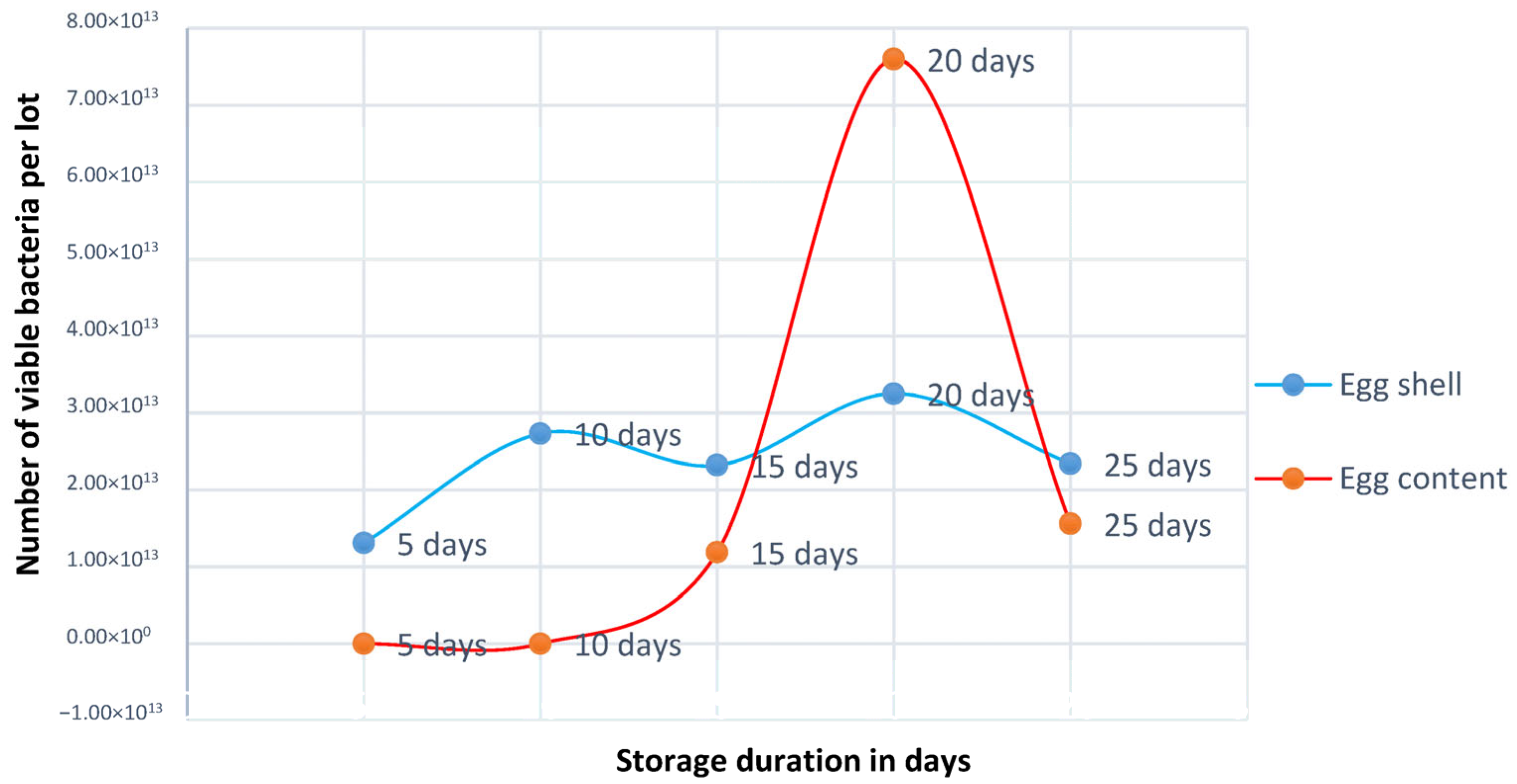
Figure 1. Total viable count/lot depending on storage duration.
Bacteria 04 00040 g001
Figure 2. PCR amplification of (A) 16s rRNA gene of E. coli at 585 bp; (B) Inv-A gene for Salmonella spp. at 796 bp; (C) Tuf gene for Staphylococcus spp. at 884 bp; Lane M = 100 bp DNA ladder; NC = Negative Control; PC = Positive Control; numeric numbers are the positive samples.
Bacteria 04 00040 g002
Figure 3. PCR amplification of (A) Simplicillium spp using genus-specific primer ITS-1 and ITS-4, showing amplification at 606 bp; (B) Saccharomyces spp using genus-specific primer ITS-1 and ITS-4, showing amplification at 550 bp; Lane M = 100 bp DNA ladder; NC = Negative Control; PC = Positive Control; numeric numbers are the positive samples.
Bacteria 04 00040 g003
Figure 4. Antibiotic susceptibility pattern of E. coli isolates.
Bacteria 04 00040 g004
Figure 5. Antibiotic susceptibility pattern of Salmonella spp. isolates.
Bacteria 04 00040 g005
Figure 6. Antibiotic susceptibility pattern of Staphylococcus spp. isolates.
Bacteria 04 00040 g006
Figure 7. Graphical representation of organoleptic properties of boiled eggs depending on storage duration.
Bacteria 04 00040 g007
Table 1. The sequence of primers for bacteria (E. coli, Salmonella spp., and Staphylococcus spp., respectively) and fungus.
Primer NameOlogonucleotide Sequence (5′–3′)Targeted GeneAmplicon Size (bp)References
16SrRNA FGACCTCGGTTTAGTTCACAGA16SrRNA585[14]
16SrRNA RCACACGCTGACGCTGACCA
Inv-A FCGG TGG TTT TAA GCG TAC TCT Tinv-A796[23]
Inv-A RCGA ATA TGC TCC ACA AGG TTA
Tseq271AAY ATG ATI ACI GGI GCI GCI CAR ATG GATuf gene884[1]
Tseq1138CCI ACI GTI CKI CCR CCY TCR CG
ITS 1TCC GTA GGT GAA CCT GCG GSimplicillium spp.~606
ITS 4TCC TCC GCT TAT TGA TAT GC
ITS 1TCC GTA GGT GAA CCT GCG GSaccharomyces spp.~550[20]
ITS 4TCC TCC GCT TAT TGA TAT GC
Table 2. Prevalence of bacteria and fungus depending on storage duration.
DaysE. coliSalmonella spp.Staphylococcus spp.Proteus spp.Fungus
EggshellEgg ContentEggshellEgg ContentEggshellEgg ContentEggshellEgg ContentEggshellEgg Content
5 days
(4 samples)
000025%000100%0
10 days
(4 samples)
00000000100%0
15 days
(8 samples)
100%050%50%50%50%3.13%0100%0
20 days
(8 samples)
62.5%12.5%12.5%50%50%12.5%00100%0
25 days
(8 samples)
50%50%100%050%000100%0
Table 3. Overall prevalence of bacteria and fungus.
ParameterPresence of E. coliPresence of Salmonella spp.Presence of Staphylococcus spp.Presence of Proteus spp.Presence of Fungus
Eggshell (32)Egg Content (32)Eggshell (32)Egg Content (32)Eggshell (32)Egg Content (32)Eggshell (32)Egg Content (32)Eggshell (32)Egg Content (32)
17513813510320
Prevalence 53.13%15.63%40.63%25%40.63%15.63%3.13%0100%0
p value 0.0016 *0.1830.026 *0.3130.000
* Means 5% level of sig. (p < 0.05).
Disclaimer/Publisher’s Note: The statements, opinions and data contained in all publications are solely those of the individual author(s) and contributor(s) and not of MDPI and/or the editor(s). MDPI and/or the editor(s) disclaim responsibility for any injury to people or property resulting from any ideas, methods, instructions or products referred to in the content.