Article

Genetic Diversity Among Independent Isolates of the Dolichocephalovirinae Subfamily

Department of Biological Sciences, University of South Carolina, Columbia, SC 29208, USA
*
Author to whom correspondence should be addressed.
Bacteria 2025, 4(1), 8; https://doi.org/10.3390/bacteria4010008
Submission received: 6 December 2024 / Revised: 6 January 2025 / Accepted: 21 January 2025 / Published: 4 February 2025

Abstract

:
Members of the Dolichocephalovirinae subfamily are giant viruses with an elongated head and a flexible tail that is used to infect Caulobacter strains. In this paper, we describe the isolation and characterization of nine newly isolated phages and present evidence that seven of these phages represent a new Dolichocephalovirinae genus that has significant differences from the four previously described Dolichocephalovirinae genera. In addition, since these new phages were isolated from a single sampling site over the course of three years, a comparison of their genome sequences reveals a low level of within-population diversity resulting from both single-nucleotide polymorphisms and insertions or deletions. A comparison of the host ranges of these phages suggests that differences in host susceptibility may be an important factor in maintaining this diversity.

1. Introduction

Bacteriophage studies are enjoying a renaissance since determining the nucleotide sequence of bacteriophage genomes has become both easy and inexpensive [1]. In addition, the continued increase in antibiotic resistance among bacterial pathogens has generated renewed interest in the use of bacteriophages as a complement to antibiotic treatment [2,3]. For instance, residues of antibiotics, agrochemicals, and heavy metals from pharmaceuticals disrupt aquatic microbial communities, driving the spread of antibiotic resistance (AMR) genes [4]. Surface water worldwide is a known reservoir for AMR genes, which spread through horizontal gene transfer [5,6]. If multidrug resistance (MDR) continues to rise, it could cause 10 million deaths annually and cost USD 100 trillion by 2050 [7]. To combat this crisis, bacteriophages offer a sustainable and effective solution. Unlike antibiotics, which kill both harmful and beneficial bacteria, phages can be used to target harmful bacteria, reducing AMR risks without harming humans, animals, or the environment [8].
The use of bacteriophages either to treat infections or for other purposes will necessitate an increased understanding of natural bacteriophage diversity and genome evolution. To help meet this need, our laboratory has initiated a long-term investigation of the bacteriophages that infect Caulobacter crescentus and are present at a streamside study site located on the University of South Carolina campus [9]. Caulobacter crescentus is a well-studied bacterium that serves as a model system for other Alphaproteobacteria. Caulobacters differ from most other bacteria since each cell division results in two distinct types of daughter cells, a stalked cell that is like the parent cell and a motile swarmer cell. The swarmer cell must go through a maturation process where it loses its flagellum and synthesizes a stalk before it can initiate a new round of cell division. Thus, numerous studies over the past 50 years have gradually elucidated the numerous regulatory circuits involved in controlling changes in gene expression that occur during the C. crescentus cell cycle [10,11,12,13].
Despite extensive laboratory studies, the interactions of Caulobacter species with other organisms in their natural environments remain poorly understood. Given the ubiquity of bacteriophages, investigating how these phages interact with Caulobacter in natural settings could be a pivotal step in unraveling the broader ecological relationships between Caulobacter and other microorganisms, including bacteria, fungi, and plants. For example, we recently identified a temperate Podovirus, designated S2B, that infects Caulobacter crescentus and can establish a lysogenic relationship by integrating into the host genome after recombination with a matching 16 bp nucleotide sequence that is present in a host tRNA-ser gene [14]. This discovery suggests that lysogenic Caulobacter strains may engage in distinct environmental interactions compared to non-lysogenic strains, opening new avenues for exploring the ecological roles of Caulobacter in diverse environments.
The most common type of Caulobacter bacteriophages is the well-studied CbK-like Dolichocephalovirinae, which have an elongated head, a flexible tail, and genomes larger than 200 kb [15,16]. The CbK phage infects swarmer cells when the flexible tail of the phage wraps itself around the swarmer cell pilus. Subsequently, the phage tail is pulled into the cell when the wrapped pilus is retracted [17]. Furthermore, the phages have a unique head filament that attaches to the flagellum and slides along the filament as it rotates [18]. Thus, the flagellum provides a big target for phage attachment and subsequent movement to the flagellar pole of the cell where the pili are located. In addition, it explains why Caulobacter non-motile mutants are partially resistant to CbK infection, as the absence of a functional flagellum disrupts the phage’s ability to efficiently reach the pili, thereby hindering infection [19].
Recently, our laboratory showed that the CbK-like bacteriophages can be grouped into four lineages that are sufficiently distinct that they represent separate genera [20]. These four lineages comprise the Dolichocephalovirinae subfamily, which was recently defined by the International Committee for the Taxonomy of Viruses (ICTV), and the four new genera were designated Shapirovirus, Bertelyvirus, Colossusvirus, and Poindextervirus. The Shapirovirus genus includes the original CbK bacteriophage [21] and most of the bacteriophages described by Johnson et al. [22], Gill et al. [15], and Ash et al. [16]. However, the Bertelyvirus, Colossusvirus, and Poindextervirus genera include the larger CbK-like phages, Colossus and Rogue, described previously [15] and four additional large phages that were described by Wilson and Ely [20]. The Bertelyvirus genus includes the largest of these phages, CcrSC and CcrBL9, with genome sizes greater than 300 kb. Since CcrSC and CcrBL9 share only 91% genome nucleotide identity, they should be considered members of two different species based on the current ICTV criteria (members of the same species are more than 95% identical at the nucleotide level throughout the length of the genome, tested reciprocally) [23].
Our analysis of additional bacteriophages isolated from our long-term study site has resulted in the identification of numerous additional CbK-like viruses. In this paper, we describe nine new isolates, seven of which represent a new genus of the Dolichocephalovirinae subfamily.

2. Materials and Methods

2.1. Bacterial Culture Preparation

All bacterial cultures used in this study were grown in peptone yeast extract (PYE) liquid media, composed of 0.2% peptone, 0.1% yeast extract, 0.5 mM CaCl2, and 0.8 mM MgSO4. Cultures were incubated at 30 °C in test tubes under constant agitation using a rotator to ensure aeration. Each culture was grown for 24 h to reach the desired cell density before further use.

2.2. Bacteriophage Isolation and Cultivation

Bacteriophages were isolated from Rocky Branch Creek water samples on the campus of the University of South Carolina using an enrichment procedure. Briefly, water samples were filtered through a 0.45 µm membrane to remove debris and most bacteria. The filtrate was then combined with 2.5 mL of 5× PYE medium and 0.6 mg of streptomycin in a sterile flask. To enrich for phages, 50 µL of SC1004, a streptomycin-resistant mutant of the Caulobacter crescentus CB15 strain, was added to the flask, and the mixture was incubated overnight at 30 °C with constant shaking.
The following day, the bacterial cultures were centrifuged at 7000× g for 10 min to pellet the bacterial cells. The resulting supernatants were treated with 1 mL of chloroform to lyse any remaining bacteria, and the mixtures were shaken thoroughly to ensure complete bacterial removal. Serial dilutions of the lysates were prepared and mixed with 100 µL of SC1004 in 3.5 mL of SSM (PYE containing 0.3% agar), which was then poured onto PYE agar plates. After overnight incubation at 30 °C, individual plaques were stabbed and suspended in 3 mL PYE. Dilutions of this phage suspension were again added to SSM and poured onto a PYE agar plate. After overnight incubation, a single plaque was cut out and resuspended in 3 mL PYE. Subsequently, a confluent lysis plate was flooded with 5 mL PYE and decanted after overnight refrigeration to provide a high titer lysate.

2.3. Host Range Determination

The host range of the phages was determined using a spotting assay. A mixture of 100 μL of a potential host culture, which included Caulobacter vibrioides strains CB2, CB13, CB15, RBW21, RBW22, and RBW29; Caulobacter segnis strains TK0059 and CBR1; Caulobacter rhizosphaerae strains RBW14, RBW23, and RBW25; Caulobacter henricii strains CB4; and 14 non-Caulobacter strains [24], was combined with 3.5 mL of molten PYE semi-solid medium (PYE SSM). This mixture was then poured onto the surface of a PYE agar plate and allowed to cool and solidify. After solidification, 10 μL aliquots of each phage lysate were spotted onto the surface of the overlay. The plates were incubated overnight at 34 °C. After incubation, the presence of clear zones around the phage spots was assessed to determine if the phages were capable of lysing the potential host cells.

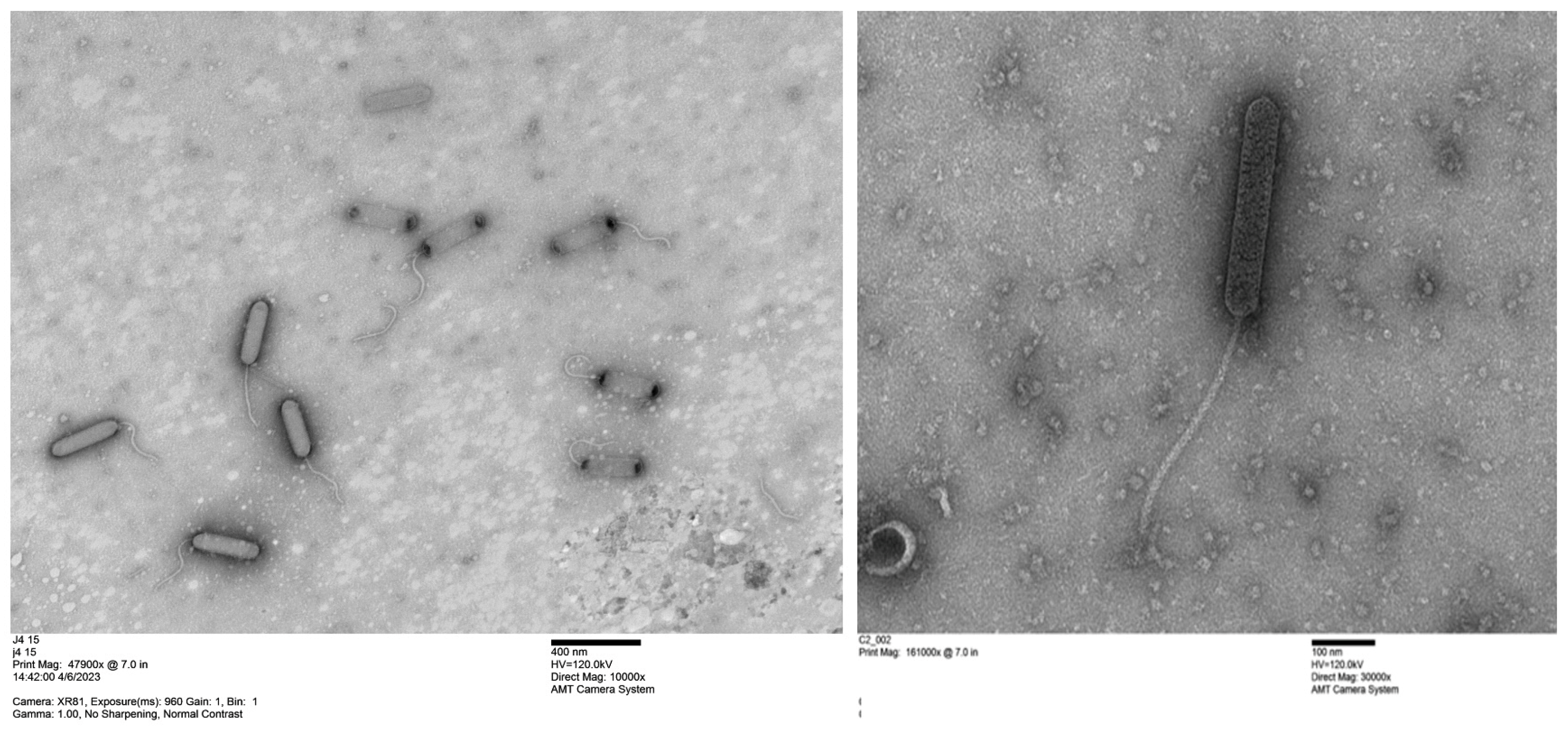
2.4. Transmission Electron Microscope Analysis of Purified Phages with Uranyl Acetate Staining

Purified phage isolates were visualized using a JEOL 200CX transmission electron microscope (TEM, JEOL Peabody, MA USA). For sample preparation, a formvar/carbon-coated 300-mesh copper grid was floated on a drop of a 1:1 mixture of 2% uranyl acetate and phage lysate for 1–2 min to allow phage adsorption. Excess liquid was gently wicked away with filter paper, and the grid was air-dried. TEM imaging was performed to examine the morphology of the phage particles.

2.5. Phage Genome Sequencing and Annotation

DNA was isolated from phage lysates using a Qiagen (Hilden, Germany) DNA isolation kit according to the manufacturer’s instructions. The resulting phage DNA was combined with other phage and bacterial DNA, and the nucleotide sequences of the combined DNAs were determined using PacBio sequencing technology and HGAP4 assembly at the Delaware University Bioinformatics Institute or the University of Maryland Institute for Genome Sciences. The nucleotide sequences of the phage genomes were annotated using the Rapid Annotation using Subsystem Technology (RAST) [25], and the annotated sequences were trimmed to remove duplicate sequences at the ends of each phage genome contig using Artemis [26]. The resulting annotated genomes were then compared using Mauve [27]. The mutational direction of individual SNPs was inferred based on the assumption that the most frequent base at a particular locus was the ancestral allele. SNPs with uncertain ancestry were omitted from the analysis. A Viridic analysis was performed to identify which phages belonged to the same genus and species [23].
NCBI accession numbers for the genome nucleotide sequences described in this paper include the following: CcrSC (MH588547), CcrBL9 (MH588546), CcrS2L (PQ287319), CcrBL57 (PQ287320), CcrC1 (MZ574429), CcrC2 (MZ574430), CcrC3 (OP620777), CcrJ4 (MZ574431), CcrBL1 (OP620778), CcrBL47 (MZ574432), CcrRB23 (MZ574433).

3. Results and Discussion

3.1. Isolation and Characterization of Ten Bacteriophages from Rocky Branch Creek

Ten new bacteriophages were isolated independently from the same site on the Rocky Branch Creek in Columbia, SC, between January 2016 and January 2020. One of these isolates, designated S2B, was the first Caulobacter lysogenic bacteriophage to be described [14]. After purification, each of the other nine phages was characterized with regard to plaque size and host range (Table 1 and Table 2). Electron microscopic observations (examples in Figure 1) demonstrated that all nine phages had an elongated head with a long, flexible tail similar to those previously observed with CbK and other members of the Dolichocephalovirinae subfamily [15,16,20,21]. The members of this subfamily are considered giant phages with genomes larger than 200 kb, and they have a large, elongated head and a long, flexible tail.

3.2. Characterization of Bacteriophage Host Ranges

Host range analysis of the newly isolated CbK-like bacteriophages revealed significant variability in their specificity, with some exhibiting broad host ranges, while others displayed more narrow host ranges (Table 2). Notably, phages such as CcrS2L, CcrBL47, CcrRB23, and CcrBL57 demonstrated the ability to infect multiple Caulobacter species, including C. vibrioides, C. segnis, and C. rhizosphaerae. These phages exhibited considerable versatility in their host range, infecting both closely related and more distantly related strains within the Caulobacter genus. The broad host range observed for these phages suggests that they may play an important role in regulating microbial populations across diverse environments, such as soil or aquatic ecosystems, where microbial diversity is a central feature. Phages with the ability to target multiple strains could be particularly useful in applications like environmental decontamination or biocontrol, where controlling a wide variety of bacterial strains is necessary to achieve effective microbial management.
These findings highlight the diverse host-targeting capabilities of the phages studied. The observed differences in host range likely contribute to the maintenance of genetic diversity within this population of Dolichocephalovirinae bacteriophages and may play a key role in their ecological adaptability and evolutionary dynamics.

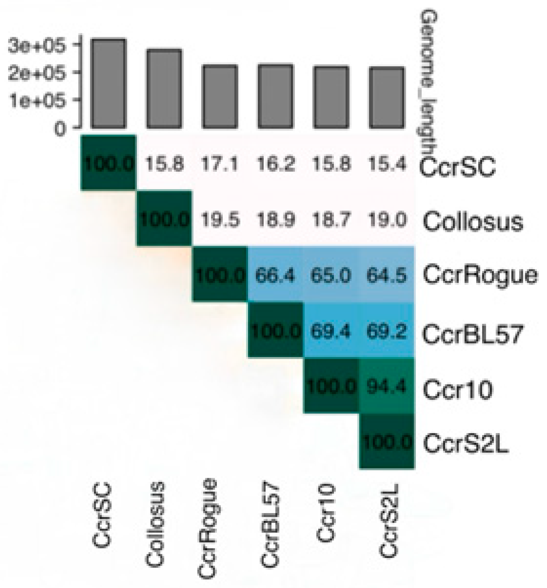
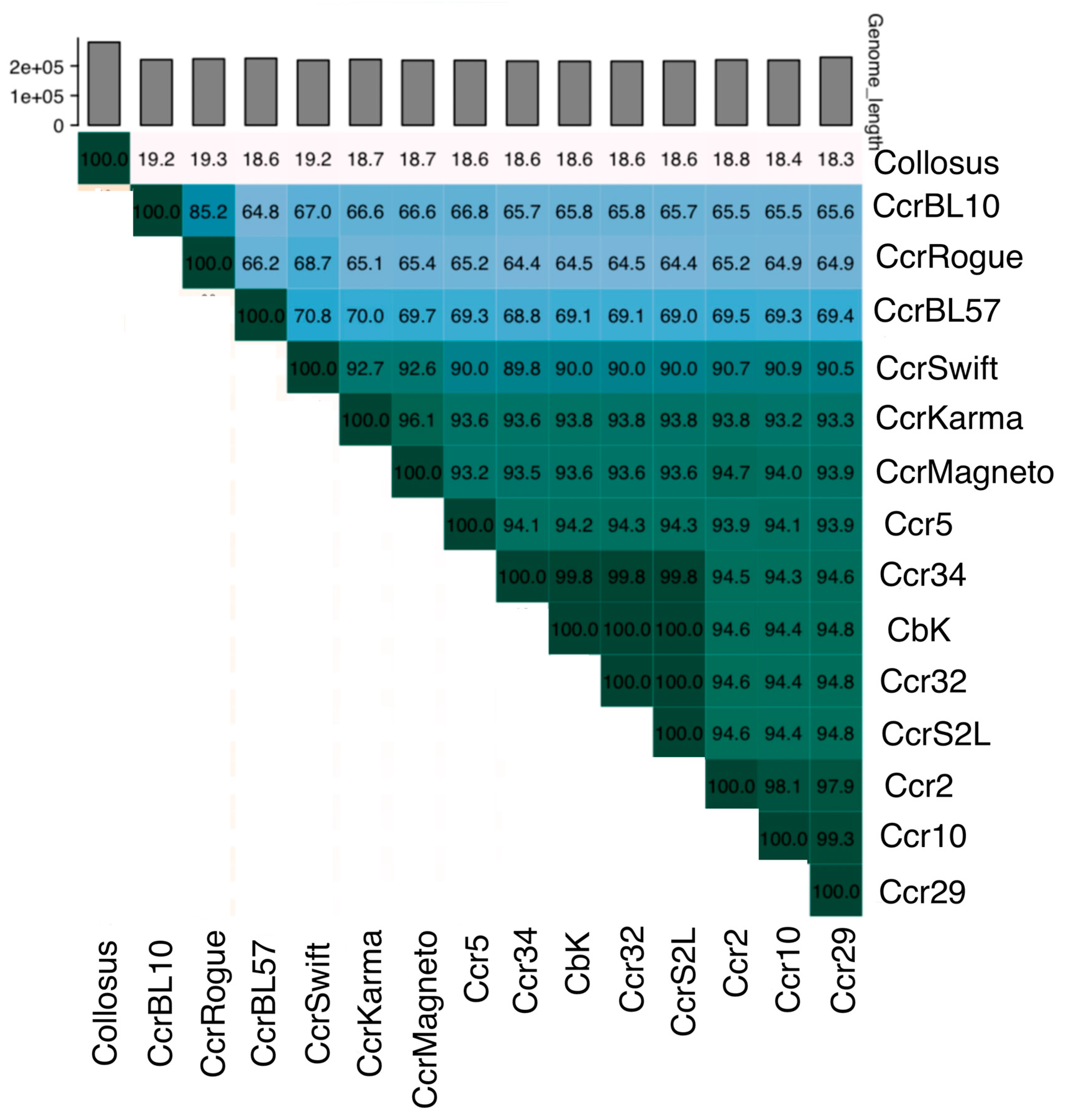
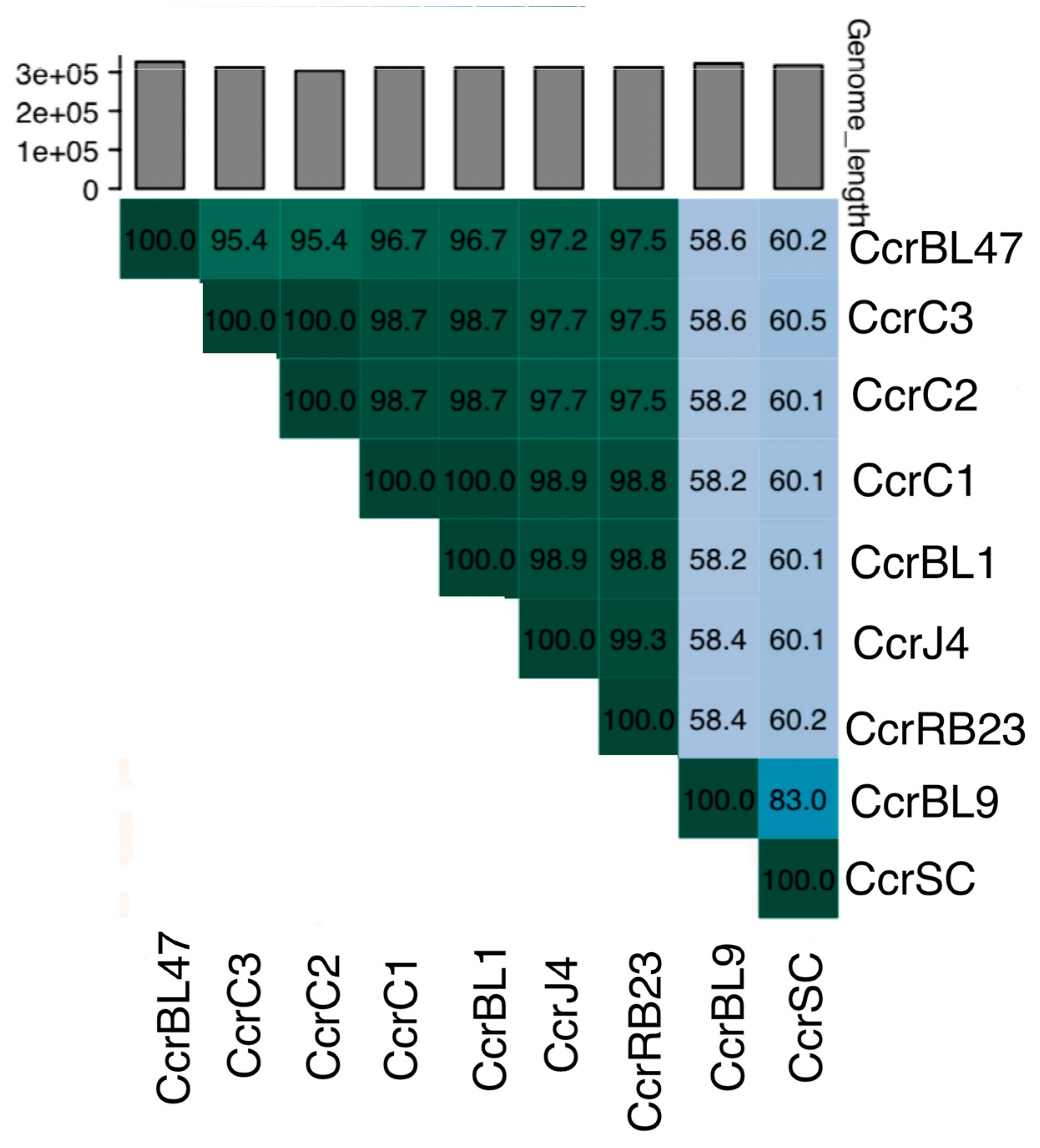
3.3. Genetic Diversity and Taxonomic Classification of Dolichocephalovirinae Phages

One objective of this study was to estimate the genetic diversity of a naturally occurring population of bacteriophages. Therefore, the nucleotide sequence of each of the nine genomes was determined. To determine how closely the new phage genomes were related to each other and to the previously described Dolichocephalovirinae genomes [15,16,20,21,22], the nine genomes were compared using the Viridic program [23]. The resulting comparisons (Figure 2) indicated that CcrBL57 might not belong to any of the four genera in the Dolichocephalovirinae subfamily since members of the same genus should have at least 70% nucleotide identity based on the Viridic comparison and the highest CcrBL57 comparison was 69.4%. However, a more detailed comparison of the CcrBL57 genome to other closely related phage genomes generated two values just above 70%. Similarly, CcrBL57 was considered a distinct species in only one of the three trees constructed using VICTOR [28]. Therefore, CcrBL57 should be considered a representative of a new species that is distantly related to the other species in the Shapirovirus genus.
In contrast, the CcrS2L genome was closely related to the Ccr10 genome, so CcrS2L should be considered part of the Shapirovirus genus (Figure 2). When we performed a more detailed comparison that included 11 Shapirovirus genomes, we found that CcrS2L should be considered part of the CbK species of the Shapirovirus genus (Figure 3). In addition, the data show that the Shapriovirus genus should include three additional species in addition to the previously named Swift and CbK species. Based on the current Shapirovirus species names, we propose that the three new species be designated Karma, CcrFive, and CcrTen. This expanded classification not only enhances our understanding of Shapirovirus diversity, but it also underscores the dynamic and evolving nature of bacteriophage taxonomy as more phage genome sequences are obtained.
The proposed CcrTen genus consists of CCr10, Ccr2, and Ccr29. The Ccr10 and Ccr2 genomes differ by only two SNPs and a series of four Gs in the Ccr2 genome and five Gs at the corresponding site in the Ccr10 genome. In contrast, the Ccr29 genome contains 11 additional SNPs and five insertions. The CbK species includes Ccr32, Ccr34, and S2L. The four genomes are highly conserved with small numbers of SNPs and one or two insertions in each genome relative to the CbK genome. In contrast, in the two-member Karma genus, the Karma genome has only 96% shared identity with the Magneto genome. The remaining two species are represented by a single genome (Figure 3).
Blast results indicated that the remaining seven phage genomes were closely related to each other and more similar to the Bertelyviruses than to any other Dolichocephalovirinae genus. In addition, the seven phage genomes had a larger genome size and a lower genomic GC content that was similar to those of the Bertelyviruses genus and different from the other Dolichocephalovirinae genera (Table 1 and reference [15]). A subsequent Viridic comparison of the remaining seven new isolates with CcrSC and CcrBL9 revealed that the seven phage genomes had greater than 95% nucleotide identity to each other, but less than 70% nucleotide identity to the CcrSC and CcrBL9 genomes (Figure 4). Recently, Turner et al. [23] suggested that the genomes of members of the same bacteriophage species should have nucleotide identities of more than 95% across the entire genome in pairwise comparisons and the genomes of members of the same genus should have more than 70% nucleotide identity. Therefore, based on these criteria, we concluded that the seven newly isolated bacteriophages are closely related isolates of a single species of a new genus of the Dolichocephalovirinae subfamily that is more closely related to the Bertelyvirus than it is to the other of the Dolichocephalovirinae genera.
Genome comparisons indicated that the seven similar phage genomes contain the same set of genes with only minor differences. However, an analysis of the 7088 single-nucleotide polymorphisms (SNPs) that we observed in a comparison of the seven new genomes showed that 2575 SNPs resulted in a loss of GC content (GC->AT) compared to 2701 SNPs that resulted in a gain of GC content. Thus, there has been a small gain of GC content since these seven phage genomes shared a common ancestor. This result is consistent with the idea that these phage genomes are slowly increasing their GC content to match the 67.2% GC in their host genomes, and it is in contrast to a recent analysis that showed that a slow loss of GC content occurred over evolutionary time for all bacterial genomes with more than 40% genomic GC content [29].
As indicated above, the seven genomes of the new Dolichocephalovirinae genus phages are most closely related to the genomes of the Bertelyviruses. When the seven new genomes were compared with the CcrSC and CcrBL9 Bertelyvirus genomes, the seven new genomes were missing two hypothetical genes, but they had two extra hypothetical genes elsewhere in the genome. In addition, the CcrSC and CcrBL9 genomes contained an eight-gene inversion relative to the seven new genomes. Furthermore, all nine phage genomes contained the inversion described by Wilson and Ely [20] that was present only in CcrSC and CcrBL9 and not any of the other members of the Dolichocephalovirinae subfamily. Thus, this inversion could be considered a defining characteristic of both the Bertelyviruses and the new seven-member genus. Similarly, the new inversion found in the seven new phage genomes may be a defining characteristic of this new Dolichocephalovirinae genus.
When the seven genomes of the new Dolichocephalovirinae genus phages were compared to each other, the CcrC2 and CcrC3 genomes were nearly identical and the CcrC1 and CcrBL1 genomes were nearly identical (Figure 3). The CcrC2 and CcrC3 genomes differed by one SNP, two 1-base indels, a 99-base insertion, and a seven-gene duplication. In contrast, the CcrC1 and CcrBL1 genomes differed by only three one-base indels in hypothetical genes. However, two of these one-base indels are in different genes in an operon that codes for tail genes. Thus, they may be responsible for the differences in host specificity for these two bacteriophages.

4. Conclusions

A total of ten new bacteriophages (nine described in this paper and one described previously [14]) were obtained from a single sampling site, and only two of these phages were shown to belong to a previously described bacteriophage genus. In addition, seven of the ten phages were closely related and provided an example of the kind of within-species genetic diversity that can be observed at a single sampling location. Some phage genome pairs differed at a few loci with single-nucleotide polymorphisms (SNPs), single-base pair indels, and larger indels while others had more extensive differences. Thus, there is remarkably little genetic variation among these seven phage genomes even though the phage isolates were isolated from separate samples obtained over a span of three years. However, despite this high degree of genome conservation, there appears to be selection for differences in the tail genes that impact host specificity.
These results highlight the significant genetic variability and dynamic evolutionary processes occurring within phage populations at localized sites. This study provides valuable insights into the diversity of bacteriophages and the factors that drive genetic variation in natural environments, furthering our understanding of phage ecology and evolution. These findings could have important implications for environmental applications and contribute to the broader field of microbial ecology, offering potential avenues for phage-based interventions and biotechnological innovations.

Author Contributions

B.E., T.M. and Q.T. participated in the experimental design and the data analysis. T.M. and B.E. wrote the manuscript. All authors have read and agreed to the published version of the manuscript.

Funding

This research received no external funding.

Institutional Review Board Statement

Not applicable.

Informed Consent Statement

Not applicable.

Data Availability Statement

All data for this study is included in this manuscript or in the Gen-Bank database as indicated in Methods.

Conflicts of Interest

The authors declare no conflicts of interest.

References

  1. Tubman, E. Bacteriophages: Methods and Protocols; Springer: New York, NY, USA, 2024; Volume 2738, pp. 1–431. [Google Scholar] [CrossRef]
  2. Kosznik-Kwasnicka, K.; Topka, G.; Mantej, J.; Grabowski, L.; Necel, A.; Wegrzyn, G.; Wegrzyn, A. Propagation, purification, and characterization of bacteriophages for phage therapy. In Bacteriophages: Methods and Protocols; Springer: New York, NY, USA, 2024; Volume 2738, pp. 357–400. [Google Scholar]
  3. Laanto, E. Overcoming bacteriophage resistance in phage therapy. In Bacteriophages: Methods and Protocols; Springer: New York, NY, USA, 2024; Volume 2738, pp. 401–410. [Google Scholar]
  4. Rout, A.K.; Dixit, S.; Tripathy, P.S.; Rout, S.S.; Parida, S.N.; Parida, P.K.; Sarkar, D.J.; Das, B.K.; Singh, A.K.; Behera, B.K. Metagenomic Landscape of Sediments of River Ganga Reveals Microbial Diversity, Potential Plastic and Xenobiotic Degradation Enzymes. J. Hazard. Mater. 2024, 471, 134377. [Google Scholar] [CrossRef]
  5. Wang, S.; Nie, W.; Gu, Q.; Wang, X.; Yang, D.; Li, H.; Wang, P.; Liao, W.; Huang, J.; Yuan, Q.; et al. Spread of antibiotic resistance genes in drinking water reservoirs: Insights from a deep metagenomic study using a curated database. Water Res 2024, 1, 121572. [Google Scholar] [CrossRef]
  6. Das, B.K.; Behera, B.K.; Chakraborty, H.J.; Paria, P.; Gangopadhyay, A.; Rout, A.K.; Nayak, K.K.; Parida, P.K.; Rai, A. Metagenomic Study Focusing on Antibiotic Resistance Genes from the Sediments of River Yamuna. Gene 2020, 758, 144951. [Google Scholar] [CrossRef]
  7. O’Neil, J. Antimicrobial Resistance: Tackling a Crisis for the Health and Wealth of Nations. The Review on Antimicrobial Resistance 2014. Available online: https://amr-review.org/sites/default/files/AMR%20Review%20Paper%20-%20Tackling%20a%20crisis%20for%20the%20health%20and%20wealth%20of%20nations_1.pdf (accessed on 4 October 2024).
  8. Wittebole, X.; De Roock, S.; Opal, S.M. A historical overview of bacteriophage therapy as an alternative to antibiotics for the treatment of bacterial pathogens. Virulence 2014, 5, 226–235. [Google Scholar] [CrossRef]
  9. Ely, B.; Hils, M.; Clarke, A.; Albert, M.; Holness, N.; Lenski, J.; Mohammadi, T. New genera and species of Caulobacter and Brevundimonas bacteriophages provide insights into phage genome evolution. Viruses 2024, 16, 641. [Google Scholar] [CrossRef]
  10. Barrows, J.M.; Goley, E.D. Synchronized swarmers and sticky stalks: Caulobacter crescentus as a model for bacterial cell biology. J. Bacteriol. 2023, 205, e00384-22. [Google Scholar] [CrossRef]
  11. Chong, T.N.; Shapiro, L. Bacterial cell differentiation enables population level survival strategies. mBio 2024, 15, e00758-24. [Google Scholar] [CrossRef]
  12. Hallgren, J.; Jonas, K. Nutritional control of bacterial DNA replication. Curr. Opin. Microbiol. 2024, 77, 102403. [Google Scholar] [CrossRef]
  13. Fröhlich, K.S.; Gomariz, M.V. RNA-controlled regulation in Caulobacter crescentus. Curr. Opin. Microbiol. 2021, 60, 1–7. [Google Scholar] [CrossRef]
  14. Ely, B.; Berrios, L.; Thomas, Q. S2B, a temperate bacteriophage that infects Caulobacter crescentus strain CB15. Curr. Microbiol. 2022, 79, 98. [Google Scholar]
  15. Gill, J.J.; Berry, J.D.; Russell, W.K.; Lessor, L.; Escobar-Garcia, D.A.; Hernandez, D.; Kane, A.; Keene, J.; Maddox, M.; Martin, R.; et al. The Caulobacter crescentus phage phiCbK: Genomics of a canonical phage. BMC Genom. 2012, 13, 542. [Google Scholar] [CrossRef] [PubMed]
  16. Ash, K.T.; Drake, K.M.; Gibbs, W.S.; Ely, B. Genomic diversity of type B3 bacteriophages of Caulobacter crescentus. Curr. Microbiol. 2017, 74, 779–786. [Google Scholar] [CrossRef] [PubMed]
  17. Dillard, R.S.; Storms, R.E.; De Masi, L.; Hampton, C.; Panis, G.; Viollier, P.H.; Wright, E.R. Analysis of phage-pilus interactions in Caulobacter crescentus. Microsc. Microanal. 2016, 22 (Suppl. S3), 202. [Google Scholar] [CrossRef]
  18. Guerrero-Ferreira, R.C.; Viollier, P.H.; Ely, B.; Poindexter, J.S.; Georgieva, M.; Jensen, G.J.; Wright, E.R. A novel mechanism for bacteriophage adsorption to the motile bacterium Caulobacter crescentus. Proc. Nat. Acad. Sci. USA 2011, 108, 9963–9968. [Google Scholar] [CrossRef]
  19. Johnson, R.C.; Ely, B. Analysis of non-motile mutants of the dimorphic bacterium Caulobacter crescentus. J. Bacteriol. 1979, 137, 627–634. [Google Scholar] [CrossRef]
  20. Wilson, K.; Ely, B. Analyses of four new Caulobacter PhiCbKviruses indicate independent lineages. J. Gen. Virol. 2019, 100, 321–331. [Google Scholar] [CrossRef]
  21. Agabian-Keshishian, N.; Shapiro, L. Stalked bacteria: Properties of deoxyribonucleic acid bacteriophage PhiCbK. J. Virol. 1970, 5, 795–800. [Google Scholar] [CrossRef]
  22. Johnson, R.; Wood, N.; Ely, B. Isolation and characterization of bacteriophages for Caulobacter crescentus. J. Gen. Virol. 1977, 37, 323–335. [Google Scholar] [CrossRef]
  23. Turner, D.; Kropinski, A.M.; Adriaenssens, E.M. A roadmap for genome-based phage taxonomy. Viruses 2021, 13, 506. [Google Scholar] [CrossRef]
  24. Mohammadi, T.; Ely, B. The isolation and characterization of novel Caulobacter and Non-Caulobacter lysogenic bacteria from soil and the discovery of broad-host-range phages infecting multiple genera. Microorganisms 2024, 12, 1894. [Google Scholar] [CrossRef]
  25. Overbeek, R.; Olson, R.; Pusch, G.D.; Olsen, G.J.; Davis, J.J.; Disz, T.; Edwards, R.A.; Gerdes, S.; Parrello, B.; Shukla, M.; et al. The SEED and the rapid annotation of microbial genomes using Subsystems Technology (RAST). Nucleic Acids Res. 2013, 42, D206–D214. [Google Scholar] [CrossRef] [PubMed]
  26. Rutherford, K.; Parkhill, J.; Crook, J.; Horsnell, T.; Rice, P.; Rajandream, M.A.; Barrell, B. Artemis: Sequence visualization and annotation. Bioinformatics 2000, 10, 944–945. [Google Scholar] [CrossRef] [PubMed]
  27. Darling, A.C.; Bau, B.; Blattner, F.R.; Perna, N.T. Mauve: Multiple alignment of conserved genomic sequence with rearrangements. Genome Res. 2004, 14, 1394–1403. [Google Scholar] [CrossRef]
  28. Meier-Kolthoff, J.P.; Göker, M. VICTOR: Genome-based phylogeny and classification of prokaryotic viruses. Bioinformatics 2017, 33, 3396–3404. [Google Scholar] [CrossRef]
  29. Ely, B. Genomic GC content drifts downward in most bacterial genomes. PLoS ONE 2021, 16, e0244163. [Google Scholar] [CrossRef]
Figure 1. Transmission electron micrographs of bacteriophages CcrJ4 and CcrC2, respectively. The scale bar represents 400 nm for the CcrJ4 phage image and 100 nm for the CcrC2 phage image.
Bacteria 04 00008 g001
Figure 2. A Viridic comparison of the nucleotide sequences of six representative Dolichocephalovirinae genome sequences.
Bacteria 04 00008 g002
Figure 3. A Viridic comparison of the nucleotide sequences of 15 Dolichocephalovirinae genomes.
Bacteria 04 00008 g003
Figure 4. A Viridic comparison of the nucleotide sequences of nine Dolichocephalovirinae genomes.
Bacteria 04 00008 g004
Table 1. Characteristics of the new bacteriophages isolated from Rocky Branch Creek.
Phage NameCcrS2LCcrC1CcrC2CcrC3CcrJ4CcrBL1Ccr BL47Ccr RB23Ccr BL57
Date collectedJanuary 2018January 2018January 2018January 2018June 2018January 2016January 2016January 2019January 2016
Plaque size (mm)0.50.50.50.50.50.50.50.50.5
Genome size (kbp)216312303312313312327312226
GC content (%)66.264.264.264.164.264.264.164.266.3
tRNA genes242525252525252525
# of genes347542519530563539581543384
Table 2. Host range results of the new bacteriophages isolated from Rocky Branch Creek.
Host CcrS2L CcrC1CcrC2CcrC3CcrJ4Ccr BL1Ccr BL49Ccr RB23Ccr BL57
CB15+++++++++
CB13+++++++++
CB2+++++++++
CBR1++++++++
TK0059++ +
RBW14++ ++-+++
RBW21++---++
RBW22+++++++++
RBW23++++++++
Disclaimer/Publisher’s Note: The statements, opinions and data contained in all publications are solely those of the individual author(s) and contributor(s) and not of MDPI and/or the editor(s). MDPI and/or the editor(s) disclaim responsibility for any injury to people or property resulting from any ideas, methods, instructions or products referred to in the content.

