Article

Identification of the Recently Described Avian Hepatitis E Genotype 7 in an Outbreak of Hepatitis-Splenomegaly Syndrome (HSS) with High Mortality and Severe Drop in Egg Production in a Parent Stock Flock in Bangladesh

1
Clinic for Poultry and Fish Medicine, University of Veterinary Medicine, Veterinaerplatz 1, 1210 Vienna, Austria
2
AgriAL Bt., Béri Balogh Ádám u. 42, 2100 Gödöllo, Hungary
3
Peoples Poultry & Hatchery Ltd., 3 Shahid Tajuddin Ahamed Sahroni, Moghbazar, Dahaka 1217, Bangladesh
*
Author to whom correspondence should be addressed.
Poultry 2025, 4(2), 16; https://doi.org/10.3390/poultry4020016
Submission received: 16 December 2024 / Revised: 10 February 2025 / Accepted: 25 March 2025 / Published: 3 April 2025
(This article belongs to the Topic Advances in Infectious and Parasitic Diseases of Animals)

Abstract

:
This study reports an outbreak of hepatitis-splenomegaly syndrome (HSS) in a color broiler parent stock flock in Bangladesh, marking the first known instance of HSS associated with avian hepatitis E virus (aHEV) genotype 7 outside Europe and only the second report of HSS in South Asia. The affected flock exhibited severe clinical signs, including a high cumulative mortality, reaching 31.6% in hens, and an abrupt decrease in egg production, dropping by over 20 percent. Histopathological analysis of liver and spleen samples revealed multifocal areas of necrosis, hemorrhages, and bacterial colonies. RT-PCR confirmed the presence of aHEV and immunohistochemistry showed signals within hepatic sinusoids and peri-ellipsoidal zones in the spleen. Complete genome sequencing of RNA from liver and bile samples on the Illumina platform established a pathogenic link to aHEV genotype 7. Despite aHEV’s known association with HSS, inconsistencies in disease manifestation suggest additional cofactors influencing pathogenesis, with secondary bacterial infections potentially contributing to clinical severity in this outbreak. Overall, this case expands the geographic distribution of aHEV genotype 7 and highlights the need for further epidemiological studies to investigate genotype–pathogenicity associations, especially in regions with limited prior data on HSS.

1. Introduction

Hepatitis-splenomegaly syndrome (HSS) impacts both layers and breeder chickens, increasing mortality and decreasing egg production. Affected birds display an accumulation of red fluid or clotted blood in the abdominal cavity and show marked liver and spleen enlargement [1]. Due to these pathological changes, the syndrome was initially termed big liver and spleen (BLS) disease, and is known to reduce egg production by up to 20% and elevate mortality rates by as much as 1% per week in broiler breeders [2]. Clinically, birds with BLS/HSS present pale combs and wattles, anorexia, lethargy, and soiled vent feathers [3]. However, most aHEV infections result in subclinical conditions, in which birds experience no obvious clinical signs. This is also the case in birds other than chickens, such as pheasants, waterfowl, and wild birds, in which aHEV has been identified but no association with HSS has been found [4,5,6,7].
HSS is caused by the avian hepatitis E virus (aHEV), a member of the Hepeviridae family, specifically classified under the genus Avihepevirus and species Avihepevirus magniiecur [8]. aHEV possesses a single-stranded, positive-sense RNA genome approximately 6.6 kb in size, and phylogenetic analyses of its genomic sequences have identified seven distinct genotypes, each with different geographical distributions: genotype 1 has been reported from Australia and Korea; genotypes 2 and 3 are found in the USA, central Europe, and Asia; genotype 4 appears in Hungary and parts of Asia; while genotypes 5 and 6 have more recently been identified in China, and genotype 7 in France and Poland [3,4,9,10].
In the present study, we report an outbreak of HSS in a parent stock flock in Bangladesh, associated with the recently identified aHEV genotype 7, expanding the geographic distribution of this genotype.

2. Materials and Methods

2.1. Description of Field Case and Clinical Investigations

A color parent stock flock in Bangladesh, consisting of 7847 hens and 1292 males, experienced an abrupt increase in mortality and a significant drop in egg production at approximately 42 weeks of age. Initial local veterinary investigations suspected an outbreak of fowl cholera. However, subsequent investigations and different antibiotic treatments, including florfenicol, sulfonamides, gentamycin, and norfloxacin, did not lead to any sustained improvement. The high mortality prompted an etiological investigation that also included PCR screening for avian influenza virus and Newcastle disease virus, though all tests were negative. The flock was slaughtered at 54 weeks of age due to continuous high mortality and production losses. Given the persistently high mortality and production decline despite extensive antibiotic treatments, and the specific necropsy findings, aHEV emerged as a potential cause of the outbreak.

2.2. Histopathology and Immunohistochemistry

Liver and spleen samples were prepared for histological examination by fixing them in a 4% neutral buffered formaldehyde solution (SAV LP GmbH, Flintsbach, Germany). This was followed by a dehydration process and embedding in paraffin. The formalin-fixed, paraffin-embedded (FFPE) tissues were then sectioned into 4 µm slices using a microtome (Microm HM 360; Microm Laborgerate GmbH, Walldorf, Germany). The sections were mounted on glass slides and stained with haematoxylin and eosin (H&E) for microscopic evaluation.
For the immunohistochemistry (IHC) detection of aHEV in tissues, additional sections of FFPE liver and spleen samples obtained (4 µm) by a microtome (Microm HM 360) were mounted on positively charged glass slides (SuperFrost plus; Menzel-Gläser, Braunschweig, Germany), and processed as previously reported [4].

2.3. RNA Extraction and RT-PCR Investigations

RNA was isolated from two liver samples, one spleen sample, and one bile sample, collected on FTA® cards using the IndiSpin Pathogen Kit (Indical Bioscience, Leipzig, Germany), following the protocol provided by the manufacturer. The extracted RNA was eluted in 50 µL of ultra-purified water or the kit’s elution buffer. These RNA samples were then subjected to RT-PCR with the OneStep RT-PCR kit (Qiagen, Vienna, Austria), adhering to the manufacturer’s guidelines. Specific primers targeting the Helicase gene (Helicase F/R) within ORF 1 and the Capsid gene (Forw/Rev1_C-BLSV) within ORF 2 were employed at a final concentration of 1 µM [11]. Each RT-PCR run included negative controls for both extraction and PCR processes. The resulting PCR products were analyzed using gel electrophoresis on a 1.5% (w/v) Tris acetate-EDTA-agarose gel at 100 V for 60 min, stained with GelRed® (Biotium, Vienna, Austria), and visualized under ultraviolet light using the BioRad Universal Hood II (Bio-Rad Laboratories, Hercules, CA, USA). The expected PCR bands were then excised, purified using the QIAquick Gel Extraction Kit (Qiagen) per the manufacturer’s protocol, and sequenced directly via the Sanger method using the PCR primers (LGC Genomics, Berlin, Germany).

2.4. Next-Generation Sequencing

Liver and bile samples from the affected flock, spotted on FTA cards, were analyzed through deep sequencing using the Illumina NextSeq2000 platform. The FTA cards were assigned the internal diagnostic number PA23–27607. RNA was extracted from the FTA cards using RNeasy Plus Kit (Qiagen) followed by genomic DNA removal using RNase-free DNaseI (Qiagen). Before library preparation, RNA samples from the liver and bile were pooled and ribosomal RNA was removed using NEBNext rRNA Depletion Kit v2 (New England Biolabs, Hitchin, UK). The sequencing library was prepared using NEBNext® Ultra™ II RNA Library Prep Kit for Illumina (New England Biolabs) and was sequenced on an Illumina NextSeq2000 platform using PE150 mode at Vienna BioCenter Core Facilities GmbH (Next Generation Sequencing Facility, Vienna, Austria). Adapters were removed from paired-end reads obtained from two independent Illumina sequencing runs using Cutadapt [12] with the following parameters: -a AGATCGGAAGAGC-A AGATCGGAAGAGC-m 20-j 16 to ensure high-quality reads with a minimum length of 20 bp. Post-adapter removal reads from each run were imported into CLC Genomics Workbench (v23) (https://digitalinsights.qiagen.com/; accessed on 29 April 2024), and de novo metagenomic assembly was performed separately on each dataset, generating two initial sets of contigs. To obtain a preliminary consensus sequence, each set of contigs was aligned to the reference sequence for avian hepatitis E virus (aHEV), GenBank accession number NC_023425, and consensus sequences were extracted from these alignments. Following this, all quality-filtered reads from both sequencing runs were aligned to the preliminary consensus sequence. A final consensus sequence was then generated using a minimum coverage threshold of 10, with gaps filled using data from the initial consensus to ensure sequence continuity, leading to the discovery assembly of contigs covering nearly the entire genome length of an aHEV strain. The remaining sequence gaps were closed by RT-PCR using the OneStep RT-PCR Kit (Qiagen) and primers listed in Table 1. All primers were used in a final concentration of 600 nM. Thermal conditions were as follows: one cycle of 50 °C for 30 min, 95 °C for 15 min, 40 cycles of 95 °C for 30 s, 49 °C or 53 °C (Table 1) for 30 s, 72 °C for 90 s, and one cycle of final elongation at 72 °C for 10 min. PCR products of the correct size were purified from agarose gel by the QIAquick Gel Extraction Kit (Qiagen) and cloned into TOPO TA-Vector (Invitrogen, Thermo Fischer Scientific, Vienna, Austria), according to the manufacturer’s instructions. Three positive clones from each cloning reaction were sequenced using Sanger sequencing and M13 primers (LGC Genomics). Each clone was sequenced in both directions. The consensus sequence from 3 independent clones was taken for assembly.

2.5. Phylogenetic Analysis

To confirm the phylogenetic relationship of the newly identified aHEV genome in sample 23–27607, all near-complete and complete genomes representing known aHEV genotypes were retrieved from GenBank (as of 12 November 2024). These sequences and the newly obtained aHEV sequence were aligned using the MAFFT method within the MegAlig Pro module of the Lasergene v17.3 software (DNASTAR, Madison, WI, USA). Conserved blocks were selected using the online tool GBlocks. Phylogenetic analysis was conducted using the Maximum Likelihood (RAxML) method implemented in MegAlign Pro of Lasergene v17.5.0 software, with tree robustness assessed through bootstrap re-sampling with 500 replicates. Bootstrap values of 75% or higher were considered significant. Sequence distances were calculated using the Uncorrected Pairwise distance metric with global gap removal.

3. Results

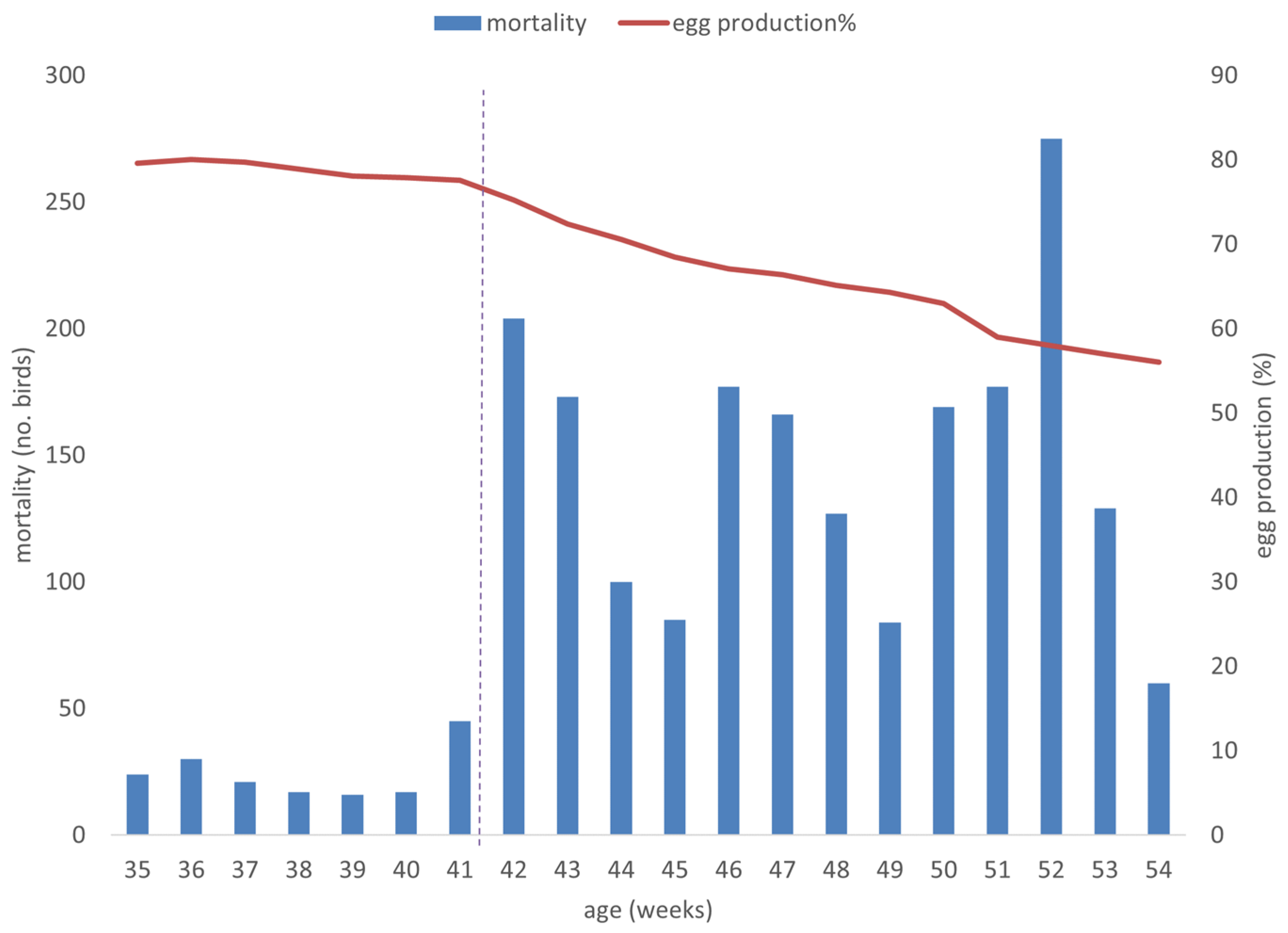
3.1. Clinical Features of the HSS Outbreak

The onset of the disease was in week 42, characterized by increasing mortality, affecting only female birds, which peaked in week 52 with 275 dead birds in one week. The clinical presentation of the outbreak was characterized by (1) high mortality, with overall mortality reaching 31.6% (Figure 1); (2) a production drop, with egg production dropping significantly from 77.56% to 59.57% during the outbreak, ultimately reducing to 55.49% by the time of slaughter (Figure 1); and (3) clinical signs consisting of pale combs, poor body condition, and no respiratory symptoms (Figure 2a). Post-mortem investigations revealed hemorrhagic hepatitis with friable and swollen livers, enlarged and marbled spleens, and ovarian atrophy (Figure 2b–d).

3.2. Histological Assessment

Histological examination of the liver and spleen samples revealed multifocal areas of coagulative or generalized necrosis, hemorrhages, and the presence of rod-shaped bacterial colonies. Immunohistochemistry (IHC) detected positive signals for aHEV ORF2 in both liver and spleen samples from affected chickens (Figure 3). In the liver, these positive signals were observed within hepatic sinusoids and clusters of mononuclear cells (Figure 3a). In the spleen, the signals were more prominent in peri-ellipsoidal zones but distributed throughout the organ (Figure 3b).

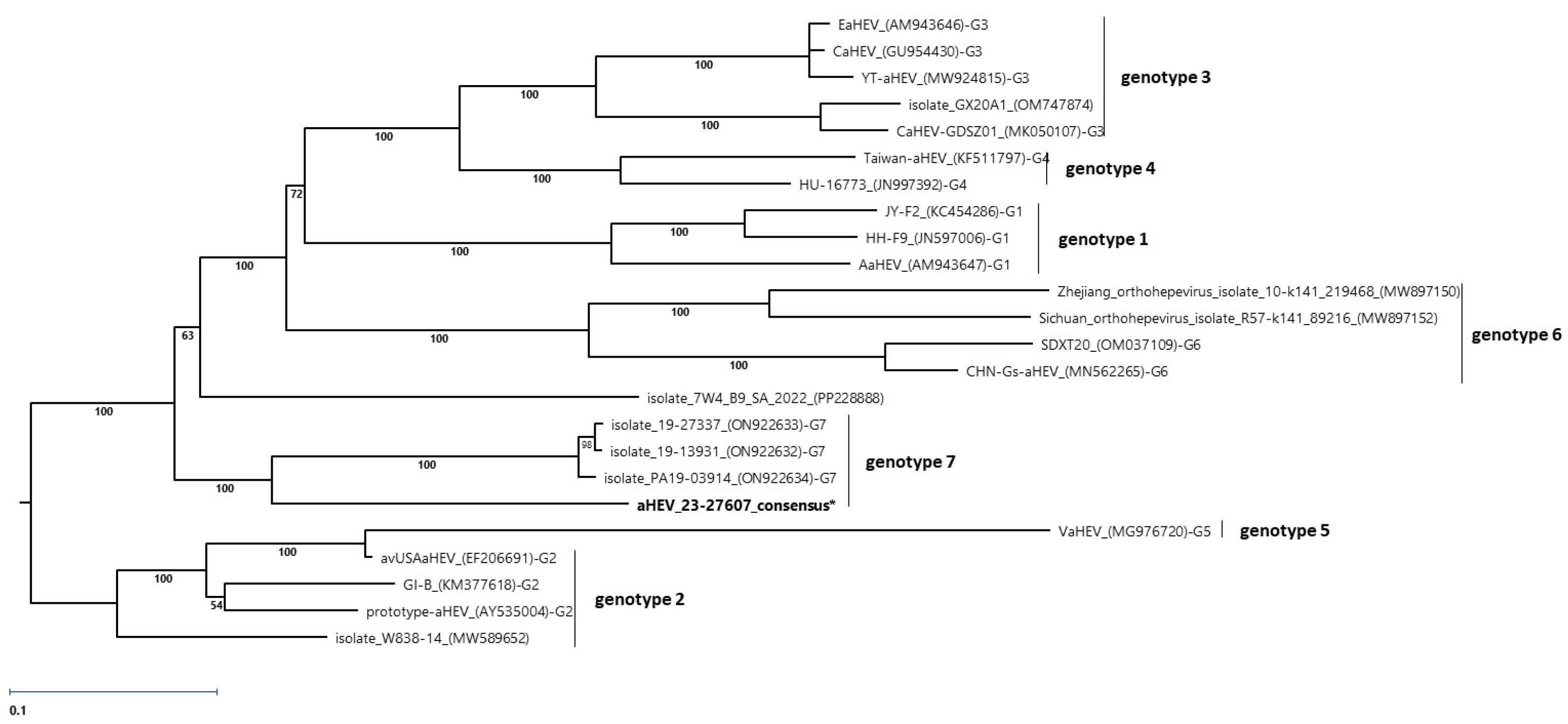
3.3. RT-PCR Investigations, Sequencing, and Phylogenetic Analysis

The RT-PCR investigation of liver and bile smears from an FTA card turned out positive for aHEV. Phylogenetic analysis of sequenced RT-PCR bands indicated the potentially novel genotype of aHEV. To gain further genomic information and to support detailed phylogenetic analysis, the virus’s complete genome sequence was determined using next-generation sequencing (NGS). De novo assembly from Illumina reads identified 3 contigs mapping to aHEV genomes, covering 5601 bp of the genome. The closure of 2 genome gaps at the N-terminal region by RT-PCR and sequencing, resulted in a final assembly of 6622 bp. The assembled sequence included all three open reading frames (ORFs) of aHEV and partial 5′- and 3′-untranslated regions (UTRs). The genome sequence—aHEV 23-27607—has been submitted to the NCBI database under the accession number (PQ605813).
Phylogenetic analysis based on the complete genome sequence positioned the newly sequenced genome on a distinct branch closely related to other genotype 7 isolates (Figure 4). The percent identity matrix based on the complete genomes showed a nucleotide identity of 83.80–83.95% with other isolates of genotype 7 and 78.85–82.99% compared to other aHEVs, supporting its classification to genotype 7 (Supplementary Table S1).

4. Discussion

Since aHEV was first identified as the causative agent of BLS disease/HSS in chickens [13,14], extensive genetic characterization has revealed considerable heterogeneity among field strains of the virus [11,15,16,17]. Initially, four distinct aHEV genotypes were identified, each associated with specific geographic regions [18]. However, subsequent analyses of complete genomes from aHEV strains identified in China led to the identification of two additional genotypes [9,10], and a seventh genotype was later documented in chickens and pheasants in Poland and France, respectively [4]. In this study, we present the first reported case of HSS in Bangladesh, which is only the second report of the syndrome in South Asia [19], and notably the first documented case of HSS caused by aHEV genotype 7 (aHEV 23-27607) outside of Europe.
One striking aspect of this outbreak is the severe impact on cumulative mortality and egg production within the affected flock, with a reduction in egg production exceeding 20%. Similar decreases in egg production have been documented in other HSS cases, with reductions of up to 20% often characterizing severe outbreaks [3]. However, the mortality observed in this case, reaching 31.6% among hens, is reasonably high for HSS and indicates a particularly severe disease presentation. In China, a severe form of HSS, termed hepatic rupture hemorrhagic syndrome, has been associated with cumulative mortality around 15% in laying chickens due to aHEV genotype 5 [20]. More recent outbreaks attributed to aHEV genotypes 5 and 6 in China have shown cumulative mortalities reaching 30% [21,22]. Conversely, outbreaks caused by aHEV genotype 7 in broiler breeders in Poland have reported mortalities of up to 9% [4]. This variability in mortality rates raises the question of whether the severity of HSS outbreaks is linked to specific aHEV genotypes or the potential pathogenicity of individual strains. Nonetheless, our understanding of aHEV pathogenesis is limited by the current lack of an effective in vitro propagation system for the virus, which hampers more detailed studies [3]. To elucidate the potential link between genotype and pathogenicity, comprehensive epidemiological studies including both clinically affected and asymptomatic birds are warranted.
Although aHEV is recognized as the etiological agent of HSS, infection with the virus does not consistently lead to disease manifestation in a flock [17,23,24,25]. Furthermore, it was detected in the metavirome of clinically healthy chickens [26,27]. Consequently, various cofactors—such as environmental stress, hormonal fluctuations, immune status, or other underlying health conditions—are often considered in discussions regarding the pathogenesis of HSS in aHEV-infected birds. In our investigation, histological analysis identified bacterial colonies within liver and spleen tissues, a finding consistent with our observations in previous cases [4]. This bacterial presence may indicate a secondary complication associated with the chronic nature of HSS and potentially compromised immune function in affected birds. However, prior studies have generally failed to establish a consistent association between HSS and the presence of specific toxins or pathogenic bacteria, further supporting the primary etiological role of aHEV in HSS [1].
In this study, IHC and RT-PCR were performed to confirm the presence of aHEV in organs displaying pathological changes. Positive signals were detected in the liver and spleen samples, specifically in regions showing necrotic and inflammatory lesions, thus reinforcing the role of aHEV in disease pathology. The localization of aHEV antigen in hepatic sinusoids and peri-ellipsoidal zones within the spleen is consistent with previous reports, which have documented similar antigen distribution patterns in these organs [4,28,29]. These findings substantiate the pathogenic association between aHEV and the lesions observed, underscoring the need for further research to clarify the complex interactions between aHEV infection and HSS severity.
In summary, our study provides the first documented case of hepatitis-splenomegaly syndrome (HSS) in Bangladesh, expanding the geographical scope of aHEV genotype 7 beyond Europe and marking only the second report from South Asia. The severe clinical presentation, characterized by high cumulative mortality and significant drops in egg production, underscores the potentially substantial impact of HSS on poultry health and productivity. Our findings, in conjunction with prior reports, suggest a possible relationship between aHEV genotype and disease severity; however, due to limitations in current knowledge on aHEV pathogenesis and pathogenicity, this association remains inconclusive. Moving forward, performing comprehensive epidemiological studies involving both diseased and healthy birds, as well as further advances in aHEV research methodologies, will be crucial for clarifying the roles of genotype variability, host factors, and potential cofactors in the manifestation and severity of HSS.

Supplementary Materials

The following supporting information can be downloaded at: https://www.mdpi.com/article/10.3390/poultry4020016/s1, Table S1: Percent identity of complete genomes.

Author Contributions

Conceptualization, L.K. and M.H.; methodology, I.B., D.L. and M.H.; software, N.P.; validation, I.B., D.L. and N.P.; formal analysis, I.B., D.L. and N.P.; investigation, I.B., L.K., R.H., D.L. and N.P.; resources, M.H.; data curation, N.P.; writing—original draft preparation, M.M. and I.B.; writing—review and editing, M.M., I.B., L.K., D.L., N.P. and M.H.; visualization, M.M.; supervision, M.H.; project administration, M.H. All authors have read and agreed to the published version of the manuscript.

Funding

This research received no external funding.

Institutional Review Board Statement

This study was conducted in accordance with the Animal Welfare Act, 2019 (Act No. 12 of 2019) of Bangladesh.

Informed Consent Statement

All veterinary procedures were performed in cooperation with licensed veterinarians responsible for the respective farms, with prior consent obtained from the farmers.

Data Availability Statement

The data are contained within the article. The original contributions presented in the study are included in the article; further inquiries can be directed to the corresponding author.

Acknowledgments

The authors extend their gratitude to Barbara Jaskulska, Irina Prokofieva, and Patricia Wernsdorf for their valuable technical assistance. Open Access Funding by the University of Veterinary Medicine Vienna.

Conflicts of Interest

László Kőrösi is employed by AgriAL Bt., Hungary; Rakibul Hasan is employed by Peoples Poultry & Hatchery Ltd Bangladesh. The remaining authors declare that the research was conducted in the absence of any commercial or financial relationships that could be construed as a potential conflict of interest.

References

  1. Meng, X.J.; Shivaprasad, H.L. Avian Hepatitis E Virus Infections. In Diseases of Poultry; Swayne, D.E., Boulianne, M., Logue, C.M., McDougald, L.R., Nair, V., Suarez, D.L., Eds.; Wiley-Blackwell: Hoboken, NJ, USA, 2020; pp. 528–534. [Google Scholar]
  2. Handlinger, J.H.; Williams, W. An Egg Drop Associated with Splenomegaly in Broiler Breeders. Avian Dis. 1988, 32, 773–778. [Google Scholar] [CrossRef] [PubMed]
  3. Sun, P.; Lin, S.; He, S.; Zhou, E.; Zhao, Q. Avian Hepatitis E Virus: With the Trend of Genotypes and Host Expansion. Front. Microbiol. 2019, 10, 1696. [Google Scholar] [CrossRef]
  4. Matos, M.; Bilic, I.; Tvarogová, J.; Palmieri, N.; Furmanek, D.; Gotowiecka, M.; Liebhart, D.; Hess, M. A Novel Genotype of Avian Hepatitis E Virus Identified in Chickens and Common Pheasants (Phasianus colchicus), Extending Its Host Range. Sci. Rep. 2022, 12, 21743. [Google Scholar] [CrossRef]
  5. Siedlecka, M.; Kublicka, A.; Wieliczko, A.; Matczuk, A.K. Molecular Detection of Avian Hepatitis E Virus (Orthohepevirus B) in Chickens, Ducks, Geese, and Western Capercaillies in Poland. PLoS ONE 2022, 17, e0269854. [Google Scholar] [CrossRef]
  6. Zhang, X.; Bilic, I.; Troxler, S.; Hess, M. Evidence of Genotypes 1 and 3 of Avian Hepatitis E Virus in Wild Birds. Virus Res. 2017, 228, 75–78. [Google Scholar] [CrossRef]
  7. Yang, C.; Wang, L.; Shen, H.; Zheng, Y.; Gauger, P.C.; Chen, Q.; Zhang, J.; Yoon, K.J.; Harmon, K.M.; Main, R.G.; et al. Detection and Genomic Characterization of New Avian-like Hepatitis E Virus in a Sparrow in the United States. Arch. Virol. 2018, 163, 2861–2864. [Google Scholar] [CrossRef] [PubMed]
  8. Purdy, M.A.; Drexler, J.F.; Meng, X.J.; Norder, H.; Okamoto, H.; Van der Poel, W.H.M.; Reuter, G.; de Souza, W.M.; Ulrich, R.G.; Smith, D.B. ICTV Virus Taxonomy Profile: Hepeviridae 2022. J. Gen. Virol. 2022, 103, 001778. [Google Scholar] [CrossRef]
  9. Liu, B.; Chen, Y.; Zhao, L.; Zhang, M.; Ren, X.; Zhang, Y.; Zhang, B.; Fan, M.; Zhao, Q.; Zhou, E.M. Identification and Pathogenicity of a Novel Genotype Avian Hepatitis E Virus from Silkie Fowl (Gallus Gallus). Vet. Microbiol. 2020, 245, 108688. [Google Scholar] [CrossRef] [PubMed]
  10. Su, Q.; Zhang, Z.; Zhang, Y.; Cui, Z.; Chang, S.; Zhao, P. Complete Genome Analysis of Avian Hepatitis E Virus from Chicken with Hepatic Rupture Hemorrhage Syndrome. Vet. Microbiol. 2020, 242, 108577. [Google Scholar] [CrossRef]
  11. Bilic, I.; Jaskulska, B.; Basic, A.; Morrow, C.J.; Hess, M. Sequence Analysis and Comparison of Avian Hepatitis E Viruses from Australia and Europe Indicate the Existence of Different Genotypes. J. Gen. Virol. 2009, 90, 863–873. [Google Scholar] [CrossRef]
  12. Martin, M. Marcel Martin Cutadapt Removes Adapter Sequences from High-Throughput Sequencing Reads. EMBnet J. 2011, 17, 10–12. [Google Scholar] [CrossRef]
  13. Haqshenas, G.; Shivaprasad, H.L.; Woolcock, P.R.; Read, D.H.; Meng, X.J. Genetic Identification and Characterization of a Novel Virus Related to Human Hepatitis E Virus from Chickens with Hepatitis-Splenomegaly Syndrome in the United States. J. Gen. Virol. 2001, 82, 2449–2462. [Google Scholar] [CrossRef] [PubMed]
  14. Payne, C.J.; Ellisa, T.M.; Planta, S.L.; Gregorya, A.R.; Wilcoxb, G.E. Sequence Data Suggests Big Liver and Spleen Disease Virus (BLSV) Is Genetically Related to Hepatitis E Virus. Vet. Microbiol. 1999, 68, 119–125. [Google Scholar] [CrossRef]
  15. Agunos, A.C.; Yoo, D.; Youssef, S.A.; Ran, D.; Binnington, B.; Hunter, D.B. Avian Hepatitis E Virus in an Outbreak of Hepatitis-Splenomegaly Syndrome and Fatty Liver Haemorrhage Syndrome in Two Flaxseed-Fed Layer Flocks in Ontario. Avian Pathol. 2006, 35, 404–412. [Google Scholar] [CrossRef] [PubMed]
  16. Huang, F.F.; Haqshenas, G.; Shivaprasad, H.L.; Guenette, D.K.; Woolcock, P.R.; Larsen, C.T.; Pierson, F.W.; Elvinger, F.; Toth, T.E.; Meng, X.J. Heterogeneity and Seroprevalence of a Newly Identified Avian Hepatitis E Virus from Chickens in the United States. J. Clin. Microbiol. 2002, 40, 4197–4202. [Google Scholar] [CrossRef] [PubMed]
  17. Sun, Z.F.; Larsen, C.T.; Dunlop, A.; Huang, F.F.; Pierson, F.W.; Toth, T.E.; Meng, X.J. Genetic Identification of Avian Hepatitis E Virus (HEV) from Healthy Chicken Flocks and Characterization of the Capsid Gene of 14 Avian HEV Isolates from Chickens with Hepatitis-Splenomegaly Syndrome in Different Geographical Regions of the United States. J. Gen. Virol. 2004, 85, 693–700. [Google Scholar] [CrossRef]
  18. Johne, R.; Dremsek, P.; Reetz, J.; Heckel, G.; Hess, M.; Ulrich, R.G. Hepeviridae: An Expanding Family of Vertebrate Viruses. Infect. Genet. Evol. 2014, 27, 212–229. [Google Scholar] [CrossRef]
  19. Iqbal, T.; Rashid, U.; Idrees, M.; Afroz, A.; Kamili, S.; Purdy, M.A. A Novel Avian Isolate of Hepatitis E Virus from Pakistan. Virol. J. 2019, 16, 142. [Google Scholar] [CrossRef] [PubMed]
  20. Su, Q.; Li, Y.; Meng, F.; Cui, Z.; Chang, S.; Zhao, P. Hepatic Rupture Hemorrhage Syndrome in Chickens Caused by a Novel Genotype Avian Hepatitis E Virus. Vet. Microbiol. 2018, 222, 91–97. [Google Scholar] [CrossRef] [PubMed]
  21. Hou, L.; Chi, Z.; Zhang, Y.; Xue, Q.; Zhai, T.; Chang, S.; Wang, J.; Zhao, P. Natural Co-Infection of Fowl Adenovirus Type E-8b and Avian Hepatitis E Virus in Parental Layer Breeders in Hebei, China. Virol. Sin. 2023, 38, 317–320. [Google Scholar] [CrossRef]
  22. Meng, K.; Liu, M.; Zhang, Y.; Yuan, X.; Xu, H. A Genetically Novel Avian Hepatitis E Virus in China. Virus Genes. 2022, 58, 589–593. [Google Scholar] [CrossRef] [PubMed]
  23. Peralta, B.; Biarnés, M.; Ordóñez, G.; Porta, R.; Martín, M.; Mateu, E.; Pina, S.; Meng, X.J. Evidence of Widespread Infection of Avian Hepatitis E Virus (Avian HEV) in Chickens from Spain. Vet. Microbiol. 2009, 137, 31–36. [Google Scholar] [CrossRef]
  24. Sprygin, A.V.; Nikonova, Z.B.; Zinyakov, N.G. Avian Hepatitis E Virus Identified in Russian Chicken Flocks Exhibits High Genetic Divergence Based on the ORF2 Capsid Gene. Avian Pathol. 2012, 41, 459–463. [Google Scholar] [CrossRef]
  25. Troxler, S.; Pać, K.; Prokofieva, I.; Liebhart, D.; Chodakowska, B.; Furmanek, D.; Hess, M. Subclinical Circulation of Avian Hepatitis E Virus within a Multiple-Age Rearing and Broiler Breeder Farm Indicates Persistence and Vertical Transmission of the Virus. Avian Pathol. 2014, 43, 310–318. [Google Scholar] [CrossRef] [PubMed]
  26. Nwokorogu, V.C.; Pillai, S.; San, J.E.; Pillay, C.; Nyaga, M.M.; Sabiu, S. A Metagenomic Investigation of the Faecal RNA Virome Structure of Asymptomatic Chickens Obtained from a Commercial Farm in Durban, KwaZulu-Natal Province, South Africa. BMC Genom. 2024, 25, 629. [Google Scholar] [CrossRef]
  27. Chen, Y.-M.; Sadiq, S.; Tian, J.-H.; Chen, X.; Lin, X.-D.; Shen, J.-J.; Chen, H.; Hao, Z.-Y.; Wille, M.; Zhou, Z.-C.; et al. RNA Viromes from Terrestrial Sites across China Expand Environmental Viral Diversity. Nat. Microbiol. 2022, 7, 1312–1323. [Google Scholar] [CrossRef]
  28. Clarke, J.K.; Allan, G.M.; Bryson, D.G.; Todd, D.; MacKie, D.P.; McFerran, J.B.; Williams, W. Big Liver and Spleen Disease of Broiler Breeders. Avian Pathol. 1990, 19, 41–50. [Google Scholar] [CrossRef] [PubMed]
  29. Morrow, C.J.; Samu, G.; Mátrai, E.; Klausz, Á.; Wood, A.M.; Richter, S.; Jaskulska, B.; Hess, M. Avian Hepatitis E Virus Infection and Possible Associated Clinical Disease in Broiler Breeder Flocks in Hungary. Avian Pathol. 2008, 37, 527–535. [Google Scholar] [CrossRef] [PubMed]
Figure 1. Graphical presentation of absolute mortality (left axis) and relative egg production (right axis), in the affected flock, between 35–54 weeks of life. The peak egg production was reached at 26 weeks of life with 83.65% (not shown). The vertical dashed line represents the onset of the outbreak.
Poultry 04 00016 g001
Figure 2. Clinical and post-mortem observations during the HSS outbreak. (a) Birds in a prostrate state with pale combs; (b) liver enlargement with hemorrhagic hepatitis identified at necropsy; (c) splenomegaly with splenitis, displaying a marbled appearance; and (d) ovarian degeneration and atrophy (arrow).
Poultry 04 00016 g002
Figure 3. IHC analysis of liver and spleen samples from chickens affected by HSS. Positive staining for aHEV ORF2 was observed within (a) hepatic sinusoids in the liver (scale bar = 50 µm) and (b) in peri-ellipsoidal zones distributed throughout the spleen (scale bar = 100 µm). The reaction was developed using a DAB Substrate Kit for peroxidase (Vector Laboratories) and subsequently counterstained with Mayer’s Hematoxylin (Merck, Darmstadt, Germany).
Poultry 04 00016 g003
Figure 4. Phylogenetic analysis of Avihepevirus magneiicur genomes. The analysis was performed using the Maximum Likelihood method (RAxML) and was based on the alignments of conserved sites that included 5507 positions in the final dataset. The unrooted tree is shown. This analysis involved 24 nucleic acid sequences. All genomes from the Genbank are labeled with the name, accession number in parentheses, and the designation of the corresponding genotype as the G-number. The sequence obtained in the present study is labeled in bold. The robustness of the tree was tested by bootstrap analysis with 500 iterations and the percentage of trees in which associated taxa clustered together is shown next to the branches. Evolutionary analysis was conducted in the MegAlign Pro module of Lasergene v17.5.0 software (DNASTAR, Madison, WI, USA).
Poultry 04 00016 g004
Table 1. List of primers used for the gap-closure RT-PCR. Primer sequences, annealing temperatures, and RT-PCR product sizes are given.
PrimerSequence (5′-3′)Annealing Temperature (°C)Product Size (bp)
N_term_aHEV-F1CCTACCTTACCCAACAGCAG491076
N_term_aHEV-R1CCTCACACCACGAGCTATAC
N_term_aHEV-F2GGTACTCAGCCGGATTCTATC531051
N_term_aHEV-R2CGGCCTTCGTTTAGTACAAAG
Disclaimer/Publisher’s Note: The statements, opinions and data contained in all publications are solely those of the individual author(s) and contributor(s) and not of MDPI and/or the editor(s). MDPI and/or the editor(s) disclaim responsibility for any injury to people or property resulting from any ideas, methods, instructions or products referred to in the content.

