Review

Physiopathological Aspects of Synovial Fluid and Membrane in Psoriatic Arthritis

Rheumatology Unit, Department of Medicine—DIMED, University of Padova, 35128 Padova, Italy
*
Author to whom correspondence should be addressed.
Rheumato 2024, 4(4), 193-202; https://doi.org/10.3390/rheumato4040015
Submission received: 11 September 2024 / Revised: 25 October 2024 / Accepted: 1 November 2024 / Published: 5 November 2024

Abstract

:
Background: Psoriatic arthritis (PsA) is a chronic inflammatory arthropathy characterized by a variety of clinical manifestations, mainly affecting joints and entheses, but also skin, nails, the eye, and the intestine. Objectives: In this review, we describe the essential characteristics of both synovial membranes and synovial fluid (SF) in PsA. Similarly to other inflammatory arthritis, the histological peculiarities in PsA synovitis are lining hyperplasia, neoangiogenesis, and sublining infiltration by immune cells and inflammatory mediators. Synovial effusions are frequent in PsA patients and SF analysis allows us to determine the pathological process occurring in the joint. Routine examinations help clinicians in defining the inflammatory status and possibly the detection of specific cell subsets. In addition, pathogenic crystals including monosodium urate and calcium pyrophosphate may be found in PsA SF. Conclusions: SF represents a potential substrate to identify the biomarkers that are useful to predict disease progression and response to medications in PsA patients, thus guiding the choice of appropriate and tailored pharmacological treatment.

1. Introduction

Psoriatic arthritis (PsA) is considered a chronic inflammatory arthropathy, characterized by the inflammation of the joints and entheses, including the axial skeleton. Nevertheless, PsA is also characterized by complex clinical heterogeneity. Clinical manifestations may involve the skin (psoriasis), nail changes, and, less frequently, the eye (uveitis) and the bowel (inflammatory bowel disease) [1,2,3]. PsA is included in the spondyloarthritis group [4] and it is distinguished from rheumatoid arthritis (RA) by sporadic positivity for rheumatoid factor (RF) and anti-cyclic citrullinated peptide antibodies (anti-CCP antibodies), as well as for the presence of specific clinical features such as an asymmetric distribution of the inflamed joints, sacroiliitis or spinal involvement, distal interphalangeal joint inflammation, and non-articular manifestations [2,3,5,6,7,8].
The pathophysiology of PsA is not yet fully understood. PsA mostly develops in patients with an established diagnosis of psoriasis; however, inflammation of the synovium can also occur regardless of skin involvement [8].
PsA has a multimodal etiology that arises from a complex interaction of environmental (exposure to smoking, infection, and trauma), genetic (HLAs), and immunological factors [9,10]. Environmental factors can act as triggers for PsA development through the activation of antigen-presenting cells (macrophages and dendritic cells) [11,12]. Antigen presentation to T cells further releases cytokines that stimulate and differentiate T cells. The activation of several immune–inflammatory pathways acts in modulating the inflammatory process in these patients, complicating PsA treatment [1,3,13,14].
Several proinflammatory mediators are released (IL-1, IL-6, TNF-α, IL-17, and IL-23), and this inflammatory cascade promotes the initiation of inflammatory (chronic synovitis) and destructive processes in the joints [1,5,13].
With this review, we aimed to analyze the pathological processes occurring in the articular environment in PsA including the synovial membrane and fluid. The first part deals with the tissue alterations that characterize synovitis, identifying lining and sublining changes, inflammatory mediator release, and bone structure remodelling. The second part describes the inflammatory aspect of SF in relation to cell composition and the presence of pathogenic crystals. We conclude by highlighting the value of SF as a substrate for the identification of potential diagnostic and predictive biomarkers in PsA, thus guiding the choice of appropriate and tailored pharmacological treatment.

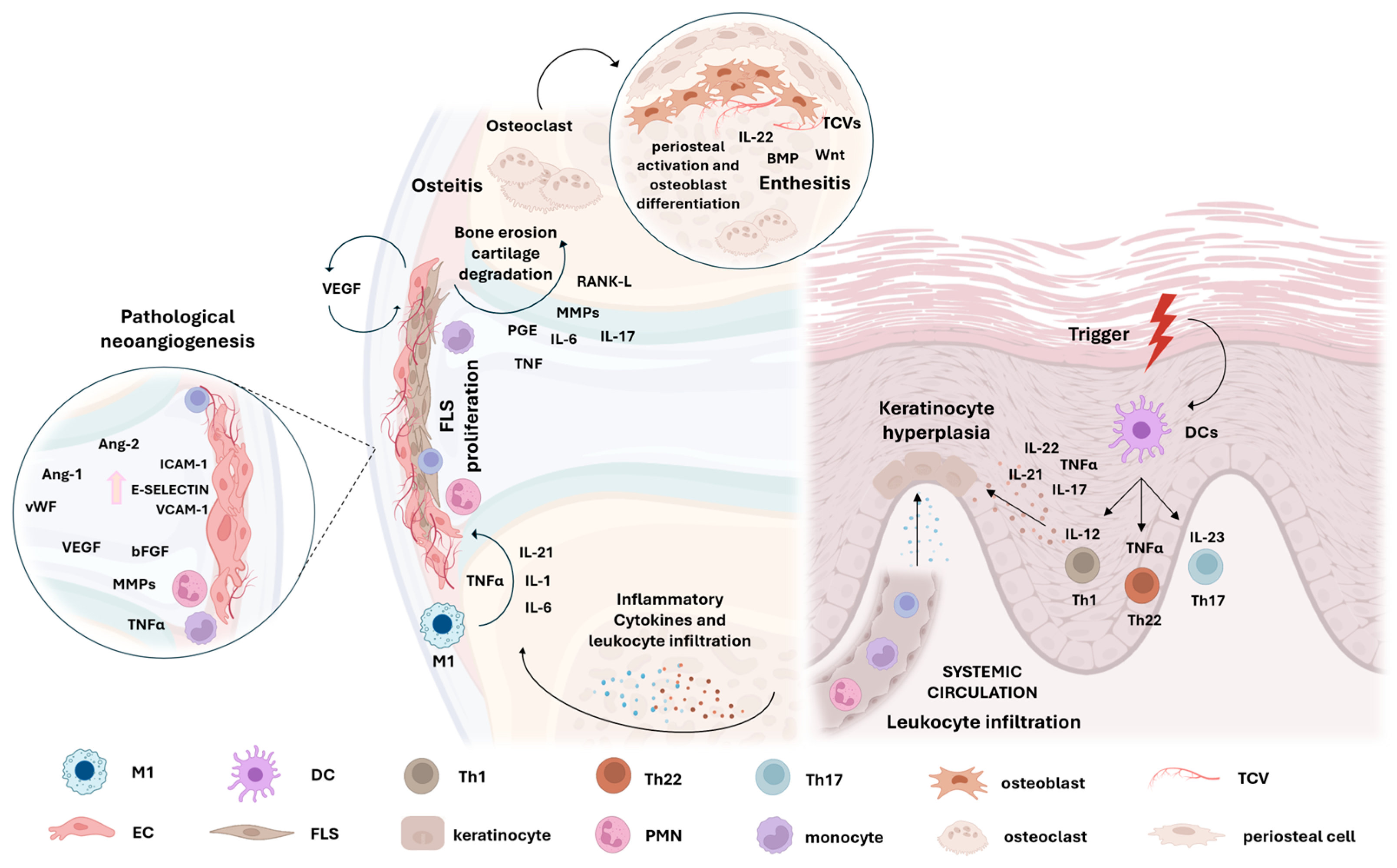
2. Synovitis in Psoriatic Arthritis

PsA synovitis has three histological characteristics that are common to other types of inflammatory arthritis—lining hyperplasia (the proliferation of fibroblast-like synoviocytes—FLSs—and the infiltration of macrophages); neoangiogenesis; and the infiltration of the sublining by inflammatory cells [3] (Figure 1).

2.1. Lining Hyperplasia

In PsA, moderate synovial lining hyperplasia is observed, compared to the important hyperplasia found in RA patients. Although, others studies have found no differences in lining between RA and PsA. Lining hyperplasia is a result of FLS activation (where they adopt an inflammatory phenotype), proliferation, and decreased FLS apoptosis. FLSs finally invade cartilage and bone, resulting in joint destruction [15,16].
Hyperplasia is also enhanced by cellular debris and fibrin. The synovial lining also presents hypertrophic synoviocytes that extend into the joint space [8,17]. The PsA lining is also characterized by a high infiltration of macrophages, which are involved in innate immune inflammation and contribute to lining thickness [3]. No differences were described in macrophage cellular density between PsA and RA synovitis and in lining layer infiltrate or thickness between disease duration and poly- or oligoarticular disease [8,17,18].

2.2. Synovial Sublining Vascularization

Pathological neoangiogenesis, i.e., the development of new blood vessels, has been reported in both synovial tissue and psoriatic skin lesions [5]. PsA synovitis has predominantly tortuous, bushy vessels, reflecting abnormal neoangiogenesis, and this characteristic is more marked in PsA than in RA synovium [3,19,20].
No differences have been shown between oligo- and polyarticular disease [21]. Vascular changes are characterized by the augmented thickening of the vascular wall and endothelial cell (EC) swelling, accompanied by polymorphonuclear (PMN), monocyte, lymphocyte, and plasma cell inflammatory infiltration in the media and adventitia [22]. Zhang et al. reported the presence of small deposits on vessel walls, such as immune complexes, microparticles, and membrane-bound vesicles, which regulate thrombosis, angiogenesis, and inflammation [8,17].
According to the vascular abnormalities found in the PsA synovium, the overexpression of vascular endothelial growth factor (VEGF) has been reported in synovial tissue and in SF (synovial fluid), together with other important factors involved in angiogenesis (angiopoietin Ang-1, Ang-2, von Willebrand’s factor, integrin αVβ3, and basic fibroblast growth factor) [5,13,20,23,24]. The activated form of Tie-2 upregulates angiogenesis through its interaction with Ang1 and Ang2, which induces inflammatory gene expression in ECs, FLSs, and macrophages [8,25,26]. Recently, we reported that SF from PsA patients includes several mediators that modulate pathological angiogenesis, including cytokines, growth factors, and metalloproteinases (MMPs), and it is able to induce in vitro EC migration and the formation of capillary-like structures [27].
Abnormal angiogenesis also promotes the infiltration of immune cells in the synovium, supporting the inflammatory process [13,21].

2.3. Sublining Influx of Innate and Adaptative Immunity Cells

Several immune cells are present in the PsA synovium, such as macrophages, neutrophils, mast cells, and lymphocytes. The activation of these cells produces multiple inflammatory and pro-angiogenic factors (cytokines, chemokines, growth factors, and MMPs), which promote the development of synovitis and joint damage [5].
Activated macrophages contribute to various proinflammatory processes in PsA. CD68+ macrophages enhance inflammation in the synovium and exhibit destructive potential [5,14,21]. CD68+ macrophage reduction is associated with a good response to therapy, suggesting the important role of these cells in PsA inflammation. In the synovium, mast cells induce angiogenesis, neutrophil influx, and the proliferation of synovial fibroblasts [28]. Furthermore, they play an important role in PsA synovial inflammation by producing IL-17A (they are the main cellular source of this cytokine) [29]. Together with mast cells, PMNs are the most common IL-17A+ cells. PMN infiltration correlates with disease activity. Indeed, their decrease after treatment is associated with a good therapeutic response (biomarker of response) [5,30].
Innate lymphoid cells (ILCs) are involved in PsA pathogenesis. ILCs produce IL-17 and IL-22, regardless of specific Ag stimulation, and their presence correlates with disease activity [31,32].
The PsA synovium is characterized by a low number of T lymphocytes; however, these cells are the main contributors to PsA pathogenesis [14,21]. The production of IL-12 and IL-23 by innate immune cells stimulates the differentiation of T cells into Th1 and Th17 helper T cells. Indeed, Th17-related cytokines IL-17 and IL-23 are highly expressed in PsA joints [33,34]. In addition, IL-17-secreting cells also include CD8+ T lymphocytes [35].

2.4. Inflammatory Mediators Involved in PsA Synovitis

An important feature of the PsA synovium is the abundant release from activated immune cells of proinflammatory cytokines (TNF-α, IL-17A, IL-23, IL-1β, IL-6, and IL-18), which are mediators of local and systemic inflammation. The efficacy of monoclonal antibodies targeting specific factors, such as IL-23, IL-17A, and TNF-α, has confirmed the importance of these cytokines in PsA, suggesting that these inhibitors block different pathogenic processes [13,21].
The contribution of the IL-23/IL-17 axis is crucial in the pathogenesis of PsA. As described previously, IL-23 plays a role in initiating disease development and it is an upstream regulator of IL-17A, while IL-17A sustains the inflammatory process and directly affects tissue physiology. Indeed, IL-23 stimulates the activation, differentiation, proliferation, and survival of Th17 cells, which further facilitate the local inflammatory process through IL-17A and IL-22 production. However, the production of IL-17 can also occur independently of IL-23 [36].
IL-17A has a multitude of effects; for example, it has an osteoclastogenic activity, up-regulates MMPs and proinflammatory cytokines (IL-1β, IL-8, and TNFα), and modulates inflammatory pain. It mediates the influx of neutrophils, as well as the expansion of Th17 cells and ILC3s, particularly in entheses, thus contributing to the generation of inflammation. Finally, it induces the production of proinflammatory mediators on keratinocytes and synovial cells, connecting the innate and adaptive immune systems to support chronic inflammation [2,37]. Elevated IL-17 levels have been found in the joints of PsA patients but not in RA patients [38].
IL-17A and TNF-α exhibit overlapping and synergistic effects. IL-17A, as well as TNF-α, is a key mediator of joint inflammation and damage in PsA patients. Through this cytokine, T1 cells promote the recruitment/activation of macrophages and neutrophilis, in addition to IL-1 and -6 production. In addition, through angiogenesis induction, it mediates the progression of joint inflammation [39]. In this context, TNF-α promotes the expression of adhesion molecules, promoting the infiltration of leukocytes. Its blockade decreases the expression of angiogenetic factors and adhesion molecules [40,41].
Similarly to IL-17A, TNF-α and IL-22 increase the production of enzymes that degrade bone and cartilage, leading to joint damage (the loss of collagen structures and cartilage surface erosion). Furthermore, these cytokines promote bone resorption via the RANKL signalling pathway, activating osteoclast precursor cells and osteoclastogenesis. Finally, IL-22 promotes osteoblast activation, leading to enhanced bone formation [42,43].
Another interesting cytokine is the TNF-like weak inducer of apoptosis (TWEAK). The TWEAK pathway promotes local chemokine and cytokine production, resulting in angiogenesis induction and the infiltration of proinflammatory cells [44].

2.5. Tissue Damage and Bone Remodelling in PsA

Tissue damage in PsA is supported by the induction of MMPs, by neutrophil influx, and by mesenchymal cell activation induced by PGE2- and IL-17-induced mesenchymal cell activation. MMPs mediate the degradation of cartilage and bone, and their expression is associated with a more destructive disease [21,24,39]. Bone remodelling in PsA is promoted by synovial inflammation. The coexistence of bone erosion and new formation represents a peculiar pattern, observed only in PsA [45]. Synovium-mediated intra-articular bone erosion is a process sustained by RANKL, MCSF, IL-17, and TNFα, leading to the differentiation of pre-osteoclasts and osteoclasts [39]. Meanwhile, bone formation driven by an inflamed synovium is associated with an increase in IL-22, which is secreted by effector cells [46,47]. In this environment, osteoblast differentiation is led by TGF-β action, through the canonical TGF-β/BMP (bone morphogenic protein) pathway and by PGE2, which also triggers pain and inflammation [48]. Instead, enthesis formation is an extra-articular process. Periosteal activation and osteoblast differentiation, mediated by BMPs, Wnt proteins, and IL-22, lead to matrix deposition and bone formation in the context of enthesitis [34].

3. Synovial Fluid in PsA

Synovitis in PsA causes an increase in SF volume, leading to joint swelling and pain. Compromised lining barrier function and increased angiogenesis allow plasma and immune cells to flow from circulation to the joint cavity [5]. As described above, synovial vascularization is particularly pronounced in PsA and this may predispose patients to develop joint swelling during disease flares or even after intense exercise or articular microtraumatic solicitation. Synovial effusions from PsA have been described to be the most abundant among all arthropathies including RA, osteoarthritis and crystal-induced arthritis [49].
SF represents an interesting reservoir of proteins originating from the synovial membrane, articular cartilage, and joint capsule, therefore reflecting the pathophysiological conditions in PsA. Furthermore, it is well established that the nature of the cells in SF reflects the pathological processes occurring in the tissue; their analysis may help clinicians in defining inflammatory status, particular early on in the disease.

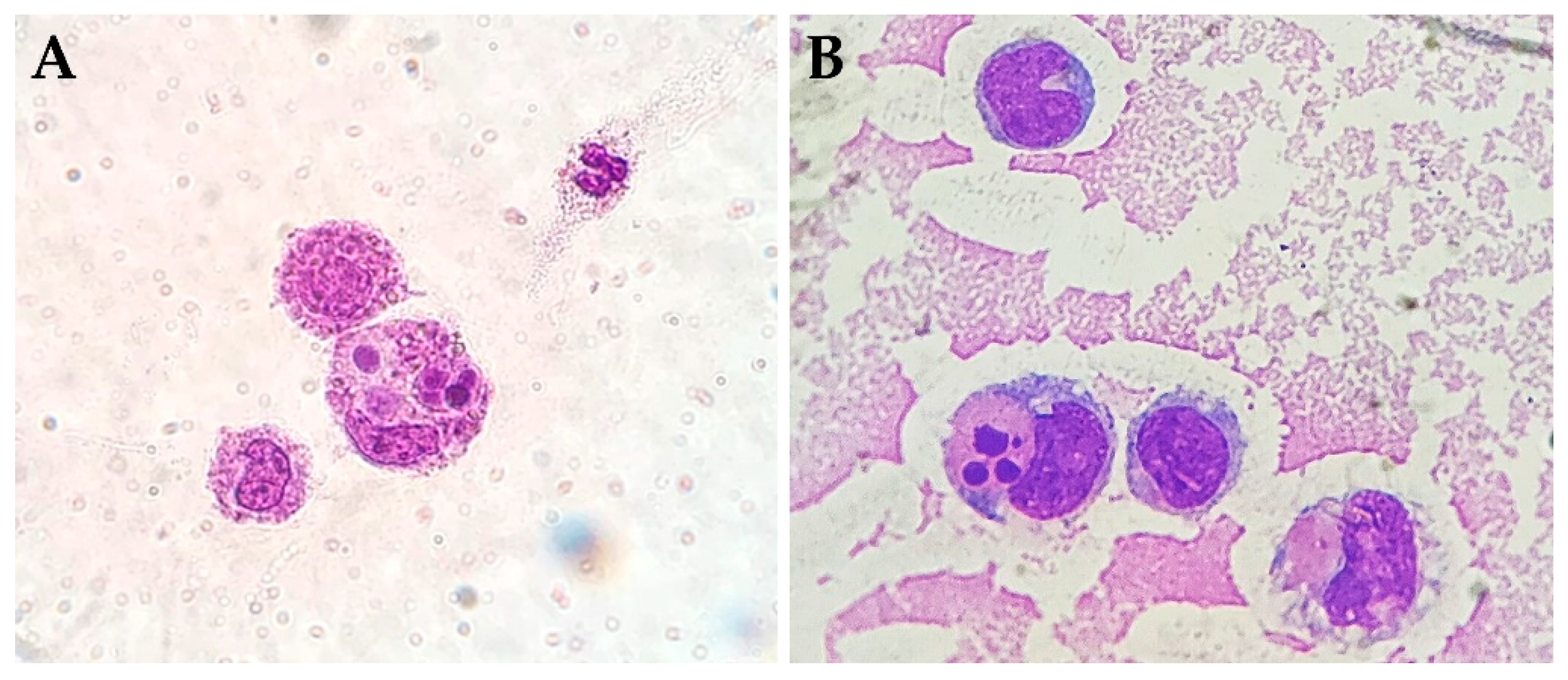
3.1. Inflammatory Cells

While a typical SF contains very few cells (<100 cells/mm3), with the prevalence of a mononuclear subset, the number of WBCs in PsA is highly variable depending on patient disease activity, joint involvement, and pharmacological treatment. Those factors also influence differential cell count, which has a great intervariability among patients. According to previous studies, WBCs in PsA range from 3000 to 20,000 cell/mm3, while the PMN percentage varies from 10 to 70 [50,51,52,53]. Results extracted from the same studies indicate similar inflammatory features in SF collected from RA, despite the more prevalent polymorphonuclear cell subset [50,51].

3.2. Cytophagocytic Mononuclear Cells

Cytophagocytic mononuclear (CPM) cells, best known as Reiter’s cells, are frequently detected in synovial fluid. They are presumably associated with apoptotic cell removal and the resolution of the articular inflammatory process. Spent neutrophils, cleared via phagocytosis by macrophages, are collected in the cytoplasm, thus forming a CPM cell that is clearly detectable during SF routine analysis (Figure 2). According to a recent review chart study, these cells have more commonly been found to be associated with spondyloarthritis, including PsA and reactive arthritis, compared to other arthropathies, and they show an interesting seasonal variation with an increased frequency during spring and autumn [54]. Given their distribution, CPM cells may represent an attractive diagnostic datapoint for the definition of SpA.

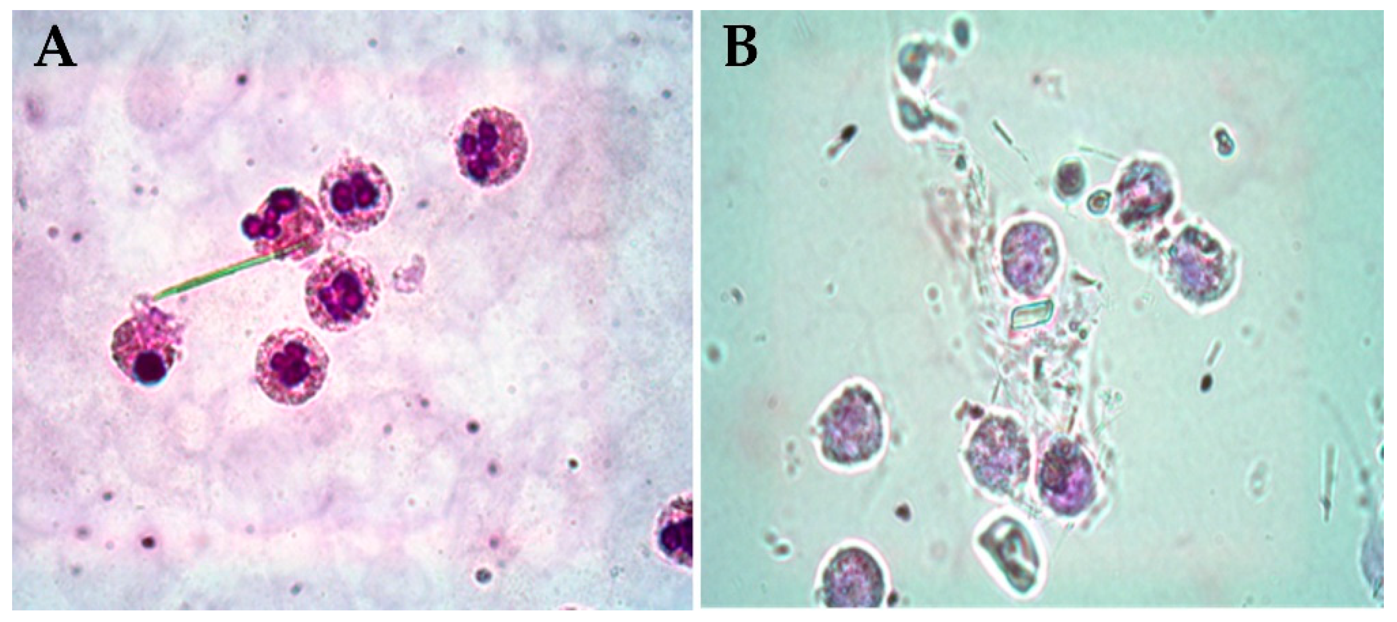
3.3. Pathogenic Crystals

PsA has been recognized as a metabolic disease given the multiple inflammatory pathways it has in common with metabolic syndrome and other comorbidities, including diabetes, cardiovascular diseases, obesity, and non-alcoholic fatty liver disease [55]. Studies have also emphasized the higher prevalence of hyperuricemia in individuals with underlying PsA compared to the general population and, consequently, a more elevated risk of developing gout. According to an epidemiological study on the frequency of pathogenic crystals, monosodium urate (MSU) (Figure 3A) has been found in 3.34% of the SF obtained from patients who have received a PsA diagnosis [50]. Other studies showed a higher prevalence of gout in PsA patients [56]. A convincing explanation and further explanation are still needed for the different rates of crystals detected in SF. As far as calcium pyrophosphate (CPP) crystals are concerned (Figure 3B), they have been evidenced in a similar percentage (3.82%) of PsA patients [50]. It could be postulated that the presence of endocrine comorbidities like hypothyroidism and osteoporosis could be involved in CPP formation within the joint. However, the identification of pathogenic crystals in SF can assist the diagnosis of possible comorbidities linked to the presence of crystals and it can lead to choosing the best pharmacological treatment for patients.

3.4. Synovial Biomarkers in SF

Early diagnosis and effective treatment is deemed essential to prevent irreversible joint damage and functional impairment in PsA. However, any validated biological molecule that is still available in PsA could help clinicians in diagnosing the disease and predicting which patients benefit from a particular treatment.
For its unique features, SF has been largely used as a substrate to identify potential diagnostic and predictive biomarkers in PsA. Different from blood, changes in SF suggest a more localized metabolism, thus reflecting joint-specific processes [57].
Markers found to be elevated in SF from PsA patients and correlating to disease activity have been postulated to be relevant targets for treatment. For instance, C5a, an important chemoattractant of granulocytes and monocytes, has been shown to play a role in driving the influx of immune cells into the joint cavity [52]. Other molecules such as cytokines of the IL-10 family [58] and IL-9, which is implicated in T cell proliferation [59], have been proposed as markers of disease progression. Fiocco et al. used SF and synovial tissue biomarkers to assess the response to intra-articular TNF-α blockers in PsA patients. These authors found some cytokines, including IL-1β, IL-1Ra, IL-6, and IL-22, to be correlated to systemic disease activity; this decreased parallelly with synovial tissue CD45 and CD3 expression after treatment [52].
Caso et al. identified soluble mediators in SF that characterize PsA and RA and may be useful for personalized precision therapies. They found that CCL-2, G-CSF, IL-1β, and TNF-α levels are specific to RA synovial fluid, whereas ICAM-1, IL-2, IL-6, IL-17A, C5a, and CXCL-9/12 levels were higher in PsA compared to RA patients. However, the potential prognostic value of SF and/or serum biomarkers requires future investigation [60].
More recent advancements in the analysis of synovial fluid through the omic methodologies have been applied to identify novel biomarkers in PsA. Cretu et al. determined upregulated proteins in PsA as potential biomarkers for the detection and treatment of the disease. Among these, proteins are related to acute-phase response signalling (CRP), immune cell trafficking (protein S100A9), granulocyte adhesion and diapedesis (MMP3), and oxidative stress (MPO) [61].
A more recent mass spectrophotometric analysis applied to SF allowed us to define two specific clusters of PsA patients with distinctive molecular patterns. Proteins including the chemokines C-X-C, motif chemokine ligand (CXCL)-9, and CXCL-11 identified a specific group of patients with higher levels of systemic inflammation and an imbalance in lipid profile [62].
In order to facilitate the goal of precision medicine, another interest aspect is to improve the knowledge on the differences between PsA forms with or without psoriasis. However, studies exploring the differences in SF between PsA patients with or without psoriasis are still limited. Data observed in a cohort of PsA patients showed that there were no differences in serum levels of TNF-α, leptin, resistin, visfatin, and ghrelin between patients with or without psoriasis. However, even though the difference was not significant, the authors reported that IL-6 concentrations were higher in PsA patients with psoriasis as compared to PsA patients without psoriasis. It would be interesting to evaluate this difference in SF to understand if it can be used to discriminate PsA patients with clinically evident psoriasis from PsA patients without psoriasis [46,63].

4. Conclusions

PsA is a multifaceted inflammatory chronic disease, whose early diagnosis is crucial to initiate early pharmacological treatment and block disease progression. Currently, its diagnosis is based on clinical and imaging tools. Although important advancements have been made in the attempt to identify biomarkers useful in PsA, so far no molecules helpful for diagnosis/treatment or that are able to predict the response to therapy have been validated. Given the frequency of effusions in patients with PsA and the expanding field of omic technologies, the measurement of biomarkers in SF may represent a potential method for biomarker discovery.

Author Contributions

Conceptualization, A.C.D., R.R., F.O. and C.B.; writing—review and editing, A.C.D., R.R., G.C., M.L., P.S., F.O. and C.B. All authors have read and agreed to the published version of the manuscript.

Funding

This work was funded by the University of Padova (grant number DOR2371827/23) and the non-profit organization “Studi in Reumatologia Antonio Spadaro” (RUNTS) (grant number PRIV23_01).

Institutional Review Board Statement

Not applicable.

Informed Consent Statement

Not applicable.

Data Availability Statement

Not applicable.

Conflicts of Interest

The authors declare no conflicts of interest.

References

  1. Jose, A.M.; Rasool, M. A glimpse on the role of IL-21 in psoriatic arthritis pathogenesis. Life Sci. 2024, 350, 122766. [Google Scholar] [CrossRef] [PubMed]
  2. Schett, G.; Rahman, P.; Ritchlin, C.; McInnes, I.B.; Elewaut, D.; Scher, J.U. Psoriatic arthritis from a mechanistic perspective. Nat. Rev. Rheumatol. 2022, 18, 311–325. [Google Scholar] [CrossRef] [PubMed]
  3. Celis, R.; Cuervo, A.; Ramírez, J.; Cañete, J.D. Psoriatic Synovitis: Singularity and Potential Clinical Implications. Front. Med. 2019, 11, 14. [Google Scholar] [CrossRef] [PubMed]
  4. Dougados, M.; Van der Linden, S.; Juhlin, R.; Huitfeldt, B.; Amor, B.; Calin, A.; Cats, A.; Dijkmans, B.; Olivieri, I.; Pasero, G.; et al. The European Spondylarthropathy Study Group preliminary criteria for the classification of spondylarthropathy. Arthritis Rheum. 1991, 34, 1218–1227. [Google Scholar] [CrossRef] [PubMed]
  5. Van Kuijk, A.W.; Tak, P.P. Synovitis in psoriatic arthritis: Immunohistochemistry, comparisons with rheumatoid arthritis, and effects of therapy. Curr. Rheumatol. Rep. 2011, 13, 353–359. [Google Scholar] [CrossRef]
  6. Mease, P. Psoriatic arthritis and spondyloarthritis assessment and management update. Curr. Opin. Rheumatol. 2013, 25, 287–296. [Google Scholar] [CrossRef]
  7. Ziade, N.; Bou, A.M.; Baraliakos, X. Peripheral spondyloarthritis and psoriatic arthritis sine psoriase: Are we dealing with semantics or clinically meaningful differences? RMD Open 2022, 8, e002592. [Google Scholar] [CrossRef]
  8. Kruithof, E.; Baeten, D.; De Rycke, L.; Vandooren, B.; Foell, D.; Roth, J.; Cañete, J.D.; Boots, A.M.; Veys, E.M.; De Keyser, F. Synovial histopathology of psoriatic arthritis, both oligo- and polyarticular, resembles spondyloarthropathy more than it does rheumatoid arthritis. Arthritis Res. Ther. 2005, 7, R569–R580. [Google Scholar] [CrossRef]
  9. Ogdie, A.; Gelfand, J.M. Clinical Risk Factors for the Development of Psoriatic Arthritis Among Patients with Psoriasis: A Review of Available Evidence. Curr. Rheumatol. Rep. 2015, 17, 64. [Google Scholar] [CrossRef]
  10. Pattison, E.; Harrison, B.J.; Griffiths, C.E.; Silman, A.J.; Bruce, I.N. Environmental risk factors for the development of psoriatic arthritis: Results from a case-control study. Ann. Rheum. Dis. 2008, 67, 672–676. [Google Scholar] [CrossRef]
  11. Thrastardottir, T.; Meer, E.; Hauksdottir, A.; Gudbjornsson, B.; Kristinsson, S.Y.; Ogdie, A.; Love, T.J. Strong Site-Specific Association of Pharyngeal Cultures with the Onset of Psoriatic Arthritis and Psoriasis, Regardless of Pathogen. Rheumatology 2023, 62, 886–893. [Google Scholar] [CrossRef] [PubMed]
  12. Azuaga, A.B.; Ramírez, J.; Cañete, J.D. Psoriatic Arthritis: Pathogenesis and Targeted Therapies. Int. J. Mol. Sci. 2023, 24, 4901. [Google Scholar] [CrossRef] [PubMed]
  13. Lee, B.W.; Moon, S.J. Inflammatory Cytokines in Psoriatic Arthritis: Understanding Pathogenesis and Implications for Treatment. Int. J. Mol. Sci. 2023, 24, 11662. [Google Scholar] [CrossRef] [PubMed]
  14. Choy, E. T cells in psoriatic arthritis. Curr. Rheumatol. Rep. 2007, 9, 437–441. [Google Scholar] [CrossRef]
  15. Dakin, S.G.; Coles, M.; Sherlock, J.P.; Powrie, F.; Carr, A.J.; Buckley, C.D. Pathogenic stromal cells as therapeutic targets in joint inflammation. Nat. Rev. Rheumatol. 2018, 14, 714–726. [Google Scholar] [CrossRef]
  16. Gao, W.; McGarry, T.; Orr, C.; McCormick, J.; Veale, D.J.; Fearon, U. Tofacitinib regulates synovial inflammation in psoriatic arthritis, inhibiting STAT activation and induction of negative feedback inhibitors. Ann. Rheum. Dis. 2016, 75, 311–315. [Google Scholar] [CrossRef]
  17. Tenazinha, C.; Barros, R.; Fonseca, J.E.; Vieira-Sousa, E. Histopathology of Psoriatic Arthritis Synovium-A Narrative Review. Front. Med. 2022, 9, 860813. [Google Scholar] [CrossRef]
  18. Zhang, L.Y.; Ogdie, A.R.; Schumacher, H.R. Light and electron microscopic features of synovium in patients with psoriatic arthritis. Ultrastruct. Pathol. 2012, 36, 207–218. [Google Scholar] [CrossRef]
  19. Reece, R.J.; Canete, J.D.; Parsons, W.J.; Emery, P.; Veale, D.J. Distinct vascular patterns of early synovitis in psoriatic, reactive, and rheumatoid arthritis. Arthritis Rheum. 1999, 42, 1481–1484. [Google Scholar] [CrossRef]
  20. Fraser, A.; Fearon, U.; Reece, R.; Emery, P.; Veale, D.J. Matrix metalloproteinase 9, apoptosis, and vascular morphology in early arthritis. Arthritis Rheum. 2001, 44, 2024–2028. [Google Scholar] [CrossRef]
  21. Espinoza, L.R.; Vasey, F.B.; Espinoza, C.G.; Bocanegra, T.S.; Germain, B.F. Vascular changes in psoriatic synovium. a light and electron microscopic study. Arthritis Rheum. 1982, 25, 677–684. [Google Scholar] [CrossRef] [PubMed]
  22. Fearon, U.; Griosios, K.; Fraser, A.; Reece, R.; Emery, P.; Jones, P.F.; Veale, D.J. Angiopoietins, growth factors, and vascular morphology in early arthritis. J. Rheumatol. 2003, 30, 260–268. [Google Scholar]
  23. Fraser, A.; Fearon, U.; Billinghurst, R.C.; Ionescu, M.; Reece, R.; Barwick, T.; Emery, P.; Poole, A.R.; Veale, D.J. Turnover of type II collagen and aggrecan in cartilage matrix at the onset of inflammatory arthritis in humans: Relationship to mediators of systemic and local inflammation. Arthritis Rheum. 2003, 48, 3085–3095. [Google Scholar] [CrossRef]
  24. Kabala, P.A.; Malvar-Fernández, B.; Lopes, A.P.; Carvalheiro, T.; Hartgring, S.A.Y.; Tang, M.W.; Conde, C.; Baeten, D.L.; Sleeman, M.; Tak, P.P.; et al. Promotion of macrophage activation by Tie2 in the context of the inflamed synovia of rheumatoid arthritis and psoriatic arthritis patients. Rheumatology 2020, 59, 426–438. [Google Scholar] [CrossRef]
  25. Krausz, S.; Garcia, S.; Ambarus, C.A.; de Launay, D.; Foster, M.; Naiman, B.; Iverson, W.; Connor, J.R.; Sleeman, M.A.; Coyle, A.J.; et al. Angiopoietin-2 promotes inflammatory activation of human macrophages and is essential for murine experimental arthritis. Ann. Rheum. Dis. 2012, 71, 1402–1417. [Google Scholar] [CrossRef] [PubMed]
  26. Baggio, C.; Boscaro, C.; Oliviero, F.; Trevisi, L.; Ramaschi, G.; Ramonda, R.; Bolego, C.; Cignarella, A. Gender differences and pharmacological regulation of angiogenesis induced by synovial fluids in inflammatory arthritis. Biomed. Pharmacother. 2022, 152, 113181. [Google Scholar] [CrossRef]
  27. Leibovich, S.J.; Polverini, P.J.; Shepard, H.M.; Wiseman, D.M.; Shively, V.; Nuseir, N. Macrophage-induced angiogenesis is mediated by tumour necrosis factor-α. Nature 1987, 329, 630–632. [Google Scholar] [CrossRef] [PubMed]
  28. Noordenbos, T.; Yeremenko, N.; Gofita, I.; van de Sande, M.; Tak, P.P.; Caňete, J.D.; Baeten, D. Interleukin-17-positive mast cells contribute to synovial inflammation in spondylarthritis. Arthritis Rheum. 2012, 64, 99–109. [Google Scholar] [CrossRef]
  29. Appel, H.; Maier, R.; Wu, P.; Scheer, R.; Hempfing, A.; Kayser, R.; Thiel, A.; Radbruch, A.; Loddenkemper, C.; Sieper, J. Analysis of IL-17+ cells in facet joints of patients with spondyloarthritis suggests that the innate immune pathway might be of greater relevance than the Th17-mediated adaptive immune response. Arthritis Res. Ther. 2011, 13, R95. [Google Scholar] [CrossRef]
  30. Leijten, E.F.; van Kempen, T.S.; Boes, M.; Michels-van Amelsfort, J.M.; Hijnen, D.; Hartgring, S.A.; van Roon, J.A.; Wenink, M.H.; Radstake, T.R. Brief report: Enrichment of activated group 3 innate lymphoid cells in psoriatic arthritis synovial fluid. Arthritis Rheumatol. 2015, 67, 2673–2678. [Google Scholar] [CrossRef]
  31. Soare, A.; Weber, S.; Maul, L.; Rauber, S.; Gheorghiu, A.M.; Luber, M.; Houssni, I.; Kleyer, A.; von Pickardt, G.; Gado, M.; et al. Cutting Edge: Homeostasis of Innate Lymphoid Cells Is Imbalanced in Psoriatic Arthritis. J. Immunol. 2018, 200, 1249–1254. [Google Scholar] [CrossRef]
  32. Di Cesare, A.; Di Meglio, P.; Nestle, F.O. The IL-23/Th17 axis in the immunopathogenesis of psoriasis. J. Investig. Dermatol. 2009, 129, 1339–1350. [Google Scholar] [CrossRef]
  33. Melis, L.; Vandooren, B.; Kruithof, E.; Jacques, P.; De Vos, M.; Mielants, H.; Verbruggen, G.; De Keyser, F.; Elewaut, D. Systemic levels of IL-23 are strongly associated with disease activity in rheumatoid arthritis but not spondyloarthritis. Ann. Rheum. Dis. 2010, 69, 618–623. [Google Scholar] [CrossRef]
  34. Menon, B.; Gullick, N.J.; Walter, G.J.; Rajasekhar, M.; Garrood, T.; Evans, H.G.; Taams, L.S.; Kirkham, B.W. Interleukin-17+CD8+ T cells are enriched in the joints of patients with psoriatic arthritis and correlate with disease activity and joint damage progression. Arthritis Rheumatol. 2014, 66, 1272–1281. [Google Scholar] [CrossRef]
  35. Blauvelt, A.; Chiricozzi, A. The Immunologic Role of IL-17 in Psoriasis and Psoriatic Arthritis Pathogenesis. Clin. Rev. Allergy Immunol. 2018, 55, 379–390. [Google Scholar] [CrossRef]
  36. Millar, N.L.; Akbar, M.; Campbell, A.L.; Reilly, J.H.; Kerr, S.C.; McLean, M.; Frleta-Gilchrist, M.; Fazzi, U.G.; Leach, W.J.; Rooney, B.P.; et al. IL-17A mediates inflammatory and tissue remodeling events in early human tendinopathy. Sci. Rep. 2016, 6, 27149. [Google Scholar] [CrossRef]
  37. Ye, P.; Rodriguez, F.H.; Kanaly, S.; Stocking, K.L.; Schurr, J.; Schwarzenberger, P.; Oliver, P.; Huang, W.; Zhang, P.; Zhang, J.; et al. Requirement of interleukin 17 receptor signaling for lung CXC chemokine and granulocyte colony- stimulating factor expression, neutrophil recruitment, and host defense. J. Exp. Med. 2001, 194, 519–527. [Google Scholar] [CrossRef]
  38. Cafaro, G.; McInnes, I.B. Psoriatic arthritis: Tissue-directed inflammation? Clin. Rheumatol. 2018, 37, 859–868. [Google Scholar] [CrossRef]
  39. Van Kuijk, A.W.; Wijbrandts, C.A.; Vinkenoog, M.; Zheng, T.S.; Reedquist, K.A.; Tak, P.P. TWEAK and its receptor Fn14 in the synovium of patients with rheumatoid arthritis compared to psoriatic arthritis and its response to tumour necrosis factor blockade. Ann. Rheum. Dis. 2010, 69, 301–304. [Google Scholar] [CrossRef]
  40. Cañete, J.D.; Pablos, J.L.; Sanmartí, R.; Mallofré, C.; Marsal, S.; Maymó, J.; Gratacós, J.; Mezquita, J.; Mezquita, C.; Cid, M.C. Antiangiogenic effects of anti-tumor necrosis factor alpha therapy with infliximab in psoriatic arthritis. Arthritis Rheum. 2004, 50, 1636–1641. [Google Scholar] [CrossRef]
  41. Nigrovic, P.A.; Lee, D.M. Mast cells in inflammatory arthritis. Arthritis Res. Ther. 2005, 7, 1. [Google Scholar] [CrossRef]
  42. Holt, V.E.; Luo, Y.; Colbert, R.A. IL-22 Effects on Osteoblast Mineralization and Metabolic Profile. J. Immunol. 2020, 204, 73–77. [Google Scholar] [CrossRef]
  43. Punzi, L.; Bertazzolo, N.; Pianon, M.; Michelotto, M.; Cesaro, G.; Gambari, P.F. The volume of synovial fluid effusion in psoriatic arthritis. Clin. Exp. Rheumatol. 1995, 13, 535–536. [Google Scholar]
  44. Min, H.K.; Won, J.-Y.; Kim, B.-M.; Lee, K.-A.; Lee, S.-J.; Lee, S.-H.; Kim, H.-R.; Kim, K.-W. Interleukin (IL)-25 suppresses IL-22- induced osteoclastogenesis in rheumatoid arthritis via STAT3 and p38 MAPK/IκBα pathway. Arthritis Res. Ther. 2020, 22, 222. [Google Scholar] [CrossRef]
  45. Belasco, J.; Wei, N. Psoriatic Arthritis: What is Happening at the Joint? Rheumatol. Ther. 2019, 6, 305–315. [Google Scholar] [CrossRef]
  46. Stober, C. Pathogenesis of psoriatic arthritis. Best Pract. Res. Clin. Rheumatol. 2021, 35, 101694. [Google Scholar] [CrossRef]
  47. Paine, A.; Ritchlin, C. Bone remodeling in psoriasis and psoriatic arthritis: An update. Curr. Opin. Rheumatol. 2016, 28, 66–75. [Google Scholar] [CrossRef]
  48. Ritchlin, C.; Rahimi, H. Altered Bone Biology in Psoriatic Arthritis. Curr. Rheumatol. Rep. 2012, 14, 349–357. [Google Scholar] [CrossRef]
  49. Oliviero, F.; Scanu, A.; Galozzi, P.; Gava, A.; Frallonardo, P.; Ramonda, R.; Punzi, L. Prevalence of calcium pyrophosphate and monosodium urate crystals in synovial fluid of patients with previously diagnosed joint diseases. Jt. Bone Spine 2013, 80, 287–290. [Google Scholar] [CrossRef]
  50. Baggio, C.; Luisetto, R.; Boscaro, C.; Scanu, A.; Ramonda, R.; Albiero, M.; Sfriso, P.; Oliviero, F. Leucocyte Abnormalities in Synovial Fluid of Degenerative and Inflammatory Arthropathies. Int. J. Mol. Sci. 2023, 24, 5450. [Google Scholar] [CrossRef]
  51. Hornum, L.; Hansen, A.J.; Tornehave, D.; Fjording, M.S.; Colmenero, P.; Wätjen, I.F.; Søe Nielsen, N.H.; Bliddal, H.; Bartels, E.M. C5a and C5aR are elevated in joints of rheumatoid and psoriatic arthritis patients, and C5aR blockade attenuates leukocyte migration to synovial fluid. PLoS ONE 2017, 12, e0189017. [Google Scholar] [CrossRef]
  52. Fiocco, U.; Sfriso, P.; Oliviero, F.; Roux-Lombard, P.; Scagliori, E.; Cozzi, L.; Lunardi, F.; Calabrese, F.; Vezzù, M.; Dainese, S.; et al. Synovial effusion and synovial fluid biomarkers in psoriatic arthritis to assess intraarticular tumor necrosis factor-α blockade in the knee joint. Arthritis Res. Ther. 2010, 12, R148. [Google Scholar] [CrossRef]
  53. Oliviero, F.; Galozzi, P.; Padoan, A.; Lorenzin, M.; Scanu, A.; Ramonda, R. Seasonal variation and disease distribution of cytophagocytic mononuclear cells in synovial fluid: A medical record study. Exp. Biol. Med. 2023, 248, 839–842. [Google Scholar] [CrossRef]
  54. Caso, F.; Chimenti, M.S.; Navarini, L.; Ruscitti, P.; Peluso, R.; Girolimetto, N.; Del Puente, A.; Giacomelli, R.; Scarpa, R.; Costa, L. Metabolic Syndrome and psoriatic arthritis: Considerations for the clinician. Expert Rev. Clin. Immunol. 2020, 16, 409–420. [Google Scholar] [CrossRef]
  55. Geneva-Popova, M.; Popova-Belova, S.; Popova, V.; Stoilov, N. Assessment of Crystals in the Synovial Fluid of Psoriatic Arthritis Patients in Relation to Disease Activity. Diagnostics 2022, 12, 1260. [Google Scholar] [CrossRef]
  56. Kalogera, S.; Jansen, M.P.; Bay-Jensen, A.-C.; Frederiksen, P.; Karsdal, M.A.; Thudium, C.S.; Mastbergen, S.C. Relevance of Biomarkers in Serum vs. Synovial Fluid in Patients with Knee Osteoarthritis. Int. J. Mol. Sci. 2023, 24, 9483. [Google Scholar] [CrossRef]
  57. Scrivo, R.; Conigliaro, P.; Riccieri, V.; Di Franco, M.; Alessandri, C.; Spadaro, A.; Perricone, R.; Valesini, G. Distribution of interleukin-10 family cytokines in serum and synovial fluid of patients with inflammatory arthritis reveals different contribution to systemic and joint inflammation. Clin. Exp. Immunol. 2015, 179, 300–308. [Google Scholar] [CrossRef]
  58. Guggino, G.; Ciccia, F.; Di Liberto, D.; Lo Pizzo, M.; Ruscitti, P.; Cipriani, P.; Ferrante, A.; Sireci, G.; Dieli, F.; Fournie, J.J.; et al. Interleukin (IL)-9/IL-9R axis drives γδ T cells activation in psoriatic arthritis patients. Clin. Exp. Immunol. 2016, 186, 277–283. [Google Scholar] [CrossRef]
  59. Mahendran, S.M.; Chandran, V. Exploring the Psoriatic Arthritis Proteome in Search of Novel Biomarkers. Proteomes. 2018, 6, 5. [Google Scholar] [CrossRef]
  60. Caso, F.; Saviano, A.; Tasso, M.; Raucci, F.; Marigliano, N.; Passavanti, S.; Frallonardo, P.; Ramonda, R.; Brancaleone, V.; Bucci, M.; et al. Analysis of rheumatoid- vs psoriatic arthritis synovial fluid reveals differential macrophage (CCR2) and T helper subsets (STAT3/4 and FOXP3) activation. Autoimmun. Rev. 2022, 21, 103207. [Google Scholar] [CrossRef]
  61. Cretu, D.; Prassas, I.; Saraon, P.; Batruch, I.; Gandhi, R.; Diamandis, E.P.; Chandran, V. Identification of psoriatic arthritis mediators in synovial fluid by quantitative mass spectrometry. Clin. Proteomics. 2014, 11, 27. [Google Scholar] [CrossRef] [PubMed]
  62. Barbarroja, N.; López-Montilla, M.D.; Cuesta-López, L.; Pérez-Sánchez, C.; Ruiz-Ponce, M.; López-Medina, C.; Ladehesa-Pineda, M.L.; López-Pedrera, C.; Escudero-Contreras, A.; Collantes-Estévez, E.; et al. Characterization of the inflammatory proteome of synovial fluid from patients with psoriatic arthritis: Potential treatment targets. Front. Immunol. 2023, 14, 1133435. [Google Scholar] [CrossRef] [PubMed]
  63. Caso, F.; Postiglione, L.; Covelli, B.; Ricciardone, M.; Di Spigna, G.; Formisano, P.; D’Esposito, V.; Girolimetto, N.; Tasso, M.; Peluso, R.; et al. Pro-inflammatory adipokine profile in psoriatic arthritis: Results from a cross-sectional study comparing PsA subset with evident cutaneous involvement and subset “sine psoriasis”. Clin. Rheumatol. 2019, 38, 2547–2552. [Google Scholar] [CrossRef] [PubMed]
Figure 1. Histological characteristic of PsA synovitis. PsA synovitis is characterized by lining hyperplasia (the proliferation of FLSs and the infiltration of macrophages), neoangiogenesis (promoted by several factors induced by inflammation), and the infiltration of the sublining by inflammatory cells. Environmental factors can act as triggers for psoriasis or PsA development via the activation of antigen-presenting cells. Antigen presentation to T cells further promotes the release of cytokines that stimulate and differentiate T cells. IL-1, IL-6, TNF-α, IL-17, and IL-23 are released, and this inflammatory cascade promotes skin lesions and, subsequently, inflammatory and destructive processes in the joints. However, the skin-to-joint inflammation transition does not always happen. Tissue damage is supported by MMPs and by neutrophils. Synovial inflammation leads to the differentiation of pre-osteoclasts and osteoclasts mediated by RANKL, MCSF, IL-17, PGE2, and TNF-α, leading to bone resorption. Periosteal activation and osteoblast differentiation lead to matrix deposition and bone formation in the context of enthesitis. TCVs connect synovial and bone marrow inflammation. BMPs, Wnt proteins, and IL-22 contribute to osteoblast differentiation and new bone formation. Abbreviations: PsA, psoriatic arthritis; RANKL, receptor activator of nuclear factor κB ligand; MCSF, macrophage-colony-stimulating factor; MMPs, matrix metalloproteinases; MBPs; bone morphogenic proteins; PGE2, prostaglandin E2; TCVs, transcortical vessels; Ang-1/Ang-2, angiopoietin; vWF, von Willebrand’s factor; bFGF, basic fibroblast growth factor; VCAM-1, vascular adhesion molecule; ICAM-1, intercellular adhesion molecule 1; VEGF, vascular endothelial growth factor.
Rheumato 04 00015 g001
Figure 2. Cytophagocytic mononuclear cells in SF samples collected from PsA patients. (A) Macrophage with two pyknotic neutrophils clearly visible inside the cytoplasm. Prestained Testsimplets® slide. (B) Macrophage with a phagocytized hyposegmented neutrophil. May–Grunwald–Giemsa staining SF smear. Oil immersion, 1000×.
Rheumato 04 00015 g002
Figure 3. Pathogenic crystals in PsA synovial fluid. (A) Monosodium and (B) pyrophosphate crystals found in SF from PsA patients. Prestained Testsimplets® slide. Oil immersion, 1000×.
Rheumato 04 00015 g003
Disclaimer/Publisher’s Note: The statements, opinions and data contained in all publications are solely those of the individual author(s) and contributor(s) and not of MDPI and/or the editor(s). MDPI and/or the editor(s) disclaim responsibility for any injury to people or property resulting from any ideas, methods, instructions or products referred to in the content.

