Review

Solid-Binding Peptide for Enhancing Biocompatibility of Metallic Biomaterials

Graduated School of Science and Engineering, Yamagata University, 3-4-16 Yonezawa, Yamagata 992-8510, Japan
SynBio 2024, 2(4), 329-343; https://doi.org/10.3390/synbio2040020
Submission received: 24 July 2024 / Revised: 11 September 2024 / Accepted: 20 September 2024 / Published: 25 September 2024
(This article belongs to the Special Issue Feature Paper Collection in Synthetic Biology)

Abstract

:
Solid-binding peptides (SBPs) are a powerful tool for surface modification of metallic biomaterials which improve the biocompatibility and functionality of medical devices. This review provides a comprehensive overview of SBP technology for metallic biomaterials. We begin with a focus on phage display technology, the cornerstone method for selecting and developing SBPs. The application of SBPs to major metallic biomaterials, including titanium, stainless steel, and cobalt–chromium alloys, is then extensively discussed with specific examples and outcomes. We also address the advantages of SBPs compared to traditional surface modification methods, such as their high specificity and biocompatibility. Furthermore, this review explores current challenges in the field, such as the integration of computational approaches for rational SBP design. To create multifunctional surfaces, the combination of SBPs with other advanced technologies is also considered. This review aims to provide a thorough understanding of the current state and future potential of SBP technology in enhancing metallic biomaterials for medical application.

1. Introduction

Solid-binding peptides (SBPs) are considered useful molecules for the surface modification of metallic biomaterials due to their high affinity for specific metal surfaces and their biodegradability [1]. SBPs are short peptides, generally selected using phage display technology, that exhibit high affinity for targeting solid materials. SBPs exhibit their high affinity through multiple contact points with material surfaces via a combination of weak interactions such as van der Waals forces, hydrogen bonding, and electrostatic forces [2]. Surface treatment using SBPs offers an alternative to traditional methods, which can overcome their limitations and enable the development of innovative metallic biomaterials with improved biocompatibility and safety [3]. Traditional methods for surface modification, such as silane coupling or the use of thiol groups, have certain limitations. For example, while the silane coupling method provides chemically stable bonds, the chemical reagents used and the process conditions can sometimes limit biocompatibility. In contrast, SBP modification offers high selectivity for specific metal surfaces, enabling precise and biocompatible surface modification. Additionally, SBP modification can be performed under mild conditions, such as room temperature and neutral pH, making it an environmentally friendly process that minimizes the impact on the material’s physical properties.
SBPs can be tailored to interact with specific biomolecules, which leads to multifunctional implants that not only replace or support damaged tissues but also promote healing and integration with the body (Figure 1). As illustrated in Figure 1, SBPs consist of a metal-binding domain and a functional motif. When applied to a metal substrate, SBPs form an adsorbed layer, which creates a biofunctional layer on the surface. This layer can enhance various biological properties of the metal surface. The lower panels in Figure 1 demonstrate three key applications of SBP-modified surfaces, clearly showing the changes in surface properties and biological responses before and after SBP modification. These examples highlight the potential of SBPs to significantly improve the functionality and biocompatibility of metallic biomaterials in diverse medical applications.
In recent years, musculoskeletal diseases have been rapidly increasing, especially in developed countries, due to changes in lifestyle. For instance, the prevalence of musculoskeletal diseases, particularly lower back pain and “text neck” associated with prolonged smartphone use, is rising [4,5,6]. Additionally, with the advancement of an aging society, the numbers of patients with age-related musculoskeletal diseases such as osteoarthritis and osteoporosis are steadily increasing [7,8]. To address these diseases, the demand for metallic biomaterials, such as artificial joints, bone fixation devices, and stents, is inevitably rising. Metals have been used as a biomaterial for their mechanical properties from ancient times to the present day. There are records from around 1500 BC of metal being used for skull repairs and foot prosthetics [9]. Although the use of metal was simple back then, advances in processing technology have led to its widespread use in the medical field. Today, metallic biomaterials are indispensable in various fields, including orthopedics, dentistry, and cardiology, and it is estimated that more than two-thirds of the materials used in the body are metal [10].
However, the biocompatibility of metals is low; therefore, surface modifications are required to improve it [11]. Various surface treatments have been investigated, including alkaline and acid treatments [12], anodizing [13], porous surface formation combined with blasting [14], ion implantation [15,16], blasting [17], mechanical polishing [18,19,20,21], and protein or peptide coatings [22,23]. Despite these efforts, each traditional surface modification method has both strengths and weaknesses, highlighting the need for new surface modification techniques that are safer and more effective [24].
This review provides an overview of the technology of metal surface modification using SBPs, discusses recent research trends, and explores future perspectives. We first explain the basic characteristics and functions of SBPs, followed by examples of their application to metallic biomaterials. We then discuss methods for designing and selecting SBPs, and optimizing the surface modification process. Finally, we consider the challenges and prospects of this technology.

2. Selection and Development of SBPs

2.1. Phage Display Technology

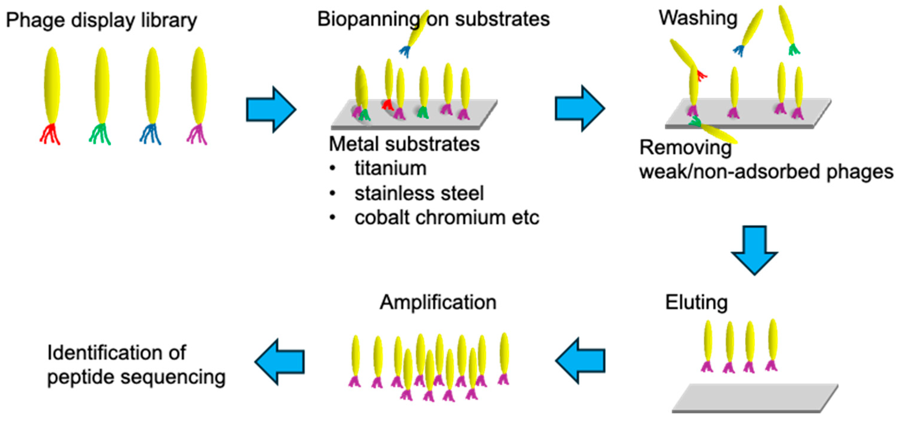
The base of SBPs lies in phage display, a technique first proposed by George P. Smith in 1985 [25,26]. This molecular evolution engineering method is a powerful molecular tool for selecting peptides with specific binding properties from a vast number of variants (libraries), as illustrated in Figure 2.
The selection process of SBPs using phage display technology generally consists of several steps. First, a phage library is exposed to the target material. Then, the bound phages are recovered and amplified over three to five rounds. With each round, the substrate is thoroughly washed to remove unbound or weakly bound phages, ensuring that only phages with high binding affinity are selected. This iterative selection process, known as biopanning, enhances the specificity and strength of the binding interactions [27,28]. Analyzing the sequences of the obtained SBPs and identifying the binding motifs provide valuable insights for applying these peptides to new materials and adding additional functionalities. Two types of bacteriophages are commonly used: M13 and T7. The M13 bacteriophage system was developed first due to its well-understood characteristics and ease of manipulation to express various peptides and proteins [29]. However, M13 is a nonlytic bacteriophage, and its recoverable peptides depend on the host’s secretion pathway, which poses limitations [30]. To address the limitations, phage display systems using T7, a lytic bacteriophage, have been developed. The T7 system can overcome the secretion pathway limitations, allowing for the display of a broader range of peptides [31].
Biomineralization is the process by which organisms form mineral structures, and understanding this process has been crucial for developing novel biomaterials. These early studies provided the foundation for using SBPs to control the deposition of minerals on materials surface, a key step in creating bioactive coatings. On the other hand, recent studies have demonstrated that chimeric molecules, which combine SBPs with functional molecules, are effective for biofunctionalizing materials [32]. This approach has generated significant interest in the surface modification of metal biomaterials. Although most identified peptides are for pure titanium, SBPs with specific high affinities for other metal materials used in medical applications, such as titanium alloys, stainless steel, and cobalt–chromium alloys, are also being discovered. The diversity of SBPs enables the customization of surface properties to meet specific clinical needs.

2.2. Computational Approaches for SBP Design and Optimization

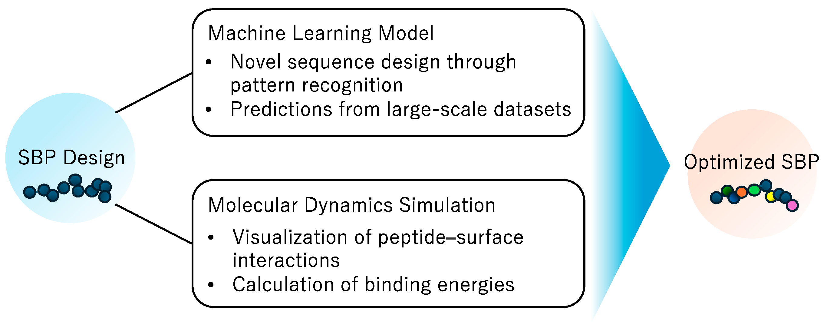
Combining phage display technology with computational approaches, such as computer-aided molecular design and machine learning, is enabling more rational and efficient design and development of SBPs [33,34]. These computational methods can predict peptide conformations and binding affinities, which reduces the time and resources needed for experimental validation. The main computational methods employed in this field include machine learning and molecular dynamics simulations.
Machine learning (ML) algorithms have demonstrated remarkable potential to predict and design novel SBPs based on existing sequence data. ML models can identify complex patterns in large datasets and predict secondary structures for enhanced binding properties [35]. For example, Janairo developed a binary ML classifier for gold-binding peptides [36]. Their support vector machine classifier, trained on 1720 peptide examples, uses Kidera factors to categorize peptides based on their binding ability. This study not only demonstrated satisfactory performance in predicting gold-binding peptides but also identified key variables, such as peptide hydrophobicity, that significantly impact binding ability.
Molecular dynamics (MD) simulations have been crucial in understanding the interactions between SBPs and material surfaces at the atomic level. For instance, Stevens et al. utilized molecular dynamics simulations alongside phage display to optimize ice-binding peptides, significantly enhancing their binding affinity and specificity [37]. Similarly, Chen et al. employed a combination of phage display and theoretical calculations to design selective covalent inhibitors, achieving high specificity and potency [38]. These approaches, while demonstrated on non-metal targets, provide a strong foundation for similar applications in the design of metal-binding peptides. Moreover, Qi and Pfaendtner developed a high-throughput computational screening method for SBPs using molecular dynamics simulations [33]. Their approach allows for the rapid estimation of binding free energy of peptide sequences to solid surfaces by leveraging the thermodynamically stable structure ensemble of each residue. This model significantly reduces the computational cost and can serve as an efficient tool for large-scale screening of SBP candidates.
This multidisciplinary approach combines the strengths of experimental and computational techniques, resulting in more effective and customized peptide solutions [39]. This combination allows the development of higher-performing SBPs. Thus, phage display technology plays a crucial role in the high-biocompatibility surface modification of materials. Its ongoing development and innovation are highly anticipated [40]. Continuous effort in phage display technology and computational sciences will improve the specificity and functionality of SBPs in biomedical applications, leading to more effective biomaterials (Figure 3).

3. Surface Modification of Metal Biomaterials Using SBPs

In the surface treatment of metallic biomaterials, SBPs offer several advantages compared to conventional methods. Specifically, (1) they have high biocompatibility, (2) they enable specific binding, and (3) modification is possible under mild conditions [41]. The general procedure for SBP-based surface modification typically involves synthesizing the selected SBP, often in combination with functional molecules to create a chimeric structure. This chimeric SBP is then applied to the metal surface under appropriate conditions (e.g., pH, temperature), ensuring that the surface is functionalized with the desired bioactive properties. Below, we provide some examples of SBP application in detail for major metallic biomaterials. Several other titanium-binding peptides have also been identified (Table 1). Peptide binding constants are typically measured using Surface Plasmon Resonance (SPR). However, due to the technical difficulties involved in coating metals onto SPR chips, binding constant data for specific metals are often not reported. As a result, binding constant data are not included in Table 1, but this remains an important issue to be addressed in future research.

3.1. Titanium and Titanium Alloys

Titanium and titanium alloys are widely used in orthopedic and dental applications due to their excellent mechanical properties, biocompatibility, and corrosion resistance. They are particularly common in artificial joints, bone fixation devices, and dental implants, making them central among metallic biomaterials [68]. The light weight and high strength of titanium and its alloys also contribute to their widespread use in medical devices, where mechanical durability is crucial. The first peptide reported to bind specifically to titanium surfaces using phage display technology was the Ti-binding peptide (TBP-1) [42]. This binding requires six residues, RKLPDA, known as minTBP-1.
While titanium and its alloys are known for their excellent biocompatibility, the application of SBPs can further enhance their surface properties and introduce additional functionalities. Despite titanium’s inherent biocompatibility, challenges such as insufficient osseointegration in some patients, the potential for bacterial colonization, and limited ability to actively promote tissue regeneration still exist [69]. SBPs offer a versatile approach to address these challenges by allowing precise control over surface chemistry and biological interactions. For instance, SBPs can be designed to promote specific cell adhesion [43] or provide antimicrobial properties [51], thus improving the overall performance of titanium implants beyond their intrinsic biocompatibility.
The effectiveness of SBPs in enhancing titanium’s biological performance has been quantitatively demonstrated in several studies. For example, Gabriel et al. reported significant improvement in endothelial cell adhesion on titanium surfaces modified with a titanium-binding peptide containing Arg-Glu-Asp-Val (REDV) [43]. Their analysis showed that unmodified titanium surface induced endothelial cell coverage of only 0.96 ± 0.38% after one day and 2.6 ± 1.6% after four days. In contrast, titanium surfaces modified with the REDV-containing peptide demonstrated significantly improved cell coverage of 3.1 ± 0.34% after one day and 6.5 ± 0.9% after four days. This represents approximately a three-fold increase in cell coverage, clearly illustrating the capacity of SBPs to enhance the biological performance of titanium beyond its inherent biocompatibility.
Titanium-binding peptides have been used to functionalize titanium surfaces by creating chimeric molecules with silk fibroin [44], insulin-like growth factor 1 (IGF-1) [45], bone morphogenetic protein 2 (BMP-2) [46], and fibronectin fragment peptides such as Arg-Gly-Asp (RGD) [56]. Biofunctional short peptides such as W9, which is derived from a TNF-a binding site on the TNF type 1 receptor, or RP1P, which is derived from the VEGF-binding domain of prominin-1, have been linked with titanium-binding peptides and coated on titanium surfaces to promote osseointegration [47]. Recent reports have focused on imparting antimicrobial properties to titanium through chimeric peptides with antimicrobial peptides [49,50,51,52,53,54]. This functionalization could reduce infection rates associated with implants, which is a significant clinical challenge.
Several peptides have also been reported to bind to titanium alloys. For instance, peptides binding to the Ti-6Al-4V alloy, which is widely used in medical fields, have shown improvements in cell adhesion [59] and reduced bacterial adhesion [60]. Additionally, peptides binding to NiTi alloys used in stents have been identified [61]. These developments extend the applications of SBPs beyond pure titanium to medically relevant alloys.
Despite the promising results of SBPs in enhancing titanium surfaces, several titanium-specific challenges remain. One such challenge is the optimizing of SBP binding to different titanium crystal phases (α, β, α + β) present in various titanium alloys. Recently, β-type titanium has garnered attention as the next generation of metallic biomaterials due to its low Young’s modulus. Consequently, the development of β-type titanium-binding peptides has become increasingly important. Additionally, further investigation into the potential influence of titanium’s surface oxide layer on SBP binding and functionality is necessary. These considerations underscore the need for continued research into titanium-specific SBP design and application methods. Such advancements could lead to more effective and versatile surface medications for titanium-based medical devices.

3.2. Stainless Steel

Stainless steel is also widely used in various medical devices due to its excellent mechanical properties and corrosion resistance. It is particularly common in orthopedic applications. The high strength and durability of stainless steel make it suitable for implants that must withstand significant mechanical stress.
In pediatric intramedullary nails, stainless steel has shown similar healing effects to titanium alloys [51]. Stainless steel costs only one-third of what titanium alloys cost [70,71,72]. In cases where it provides equivalent healing effects, stainless steel can be considered superior to titanium alloys due to its lower cost and ease of manufacturing. However, some form of surface modification is necessary to improve its biocompatibility [73]. This is especially critical as stainless steel can sometimes provoke adverse biological responses if not properly treated.
While several peptides binding to SUS316 have been reported [64], there are few examples of using peptides specifically designed for SUS316L, which is commonly used in medical applications, to impart biological functionality. This scarcity may be due to the complexity of stainless steel’s surface chemical composition.
Mikami-Sakaguchi et al. identified SUS316L-binding peptides (SBP) using phage display technology [63]. These peptides stably bind to the SUS316L surface at pH 4.0–9.0, potentially accommodating pH changes expected in conditions like infusion [74]. They demonstrated that biotinylating the peptide and attaching streptavidin-modified anti-ICAM antibodies created a chimeric antibody that facilitated stable adhesion of HUVECs on SUS316L. This represents the first instance of SBP-based SUS316L stents, as they described.
Despite these advancements, several challenges specific to stainless steel SBPs remain. The complex surface chemistry of stainless steel, including various alloying elements and their oxides, presents a unique challenge for SBP design and binding optimization. Furthermore, the potential for metal ion release from stainless steel implants [75] necessitates the development of SBPs that can not only enhance biocompatibility but also potentially reduce ion release. Further research directions may include the development of multifunctional SBPs that can simultaneously improve biocompatibility, reduce bacterial adhesion, and minimize metal ion release. Additionally, investigating the long-term stability of SBP coatings on stainless steel under physiological conditions and their performance in dynamic mechanical environments is crucial for advancing their clinical application.

3.3. Cobalt–Chromium Alloys

Cobalt–chromium (Co-Cr) alloys are widely used in orthopedic applications due to their high wear resistance and excellent corrosion resistance in biological environments [76]. They are particularly suitable for use in high-load areas, primarily in artificial joints and bone fixation devices [77,78]. Furthermore, Co-Cr alloys are used for stents [79].
While the biocompatibility of these alloys has been studied, bone growth on Co-Cr alloys can be compromised, leading to decreased bone quality. To solve this problem, it is crucial to ensure the integration of Co-Cr implants with surrounding bone tissue, promoting long-term stability and functionality. Therefore, covalently binding methods for peptides derived from BMP7 to the Co-Cr surface are being explored [80,81,82]. These methods aim to enhance the osteogenic potential of Co-Cr implants by facilitating bone growth and integration.
However, there are very few reports on Co-Cr-binding peptides. Identification of peptides that can specifically bind to Co-Cr surface and enhance their biological performance is essential for improving the effectiveness of biomedical implants. Migita et al. identified a peptide specific to cobalt–chromium–molybdenum (Co-Cr-Mo) alloy, which is widely used in dental materials and artificial joints, using a phage display system [64]. They created a chimeric Co-Cr-Mo-binding peptide (CBP) with RGD peptides, which enhanced endothelial cell adhesion on CCM alloy surfaces. This innovation could potentially improve the overall success rate of Co-Cr-based implants. They also demonstrated that this peptide remained stable adsorbed in cell culture conditions for at least two weeks [65]. This binding stability is crucial for maintaining the long-term effectiveness of the surface modification. This chimeric peptide also promoted osteoblast differentiation, showing significant potential for practical surface design.
Future research directions may include the design of multifunctional SBPs that can simultaneously enhance osteoblast differentiation, improve endothelialization for cardiovascular applications, and reduce metal ion release from the alloy. Furthermore, investigating the performance of SBP-modified Co-Cr surfaces under dynamic mechanical loading and wear conditions is crucial for advancing their application in high-stress environments such as artificial joints. The development of Co-Cr-specific SBPs also opens possibilities for creating smart implant surfaces that can respond to local biological cues, potentially leading to more integrated and functional implants.

3.4. Other Metallic Biomaterials

While titanium, stainless steel, and Co-Cr alloys are the most used metallic biomaterials, research on SBPs for other metals is also progressing. For example, gold (Au) alloys are frequently used in dental applications due to their excellent biocompatibility [83,84]. Au-binding peptides have been identified for their potential to improve the surface properties of dental implants and restorations [32,66]. The ability of these peptides to bind with Au surfaces can improve the durability and effectiveness of dental prostheses. Similarly, platinum (Pt) alloys are used for electrode materials in medical and clinical applications, due to their remarkable thermal and electrical conductivity, ductility, and corrosion resistance [85]. Research into SBPs specific to Pt alloys is underway to further their integration with biological tissues [67,86]. The chemical stability and corrosion resistance of Pt make it suitable for a range of dental and medical applications, including electrodes and catheters. While mercury amalgam has historically been used in dental fillings due to its durability and ease of application, its use has decreased due to concerns about mercury exposure [87]. As a result, research into SBPs for mercury amalgam is not as prominent as for other metallic biomaterials. Alternative materials that mitigate the health risks associated with mercury exposure are being sought, reducing the emphasis on SBPs for this application.
Thus, the development of SBPs for various metallic biomaterials enables the creation of advanced medical applications. These efforts aim to improve the performance and safety of medical devices, ultimately benefiting patient outcomes.

3.5. Advantages and Challenges of Surface Modification Using SBPs

Surface modification of metallic biomaterials using SBPs has several significant advantages compared to traditional methods. Firstly, SBPs can specifically bind to the target metal surface, enabling precise surface modification. Additionally, modification can be performed under mild conditions such as room temperature and neutral pH, minimizing adverse effects on the material and the surrounding environment [88]. Furthermore, peptides themselves possess high biocompatibility, reducing the likelihood of triggering immune reactions and providing superior safety.
However, there are also several challenges associated with surface modification using SBPs. Some SBPs exhibit insufficient long-term stability, which necessitates the development of modification methods to ensure their durability in vivo. Additionally, it is difficult to elucidate the detailed mechanisms of binding between SBPs and metals, which complicates the design of new SBPs and the improvement of existing ones [89].
Further research is needed to address these challenges and fully realize the potential of SBPs in medical applications. Addressing these challenges is expected to advance SBP-based surface modification technology and expand its application. Specifically, improving long-term stability, establishing efficient production methods, and elucidating binding mechanisms are crucial steps toward the practical implementation of this technology.
Collaborative efforts between material scientists, biologists, and medical professionals will be essential in overcoming these obstacles and advancing the field. The integration of computational modeling and experimental approaches will also play a vital role in optimizing SBP design and application.

4. Prospects

This section explores the future directions and potential advancements in SBP technology for metallic biomaterials. We discuss three key areas of development: the identification of new SBP sequences, strategies for enhancing peptide binding stability, and the creation of multifunctional surfaces through integration with other technologies. These areas represent interconnected challenges and opportunities that will shape the future of SBP-based surface modification.

4.1. Identification of Peptide Sequences Binding to New Metallic Biomaterials

Co-Cr alloys, which were previously difficult to handle in the fabrication process, can now be easily manufactured into devices using 3D printing technology [90,91]. This technological advancement enables the production of complex and customized medical devices, expanding the application potential of Co-Cr alloys in biomaterials. This advancement will increase the application of Co-Cr alloys in biomaterials, making the study of peptide binding to these alloys essential.
Tantalum is actively studied as an implant material due to its high corrosion resistance, potentially replacing titanium [92,93,94]. The ability of tantalum to promote osseointegration and its excellent biocompatibility make it a promising candidate for various implants. Identifying peptides that bind to tantalum could be highly beneficial. Additionally, zirconium alloys are investigated for medical applications due to their potential to reduce MRI artifacts [95,96,97,98]. However, zirconium is also a bioinert material, and surface modification is required to improve its biocompatibility, such as creating roughened surface [99]. Peptides binding yttria-stabilized zirconia (YSZ) have already been identified [100,101] and may be applicable to zirconium alloys as well.
It is also necessary to verify whether existing SBPs can adapt to changes in surface roughness and surface chemical composition resulting from surface treatments. Currently, biocompatibility is being adjusted by roughening the titanium surface [102]. Surface roughening techniques enhance cell adhesion and proliferation, crucial for successful implant integration. It must be examined whether existing titanium-binding peptides can be applied to titanium with such nanostructures. Furthermore, research is ongoing to impart antimicrobial properties to titanium by supporting silver or copper on its surface [103]. These antimicrobial coatings aim to prevent postsurgical infections, a common complication in implant surgeries. It is also crucial to verify whether these peptides can be applied to titanium with altered surface chemical compositions.
One of the metal-based biomaterials that has attracted significant attention in recent years is magnesium alloys. Magnesium is a metal that can be degraded and absorbed within the body, making it suitable for temporary bone fixation devices that require only short-term functionality [104,105,106]. The biodegradability of magnesium eliminates the need for a second surgery to remove implants, reducing patient risk and healthcare costs. However, excessive degradation of magnesium can produce hydrogen gas, posing a risk of damaging surrounding tissues [107,108,109]. By applying surface modification with SBPs, it is expected that the degradation rate of magnesium alloys can be controlled while promoting the adhesion and proliferation of osteoblasts, thereby significantly improving the biocompatibility of magnesium alloys. This approach aims to balance the degradation process, supporting bone healing while minimizing adverse effects.

4.2. Strategies for Enhanced Stability of Peptide Binding

Retaining SBPs on material surfaces for extended periods is crucial for maintaining the biofunctionality of the materials. To maintain biofunctionality, the search for peptides that can bind stably over the long term is essential. Stable peptide binding can prevent the loss of bioactive molecules and ensure consistent therapeutic effects.
One promising approach involves multivalent peptides, which have been reported to significantly improve binding stability [40]. For example, using branched lysine peptides to create a tetravalent titanium-binding peptide demonstrated stable functionality on titanium surface for over two weeks [110]. This enhanced stability is attributed to the increased number of contact points between the peptide and the metal surface, which reduces the likelihood of peptide desorption. This combination of organic synthetic chemistry and peptide engineering might be a highly effective strategy. Future research should explore the potential of other branched or multivalent peptides structures to further improve the stability and functionality of SBPs on various metallic surfaces.
One of the challenges in studying these material-binding peptides is accurately determining their binding constants using techniques like Surface Plasmon Resonance (SPR). Typically, SPR is used to measure binding constants by immobilizing proteins on a gold chip and monitoring the adsorption of peptides. For metal-binding peptides, this process requires coating the SPR chip with metal, which is technically challenging. Sputtering can be used for some metals, but for materials that cannot be sputtered or where the coating interferes with plasmon resonance, measuring binding constants becomes difficult. As a result, binding constant data for certain material-binding peptides are often not reported, and developing more reliable methods to obtain these data is an important area for future research.

4.3. Multifunctional Surface through Integration with Other Surface Modification Technologies

Surface modification technology using SBPs can be further enhanced by combining it with other surface treatment technologies. This integration approach allows for the creation of multifunctional biointerfaces with improved performance and versatility. One promising combination is the use of SBPs with bioactive ceramic coatings. Specifically, hydroxyapatite coatings for modifying titanium surface can improve osteoconductivity on the surface [111,112,113]. By combining such bioactive coatings with SBPs, we can potentially develop implants that not only integrate better with surrounding bone tissue but also possess additional functionalities conferred by the peptides.
The concept of multifunctional surface can be further extended by incorporating bioactive proteins alongside SBPs. For example, Xu et al. developed a fusion peptide consisting of an osteogenic functional sequence (P-15) and a bone-specific binding sequence (Asp-6), encapsulated in chitosan-modified poly (lactic acid-glycolic acid) (PLGA) microspheres [114]. This approach addressed multiple challenges in bone regeneration. The fusion peptide delivered specific osteogenic stimulation, while the chitosan-modified PLGA offered antimicrobial properties and controlled release. Their system demonstrated a multistage effect, initially promoting infection control and soft tissue healing, followed by enhanced bone regeneration.
Another exciting topic is the integrating of SBPs with a drug delivery system (DDS). This combination enables the creation of biomaterials with both tailored surface properties and controlled release of therapeutic agents. Li et al. demonstrated this concept by creating a chimera of titanium-binding peptides and exosome-binding peptides, enabling exosome-derived drug delivery on titanium implants [48]. This approach holds promise for long-term effective treatments in implant therapy, potentially reducing the risk of implant-associated infections and promoting faster healing.
Furthermore, the concept of multifunctional surfaces can be extended by combining multiple bioactive peptides with SBPs on implant surfaces. For instance, Bell et al. demonstrated that while RGD peptides enhance cell attachment, they may inhibit osteoblast differentiation [115]. Conversely, they found that KSSR, initially used as control, unexpectedly increased osteoblast differentiation. These findings highlight the complexity of cell–surface interactions and the importance of carefully selecting peptide combinations. Building on this concept, recent studies have explored the synergistic effects of multiple peptides. For example, Chausse et al. investigated a dual cell-adhesive platform combining RGDS and YIGSR sequences on bioresorbable stents [116]. This combination aimed to improve endothelialization while preventing platelet activation, demonstrating the potential of multi-peptide functionalization in creating surfaces with multiple desired properties.
These studies underscore the need for a nuanced approach in designing multifunctional surfaces. By carefully selecting various bioactive molecules and combining them with SBPs, we can potentially create implant surfaces that more closely mimic the complex extracellular matrix environment. This strategy could lead to the development of next-generation implants that not only integrate well with surrounding tissues but also actively promote desired cellular responses for enhanced healing and tissue regeneration.

5. Conclusions

This review has provided a comprehensive overview of the surface modification technology for metallic biomaterials using SBPs selected through phage display technology, detailing its principles, recent research trends, and prospects.
SBPs are short amino acid sequences that exhibit high affinity and selectivity for specific metal surfaces. They artificially evolved and selected from randomly designed peptide libraries using phage display technology. It has been demonstrated that modifying implant surfaces with SBPs significantly enhances the biocompatibility of metals, promoting remarkable cell adhesion and proliferation.
Numerous SBPs have already been identified for various metal materials, including titanium, stainless steel, and Co-Cr-Mo alloys, and their surface modification effects have been experimentally confirmed. Moreover, their application to actual medical devices, such as stents and artificial joints, is progressing, with significant improvements in biocompatibility, such as enhanced thromboresistance and osteoconductivity, observed. The integration of SBP technology with other advanced materials and technologies, such as combining SBPs with drug delivery systems, could provide localized and controlled release of therapeutic agents, enhancing treatment efficacy and minimizing side effects.
To achieve further performance enhancements and rational molecular design of SBPs, integration with theoretical approaches like computational science is explored. Computational models can predict peptide–material interactions and guide the design of peptides with optimized binding properties.
SBPs based on phage display technology are poised to play a central role pioneering the next generation of metallic biomaterials, combining excellent biocompatibility with high functionality. While the future development of this technology is highly anticipated, it is also essential to steadily address the various challenges it faces. These include ensuring long-term stability of peptide binding and understanding the detailed mechanisms of peptide–material interactions.

Funding

This research received no external funding.

Conflicts of Interest

The author declares no conflict of interest.

References

  1. Sarikaya, M.; Tamerler, C.; Schwartz, D.T.; Baneyx, F. Materials Assembly and Formation Using Engineered Polypeptides. Annu. Rev. Mater. Res. 2004, 34, 373–408. [Google Scholar] [CrossRef]
  2. Ruan, Y.; Sohail, M.; Zhao, J.; Hu, F.; Li, Y.; Wang, P.; Zhang, L. Applications of Material-Binding Peptides: A Review. ACS Biomater. Sci. Eng. 2022, 8, 4738–4750. [Google Scholar] [CrossRef]
  3. Yucesoy, D.T.; Khatayevich, D.; Tamerler, C.; Sarikaya, M. Rationally Designed Chimeric Solid-Binding Peptides for Tailoring Solid Interfaces. Med. Devices Sens. 2020, 3, e10065. [Google Scholar] [CrossRef]
  4. Gustafsson, E.; Thomée, S.; Grimby-Ekman, A.; Hagberg, M. Texting on Mobile Phones and Musculoskeletal Disorders in Young Adults: A Five-Year Cohort Study. Appl. Ergon. 2017, 58, 208–214. [Google Scholar] [CrossRef] [PubMed]
  5. Baradaran Mahdavi, S.; Riahi, R.; Vahdatpour, B.; Kelishadi, R. Association between Sedentary Behavior and Low Back Pain; A Systematic Review and Meta-Analysis. Health Promot. Perspect. 2021, 11, 393–410. [Google Scholar] [CrossRef]
  6. Alsiwed, K.T.; Alsarwani, R.M.; Alshaikh, S.A.; Howaidi, R.A.; Aljahdali, A.J.; Bassi, M.M. The Prevalence of Text Neck Syndrome and Its Association with Smartphone Use among Medical Students in Jeddah, Saudi Arabia. J. Musculoskelet. Surg. Res. 2021, 5, 266–272. [Google Scholar] [CrossRef]
  7. Briggs, A.M.; Woolf, A.D.; Dreinhöfer, K.; Homb, N.; Hoy, D.G.; Kopansky-Giles, D.; Åkesson, K.; March, L. Reducing the Global Burden of Musculoskeletal Conditions. Bull. World Health Organ. 2018, 96, 366–368. [Google Scholar] [CrossRef]
  8. Briggs, A.M.; Cross, M.J.; Hoy, D.G.; Sànchez-Riera, L.; Blyth, F.M.; Woolf, A.D.; March, L. Musculoskeletal Health Conditions Represent a Global Threat to Healthy Aging: A Report for the 2015 World Health Organization World Report on Ageing and Health. Gerontologist 2016, 56, S243–S255. [Google Scholar] [CrossRef]
  9. Marin, E.; Boschetto, F.; Pezzotti, G. Biomaterials and Biocompatibility: An Historical Overview. J. Biomed. Mater. Res. 2020, 108, 1617–1633. [Google Scholar] [CrossRef]
  10. Bandyopadhyay, A.; Mitra, I.; Goodman, S.B.; Kumar, M.; Bose, S. Improving Biocompatibility for next Generation of Metallic Implants. Prog. Mater. Sci. 2023, 133, 101053. [Google Scholar] [CrossRef]
  11. Hanawa, T. Metals and Medicine. Mater. Trans. 2021, 62, 139–148. [Google Scholar] [CrossRef]
  12. Pisarek, M.; Roguska, A.; Andrzejczuk, M.; Marcon, L.; Szunerits, S.; Lewandowska, M.; Janik-Czachor, M. Effect of Two-Step Functionalization of Ti by Chemical Processes on Protein Adsorption. Appl. Surf. Sci. 2011, 257, 8196–8204. [Google Scholar] [CrossRef]
  13. Minagar, S.; Wang, J.; Berndt, C.C.; Ivanova, E.P.; Wen, C. Cell Response of Anodized Nanotubes on Titanium and Titanium Alloys. J. Biomed. Mater. Res. 2013, 101A, 2726–2739. [Google Scholar] [CrossRef]
  14. Berger, M.B.; Cohen, D.J.; Bosh, K.B.; Kapitanov, M.; Slosar, P.J.; Levit, M.M.; Gallagher, M.; Rawlinson, J.J.; Schwartz, Z.; Boyan, B.D. Bone Marrow Stromal Cells Generate an Osteoinductive Microenvironment When Cultured on Titanium–Aluminum–Vanadium Substrates with Biomimetic Multiscale Surface Roughness. Biomed. Mater. 2023, 18, 035001. [Google Scholar] [CrossRef]
  15. Rautray, T.R.; Narayanan, R.; Kwon, T.; Kim, K. Surface Modification of Titanium and Titanium Alloys by Ion Implantation. J. Biomed. Mater. Res. 2010, 93B, 581–591. [Google Scholar] [CrossRef]
  16. Won, S.; Huh, Y.-H.; Cho, L.-R.; Lee, H.-S.; Byon, E.-S.; Park, C.-J. Cellular Response of Human Bone Marrow Derived Mesenchymal Stem Cells to Titanium Surfaces Implanted with Calcium and Magnesium Ions. Tissue Eng. Regen. Med. 2017, 14, 123–131. [Google Scholar] [CrossRef] [PubMed]
  17. Souza, J.C.M.; Sordi, M.B.; Kanazawa, M.; Ravindran, S.; Henriques, B.; Silva, F.S.; Aparicio, C.; Cooper, L.F. Nano-Scale Modification of Titanium Implant Surfaces to Enhance Osseointegration. Acta Biomater. 2019, 94, 112–131. [Google Scholar] [CrossRef]
  18. Migita, S.; Araki, K. Effect of Nanometer Scale Surface Roughness of Titanium for Osteoblast Function. AIMS Bioeng. 2017, 4, 162–170. [Google Scholar] [CrossRef]
  19. Migita, S.; Wakabayashi, K. Analysis of Disordered Abrasive Scratches on Titanium Surfaces and Their Impact on Nuclear Translocation of Yes-Associated Protein. Sci. Rep. 2022, 12, 21705. [Google Scholar] [CrossRef]
  20. Migita, S.; Okuyama, S.; Araki, K. Sub-Micrometer Scale Surface Roughness of Titanium Reduces Fibroblasts Function. J. Appl. Biomater. Funct. Mater. 2016, 14, 65–69. [Google Scholar] [CrossRef]
  21. Migita, S. Surface Roughness and Its Role in Mediating Cell Adhesion on Cobalt-Chromium-Molybdenum Alloys. Biosurf. Biotribol. 2023, 9, 161–168. [Google Scholar] [CrossRef]
  22. Morra, M. Biochemical Modification of Titanium Surfaces: Peptides and ECM Proteins. Eur. Cell Mater. 2006, 12, 1–15. [Google Scholar] [CrossRef] [PubMed]
  23. Xin, H.; Chen, J.; Li, T.; Hu, G.; Fang, Z.; Zhou, H.; Guo, K.; Wang, L.; Wang, Y. One-Step Preparation of the Engineered Titanium Implant by Rationally Designed Linear Fusion Peptides with Spacer-Dependent Antimicrobial, Anti-Inflammatory and Osteogenic Activities. Chem. Eng. J. 2021, 424, 130380. [Google Scholar] [CrossRef]
  24. Civantos, A.; Martínez-Campos, E.; Ramos, V.; Elvira, C.; Gallardo, A.; Abarrategi, A. Titanium Coatings and Surface Modifications: Toward Clinically Useful Bioactive Implants. ACS Biomater. Sci. Eng. 2017, 3, 1245–1261. [Google Scholar] [CrossRef]
  25. Smith, G.P. Filamentous Fusion Phage: Novel Expression Vectors That Display Cloned Antigens on the Virion Surface. Science 1985, 228, 1315–1317. [Google Scholar] [CrossRef]
  26. Smith, G.P.; Petrenko, V.A. Phage Display. Chem. Rev. 1997, 97, 391–410. [Google Scholar] [CrossRef]
  27. Davidson, T.A.; McGoldrick, S.J.; Kohn, D.H. Phage Display to Augment Biomaterial Function. Int. J. Mol. Sci. 2020, 21, 5994. [Google Scholar] [CrossRef]
  28. Zambrano-Mila, M.S.; Blacio, K.E.S.; Vispo, N.S. Peptide Phage Display: Molecular Principles and Biomedical Applications. Ther. Innov. Regul. Sci. 2020, 54, 308–317. [Google Scholar] [CrossRef]
  29. Sidhu, S.S. Phage Display in Pharmaceutical Biotechnology. Curr. Opin. Biotechnol. 2000, 11, 610–616. [Google Scholar] [CrossRef]
  30. Kehoe, J.W.; Kay, B.K. Filamentous Phage Display in the New Millennium. Chem. Rev. 2005, 105, 4056–4072. [Google Scholar] [CrossRef]
  31. Krumpe, L.R.H.; Atkinson, A.J.; Smythers, G.W.; Kandel, A.; Schumacher, K.M.; McMahon, J.B.; Makowski, L.; Mori, T. T7 Lytic Phage-Displayed Peptide Libraries Exhibit Less Sequence Bias than M13 Filamentous Phage-Displayed Peptide Libraries. Proteomics 2006, 6, 4210–4222. [Google Scholar] [CrossRef] [PubMed]
  32. Khatayevich, D.; Gungormus, M.; Yazici, H.; So, C.; Cetinel, S.; Ma, H.; Jen, A.; Tamerler, C.; Sarikaya, M. Biofunctionalization of Materials for Implants Using Engineered Peptides. Acta Biomater. 2010, 6, 4634–4641. [Google Scholar] [CrossRef] [PubMed]
  33. Qi, X.; Pfaendtner, J. High-Throughput Computational Screening of Solid-Binding Peptides. J. Chem. Theory Comput. 2024, 20, 2959–2968. [Google Scholar] [CrossRef] [PubMed]
  34. Bergman, M.T.; Xiao, X.; Hall, C.K. In Silico Design and Analysis of Plastic-Binding Peptides. J. Phys. Chem. B 2023, 127, 8370–8381. [Google Scholar] [CrossRef] [PubMed]
  35. Zhang, H.; Saravanan, K.M.; Wei, Y.; Jiao, Y.; Yang, Y.; Pan, Y.; Wu, X.; Zhang, J.Z.H. Deep Learning-Based Bioactive Therapeutic Peptide Generation and Screening. J. Chem. Inf. Model. 2023, 63, 835–845. [Google Scholar] [CrossRef]
  36. Janairo, J.I.B. A Machine Learning Classification Model for Gold-Binding Peptides. ACS Omega 2022, 7, 14069–14073. [Google Scholar] [CrossRef]
  37. Stevens, C.A.; Bachtiger, F.; Kong, X.-D.; Abriata, L.A.; Sosso, G.C.; Gibson, M.I.; Klok, H.-A. A Minimalistic Cyclic Ice-Binding Peptide from Phage Display. Nat. Commun. 2021, 12, 2675. [Google Scholar] [CrossRef]
  38. Chen, S.; Lovell, S.; Lee, S.; Fellner, M.; Mace, P.D.; Bogyo, M. Identification of Highly Selective Covalent Inhibitors by Phage Display. Nat. Biotechnol. 2021, 39, 490–498. [Google Scholar] [CrossRef]
  39. Li, B.; Chen, H.; Huang, J.; He, B. CD47Binder: Identify CD47 Binding Peptides by Combining Next-Generation Phage Display Data and Multiple Peptide Descriptors. Interdiscip. Sci. Comput. Life Sci. 2023, 15, 578–589. [Google Scholar] [CrossRef]
  40. Alvisi, N.; De Vries, R. Biomedical Applications of Solid-Binding Peptides and Proteins. Mater. Today Bio 2023, 19, 100580. [Google Scholar] [CrossRef]
  41. Care, A.; Bergquist, P.L.; Sunna, A. Solid-Binding Peptides: Smart Tools for Nanobiotechnology. Trend Biotechnol. 2015, 33, 259–268. [Google Scholar] [CrossRef]
  42. Sano, K.-I.; Shiba, K. A Hexapeptide Motif That Electrostatically Binds to the Surface of Titanium. J. Am. Chem. Soc. 2003, 125, 14234–14235. [Google Scholar] [CrossRef] [PubMed]
  43. Gabriel, M.; Chopra, A.; Schmidt, F. Combined Endothelialization Promoting and Surface Binding Chimeric Conjugate with Low Thrombogenicity. Bioconjug. Chem. 2021, 32, 1602–1605. [Google Scholar] [CrossRef]
  44. Watanabe, M.; Bhawal, U.K.; Takemoto, S.; Nishiyama, N.; Nakahara, Y.; Tatematsu, K.; Sezutsu, H.; Kuwabara, N.; Minamisawa, T.; Shiba, K.; et al. Bio-Functionalized Titanium Surfaces with Modified Silk Fibroin Carrying Titanium Binding Motif to Enhance the Ossific Differentiation of MC3T3-E1. Biotechnol. Bioeng. 2021, 118, 2585–2596. [Google Scholar] [CrossRef] [PubMed]
  45. Wang, J.-J.; Xue, Q.; Wang, Y.-J.; Zhang, M.; Chen, Y.-J.; Zhang, Q. Engineered Chimeric Peptides with IGF-1 and Titanium-Binding Functions to Enhance Osteogenic Differentiation In Vitro under T2DM Condition. Materials 2022, 15, 3134. [Google Scholar] [CrossRef] [PubMed]
  46. Yuasa, K.; Kokubu, E.; Kokubun, K.; Matsuzaka, K.; Shiba, K.; Kashiwagi, K.; Inoue, T. An Artificial Fusion Protein between Bone Morphogenetic Protein 2 and Titanium-Binding Peptide Is Functional In Vivo. J. Biomed. Mater. Res. 2014, 102, 1180–1186. [Google Scholar] [CrossRef]
  47. Zhao, Z.; Ma, S.; Wu, C.; Li, X.; Ma, X.; Hu, H.; Wu, J.; Wang, Y.; Liu, Z. Chimeric Peptides Quickly Modify the Surface of Personalized 3D Printing Titanium Implants to Promote Osseointegration. ACS Appl. Mater. Interfaces 2021, 13, 33981–33994. [Google Scholar] [CrossRef]
  48. Li, X.; Liu, Z.; Xu, S.; Ma, X.; Zhao, Z.; Hu, H.; Deng, J.; Peng, C.; Wang, Y.; Ma, S. A Drug Delivery System Constructed by a Fusion Peptide Capturing Exosomes Targets to Titanium Implants Accurately Resulting the Enhancement of Osseointegration Peri-Implant. Biomater. Res. 2022, 26, 89. [Google Scholar] [CrossRef]
  49. Geng, H.; Sun, X.; Zhang, X.; Yuan, Y. Efficient Titanium Surface Modified Using Bifunctional Chimeric Peptides to Prevent Biofilm Formation by Multiple Microorganisms. Colloids Surf. B Biointerfaces 2023, 230, 113534. [Google Scholar] [CrossRef]
  50. Drexelius, M.; Arnold, R.; Meinberger, D.; Wilhelm, M.; Mathur, S.; Neundorf, I. Rational Design of Bifunctional Chimeric Peptides That Combine Antimicrobial and Titanium Binding Activity. J. Pept. Sci. 2023, 29, e3481. [Google Scholar] [CrossRef]
  51. Yucesoy, D.T.; Hnilova, M.; Boone, K.; Arnold, P.M.; Snead, M.L.; Tamerler, C. Chimeric Peptides as Implant Functionalization Agents for Titanium Alloy Implants with Antimicrobial Properties. JOM 2015, 67, 754–766. [Google Scholar] [CrossRef]
  52. Wisdom, C.; Chen, C.; Yuca, E.; Zhou, Y.; Tamerler, C.; Snead, M.L. Repeatedly Applied Peptide Film Kills Bacteria on Dental Implants. JOM 2019, 71, 1271–1280. [Google Scholar] [CrossRef]
  53. Yazici, H.; O’Neill, M.B.; Kacar, T.; Wilson, B.R.; Oren, E.E.; Sarikaya, M.; Tamerler, C. Engineered Chimeric Peptides as Antimicrobial Surface Coating Agents toward Infection-Free Implants. ACS Appl. Mater. Interfaces 2016, 8, 5070–5081. [Google Scholar] [CrossRef]
  54. Wisdom, E.C.; Zhou, Y.; Chen, C.; Tamerler, C.; Snead, M.L. Mitigation of Peri-Implantitis by Rational Design of Bifunctional Peptides with Antimicrobial Properties. ACS Biomater. Sci. Eng. 2020, 6, 2682–2695. [Google Scholar] [CrossRef]
  55. Yazici, H.; Fong, H.; Wilson, B.; Oren, E.E.; Amos, F.A.; Zhang, H.; Evans, J.S.; Snead, M.L.; Sarikaya, M.; Tamerler, C. Biological Response on a Titanium Implant-Grade Surface Functionalized with Modular Peptides. Acta Biomater. 2013, 9, 5341–5352. [Google Scholar] [CrossRef]
  56. Vidal, G.; Blanchi, T.; Mieszawska, A.J.; Calabrese, R.; Rossi, C.; Vigneron, P.; Duval, J.-L.; Kaplan, D.L.; Egles, C. Enhanced Cellular Adhesion on Titanium by Silk Functionalized with Titanium Binding and RGD Peptides. Acta Biomater. 2013, 9, 4935–4943. [Google Scholar] [CrossRef]
  57. Dickerson, M.B.; Jones, S.E.; Cai, Y.; Ahmad, G.; Naik, R.R.; Kröger, N.; Sandhage, K.H. Identification and Design of Peptides for the Rapid, High-Yield Formation of Nanoparticulate TiO2 from Aqueous Solutions at Room Temperature. Chem. Mater. 2008, 20, 1578–1584. [Google Scholar] [CrossRef]
  58. Liu, Y.; Mao, J.; Zhou, B.; Wei, W.; Gong, S. Peptide Aptamers against Titanium-Based Implants Identified through Phage Display. J. Mater. Sci Mater. Med. 2010, 21, 1103–1107. [Google Scholar] [CrossRef]
  59. Panayotov, I.V.; Végh, A.G.; Martin, M.; Vladimirov, B.; Larroque, C.; Gergely, C.; Cuisinier, F.J.G.; Estephan, E. Improving Dental Epithelial Junction on Dental Implants with Bioengineered Peptides. Front. Bioeng. Biotechnol. 2023, 11, 1165853. [Google Scholar] [CrossRef]
  60. Khoo, X.; Hamilton, P.; O’Toole, G.A.; Snyder, B.D.; Kenan, D.J.; Grinstaff, M.W. Directed Assembly of PEGylated-Peptide Coatings for Infection-Resistant Titanium Metal. J. Am. Chem. Soc. 2009, 131, 10992–10997. [Google Scholar] [CrossRef]
  61. Kodama, T.; Yoshihara, A.; Goel, I.; Sekino, M.; Kuwahata, A.; Yoshimori, A.; Murayama, Y.; Ishihara, K.; Ekdahl, K.N.; Nilsson, B.; et al. Identification of Metal-Binding Peptides and Their Conjugation onto Nanoparticles of Superparamagnetic Iron Oxides and Liposomes. ACS Appl. Mater. Interfaces 2020, 12, 24623–24634. [Google Scholar] [CrossRef] [PubMed]
  62. Vreuls, C.; Zocchi, G.; Genin, A.; Archambeau, C.; Martial, J.; De Weerdt, C.V. Inorganic-Binding Peptides as Tools for Surface Quality Control. J. Inorg. Biochem. 2010, 104, 1013–1021. [Google Scholar] [CrossRef] [PubMed]
  63. Sakaguchi-Mikami, A.; Fujimoto, K.; Taguchi, T.; Isao, K.; Yamazaki, T. A Novel Biofunctionalizing Peptide for Metallic Alloy. Biotechnol. Lett. 2020, 42, 747–756. [Google Scholar] [CrossRef] [PubMed]
  64. Migita, S.; Sakashita, K.; Saito, Y.; Suyalatu; Yamazaki, T. Co–Cr–Mo Alloy Binding Peptide as Molecular Glue for Constructing Biomedical Surfaces. J. Appl. Biomater. Funct. Mater. 2020, 18, 228080002092473. [Google Scholar] [CrossRef]
  65. Migita, S.; Wakabayashi, K.; Yamazaki, T. Binding Stability of Peptides to Co–Cr–Mo Alloy Affects Proliferation and Differentiation of Osteoblast. Biotechnol. Bioeng. 2022, 119, 1157–1163. [Google Scholar] [CrossRef]
  66. Hnilova, M.; Oren, E.E.; Seker, U.O.S.; Wilson, B.R.; Collino, S.; Evans, J.S.; Tamerler, C.; Sarikaya, M. Effect of Molecular Conformations on the Adsorption Behavior of Gold-Binding Peptides. Langmuir 2008, 24, 12440–12445. [Google Scholar] [CrossRef]
  67. Seker, U.O.S.; Wilson, B.; Dincer, S.; Kim, I.W.; Oren, E.E.; Evans, J.S.; Tamerler, C.; Sarikaya, M. Adsorption Behavior of Linear and Cyclic Genetically Engineered Platinum Binding Peptides. Langmuir 2007, 23, 7895–7900. [Google Scholar] [CrossRef] [PubMed]
  68. Marin, E.; Lanzutti, A. Biomedical Applications of Titanium Alloys: A Comprehensive Review. Materials 2023, 17, 114. [Google Scholar] [CrossRef]
  69. Hanawa, T. Titanium–Tissue Interface Reaction and Its Control With Surface Treatment. Front. Bioeng. Biotechnol. 2019, 7, 170. [Google Scholar] [CrossRef]
  70. Mohamed, A.; Rajeev, A.S. Clinical Outcomes and Complications of Titanium versus Stainless Steel Elastic Nail in Management of Paediatric Femoral Fractures—A Systematic Review. Eur. J. Orthop. Surg. Traumatol. 2017, 27, 157–167. [Google Scholar] [CrossRef]
  71. Goyal, N.; Aggarwal, A.N.; Mishra, P.; Jain, A. Randomized Controlled Trial Comparing Stabilization of Fresh Close Femoral Shaft Fractures in Children with Titanium Elastic Nail System versus Stainless Steel Elastic Nail System. Acta Orthop. Belg. 2014, 80, 69–75. [Google Scholar] [PubMed]
  72. Wall, E.J.; Jain, V.; Vora, V.; Mehlman, C.T.; Crawford, A.H. Complications of Titanium and Stainless Steel Elastic Nail Fixation of Pediatric Femoral Fractures. J. Bone Jt. Surg. 2008, 90, 1305–1313. [Google Scholar] [CrossRef] [PubMed]
  73. Mojarad Shafiee, B.; Torkaman, R.; Mahmoudi, M.; Emadi, R.; Derakhshan, M.; Karamian, E.; Tavangarian, F. Surface Modification of 316L SS Implants by Applying Bioglass/Gelatin/Polycaprolactone Composite Coatings for Biomedical Applications. Coatings 2020, 10, 1220. [Google Scholar] [CrossRef]
  74. Stranz, M.; Kastang, E.S. A Review of pH and Osmolarity. Int. J. Pharm. Compd. 2002, 6, 216–220. [Google Scholar]
  75. Varmaziar, S.; Atapour, M.; Hedberg, Y.S. Corrosion and Metal Release Characterization of Stainless Steel 316L Weld Zones in Whey Protein Solution. NPJ Mater. Degrad. 2022, 6, 19. [Google Scholar] [CrossRef]
  76. Aherwar, A.; Singh, A.K.; Patnaik, A. Cobalt Based Alloy: A Better Choice Biomaterial for Hip Implants. Trends Biomater. Artif. Organs 2016, 30, 50–55. [Google Scholar]
  77. Fu, W.; Liu, S.; Jiao, J.; Xie, Z.; Huang, X.; Lu, Y.; Liu, H.; Hu, S.; Zuo, E.; Kou, N.; et al. Wear Resistance and Biocompatibility of Co-Cr Dental Alloys Fabricated with CAST and SLM Techniques. Materials 2022, 15, 3263. [Google Scholar] [CrossRef] [PubMed]
  78. Vieweg, U.; Keck, J.; Krüger, S.; Arabmotlagh, M.; Rauschmann, M.; Schilling, C. Biomechanical Comparison of Different Rod-to-Rod Connectors to a Conventional Titanium- and Cobalt Chromium Posterior Spinal Fixation System. Brain Spine 2023, 3, 101708. [Google Scholar] [CrossRef]
  79. Song, Z.; Zhao, Y.; Li, Z.; Cao, C.; Liu, G.; Liu, Q.; Zhang, X.; Dai, D.; Zheng, Z.; Zhao, C.; et al. Study on the Micro Removal Process of Inner Surface of Cobalt Chromium Alloy Cardiovascular Stent Tubes. Micromachines 2022, 13, 1374. [Google Scholar] [CrossRef]
  80. Poh, C.K.; Shi, Z.; Tan, X.W.; Liang, Z.C.; Foo, X.M.; Tan, H.C.; Neoh, K.G.; Wang, W. Cobalt Chromium Alloy with Immobilized BMP Peptide for Enhanced Bone Growth. J. Orthop. Res. 2011, 29, 1424–1430. [Google Scholar] [CrossRef]
  81. Tan, H.C.; Poh, C.K.; Cai, Y.; Soe, M.T.; Wang, W. Covalently Grafted BMP-7 Peptide to Reduce Macrophage/Monocyte Activity: An in Vitro Study on Cobalt Chromium Alloy. Biotechnol. Bioeng. 2013, 110, 969–979. [Google Scholar] [CrossRef] [PubMed]
  82. Tan, H.C.; Poh, C.K.; Cai, Y.; Wang, W. Anti-Fibrosis Effect of BMP-7 Peptide Functionalization on Cobalt Chromium Alloy. J. Orthop. Res. 2013, 31, 983–990. [Google Scholar] [CrossRef]
  83. Fu, S.; Zhao, X.; Yang, L.; Qin, G.; Zhang, E. A Novel Ti-Au Alloy with Strong Antibacterial Properties and Excellent Biocompatibility for Biomedical Application. Biomater. Adv. 2022, 133, 112653. [Google Scholar] [CrossRef]
  84. Rudolf, R.; Majerič, P.; Lazić, V.; Grgur, B. Development of a New AuCuZnGe Alloy and Determination of Its Corrosion Properties. Metals 2022, 12, 1284. [Google Scholar] [CrossRef]
  85. Liu, Y.; Yu, Q.; Yang, L.; Cui, Y. Materials and Biomedical Applications of Implantable Electronic Devices. Adv. Mater. Technol. 2023, 8, 2200853. [Google Scholar] [CrossRef]
  86. Pramounmat, N.; Yan, K.; Wolf, J.; Renner, J. Platinum-Binding Peptides: Understanding of Selective Binding and Multifunctionality. Multifunct. Mater. 2022, 5, 012002. [Google Scholar] [CrossRef]
  87. Hörsted-Bindslev, P. Amalgam Toxicity—Environmental and Occupational Hazards. J. Dent. 2004, 32, 359–365. [Google Scholar] [CrossRef]
  88. Sarikaya, M.; Tamerler, C.; Jen, A.K.-Y.; Schulten, K.; Baneyx, F. Molecular Biomimetics: Nanotechnology through Biology. Nat. Mater. 2003, 2, 577–585. [Google Scholar] [CrossRef] [PubMed]
  89. Bansal, R.; Care, A.; Lord, M.S.; Walsh, T.R.; Sunna, A. Experimental and Theoretical Tools to Elucidate the Binding Mechanisms of Solid-Binding Peptides. New Biotechnol. 2019, 52, 9–18. [Google Scholar] [CrossRef]
  90. Takaichi, A.; Suyalatu; Nakamoto, T.; Joko, N.; Nomura, N.; Tsutsumi, Y.; Migita, S.; Doi, H.; Kurosu, S.; Chiba, A.; et al. Microstructures and Mechanical Properties of Co–29Cr–6Mo Alloy Fabricated by Selective Laser Melting Process for Dental Applications. J. Mech. Behav. Biomed. Mater. 2013, 21, 67–76. [Google Scholar] [CrossRef]
  91. Migita, S.; Sakashita, K.; Suyalatu. Osteoblast Compatibility of 3D Printed Co–Cr–Mo Alloys with Different Building Direction. Biosurf. Biotribol. 2019, 5, 67–70. [Google Scholar] [CrossRef]
  92. Patel, M.S.; McCormick, J.R.; Ghasem, A.; Huntley, S.R.; Gjolaj, J.P. Tantalum: The next Biomaterial in Spine Surgery? J. Spine Surg. 2020, 6, 72–86. [Google Scholar] [CrossRef] [PubMed]
  93. Mani, G.; Porter, D.; Grove, K.; Collins, S.; Ornberg, A.; Shulfer, R. A Comprehensive Review of Biological and Materials Properties of Tantalum and Its Alloys. J. Biomed. Mater. Res. 2022, 110, 1291–1306. [Google Scholar] [CrossRef] [PubMed]
  94. Li, H.; Yao, Z.; Zhang, J.; Cai, X.; Li, L.; Liu, G.; Liu, J.; Cui, L.; Huang, J. The Progress on Physicochemical Properties and Biocompatibility of Tantalum-Based Metal Bone Implants. SN Appl. Sci. 2020, 2, 671. [Google Scholar] [CrossRef]
  95. Zhou, W.; Sun, X.; Tsutsumi, Y.; Nomura, N.; Hanawa, T. Bioinspired Low-Magnetic Zr Alloy with High Strength and Ductility. Scr. Mater. 2021, 199, 113856. [Google Scholar] [CrossRef]
  96. Suzuki, A.K.; Campo, K.N.; Fonseca, E.B.; Araújo, L.C.; Gandra, F.C.G.; Lopes, É.S.N. Appraising the Potential of Zr-Based Biomedical Alloys to Reduce Magnetic Resonance Imaging Artifacts. Sci. Rep. 2020, 10, 2621. [Google Scholar] [CrossRef]
  97. Xue, R.; Wang, D.; Yang, D.; Zhang, L.; Xu, X.; Liu, L.; Wu, D. Novel Biocompatible Zr-Based Alloy with Low Young’s Modulus and Magnetic Susceptibility for Biomedical Implants. Materials 2020, 13, 5130. [Google Scholar] [CrossRef]
  98. Sun, X.; Liu, D.; Chen, M.; Zhou, W.; Nomura, N.; Hanawa, T. Influence of Annealing Treatment on the Microstructure, Mechanical Performance and Magnetic Susceptibility of Low Magnetic Zr–1Mo Parts Manufactured via Laser Additive Manufacturing. Mater. Sci. Eng. A 2021, 804, 140740. [Google Scholar] [CrossRef]
  99. Chopra, D.; Gulati, K.; Ivanovski, S. Micro + Nano: Conserving the Gold Standard Microroughness to Nanoengineer Zirconium Dental Implants. ACS Biomater. Sci. Eng. 2021, 7, 3069–3074. [Google Scholar] [CrossRef]
  100. Rothenstein, D.; Shopova-Gospodinova, D.; Bakradze, G.; Jeurgens, L.P.H.; Bill, J. Generation of Luminescence in Biomineralized Zirconia by Zirconia-Binding Peptides. CrystEngComm 2015, 17, 1783–1790. [Google Scholar] [CrossRef]
  101. Hashimoto, K.; Yoshinari, M.; Matsuzaka, K.; Shiba, K.; Inoue, T. Identification of Peptide Motif That Binds to the Surface of Zirconia. Dent. Mater. J. 2011, 30, 935–940. [Google Scholar] [CrossRef] [PubMed]
  102. Sharath Kumar, J.; Kumar, R.; Verma, R. Surface Modification Aspects for Improving Biomedical Properties in Implants: A Review. Acta Metall. Sin. (Engl. Lett.) 2023, 37, 213–241. [Google Scholar] [CrossRef]
  103. Shimabukuro, M. Antibacterial Property and Biocompatibility of Silver, Copper, and Zinc in Titanium Dioxide Layers Incorporated by One-Step Micro-Arc Oxidation: A Review. Antibiotics 2020, 9, 716. [Google Scholar] [CrossRef] [PubMed]
  104. Amukarimi, S.; Mozafari, M. Biodegradable Magnesium-Based Biomaterials: An Overview of Challenges and Opportunities. MedComm 2021, 2, 123–144. [Google Scholar] [CrossRef]
  105. He, M.; Chen, L.; Yin, M.; Xu, S.; Liang, Z. Review on Magnesium and Magnesium-Based Alloys as Biomaterials for Bone Immobilization. J. Mater. Res. Technol. 2023, 23, 4396–4419. [Google Scholar] [CrossRef]
  106. Gutiérrez Púa, L.D.C.; Rincón Montenegro, J.C.; Fonseca Reyes, A.M.; Zambrano Rodríguez, H.; Paredes Méndez, V.N. Biomaterials for Orthopedic Applications and Techniques to Improve Corrosion Resistance and Mechanical Properties for Magnesium Alloy: A Review. J. Mater. Sci. 2023, 58, 3879–3908. [Google Scholar] [CrossRef]
  107. Sun, Y.; Helmholz, H.; Willumeit-Römer, R. Peri-Implant Gas Accumulation in Response to Magnesium-Based Musculoskeletal Biomaterials: Reframing Current Evidence for Preclinical Research and Clinical Evaluation. J. Magnes. Alloys 2024, 12, 59–71. [Google Scholar] [CrossRef]
  108. Tsakiris, V.; Tardei, C.; Clicinschi, F.M. Biodegradable Mg Alloys for Orthopedic Implants—A Review. J. Magnes. Alloys 2021, 9, 1884–1905. [Google Scholar] [CrossRef]
  109. Yamamoto, A.; Kikuta, A. Development of a Model System for Gas Cavity Formation Behavior of Magnesium Alloy Implantation. ACS Biomater. Sci. Eng. 2022, 8, 2437–2444. [Google Scholar] [CrossRef]
  110. Khoo, X.; O’Toole, G.A.; Nair, S.A.; Snyder, B.D.; Kenan, D.J.; Grinstaff, M.W. Staphylococcus Aureus Resistance on Titanium Coated with Multivalent PEGylated-Peptides. Biomaterials 2010, 31, 9285–9292. [Google Scholar] [CrossRef]
  111. Huang, J.; Best, S.M.; Bonfield, W.; Buckland, T. Development and Characterization of Titanium-Containing Hydroxyapatite for Medical Applications. Acta Biomater. 2010, 6, 241–249. [Google Scholar] [CrossRef] [PubMed]
  112. Cook, S.D.; Thomas, K.A.; Kay, J.F.; Jarcho, M. Hydroxyapatite-Coated Titanium for Orthopedic Implant Applications. Clin. Orthop. Relat. Res. 1988, 232, 225–243. [Google Scholar] [CrossRef]
  113. Zheng, X.; Huang, M.; Ding, C. Bond Strength of Plasma-Sprayed Hydroxyapatite/Ti Composite Coatings. Biomaterials 2000, 21, 841–849. [Google Scholar] [CrossRef] [PubMed]
  114. Xu, S.; Tian, G.; Zhi, M.; Liu, Z.; Du, Y.; Lu, X.; Li, M.; Bai, J.; Li, X.; Deng, J.; et al. Functionalized PLGA Microsphere Loaded with Fusion Peptide for Therapy of Bone Defects. ACS Biomater. Sci. Eng. 2024, 10, 2463–2476. [Google Scholar] [CrossRef]
  115. Bell, B.F.; Schuler, M.; Tosatti, S.; Textor, M.; Schwartz, Z.; Boyan, B.D. Osteoblast Response to Titanium Surfaces Functionalized with Extracellular Matrix Peptide Biomimetics: Osteoblast Response to Ligand-Functionalized Titanium. Clin. Oral. Implant. Res. 2011, 22, 865–872. [Google Scholar] [CrossRef]
  116. Chausse, V.; Mas-Moruno, C.; Martin-Gómez, H.; Pino, M.; Díaz-Ricart, M.; Escolar, G.; Ginebra, M.-P.; Pegueroles, M. Functionalization of 3D Printed Polymeric Bioresorbable Stents with a Dual Cell-Adhesive Peptidic Platform Combining RGDS and YIGSR Sequences. Biomater. Sci. 2023, 11, 4602–4615. [Google Scholar] [CrossRef]
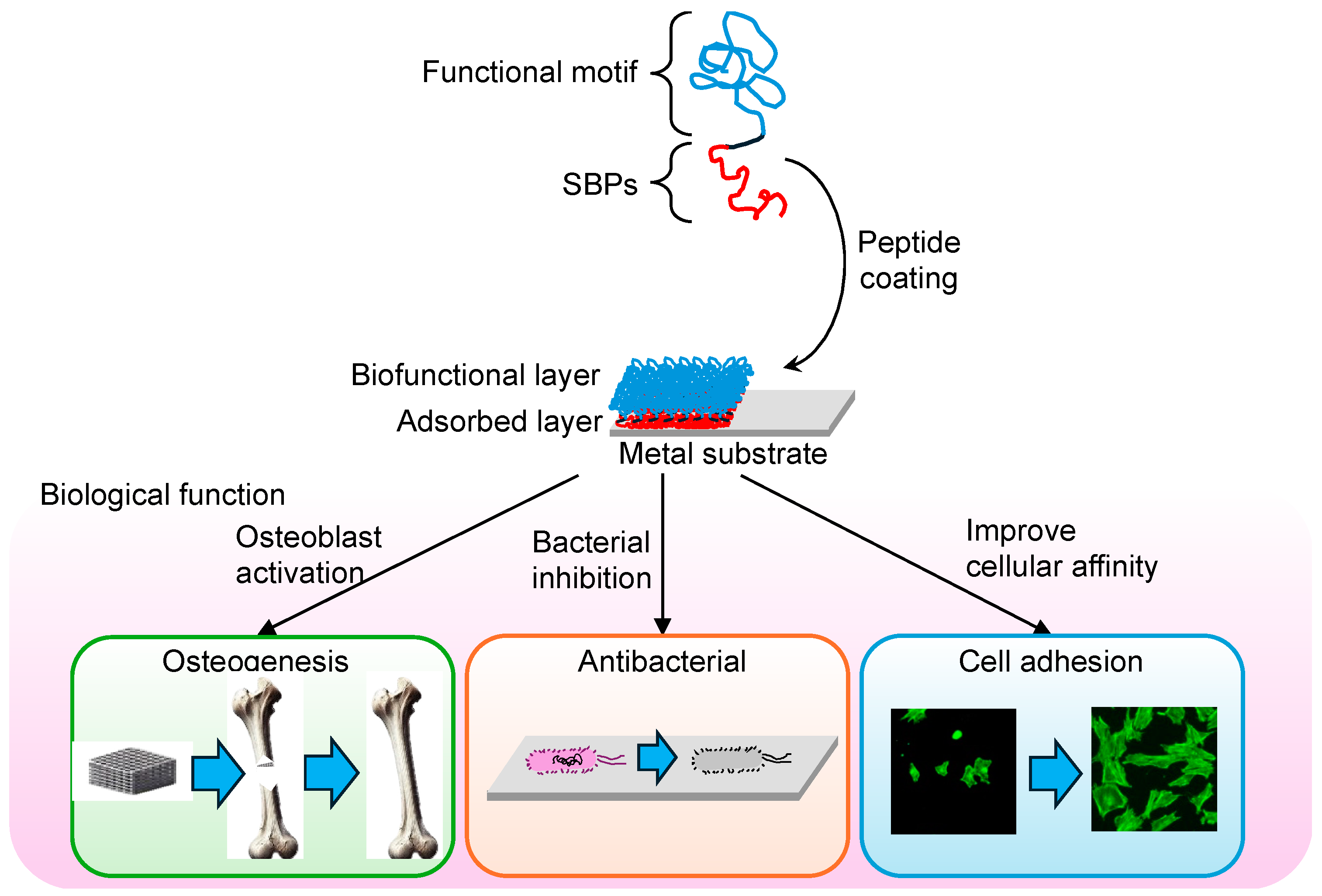
Figure 1. The process of surface modification using solid-binding peptides (SBPs) and its biological effects. SBPs, consisting of a metal-binding domain and a functional motif, are designed to adsorb onto the metal substrate. This adsorption creates an adsorbed layer, which forms a biofunctional layer on the surface, which can enhance various biological properties. The lower panels illustrate three key applications of SBP-modified surfaces: promoting osteogenesis through osteoblast activation, providing antibacterial properties promoting osteogenesis through osteoblast activation, providing antibacterial properties via bacterial inhibition, and improving cell adhesion by increasing cellular affinity.
Synbio 02 00020 g001
Figure 2. Selection process of SBPs using phage display technology. The color variations in the peptides represents different sequences. After several rounds of selection, only the peptides with the strongest binding affinity (shown in purple) remain, indicating optimized binding properties to the target surface.
Synbio 02 00020 g002
Figure 3. Computational approaches for SBP design and optimization. The color change in the peptide reflects the optimization process, resulting in improved binding properties.
Synbio 02 00020 g003
Table 1. Sequences of SBPs for various metallic biomaterials.
MaterialSequenceRefs.
TiRKLPDA[42,43,44,45,46,47,48]
RKLPDAPGMHTW[42,49,50]
RPRENRGRERGL[48,50,51,52,53,54,55]
SRPNGYGGSESS[50,51,55]
HAYKQPVLSTPF[51]
CGHTHYHAVRTQT[56,57]
ATWVSPY[48,58]
Ti-6Al-4VSVSVGMKPSPRP[59]
WDPPTLKRPVSP[59]
SHKHPVTPRFFVVESK[60]
NiTiNHHMMPAWNVKH[61]
SUS 316MTWDPSLASPRS[62]
SUS 316LVQHNTKYSVVIR[63]
CoCr alloyTSNLWRYDRLTM[61]
CoCrMo alloyQHKYTPIHEGRW[64,65]
AuWALRRSIRRQSY[66]
WAGAKRLVLRRE[66]
PtPTSTGQA[67]
PtWLTPHKHHKHLHA[61]
Disclaimer/Publisher’s Note: The statements, opinions and data contained in all publications are solely those of the individual author(s) and contributor(s) and not of MDPI and/or the editor(s). MDPI and/or the editor(s) disclaim responsibility for any injury to people or property resulting from any ideas, methods, instructions or products referred to in the content.