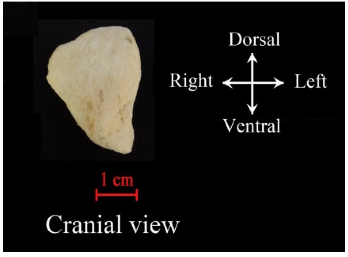
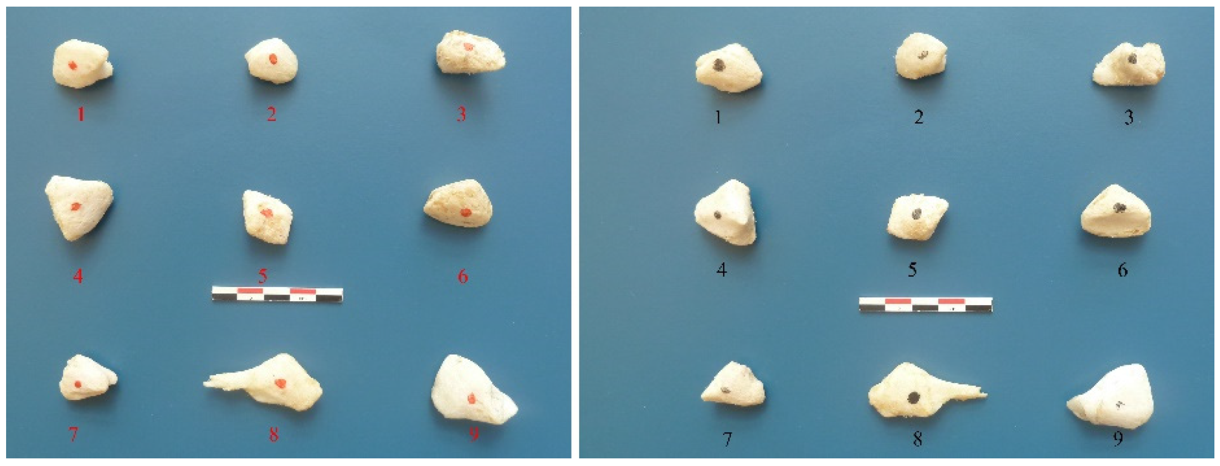
Diaphragm Bone in Dromedary (Camelus dromedarius L., 1758): Anatomy and Investigation Using Computed Tomography Imaging †
Abstract
:1. Introduction
2. Materials and Methods
3. Results and Discussion
4. Conclusions
Author Contributions
Funding
Institutional Review Board Statement
Informed Consent Statement
Data Availability Statement
Conflicts of Interest
References
- Guintard, C.; Babelhadj, B. Morphotypes et force animale développée. Comparaison de deux populations de dromadaires algériens: La Sahraoui et la Targui (Camelus dromedarius, L.). In Animal Source D’énergie: Enquêtes dans l’Europe Pré-Industrielle; Guizard, F., Beck, C., Eds.; Presses Universitaires de Valenciennes: Valenciennes, France, 2018; pp. 133–147. [Google Scholar]
- Faye, B. Statut Nutritionnel du Bétail Dans la République de Djibouti; Ministère de la Coopération: Paris, France, 1989. [Google Scholar]
- Narjisse, H. Nutrition et production laitière chez le dromadaire. Options Méditerranéennes-Série Séminaires 1989, 2, 163–166. [Google Scholar]
- Maskar, U. Bones of the diaphragm in camels. Acta Anat. 1957, 30, 461–471. [Google Scholar] [CrossRef] [PubMed]
- Namshir, N. Regional anatomy of camel internal organs. J. Agric. 1982, 3, 25. [Google Scholar]


| Bone | CC (cm) | PD (cm) | LR (cm) | UH max (ext.) | UH max (int.) | UH min (int.) | UH mean (int.) |
|---|---|---|---|---|---|---|---|
| 1 | 0.99 | 2.45 | 1.75 | 2664 | 584 | −981 | −60 |
| 2 | 1.00 | 1.55 | 1.78 | 2334 | 654 | −1024 | −65 |
| 3 | 1.09 | 1.61 | 2.39 | 1979 | 381 | −1024 | −263 |
| 4 | 1.40 | 2.38 | 2.45 | 2470 | 509 | −1024 | −684 |
| 5 | 0.89 | 1.78 | 2.43 | 2597 | 915 | −1012 | −58 |
| 6 | 1.24 | 1.92 | 2.65 | 2315 | 605 | −1024 | −274 |
| 7 | 1.06 | 1.95 | 2.21 | 2328 | 526 | −1024 | −325 |
| 8 | 0.81 | 2.22 | 4.49 | 2063 | 761 | −1007 | 59 |
| 9 | 1.08 | 2.34 | 3.21 | 2127 | 938 | −884 | 88 |
| Mean | 1.06 | 2.02 | 2.59 | 2320 | 653 | −1000 | −176 |
| Max | 1.40 | 2.45 | 4.49 | 2664 | 938 | −884 | 88 |
| Min | 0.81 | 1.55 | 1.75 | 1979 | 381 | −1024 | −684 |
| SD | 0.18 | 0.34 | 0.84 | 234 | 187 | 46 | 240 |
Disclaimer/Publisher’s Note: The statements, opinions and data contained in all publications are solely those of the individual author(s) and contributor(s) and not of MDPI and/or the editor(s). MDPI and/or the editor(s) disclaim responsibility for any injury to people or property resulting from any ideas, methods, instructions or products referred to in the content. |
© 2023 by the authors. Licensee MDPI, Basel, Switzerland. This article is an open access article distributed under the terms and conditions of the Creative Commons Attribution (CC BY) license (https://creativecommons.org/licenses/by/4.0/).
Share and Cite
Tekkouk-Zemmouchi, F.; Babelhadj, B.; Ridouh, R.; Betti, E.; Adamou, A.; Benhamza-Manssar, L.; Poisson, A.; Benaissa, A.; Altoama, K.; Tavernier, C.; et al. Diaphragm Bone in Dromedary (Camelus dromedarius L., 1758): Anatomy and Investigation Using Computed Tomography Imaging. Biol. Life Sci. Forum 2023, 22, 17. https://doi.org/10.3390/blsf2023022017
Tekkouk-Zemmouchi F, Babelhadj B, Ridouh R, Betti E, Adamou A, Benhamza-Manssar L, Poisson A, Benaissa A, Altoama K, Tavernier C, et al. Diaphragm Bone in Dromedary (Camelus dromedarius L., 1758): Anatomy and Investigation Using Computed Tomography Imaging. Biology and Life Sciences Forum. 2023; 22(1):17. https://doi.org/10.3390/blsf2023022017
Chicago/Turabian StyleTekkouk-Zemmouchi, Faiza, Baaissa Babelhadj, Rania Ridouh, Eric Betti, Abdelkader Adamou, Louiza Benhamza-Manssar, Archibald Poisson, Atika Benaissa, Kassem Altoama, Christian Tavernier, and et al. 2023. "Diaphragm Bone in Dromedary (Camelus dromedarius L., 1758): Anatomy and Investigation Using Computed Tomography Imaging" Biology and Life Sciences Forum 22, no. 1: 17. https://doi.org/10.3390/blsf2023022017
APA StyleTekkouk-Zemmouchi, F., Babelhadj, B., Ridouh, R., Betti, E., Adamou, A., Benhamza-Manssar, L., Poisson, A., Benaissa, A., Altoama, K., Tavernier, C., & Guintard, C. (2023). Diaphragm Bone in Dromedary (Camelus dromedarius L., 1758): Anatomy and Investigation Using Computed Tomography Imaging. Biology and Life Sciences Forum, 22(1), 17. https://doi.org/10.3390/blsf2023022017


