Proceeding Paper

Antioxidant Properties of Chickpea (Cicer arietinum L.) Protein Hydrolysates: The In Vitro Evaluation of SOD Activity in THP-1 Cell Line †

by
Noelia María Rodríguez-Martin
1,
José Carlos Marquez
1,
Álvaro Villanueva
1,
Francisco Millán
1,
María del Carmen Millán-Linares
2 and
Justo Pedroche
1,*
1
Department of Food & Health, Instituto de la Grasa, CSIC. Ctra. de Utrera Km. 1, 41013 Seville, Spain
2
Department of Medical Biochemistry, Molecular Biology, and Immunology, School of Medicine, Universidad de Sevilla, Av. Dr. Fedriani 3, 41071 Seville, Spain
*
Author to whom correspondence should be addressed.
Presented at the IV Conference Ia ValSe-Food CYTED and VII Symposium Chia-Link, La Plata and Jujuy, Argentina, 14–18 November 2022.
Biol. Life Sci. Forum 2022, 17(1), 12; https://doi.org/10.3390/blsf2022017012
Published: 27 October 2022
(This article belongs to the Proceedings of IV Conference Ia ValSe-Food CYTED and VII Symposium Chia-Link)

Abstract

:
Chickpeas (Cicer arietinum L.) are the third most important grain legume in the world in terms of their area coverage, the volume of production and the amount of trade. The crop is primarily grown in India, being USA, Argentina and Mexico the main producers of chickpea in the American continent. It can be considered as a cheap, sustainable, and healthy source of nutrients with a high content of proteins. In addition, the hydrolysis of chickpea proteins (CPH) can release bioactive peptides with immunomodulatory properties. Hence, the in vitro study of CPH in the monocytic cell line would be a relevant strategy to recognise the immunomodulatory hydrolysates. The main aim of this study was to evaluate the immunomodulatory potential of CPH. A chickpea protein concentrate was hydrolysed using Bioprotease-660LA under specific conditions. The resulting hydrolysates were evaluated to search for the potentially bioactive CPHs. The study led to the identification of one bioactive hydrolysate, which was used on THP-1 cell line as it was stimulated with lipopolysaccharide (LPS) to evaluate the inflammatory status of it. The ELISA and RT-qPCR techniques were used to analyse the levels of inflammatory cytokine production. The total superoxide dismutase (SOD) activity was also evaluated using a commercial determination kit. Our data showed that the selected CPH down-regulated the mRNA transcriptional levels of cytokines IL-1β and TNF-α in the LPS-stimulated THP-1 cell line. In addition, CPH increased the SOD activity, in contrast to the LPS control. This study suggests that CPH may improve the inflammatory state and play a significant role in the regulation of the SOD signalling pathways in THP-1 cell lines.

1. Introduction

Chickpeas (Cicer arietinum L.) are the third most important grain legume in the world in terms of their area coverage, the volume of production and the amount of trade [1]. The crop is primarily grown in India, being USA, Argentina and Mexico the main producers of chickpea in the American continent. [1]. It can be considered a cheaper, sustainable, and healthy source of nutrients with a high content of immunonutrients. Chickpea varieties contain 15–40% proteins, 15–68% carbohydrates, and 7% lipids. Nutritionally, this legume has a good composition of the essential amino acids with a high bioavailability, and a low allergenicity when it is compared to others such as soybeans or lupins. Furthermore, chickpea protein hydrolysates (CPHs) have better technological properties than chickpea concentrates and isolates do, in addition to the possibility of them producing bioactive peptides [2,3].
Inflammation is a normal process that removes the injurious infection and promotes the tissue repair and recovery related to several inflammatory pathways and involving Reactive Oxygen Species (ROS) [4,5,6,7,8,9]. However, the presence of certain factors has been linked to the inhibition of the inflammation resolution, thereby causing a chronical low-grade inflammation. This chronical inflammatory status causes several alterations to the normal cellular physiology, thus increasing the risk of non-communicable disease, infectious susceptibility, and cancer development risks [10]. There are multiple possible causes for the development of a chronical inflammatory status: bad diet habits, intestinal dysbiosis, obesity, stress, and a high level of physical activity. The clinical consequences of this chronical inflammatory status are associated with a higher risk of metabolic syndrome and its related comorbidities, cardiovascular disease, cancer, neurodegenerative disorders, autoimmune diseases, among others [10].
The study of CPH in the THP-1 cell line is a relevant strategy to recognise the antioxidant and anti-inflammatory immunonutrient in the treatment of inflammatory diseases.

2. Materials and Methods

2.1. Material and Reagent

Chickpea protein hydrolysate (CPH) was supplied by the Group of Plant Proteins at the Instituto de la Grasa-CSIC (Spain). THP-1 human cell line was supplied by the Unit of Cell Biology of the Instituto de la Grasa-CSIC (Spain). All of the solvents and buffers that were used were of biomolecular grade, and all of the disposable plastics were DNase, RNase-free and sterile.

2.2. Cell Culture and Treatment

THP-1 cell line was cultured in RPMI-1640, 10% FBS, and it was 1% penicillin-streptomycin-supplemented. After seeding THP-1 cells into the 12 well plates at concentration of 500,000 cell/mL, the cells were stimulated with 0.1 µg/mL LPS with the exception of the negative control. The stimulated cells were treated with CPH at different doses (50 and 100 µg/mL), except for the negative (no LPS, No CPH) and positive control (LPS; no CPH), and they were incubated for 24 h (37 °C, 5% CO2).

2.3. Cell Cytotoxicity MTT Assay

THP-1 cell line was seeded in a 96 well plate at concentration of 500,000 cell/mL. After seeding, the cells were subjected to different concentrations of CPH (0, 25, 50, 100, 250, 500 and 1000 µg/mL) and incubated for 24 h. Afterwards, MTT solution was applied at these concentrations to each well and incubated for 3 h. Then, DMSO was applied to dissolve the formazan crystals. The absorbance was obtained using a microplate reader at 570 nm and it was corrected to 650 nm. The cell cytotoxicity of CPH at different concentrations was compared to the negative (non-treated cells) and positive controls (Triton).

2.4. Gene Expression Analysis and Cytokine Measurement

Real-time quantitative polymerase chain reaction (qRT-PCR) was used to measure the levels of the messenger RNA (mRNA). The cells were recovered, and the total RNA was extracted using TRIsure reagent. Complementary DNA (cDNA) were obtained using the iScript cDNA Synthesis Kit from the extracted RNA samples. The performed primer pairs and SYBR green master mix were used to carry out RT-PCR. Finally, the TNF-α, IL-1β, SOD1, and SOD2 gene expression were normalised using the internal control GADPH (Table 1).
After the treatments were performed, the supernatants of cells containing only culture media were collected. TNF-α and IL-1β concentrations were measured using human ELISA kits following the manufacturer’s instructions (Diaclone, Besancon, France).

2.5. Nitric Oxide Measurement

The total NO levels were obtained from the supernatants by the Griess reaction that is based on the reduction of nitrates to nitrites. Briefly, 100 µL of Griess reagent was added to 100 µL of sample. The Griess reagent consist of a mixture of equal volumes of N-(1-naphthyl) ethylenediamine (0.1%) in deionised water and sulfanilamide in 5% H3PO4 (1%). Absorbance at 546 nm was measured using a microplate reader. The nitrite concentration was calculated using a NaNO2 standard curve.

2.6. Superoxide Dismutase Assay

After the seeding procedure, the LPS-activated THP-1 cells in a 96-well plate, and concentrations of 50 and 100 µg/mL of CPH were used and incubated for 24 h. The total activity of antioxidant enzyme SOD was measured in cellular homogenates using commercial colorimetric assay. The assay procedure was prepared according to the manufacturer’s protocol (Abcam, Boston, MA, USA).

2.7. Statistical Analysis

Data are expressed as arithmetic means with standard deviations (SD). Graph Pad Prism Version 8.0.1 software (San Diego, CA, USA) was used to evaluate the data. One-way ANOVA following Tukey’s test for multiple comparison and post hoc test were used to calculate the statistical significance of differences. P-value less than 0.05 was considered statistically significant. All of the assays were performed in quadruplicate.

3. Results and Discussion

Chickpeas have been shown to have further biological activities by different active compounds such as peptides, phenolic compounds, and others. Several chickpea-derivate products have been identified as compounds with activities such as antioxidant, antihypertensive, hypocholesterolemia, and anticancer [3]. In addition, the bioactive peptides, protein hydrolysates, and protein extracts from the chickpeas have also showed the properties that have been mentioned before [2,3,4]. In this sense, the biological activity of a CPH might be related to the group of peptides that were obtained during the enzyme hydrolysis where the amino acid composition, conformation, length, and sequence are key factors that produce the biological activity.

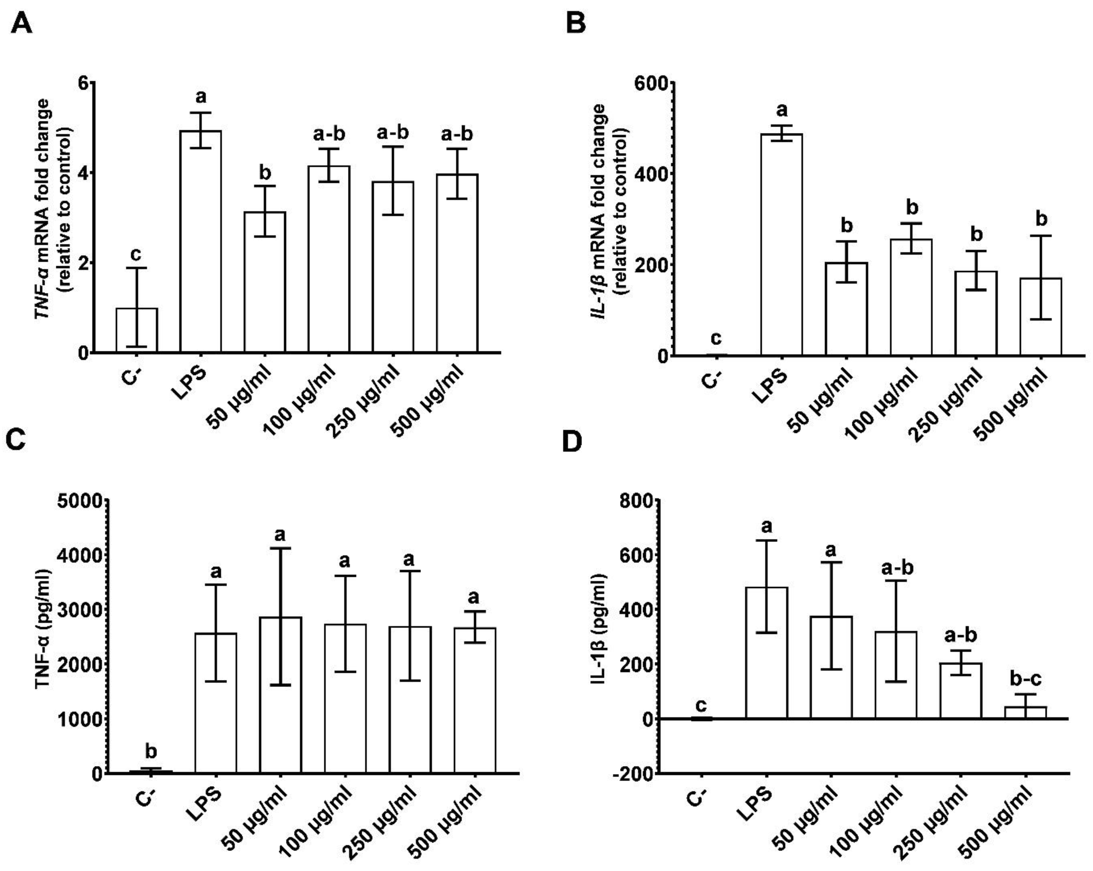
3.1. CPH Diminishing Inflammation in LPS Stimulated THP-1 Cells

The cell viability that was evaluated by using the MTT method was not affected by any of the CPH concentrations that were used. It may, therefore, be concluded that the use of CPH could be of interest in human nutrition as a non-toxic active ingredient. Thus, 50, 100, 250 and 500 µg/mL CPH were selected for the experiments (Figure 1).
To evaluate the immunomodulatory properties of CPH, the gene expression of the proinflammatory cytokines was measured by RT-qPCR in the THP-1 cells that were stimulated with LPS after 24 h of exposure. The CPH treatment reduced the statistically significant (p < 0.05) expression of TNF-α by 20% at a dose of 50 µg/mL, as shown in Figure 2A. This effect was particularly important in the case of IL-1β, where the IL-1β mRNA expression decreased by 58%, 48%, 62% and 64% with 50, 100, 250 and 500 µg/mL CPH, respectively (p < 0.0001) in comparison that which was observed in the LPS-treated cells (Figure 2B). Furthermore, the cytokine release that was measured by the ELISA method, which is shown in Figure 2D, was consistent with these RT-qPCR results. The major effect of CPH in IL-1β suggests the inhibition of the inflammatory pathways that are involved in the release of IL-1β. In the case of the LPS stimulation, the proinflammatory status has been produced by the binding of it with Toll Like Receptor 4 (TLR4). When the TLR4 receptor signalling pathway is activated, the aNF-κB protein complex is assembled, and it acts as transcriptional factor inside the nucleus and promotes the expression of the pro-inflammatory genes such as Pro-Il-1β, Pro-Il-18, Pro-Caspase-1, Asc, and Nlrp3. The expression of these proteins leads to the inflammasome pathway activation and the assembly of NLRP3, ASC, and Pro-Caspase-1, thereby conforming the NLRP3 inflammasome. Caspase-1 is released from the inflammasome complex, and this promotes the activation of the Il-1β and Il-18 cytokines from their precursors which are secreted to the extracellular medium [6]. Hence, the CPH could act as a targeting drug by inhibiting the Il-1β expression and the release of the NF-κB transcriptional effect activation, the inflammasome assembly or a mix of both pathways. However, these pathways are too long, and it is necessary that we conduct more research to verify the inflammatory markers and pathway that are mentioned above.

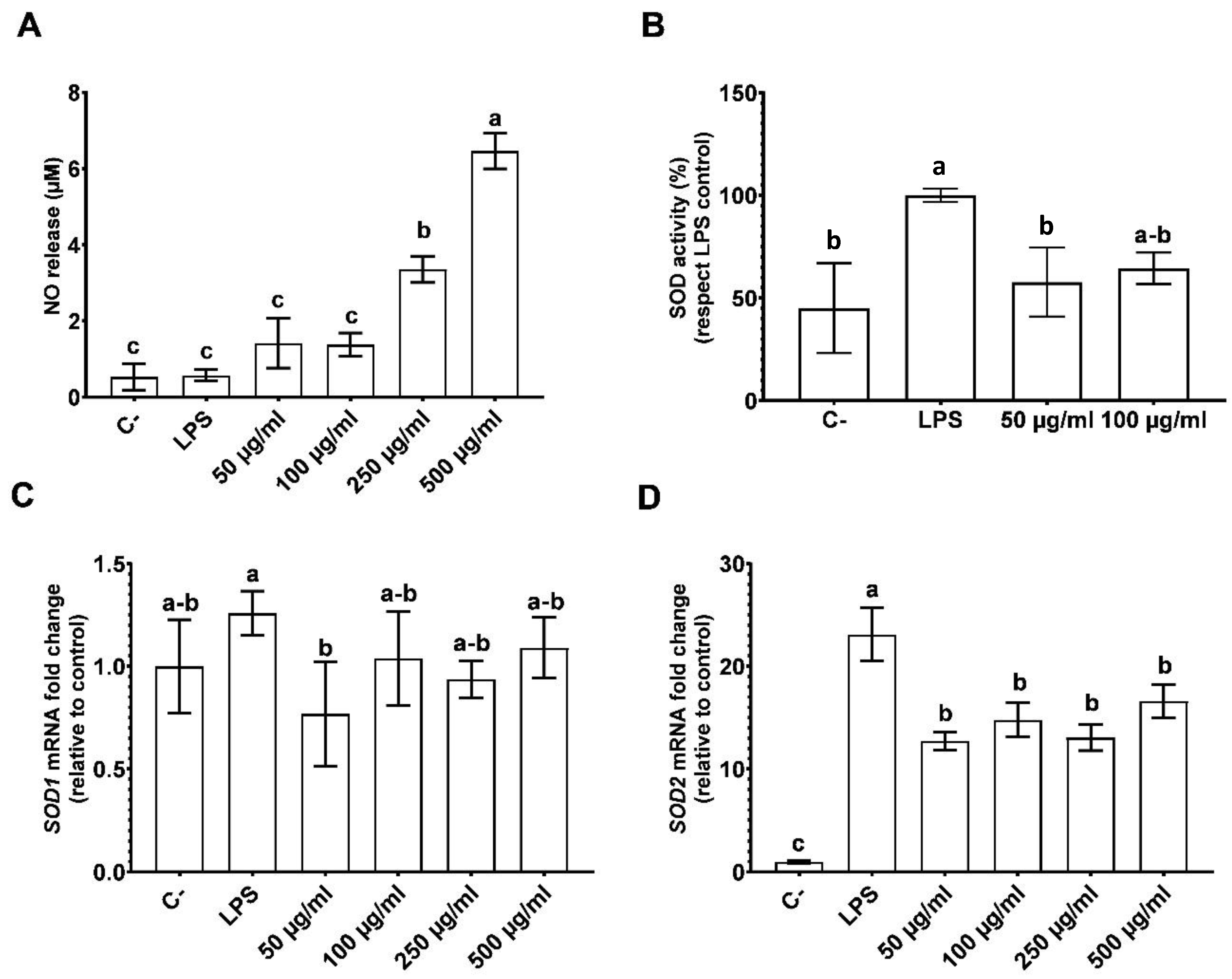
3.2. The Effects of CPH on Oxidative Stress

To investigate the antioxidant cellular responses to CPH, the reactive nitrogen species levels in the supernatants (NO), the total SOD inhibition rate in the cellar homogenates and the extracellular SOD (SOD1) and the mitochondrial SOD (SOD2) transcriptional levels were determined. The results revealed that CPH influences the NO levels which revealed a dose-dependent increase in the amount of extracellular NO (Figure 3A). Likewise, the SOD activity showed a recovery of the SOD levels (p < 0.05) (Figure 3B). Finally, the mRNA of the SOD isoforms revealed that the effect of the SOD1 mRNA expression was similar to the negative control (Figure 3C). In the case of the levels of SOD2, a decrease in the mRNA expression of this isoform was observed which accounts for about 55% of it (p < 0.0001), 64% of it (p < 0.0001), 56% of it (p < 0.0001), and 72% of it (p < 0.0005), respectively, for each dose in contrast to the LPS control (Figure 3D).
NO, NO2, and ONOO are reactive nitrogen/oxygen species which are considered both as signalling molecules and potentially cytotoxic ones. The superoxide molecules react with NO, forming ONOO, and this reaction is catalysed by the SODs, specially SOD1 in the cytoplasm. The ONOO molecule is considered as a strong oxidant and nitrating agent, but it is also is stable at physiological conditions [7]. The intracellular ROS, specifically the mitochondrial ROS are new actors enhancing the proinflammatory pathways including the NF-κB and NLRP3 inflammasome. The cellular SOD activity that is shown in Figure 3B suggests that there was a decrease of the SOD levels which could be indicated as a restoration of intracellular ROS basal levels. Similar results appeared in the SOD1 and SOD2 transcriptional levels which may be traduced in an intracellular ROS and mitochondrial ROS stress decrease.
Besides this, NO·is considered to be a widespread signalling molecule in mammals. NO has been shown to be a cell-to-cell messenger, being responsible for the modulating processes that are related to the circulation as vasodilation and relaxation of the smooth muscles [8]. SOD enzymes also participate in cell signalling pathways, regulating NO release [8,9]. As it was observed in Figure 3A, the NO mRNA expression with the negative and positive controls showed similar values to those of the NO mRNA expression with 50 and 100 µg/mL of the CPH doses. In addition, in the tolerance studies of low-dose LPS in the THP-1 cells, it has been reported that the NO levels increase, and the down-regulating of cytokines occurs [9]. This tolerance to LPS is associated with the epigenetic changes in the chromatin [9]. The CPH could generate the epigenetic changes that mimic to LPS low-dose-tolerance, thereby affecting the NO production and cytokine transcription.

4. Conclusions

Our data showed that the CPH down-regulated the mRNA transcriptional levels of the proinflammatory cytokines in THP-1 cell line that was stimulated with LPS, and particularly, the levels of IL-1β may indicate the NLRP3 inflammasome inhibition by the repression of the NF-κB inflammatory pathway. In addition, CPH has restored the intracellular SOD activity, in contrast to that which was achieved by the LPS control, mainly by the use of the mitochondrial SOD which removed the ROS stress and prevented the NF-κB and NLRP3 inflammasome feeding. Finally, the increase of the NO release at the highest CPH doses could indicate a possible cell-to-cell signal to restore the non-oxidative and non-inflammatory cellular status. Herein, this study suggests that CPH may improve the inflammatory states, and that it has a role in the SOD-derived oxidative stress in the THP-1 cell line.

Author Contributions

Conceptualization, J.P. and N.M.R.-M.; methodology, N.M.R.-M., and J.C.M.; software, J.C.M.; validation, J.P., M.d.C.M.-L. and Á.V.; formal analysis, N.M.R.-M.; investigation, J.P.; resources, J.P.; data curation, N.M.R.-M.; writing—original draft preparation, N.M.R.-M.; writing—review and editing, J.P.; visualization, J.C.M.; supervision, F.M.; project administration, J.P.; funding acquisition, J.P., and F.M. All authors have read and agreed to the published version of the manuscript.

Funding

This work was supported by grant Ia ValSe-Food-CYTED (119RT0567), by the “Centro para el Desarrollo Tecnológico Industrial” (CDTI), project no: IDI-20200562 (Diseño y evaluacion de nuevos ingredientes aplicados a alimentacion deportiva basados en hidrolizados proteicos vegetales (PLANTPROSPORT)), as well as the “Ministerio de Ciencia, Innovación y Universidades”, and the “Agencia Estatal de Investigación”, project no: PID2019-111368RB-I00 (Evaluation of new disruptive technologies (steam explosion) in the design of tailor-made plant protein hydrolysates applied to sport nutrition (PROVERDE)).

Institutional Review Board Statement

Not applicable.

Informed Consent Statement

Not applicable.

Data Availability Statement

Not applicable.

Conflicts of Interest

The authors declare no conflict of interest.

References

  1. FAOSTAT. Available online: https://www.fao.org/faostat/en (accessed on 1 August 2022).
  2. Boukid, F. Chickpea (Cicer arietinum L.) protein as a prospective plant-based ingredient: A review. Int. J. Food Sci. Technol. 2021, 56, 5435–5444. [Google Scholar] [CrossRef]
  3. Faridy, J.-C.M.; Stephanie, C.-G.M.; Gabriela, M.-M.O.; Cristian, J.-M. Biological Activities of Chickpea in Human Health (Cicer arietinum L.). A Review. Plant Foods Hum. Nutr. 2020, 75, 142–153. [Google Scholar] [CrossRef] [PubMed]
  4. Guilliams, M.; Mildner, A.; Yona, S. Developmental and Functional Heterogeneity of Monocytes. Immunity 2018, 49, 595–613. [Google Scholar] [CrossRef] [Green Version]
  5. Liu, T.; Zhang, L.; Joo, D.; Sun, S.-C. NF-κB signaling in inflammation. Signal Transduct. Target. Ther. 2017, 2, 17023. [Google Scholar] [CrossRef] [Green Version]
  6. Swanson, K.V.; Deng, M.; Ting, J.P.Y. The NLRP3 inflammasome: Molecular activation and regulation to therapeutics. Nat. Rev. Immunol. 2019, 19, 477–489. [Google Scholar] [CrossRef] [PubMed]
  7. Duez, H.; Pourcet, B. Nuclear Receptors in the Control of the NLRP3 Inflammasome Pathway. Front. Endocrinol. 2021, 12, 630536. [Google Scholar] [CrossRef] [PubMed]
  8. Wang, Y.; Branicky, R.; Noë, A.; Hekimi, S. Superoxide dismutases: Dual roles in controlling ROS damage and regulating ROS signaling. J. Cell Biol. 2018, 217, 1915–1928. [Google Scholar] [CrossRef] [PubMed] [Green Version]
  9. Rios, E.C.S.; de Lima, T.M.; Moretti, A.I.S.; Soriano, F.G. The role of nitric oxide in the epigenetic regulation of THP-1 induced by lipopolysaccharide. Life Sci. 2016, 147, 110–116. [Google Scholar] [CrossRef]
  10. Furman, D.; Campisi, J.; Verdin, E.; Carrera-Bastos, P.; Targ, S.; Franceschi, C.; Ferrucci, L.; Gilroy, D.W.; Fasano, A.; Miller, G.W.; et al. Chronic inflammation in the etiology of disease across the life span. Nat. Med. 2019, 25, 1822–1832. [Google Scholar] [CrossRef]
Figure 1. Effect of CPH on THP-1 cells viability. THP-1 cells were cultured with different doses of CPH (25, 50, 100, 250, 500 and 1000 µg/mL) for 24 h. Data are expressed as mean ± SD of four separate experiments. p value < 0.0001 was considered statistical significance (****). C+, live cell control; C− death cell control.
Blsf 17 00012 g001
Figure 2. Effect of CPH on LPS-induced cytokines mRNA relative expression (TNF-α expression were significantly lower in the C−, and 50 µg/mL (A), and IL-1β which shown a significant expression decrease in all the concentrations as well as C− (B)) in THP-1 cells, and effect of CPH on LPS-induced protein level (which did not show statistical significance in the TNF-α release for any concentration of product (C), in contrast to the release of IL-1β which were shown a significant decrease in a dose dependant manner (D)). THP-1 cells previously stimulated with LPS were cultured with different doses of CPH (50, 100, 250 and 500 µg/mL) for 24 h. Data are expressed as mean ± SD of four separate experiments. p value < 0.05 was considered statistical significance and marked with different letters. C−, live cell control; LPS, lipopolysaccharide-treated cell.
Blsf 17 00012 g002
Figure 3. The effect of CPH was high in a concentration dependant manner on induced NO release (A), a restore of the SOD activity was observed in contrast to the LPS control (B), and LPS-induced ROX enzymes mRNA relative expression, SOD1 (C) and a lower SOD2 expression was observed when the cells were treated with CPH at different doses (D) in THP-1 cells. THP-1 cells previously stimulated with LPS were cultured with different doses of CPH (50, 100, 250 and 500 µg/mL) for 24 h (in exception of SOD activity assay). Data are expressed as mean ± SD of four separate experiments. p value < 0.05 was considered statistical significance and marked with different letters. C− live cell control; LPS, lipopolysaccharide-treated cell.
Blsf 17 00012 g003
Table 1. Primer sequences for real time PCR.
Gene NameNM CodeForward SequenceReverse Sequence
IL-1βNM_000576.25′-TCCTTCAGACACCCTCAACC-3′5′-AGGCCCCAGTTTGAATTCTT-3′
TNF-αNM_000594.35′-CTGTCCTGCGTGTTGAAAGA-3′5′-TTCTGCTTGAGAGGTGCTGA-3′
SOD1NM_000454.55′-ACAAAGATGGTGTGGCCGAT-3′5′-AACGACTTCCAGCGTTTCCT-3′
SOD2NM_000636.45′-AACAACCTGAACGTCACCGA-3′5′-CACGTTTGATGGCTTCCAGC-3′
GADPHNM_002046.65′-GAGTCAACGGATTTGGTCGT-3′5′-GACAAGCTTCCCGTTCTCAG-3′
Abbreviations: IL (Interleukine), TNF (tumor Necrosis Factor), SOD (Super Oxide Dismutase), SOD1 (Mn like SOD or extracellular SOD), SOD2 (Cu/Zn like SOD or mitochondrial SOD), GADPH (Glyceraldehyde-3-Phosphate Dehydrogenase).
Publisher’s Note: MDPI stays neutral with regard to jurisdictional claims in published maps and institutional affiliations.

