Proceeding Paper

Bladder Cancer and Fluorescence Cystoscopy †

1
English Division Science Club, Medical College, University of Rzeszów, Kopisto 2A, 35-310 Rzeszów, Poland
2
Department of Photomedicine and Physical Chemistry, Medical College, University of Rzeszów, Kopisto 2A, 35-310 Rzeszów, Poland
*
Author to whom correspondence should be addressed.
Presented at the 2nd International Electronic Conference on Nutrients, 15–31 March 2022; Available online: https://iecn2022.sciforum.net/.
Biol. Life Sci. Forum 2022, 12(1), 4; https://doi.org/10.3390/IECN2022-12402
Published: 14 March 2022
(This article belongs to the Proceedings of The 2nd International Electronic Conference on Nutrients)

Abstract

:
Bladder cancer is one of the ten most common types of cancer diagnosed in the world. During this time, an unbalanced state of reactive oxygen species’ (ROS) production and antioxidant capacity occurs that causes oxidative damage, cell damage, and eventually the death of the cells. The aim of this research was to explain what fluorescence diagnostics is and to present the techniques of modern bladder tumor fluorescence diagnostics that uses the phenomenon of oxidative stress. Modern fluorescence diagnostics of bladder neoplasms combining fluorescence cystoscopy with other methods, shows great potential both in basic biomedical research and in the clinical practice of diagnosis and bladder cancer treatment. In this paper, we present the current research reports on the generation and operation of ROS in bladder cancer.

1. Introduction

Bladder cancer is one of the ten most common types of cancer diagnosed in the world, with the highest number of cancers diagnosed in developed countries [1,2,3]. During this period, reactive oxygen species’ (ROS) production and antioxidant capacity are out of balance, leading to oxidative damage, cell damage, and ultimately cell death. Oxidative stress mediates the development of several diseases, including urothelial carcinoma. Fluorescence diagnosis of bladder tumors, namely photodynamic diagnosis (PDD) and photodynamic therapy (PDT), has shown great potential in both basic biomedical research and clinical practice. The aim of this study was to explain what fluorescence cystoscopy is and how the phenomenon of oxidative stress works. So far, several mechanistic pathways have been proposed and oxidative stress has been shown experimentally to cause epigenetic changes. Many diagnostic and therapeutic methods are known for the treatment of bladder cancer, including the modern diagnostic methods used to detect bladder cancer: cystoscopy, biopsy, cytology, and ultrasound, computed tomography (CT), intravenous urography (IVU), and magnetic resonance imaging (MRI). However, cystoscopy is considered the gold standard for the initial treatment of bladder cancer. In addition to the above methods, bladder cancer can also be detected using fluorescence cystoscopy (PDD) [4]. Bladder cancer diagnosis under fluorescence control has excellent sensitivity and specificity [5,6]. Studies comparing white light cystoscopy with fluorescence cystoscopy confirmed an approximately 30% increase in the frequency of in situ detection of bladder cancer [7]. Narrow-band imaging improved detection rates and reduced the risk of recurrence after 3 and 12 months. Blue light cystoscopy (BLC) can detect up to 14% of Ta/T1 papillary lesions and 40% of in situ neoplastic lesions, which are overlooked in conventional cystoscopy [8]. The photodynamic diagnostic method for tumors is the most accurate method for imaging tumor tissue and, unlike conventional cystoscopy, allows a visualization of all tumor foci at the earliest stages of their formation [9].

2. Methods

Cystoscopy is an endoscopy that examines the mucosa of the bladder. Standard cystoscopy is performed using white light. Fluorescence cystoscopy, on the other hand, involves the use of light of an appropriate wavelength after prior intravesical administration of a photosensitizer (PS). The reaction takes place in the presence of oxygen. This approach requires a preoperative intravesical infusion of fluorophores that are captured by urothelial cells and preferentially metabolized by dysplastic cells. Under the influence of light of the appropriate wavelength, the dysplastic cells emit a characteristic red fluorescence. Current commonly used photosensitizers for PDD are 5-aminolevulinic acid (5-ALA) or 5-aminolevulinic acid (HAL) [10,11,12]. Both drugs (5-ALA and HAL) can be instilled topically into the bladder without causing any systemic or local toxicity. The applications of the two compounds vary widely [13].

2.1. 5-Aminolevulinic Acid

5-ALA (Figure 1) is the first locally administered prodrug for bladder tumor photodiagnosis. 5-ALA acid has the ability to selectively accumulate in cancer cells. This enables visualization of small or flat tumors that do not grow into the bladder lumen under fluorescent light, which significantly improves the detectability of bladder cancer compared to normal cystoscopy under white light. Multiple studies have demonstrated that 5-ALA fluorescence cystoscopy significantly improves the overall diagnosis of malignant bladder lesions compared with standard white light cystoscopy [14,15,16].
We were able to employ cystoscopy to inspect tumor cells under the impact of blue light due to the features of 5-ALA, which causes an excessive accumulation of intracellular levels of the fluorescent substrate protoporphyrin IX (PpIX). This is due to the fact that, unlike normal tissues, they have a proclivity for accumulating extra PpIX. As a result, 5-ALA can be employed to cause red fluorescence in neoplastic and precancerous tumors in order to identify them [17]. The relative insolubility of 5-ALA in tissues is one of its drawbacks. This disadvantage is not present in hexyl 5-aminolevulinic acid (HAL), a hexyl ester derivative with improved lipophilic characteristics [10].

2.2. 5-Aminolevulinic Acid Hexyl

HAL (Figure 2) is a lipophilic derivative of 5-ALA that is more potent. It has the benefits of 5-ALA in that it increases the number of photoactive porphyrins in a smaller dose and in a shorter amount of time. This allows for faster substance instillation into the bladder and better detection of photosensitive tumors [18,19]. The results of a prospective multicenter US registry from 2018 confirm that BLC with HAL improves the detection of all malignancies by 23 percent, papillary by at least 12 percent, and CIS by 43 percent compared to conventional WLC [20]. Moreover, many randomized controlled trials have shown that BLC with HAL aids in the detection of bladder tumors [21,22,23].

2.3. Hypericin and Other Photosensitizers

Hypericin is another PS that is a natural chemical. Hypericin is a polycyclic aromatic molecule with significant photoactive characteristics that belongs to the class of quinone derivatives [24,25]. When compared to 5-ALA and HAL, hypericin is less susceptible to photobleaching and has better sensitivity and specificity of 94 percent and 95 percent, respectively [26]. Hypericin has a high tolerance, according to research, and the fluorescence can continue up to 16 h after instillation. Hypericin’s low water solubility, which can be changed using solvents such as polyvinylpyrrolidone (PVP) and albumin [27], is likely a constraint. Although thousands of compounds have been found to be photosensitizing agents, the most often used photosensitizers are 5-aminolevulinic acid (5-ALA) and 5-aminolevulinic acid (HAL).
A successful photosensitizer in the clinic has the majority of the following characteristics: it is non-toxic until activated, it is hydrophilic for easy systemic distribution, it is activated by a therapeutically useful wavelength of light, and it consistently induces a photodynamic response. Furthermore, it is concentrated in the neoplasm, cleanses normal tissue, is rapidly expelled from the patient’s body, is a non-toxic breakdown product with concomitant easy synthesis, and, most importantly, is commercially available [28]. For proper activation, each PS requires a specific light wavelength and intensity. Red light with a wavelength of 630 nm has been shown in clinical studies to penetrate tissue up to 0.5 cm, allowing lighting of both the surface and deeper situated tumors [29,30].
Essentially, light of a specific wavelength activates the PS, which then initiates a sequence of photochemical processes that, in theory, allow the tumor to be destroyed without causing undue damage to healthy tissue. By transferring an electron from the photon of light to the PS, the light energy modifies the neutral PS. After that, the active PS might squander energy in a variety of ways. The emission of light might result in energy loss. The light phenomena that can be seen enables tumor identification and delineation [29,31,32].
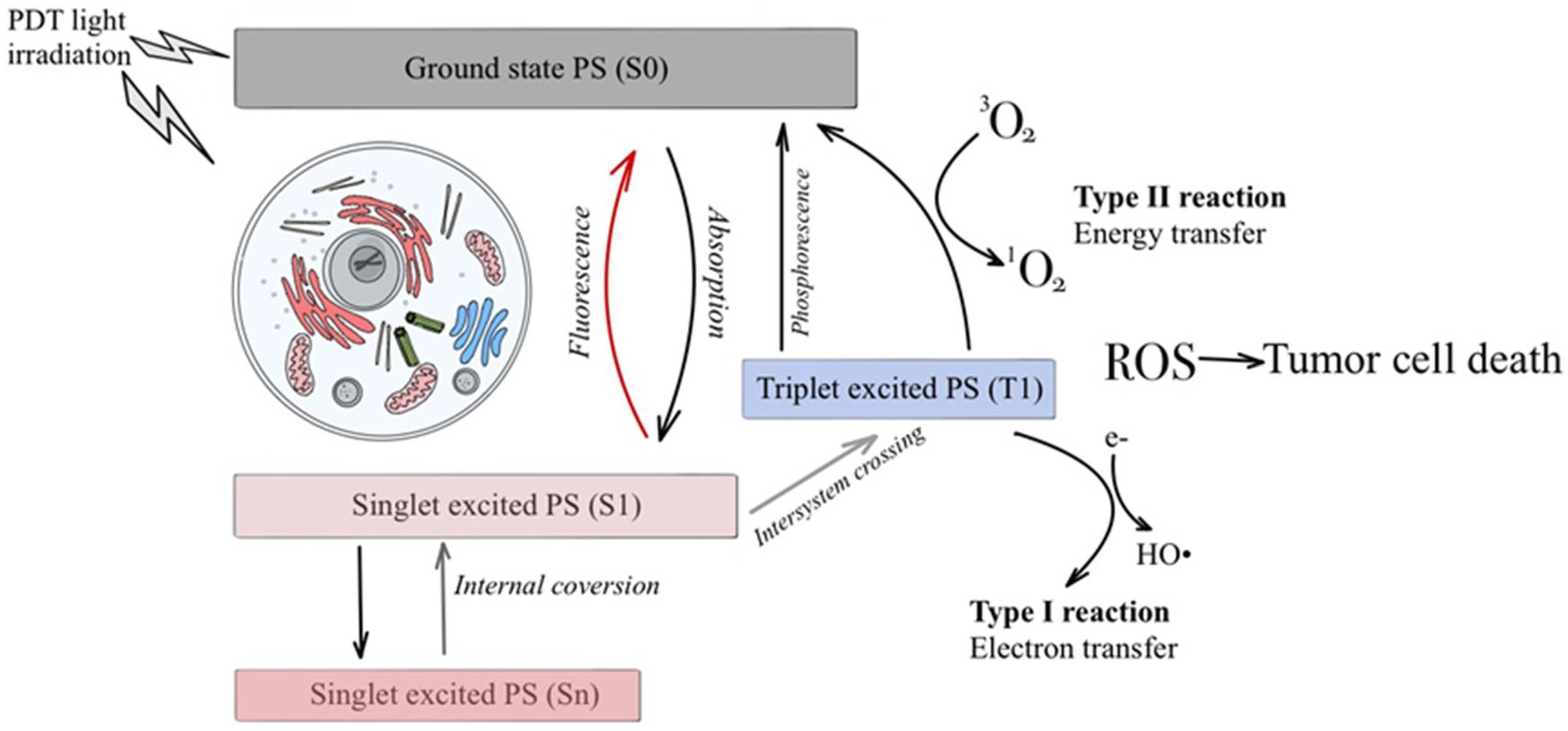
The photodynamic response has two major processes, both of which are reliant on the oxygen molecules located inside the cells (Figure 3). Both processes have a similar first step. Type I and type II photoprocesses are the two types of photoprocesses used in PDT. Type I and type II processes are completely reliant on molecular oxygen. Type II entails energy transfer from the photosensitizer to ground-state 3O2 to produce singlet oxygen (1O2), whereas type I entails photoinduced electron transfer to produce superoxide (O2) or hydroperoxyl radicals (HO2•). Type I and type II photosensitized reactions both result in biomolecule breakdown and, as a result, tissue damage/destruction [33].
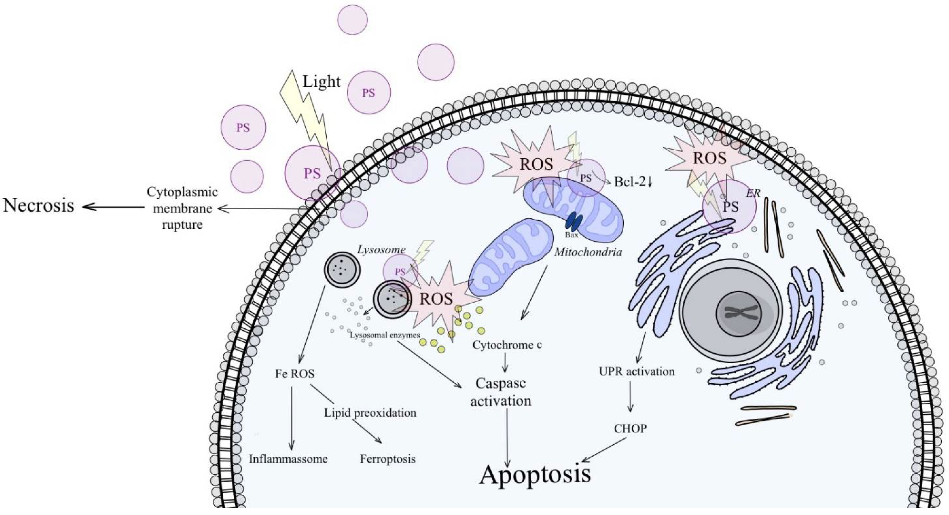
Photon absorption by PS transforms the pivot states (S0) of the PS into an excited unstable state (S1) that undergoes a triplet state intersystem transition (T1). Alternatively, the photon passes from S0 to other regularly repeating molecules, forming a cytotoxic radical knot [34]. In any case, the test radicals produced react aggressively with nu[cleic, developmental, and other lipids, as well as other substrates containing numerous additional carcinogens. In the initial episode of death, photodamage by ROS can cause apoptosis and necrosis [35]. The active phase after PDT treatment is defined by type, and death is determined by PDT dosimetry (e.g., PS causes apoptosis in mitochondria, although it can begin necrosis in the cell membrane following irradiation (Figure 4)). Overall, PDT-induced increased apoptosis when the shift to shift changed [36].
Apoptosis is a tightly controlled cell death process. Following the destruction of multiple organelles, it entails the beginning of several pathways [37]. Mitochondria are important regulators of apoptosis and any PS found on the organelle promoted the process. Caspase proteins can be activated by cytochrome c leakage from mitochondria into the cytosol, resulting in signal transduction and apoptosis [38].
Necrosis causes significant damage to cell components at the PS site of action, resulting in intracellular material leaking that could induce inflammation. When PS localizes in the plasma membrane and PDT with greater doses of light and PS, this mechanism happens [39]. Beclin-1 is a tumor suppressor protein that is linked to autophagy. The autophagic mechanism is diverse in its impact and Beclin-1 production is aimed at tumor growth inhibition. However, as the tumor progresses, a different autophagy pathway is activated to help the cells in the core and the low-nutrient region of the tumor receives the energy it requires to stay alive [40]. Photodamaged cells recycle their damaged organelles and cytoplasmic components in the subsequent stages to become resistant to PDT [41]. Autophagy may either be engaged to recycle the damaged organelle, causing cell survival (low dose PDT), or it can be induced to cause full cell organelle destruction, causing cell death (high dose PDT). Mild PDT, on the other hand, is induced by insufficient PS localization, PS efflux out of the cell, and low light which triggers autophagy’s defensive potential to repair the damage, resulting in PDT resistance [42].
An important role in the accumulation of PpIX in cells is played by nitric oxide (NO), which interacts with iron and prosthetic groups of proteins and iron-containing enzymes (heme, cytochrome C). This can lead to an increase in oxidative stress (Figure 5), a decrease in intracellular iron concentration, and a decrease in the activity of mitochondrial enzymes, including ferrochelatase, which, in turn, leads to the accumulation of PpIX. It was also observed that the expression of nitric oxide synthase is increased in neoplastic cells, which may explain the selective accumulation of PpIX in neoplastic tissue. Therefore, endogenously produced NO may be an important factor regulating cell response to ALA-PDT [43].
Photodynamic therapy and photodynamic diagnostics are often considered together. This is due to the fact that the same PS can be used first for diagnosis and then for therapy. The essence of PDD is the emission of light after the irradiation of tissues containing natural fluorochromes or introduced photosensitizers. The concentrations of fluorochromes in the neoplastic and healthy tissue are different, which causes the difference in the intensity of fluorescence emitted by these tissues. The optical effect used in diagnostics is the emission of red light by the neoplastic tissue and green light by the healthy tissue [44]. The drug that accumulated mainly in the neoplastic tissue is irradiated, usually after 24–72 h, with a beam of appropriate intensity. Activation of PS results in the production of ROS that damage cell organelles, leading to cell death. PDT and PDD allow the detection and treatment of diffuse multifocal lesions, with the possibility of multiple repetitions of the procedure, without the risk of accumulation of a toxic agent.
Unlike other kinds of cancer therapy (chemotherapy and radiation), these procedures do not lead to the establishment of secondary neoplasms [45].
In practice, the bladder wall must be smoothly unfurled and extended during fluorescence cystoscopy to decrease the danger of false positives caused by tangential lighting of the mucosal folds. Furthermore, it is critical to maintain a clean environment inside the bladder, as any bleeding traps the blue light and significantly lowers the stimulation of the bladder wall. As a result, any biopsy or resection region should be meticulously coagulated before surgery, and any questionable area spotted by fluorescence should be biopsied as soon as possible, as photobleaching (natural fluorescence decay) could distort the results. This danger is reduced by the increased fluorescence intensity produced with HAL compared to ALA [43].

3. Discussion

PDT and PDD are safe, well-tested, and effective in the treatment of non-infiltrating bladder cancer with varied risks. Since they allow for a decrease in relapses, urologists and patients should favorably evaluate the possibility of progression reduction [46]. When compared to white light cystoscopy, PDD improves the in situ detection of cancer and papillary lesions [47]. PDD is a highly specific, sensitive, and successful diagnostic tool. Improved diagnostic capacity, higher tumor resection, and a moderate but substantial decrease in relapse-free survival have all been demonstrated in prospective phase III clinical studies. During diagnostic cystoscopy and endoscopic resection, improved cystoscopy or blue light cystoscopy gives superior sensitivity and specificity for finding bladder tumors. The reported side effects are similar to those seen with regular white light tumor cystoscopy [12].
The disadvantage of the procedures described above is that they can lead to an erroneous false-positive diagnosis. The inadequate expertise of the surgeon conducting the examination, autofluorescence, tangential imaging, and bladder inflammation are all factors that could affect this. The depth of penetration of both ALA and HAL is restricted, limiting the examination of more invasive lesions [48]. Furthermore, any flaw in the light source’s energy or transmission owing to damage to the light cables could diminish the excitation strength of the light at the endoscope’s tip, lowering picture quality.
PDD has the potential drawback of requiring the patient to use a urethral catheter prior to surgery. However, as verified postoperatively, the catheter causes no discomfort to patients, thus this should be considered if the above-mentioned advantages are still evident. Finally, bladder cancer is the most costly malignancy from diagnosis to death, according to various research [49,50].

4. Conclusions and Future Perspectives

Fluorescence cystoscopy is a safe, well-tested, and effective therapy for bladder cancer, with strong evidence to back it up. Combining PDD with other bladder cancer diagnosis methods is the currently explored strategy, which is accessible and implementable while also being more successful.
There is space for more development and study in this field, as well as a need for it in order to remove obstacles and provide new, enhanced diagnostic procedures. New approaches to bladder cancer fluorescence diagnostics, without a doubt, offer promise for the future in this field of medicine and a reduction in the challenges connected with the use of PDD. Technical considerations, associated costs, a steep learning curve, and, most importantly, clinical success in practice are key obstacles to the widespread clinical application of these revolutionary procedures in the foreseeable future.

Author Contributions

Conceptualization, D.A.; methodology, M.P.; validation, D.A.; formal analysis, M.P.; investigation, M.P.; resources, D.A.; data curation, D.A.; writing—original draft preparation, M.P.; writing—review and editing, M.P. and D.A.; visualization, D.A.; supervision, D.A.; project administration, D.A. All authors have read and agreed to the published version of the manuscript.

Funding

This research received no external funding.

Institutional Review Board Statement

Not applicable.

Informed Consent Statement

Not applicable.

Conflicts of Interest

The authors declare no conflict of interest.

References

  1. Siegel, R.L.; Miller, K.D.; Jemal, A. Cancer statistics. CA Cancer J. Clin. 2020, 70, 7–30. [Google Scholar] [CrossRef] [PubMed]
  2. Richters, A.; Aben, K.K.; Kiemeney, L.A. The global burden of urinary bladder cancer: An update. World J. Urol. 2020, 38, 1895–1904. [Google Scholar] [CrossRef] [PubMed] [Green Version]
  3. Sung, H.; Ferlay, J.; Siegel, R.L.; Laversanne, M.; Soerjomataram, I.; Jemal, A.; Bray, F. Global Cancer Statistics 2020: GLOBOCAN Estimates of Incidence and Mortality Worldwide for 36 Cancers in 185 Countries. CA Cancer J. Clin. 2021, 71, 209–249. [Google Scholar] [CrossRef] [PubMed]
  4. Koenig, F.; McGovern, F.J.; Larne, R.; Enquist, H.; Schomacker, K.T.; Deutsch, T.F. Diagnosis of bladder carcinoma using protoporphyrin IX fluorescence induced by 5-aminolaevulinic acid. BJU Int. 1999, 83, 129–135. [Google Scholar] [CrossRef] [PubMed] [Green Version]
  5. D’Hallewin, M.A.; Bezdetnaya, L.; Guillemin, F. Fluorescence Detection of Bladder Cancer: A Review. Eur. Urol. 2002, 42, 417–425. [Google Scholar] [CrossRef]
  6. Makito, M.; Yasushi, N.; Satoshi, A.; Yoshihiro, T.; Masaomi, K.; Sayuri, O.; Yoshitomo, C.; Nobumichi, T.; Yoshihiko, H.; Kiyohide, F. Diagnostic approach for cancer cells in urine sediments by 5-aminolevulinic acid-based photodynamic detection in bladder cancer. Cancer Sci. 2014, 105, 616–622. [Google Scholar]
  7. Fradet, Y.; Grossman, H.B.; Gomella, L.; Lerner, S.; Cookson, M.; Albala, D.; Droller, M.J.; PC B302/01 Study Group. A comparison of hexaminolevulinate fluorescence cystoscopy and white light cystoscopy for the detection of carcinoma in situ in patients with bladder cancer: A phase III, multicenter study. J. Urol. 2007, 178, 68–73. [Google Scholar] [CrossRef]
  8. Lenis, A.T.; Lec, P.M.; Chamie, K.; MSHS, M. Bladder Cancer: A Review. JAMA 2020, 324, 1980–1991. [Google Scholar] [CrossRef]
  9. Lipiński, M. Diagnostyka fluorescencyjna nowotworów pęcherza moczowego. Przegląd Urol. 2013, 2, 78. [Google Scholar]
  10. Kennedy, J.C.; Pottier, R.H.; Pross, D.C. Photodynamic therapy with endogenous protoporphyrin IX: Basic principles and present clinical experience. J. Photochem. Photobiol. B 1990, 6, 143–148. [Google Scholar] [CrossRef]
  11. Chatterton, K.; Ray, E.; O’Brien, T.S. Fluorescence diagnosis of bladder cancer. Br. J. Nurs. 2006, 15, 595–597. [Google Scholar] [CrossRef] [PubMed]
  12. Lerner, S.P.; Goh, A. Novel endoscopic diagnosis for bladder cancer. Cancer 2015, 121, 169–178. [Google Scholar] [CrossRef] [PubMed] [Green Version]
  13. Jichlinski, P.; Leisinger, H. Fluorescence Cystoscopy in the Management of Bladder Cancer: A Help for the Urologist! Urol. Int. 2005, 74, 97–101. [Google Scholar] [CrossRef] [PubMed]
  14. Burger, M.; Stief, C.G.; Zaak, D.; Stenzl, A.; Wieland, W.F.; Jocham, D.; Otto, W.; Denzinger, S. Hexaminolevulinate is equal to 5-aminolevulinic acid concerning residual tumor and recurrence rate following photodynamic diagnostic assisted transurethral resection of bladder tumors. Urology 2009, 74, 1282–1286. [Google Scholar] [CrossRef]
  15. Zaak, D.; Kriegmair, M.; Stepp, H.; Stepp, H.; Baumgartner, R.; Oberneder, R.; Schneede, P.; Corvin, S.; Frimberger, D.; Knuchel, R.; et al. Endoscopic detection of transitional cell carcinoma with 5-aminolevulinic acid: Results of 1012 fluorescence endoscopies. Urology 2001, 57, 690–694. [Google Scholar] [CrossRef]
  16. Zumbragel, A.; Bichler, K.; Stenzl, A. 5-ALA induced fluorescence cystoscopy for superficial bladder cancer: Five year results. Eur. Urol. 2003, 1 (Suppl 2), 111. [Google Scholar] [CrossRef]
  17. Batlle, A.M. Porphyrins, porphyrias, cancer and photodynamic therapy—A model for carcinogenesis. J. Photochem. Photobiol. B 1993, 20, 5–22. [Google Scholar] [CrossRef]
  18. Lange, N.; Jichlinski, P.; Zellweger, M.; Forrer, M.; Marti, A.; Guillou, L.; Kucera, P.; Wagnières, G.; van den Bergh, H. Photodetection of early human bladder cancer based on the fluorescence of 5-aminolevulinic acid hexyl ester-induced protoporphyrin IX: A pilot study. Br. J. Cancer 1999, 80, 185–193. [Google Scholar] [CrossRef] [Green Version]
  19. Jichlinski, P.; Guillou, L.; Karlsen, S.J.; Malmstrom, P.U.; Jocham, D.; Brennhovd, B.; Johansson, E.; Gartner, T.; Lange, N.; van den Bergh, H.; et al. Hexaminolevulinate fluorescence cystoscopy: New diagnostic tool for photodiagnosis of superficial bladder cancer—A multicenter study. J. Urol. 2003, 170, 226–229. [Google Scholar] [CrossRef] [Green Version]
  20. Daneshmand, S.; Bazargani, S.T.; Bivalacqua, T.J.; Holzbeierlein, J.M.; Willard, B.; Taylor, J.M.; Liao, J.C.; Pohar, K.; Tierney, J.; Konety, B. Blue Light Cystoscopy with Cysview Registry Group. Blue light cystoscopy for the diagnosis of bladder cancer: Results from the US prospective multicenter registry. Urol. Oncol. 2018, 36, 361.e1–361.e6. [Google Scholar] [CrossRef]
  21. Jocham, D.; Witjes, F.; Wagner, S.; Zeylemaker, B.; Van Moorselaar, J.; Grimm, M.O.; Muschter, R.; Popken, G.; König, F.; Knüchel, R.; et al. Improved detection and treatment of bladder cancer using hexaminolevulinate imaging: A prospective, phase III multicenter study. J. Urol. 2005, 174, 862–866. [Google Scholar] [CrossRef] [PubMed] [Green Version]
  22. Grossman, H.B.; Gomella, L.; Fradet, Y.; Morales, A.; Presti, J.; Ritenour, C.; Nseyo, U.; Droller, M.J.; PC B302/01 Study Group. A phase III, multicenter comparison of hexaminolevulinate fluorescence cystoscopy and white light cystoscopy for the detection of superficial papillary lesions in patients with bladder cancer. J. Urol. 2007, 178, 62–67. [Google Scholar] [CrossRef] [PubMed]
  23. Hermann, G.G.; Mogensen, K.; Carlsson, S.; Marcussen, N.; Duun, S. Fluorescence-guided transurethral resection of bladder tumours reduces bladder tumour recurrence due to less residual tumour tissue in Ta/T1 patients: A randomized two-centre study. BJU Int. 2011, 108, E297–E303. [Google Scholar] [CrossRef] [PubMed] [Green Version]
  24. D’Hallewin, M.A.; De Witte, P.; Waelkens, E.; Merlevede, W.; Baert, L. Fluorescence detection of flat bladder carcinoma in situ after intravesical instillation of hypericin. J. Urol. 2000, 164, 349–351. [Google Scholar] [CrossRef]
  25. D’Hallewin, M.A.; Kamuhabwa, A.R.; Roskams, T.; de Witte, P.A.; Baert, L. Hypericin-based fluorescence diagnosis of bladder carcinoma. BJU 2002, 89, 760–763. [Google Scholar] [CrossRef]
  26. Kubin, A.; Meissner, P.; Wierrani, F.; Burner, U.; Bodenteich, A.; Pytel, A.; Schmeller, N. Fluorescence diagnosis of bladder cancer with new water soluble hypericin bound to polyvinylpyrrolidone: PVP-hypericin. Photochem. Photobiol. 2008, 84, 1560–1563. [Google Scholar] [CrossRef]
  27. Vandepitte, J.; Van Cleynenbreugel, B.; Hettinger, K.; Van Poppel, H.; de Witte, P.A. Biodistribution of PVP-hypericin and hexaminolevulinate-induced PpIX in normal and orthotopic tumor-bearing rat urinary bladder. Cancer Chemother. Pharmacol. 2011, 67, 775–781. [Google Scholar] [CrossRef] [Green Version]
  28. Wang, J.; Fang, R.; Wang, L.; Chen, G.; Wang, H.; Wang, Z.; Zhao, D.; Pavlov, V.N.; Kabirov, I.; Wang, Z.; et al. Identification of Carbonic Anhydrase IX as a Novel Target for Endoscopic Molecular Imaging of Human Bladder Cancer. Cell Physiol. Biochem. 2018, 47, 1565–1577. [Google Scholar] [CrossRef]
  29. Allison, R.R.; Downie, G.H.; Cuenca, R.; Hu, X.H.; Childs, C.J.; Sibata, C.H. Photosensitizers in clinical PDT. Photodiagnosis Photodyn. Ther. 2004, 1, 27–42. [Google Scholar] [CrossRef]
  30. Mang, T.S. Lasers and light sources for PDT: Past, present and future. Photodiagnosis Photodyn. Ther. 2004, 1, 43–48. [Google Scholar] [CrossRef]
  31. Brancaleon, L.; Moseley, H. Laser and non-laser light sources for photodynamic therapy. Lasers Med. Sci. 2002, 17, 173–186. [Google Scholar] [CrossRef] [PubMed]
  32. Moan, J.; Berg, K. The photodegradation of porphyrins in cells can be used to estimate the lifetime of singlet oxygen. Photochem. Photobiol. 1991, 53, 549–553. [Google Scholar] [CrossRef] [PubMed]
  33. Baptista, M.S.; Cadet, J.; di Mascio, P.; Ghogare, A.A.; Greer, A.; Hamblin, M.R.; Lorente, C.; Nunez, S.C.; Ribeiro, M.S.; Thomas, A.H.; et al. Type I and Type II Photosensitized Oxidation Reactions: Guidelines and Mechanistic Pathways. Photochem. Photobiol. 2017, 93, 912–919. [Google Scholar] [CrossRef] [PubMed] [Green Version]
  34. Robertson, C.A.; Evans, D.H.; Abrahamse, H. Photodynamic therapy (PDT): A short review on cellular mechanisms and cancer research applications for PDT. J. Photochem. Photobiol. B Biol. 2009, 96, 1–8. [Google Scholar] [CrossRef]
  35. Olsen, C.E.; Weyergang, A.; Edwards, V.T.; Berg, K.; Brech, A.; Weisheit, S.; Høgset, A.; Selbo, P.K. Development of resistance to photodynamic therapy (PDT) in human breast cancer cells is photosensitizer-dependent: Possible mechanisms and approaches for overcoming pdt-resistance. Biochem. Pharmacol. 2017, 144, 63–77. [Google Scholar] [CrossRef] [Green Version]
  36. Agostinis, P.; Buytaert, E.; Breyssens, H.; Hendrickx, N. Regulatory pathways in photodynamic therapy induced apoptosis. Photochem. Photobiol. Sci. 2004, 3, 721–729. [Google Scholar] [CrossRef]
  37. Aniogo, E.C.; George, B.P.A.; Abrahamse, H. Role of Bcl-2 Family Proteins in Photodynamic Therapy Mediated Cell Survival and Regulation. Molecules 2020, 25, 5308. [Google Scholar] [CrossRef]
  38. Mayer, B.; Oberbauer, R. Mitochondrial regulation of apoptosis. Physiology 2003, 18, 89–94. [Google Scholar] [CrossRef] [Green Version]
  39. Mroz, P.; Yaroslavsky, A.; Kharkwal, G.B.; Hamblin, M.R. Cell death pathways in photodynamic therapy of cancer. Cancers 2011, 3, 2516–2539. [Google Scholar] [CrossRef] [Green Version]
  40. Maiuri, M.C.; Criollo, A.; Kroemer, G. Crosstalk between apoptosis and autophagy within the Beclin 1 interactome. EMBO J. 2010, 29, 515–516. [Google Scholar] [CrossRef] [Green Version]
  41. Mohammad-Hadi, L.; MacRobert, A.J.; Loizidou, M.; Yaghini, E. Photodynamic therapy in 3D cancer models and the utilisation of nanodelivery systems. Nanoscale 2018, 10, 1570–1581. [Google Scholar] [CrossRef] [PubMed] [Green Version]
  42. Broekgaarden, M.; Weijer, R.; van Gulik, T.M.; Hamblin, M.R.; Heger, M. Tumor cell survival pathways activated by photodynamic therapy: A molecular basis for pharmacological inhibition strategies. Cancer Metastasis Rev. 2015, 34, 643–690. [Google Scholar] [CrossRef] [PubMed] [Green Version]
  43. Yamamoto, F.; Ohgari, Y.; Yamaki, N.; Kitajima, S.; Shimokawa, O.; Matsui, H.; Taketani, S. The role of nitric oxide in δ-aminolevulinic acid (ALA)-induced photosensitivity of cancerous cells. Biochem. Biophys. Res. Commun. 2007, 353, 541–546. [Google Scholar] [CrossRef] [PubMed]
  44. Stringer, M.; Moghissi, K. Photodiagnosis and fluorescence imaging in clinical practice. Photodiagn. Photodyn. Ther. 2004, 1, 9–12. [Google Scholar] [CrossRef]
  45. Sieroń, A.; Stręk, W.; Podbielska, H. Diagnostyka i Terapia Fotodynamiczna; Wydawnictwo Medyczne Urban & Partner: Wrocław, Poland, 2004; pp. 185–278. [Google Scholar]
  46. Lotan, Y. Promises and challenges of fluorescence cystoscopy. Urol. Oncol. 2015, 33, 261–264. [Google Scholar] [CrossRef]
  47. Kriegmair, M.C.; Rother, J.; Grychtol, B.; Theuring, M.; Ritter, M.; Günes, C.; Michel, M.S.; Deliolanis, N.C.; Bolenz, C. Multiparametric Cystoscopy for Detection of Bladder Cancer Using Real-time Multispectral Imaging. Eur. Urol. 2020, 77, 251–259. [Google Scholar] [CrossRef]
  48. Steinbach, P.; Weingandt, H.; Baumgartner, R.; Kriegmair, M.; Hofstadter, F.; Knuchel, R. Cellular fluorescence of the endogenous photosensitizer protoporphyrin IX following exposure to 5-aminolevulinic acid. Photochem. Photobiol. 1995, 62, 887–895. [Google Scholar] [CrossRef]
  49. Avritscher, E.B.; Cooksley, C.D.; Grossman, H.B.; Sabichi, A.L.; Hamblin, L.; Dinney, C.P.; Elting, L.S. Clinical model of lifetime cost of treating bladder cancer and associated complications. Urology 2006, 68, 549–553. [Google Scholar] [CrossRef]
  50. Konety, B.R.; Joyce, G.F.; Wise, M. Bladder and upper tract urothelial cancer. J. Urol. 2007, 177, 1636–1645. [Google Scholar] [CrossRef]
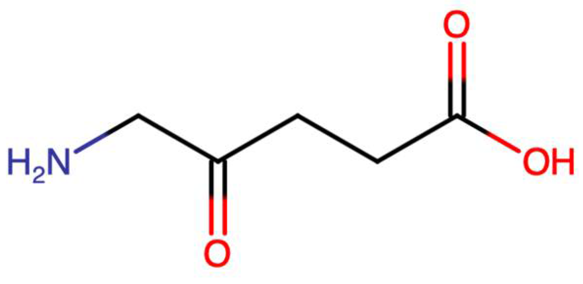
Figure 1. 5-aminolevulinic acid (5-ALA) chemical formula.
Blsf 12 00004 g001
Figure 2. Hexyl of 5-aminolevulinic acid (HAL) chemical formula.
Blsf 12 00004 g002
Figure 3. Type I and type II photosensitized reactions.
Blsf 12 00004 g003
Figure 4. Ways of bladder cancer cell death after PDT.
Blsf 12 00004 g004
Figure 5. Oxidative stress caused by PDT.
Blsf 12 00004 g005
Publisher’s Note: MDPI stays neutral with regard to jurisdictional claims in published maps and institutional affiliations.