Proceeding Paper

Oral Administration of Rauwolfia serpentina Plant Extract Mitigated Immobilization Stress-Induced Behavioral and Biochemic and Deficits in Rats †

Neurochemistry and Biochemical Neuropharmacology Research Laboratory, Department of Biochemistry, University of Karachi, Karachi 75270, Pakistan
*
Authors to whom correspondence should be addressed.
Presented at the 2nd International Electronic Conference on Nutrients, 15–31 March 2022; Available online: https://iecn2022.sciforum.net/.
Biol. Life Sci. Forum 2022, 12(1), 32; https://doi.org/10.3390/IECN2022-12393
Published: 14 March 2022
(This article belongs to the Proceedings of The 2nd International Electronic Conference on Nutrients)

Abstract

:
Objectives: Nowadays, the global population is moving towards herbal drugs, which contain bioactive compounds, to cure diseases. Rauwolfia serpentina is a medicinally important herb that is mainly effective in the treatment of hypertension and psychotic disorders. The present study was designed to investigate the effects of Rauwolfia serpentina on acute stress. The herb extract was orally administered before immobilization for 2 h only, to monitor any change in behavioral activities. We also evaluated the role of Rauwolfia serpentina in oxidative stress, including its effect on antioxidant enzymes’ activities, such as catalase and superoxide dismutase, and also on plasma glucose, corticosterone and leptin levels. Methods: Animals were divided into four groups, which were (i) saline unstressed, (ii) Rauwolfia serpentina unstressed, (iii) saline stressed and (iv) Rauwolfia serpentina stressed, which were injected accordingly with saline (1 mL/kg) or Rauwolfia serpentina (30 mg/kg). Animals of the stressed group received immobilization for 2 h. Behavioral analysis was performed after the termination of the 2 h immobilization period. Animals were then decapitated and plasma samples were collected for CAT, SOD, corticosterone, leptin and glucose estimation. Results: Results showed that Rauwolfia serpentina is an effective anxiolytic agent as it attenuates stress-induced behavioral deficits and improves locomotor activity. On the other hand, it provides positive outcomes regarding the antioxidant enzymes levels of stressed animals. Conclusion: Rauwolfia Serpentina was found to prevent the stress-induced increase in corticosterone, and an increase in the levels of endogenous leptin attenuates the stress-induced activity of the HPA axis. It is also concluded that 30 mg/kg of Rauwolfia serpentina is not sufficient to produce hypoglycemic effects. However, more studies are recommended to explain the particular action by which Rauwolfia serpentina produces its effects.

Published: 14 March 2022

1. Introduction

Stress has exhibited an imperative role in the etiology, exacerbation and cure of affective psychopathology, suggesting close interplay between the two [1]. Acute stress is a result of a traumatic event that causes a person to feel fear and helplessness [2]. A variety of diverse environmental and stressful stimuli have also been reported to alter behavioral patterns, neurotransmitter levels and oxidative damage in discrete areas of the brain [3,4]. However, the effects of stress on the brain have long been associated with the onset and exacerbation of several neuropsychiatric disorders, such as depression, anxiety, drug addiction and epilepsy [5]. Parallel studies on experimental animals showed that an uncontrollable stressor produced neurochemical changes and behavioral deficits [6,7]. Several investigators have suggested a link between oxidative stress and certain anxiety disorders, such as obsessive compulsive disorder and panic disorder, indicating that the oxidative metabolism can affect the regulation of anxiety.
Recognizing elements that contribute to neurodegenerative progression in the brain is one of the chief goals of contemporary medicine. There are several hypotheses regarding the mechanisms that lead to the damage and death of brain cells in neurodegenerative diseases [8], such as excitotoxic effects by excitatory amino acids [9], impairment in cellular energy metabolism [10,11] and oxidative stress (OS),which is caused by free radicals or other reactive molecules [9,12]. The results of many in vitro and in vivo preclinical and clinical studies have consistently demonstrated that OS is one of the crucial players in the degeneration that occurs in the nervous system. The imbalance between OS and antioxidant defense systems seems to be a universal condition in neurodegeneration [13]. Clinical and preclinical studies indicate that neurodegenerative diseases are characterized by higher levels of OS biomarkers and by lower levels of antioxidant defense biomarkers in the brain and peripheral tissues [14]. There is now increasing evidence that reactive oxygen species (ROS) generation is involved in the regulation of neurotransmission, particularly glutamate release, which most likely plays an important role in the ‘‘fight or flight response’’ [15]. Oxidative stress creates a state of cellular imbalance, in which reactive oxygen species (ROS) production surpasses the antioxidant response mechanisms that help to neutralize ROS-mediated oxidative damage to DNA, RNA and lipids, leading to innumerable pathophysiological consequences [16,17]. In order to counterbalance the free-radical-induced damage of biological molecules, antioxidant mechanisms and enzymes are activated. Superoxide dismutase (SOD), catalase (CAT) and glutathione peroxidase (GPx) were identified as antioxidant enzymes that act as the body’s first line of defense against ROS by catalyzing their conversion to less reactive or inert species [18].Consequently, research has revealed that individuals with anxiety or depression show an extensive range of abnormalities in controlling fear-related responses, suggesting that deficits in emotion regulation may be linked to neurobiological differences in response to stress [1].
Nowadays, the global population is moving towards herbal drugs, which contain bioactive compounds, to cure diseases [19]. Rauwolfia serpentina, belonging to the family Apocynaceae, is an important medicinal plant in the pharmaceutical world due to its immense therapeutic properties [20,21]. It is effective in the treatment of hypertension and psychotic disorders such asschizophrenia, anxiety, insomnia, insanity and so forth [22]. Various indole alkaloids and related constituents have been isolated from the roots of this plant, which have significant biological activities [23]. An in vitro study described the antimicrobial and antioxidant activities of the leaf extract of this plant [24]. The principle alkaloid of Rauwolfia serpentina is reserpine [24]. It is present in the root, stem and leaves of the plant. It contains less than 0.15% of reserpine and rescinnamine group alkaloids [25].
Previously, numerous studies have been reported from our laboratory that establish the capability of phytochemicals present in rice bran oil [26], olive oil [27] and the aqueous fruit extract of sea buckthorn [28] to attenuate/or reverse anxiety in rats. Similarly, our laboratory also observed that the oral administration of red rice bran oil averted haloperidol-induced anxiety syndrome in rats [29]. Conversely, oral administration of Nigella sativa (NS) and Olea europaea (OE) oil did not show anxiolytic effects in rats [30]. In continuation of our research on the plant, the present study was designed to investigate the neuroprotective effects of Rauwolfia serpentina following acute exposure to immobilization stress in rats. The herb extract was orally administered at a non-sedative dose of 30 mg/kg [31] before immobilization for 2 h to monitor any change in behavioral activities. The neuroprotective efficacy of the plant extract was assessed in terms of its potency to attenuate oxidative-stress-induced alterations of antioxidant enzymes’ activities, such as CAT and SOD, and locomotor deficits. In order to obtain an insight into the role of Rauwolfia serpentina in the HPA axis, we also monitored plasma leptin, corticosterone and glucose levels. The study establishes that Rauwolfia serpentina plant extract may have potential therapeutic significance for the management of stress and related disorders.

2. Materials and Methods

2.1. Animals

Locally bred albino Wistar rats, weighing 180–200 g and purchased from PCSIR, were housed individually on a 12 h light/dark cycle in a temperature-controlled room (24 ± 2 °C), with free access to tap water and cubes of standard rodent diet, for at least 7 days before the start of the experiment (establishing familiarity with the environment). All procedures conducted were approved by the Local Institutional Animal Care and Use Committee at the University of the Health Sciences and conducted in full compliance with the National Institutes of Health Guide for the Care and Use of Laboratory Animals.

2.2. Prepararation of Plant Extract

Thirty grams of ground powder of the roots of Rauwolfia serpentina was extracted with methanol (1 L; 95%) overnight and filtered through Whatman No.1 filter paper twice. The filtrate was then concentrated at 40 °C till dryness in a rotary vacuum evaporator (Eyela-NE) to obtain a brown residue that was referred to as methanolic root extract (MREt) [22]. This procedure yielded 3–4% (w/w) of the dry root. The MREt was stored in an airtight container in a refrigerator, below 10 °C, until use.

2.3. Immobilization Procedure

The animals of the stress groups were subjected to a single exposure of immobilization stress for 2 h. Immobilization was done in a separate room to prevent unstressed animals from being placed under stressful conditions due to disturbance. The animals were immobilized by an approved procedure [32,33]. Wire grids fitted with a Perspex plate, as described earlier [33], were used. Immobilization was affected by pressing the legs of the rats through the gaps in the metal grid and taping them together with zincoxide plaster. Hind limbs were also taped and the head of the animal rested on the Perspex plate. After 2 h of immobilization stress, animals were released by applying acetone to the tape and returned to their home cage.

2.4. Behavioral Analysis

2.4.1. Activity in a Novel Environment (Open Field)

The locomotor activity of control and test rats was monitored in an open field apparatus. The open field was a square area of 76 × 76 cm with opaque walls of 42 cm height. The floor was divided by lines into 25 equal squares. The test was performed in a quiet room under white light to avoid any noise effect, as described earlier [34,35]. Animals were placed in the center square of the open field (one at a time). Activity in the open field was determined by counting the number of squares crossed for 5 min [36]. Exploratory activity of control rats and test rats was monitored in a balance design to avoid order effects.

2.4.2. Light–Dark Transition Test

The light–dark transition test, a behavioral test used to monitor the anxiolytic effects of drugs in preclinical investigations, is based on the innate aversion of rodents to brightly illuminated areas. The test procedure was essentially the same as described earlier [37]. The apparatus used in the present investigation was a two-compartment light–dark box. Both the light compartments (composed of transparent plastic) measured 26 × 26 × 26 cm and access between the two compartments was provided by a 12 × 12 cm passageway. The experiment was performed in a quiet, air-conditioned room, and the apparatus was placed under white light. An animal was introduced into the apparatus via the light compartment. Cumulative time spent in the light compartment and the numbers of entries into the light compartment were monitored for a period of 5 min. An entry was defined as all four paws being positioned within the light compartment. The degree of anxiety was assessed by a decrease in time passed in the light compartment and also by a decrease in the number of entries made to the light compartment.

2.5. Blood Sample Collection

Blood was collected from rats in heparinized centrifuge tubes. Centrifugation was done for 10 min. Plasma was collected and stored at −70 Celsius till biochemical estimation of the plasma glucose concentration in mg/dL, corticosterone concentration in μg%, leptin concentration in ng/mL and catalase and superoxide dismutase.

2.6. Biochemical Estimation of Glucose, Catalase and Superoxide Dismutase in Plasma

2.6.1. Determination of Catalase (EC1.11.1.6)

CAT activity was estimated by the method of Patterson [38]. The decomposition of H2O2 was measured at 240 nm, taking De at 240 nm as 43.6 mMcm–1. Reaction mixture (3.0 mL) consisted of 10.5 mM H2O2 in 0.05 M potassium phosphate buffer (pH 7.0) and the reaction was initiated after the addition of 0.1 mL enzyme extract at 25 °C. The decrease in absorbance at 240 nm was used to calculate the activity. One unit of CAT activity is defined as the amount of enzyme that catalyzes the conversion of 1 mM of H2O2 min–1 at 25 °C [39].

2.6.2. Determination of Superoxide Dismutase (EC.1.15.1.1)

The assay for SOD activity was performed by the method of [40]. The assay mixture consisted of 27.0 mL of 0.05 M potassium phosphate buffer (pH 7.8), 1.5 mL of L-methionine (300 mg per 2.7 mL), 1.0 mL of nitroblue tetrazolium salt (14.4 mg per 10 mL) and 0.75 mL of Triton X-100. Aliquots (1.0 mL) of this mixture were delivered into small glass tubes, followed by the addition of 20 mL enzyme extract and 10 mL of riboflavin (4.4 mg per 100 mL). The cocktail was mixed and then illuminated for 15 min in an aluminum foil-lined box, containing 25 W fluorescent tubes. In a control tube, the sample was replaced by 20 mL of buffer and the absorbance was measured at 560 nm. The reaction was stopped by switching off the light and placing the tubes in the dark. The increase in absorbance due to the formation of formazan was measured at 560 nm. Under the described conditions, the increase in absorbance in the control was taken as 100% and the enzyme activity in the samples was calculated by determining the percentage inhibition per minute. One unit of SOD is the amount of enzyme that causes a 50% inhibition of the rate for reduction of nitroblue tetrazolium salt under the conditions of the assay [39].

2.6.3. Estimation of Glucose in Plasma by GOD-PAP Method

The concentration of glucose in plasma was measured by using the glucose oxidase method (GOD-PAP, Solo per USO diagnostico in vitro).

2.6.4. Estimation of Leptin and Corticosterone in Plasma by ELISAKit

Animals were decapitated, followed by the collection of blood in heparinized centrifuge tubes. Centrifugation proceeded for 20 min at 2000× g and 4 °C to obtain plasma. The samples were stored at −70 degrees Celsius until the assay of plasma leptin and corticosterone using an ELISA kit (Cat # EZRL-83K).

3. Experimental Protocol

Twenty-four animals randomly divided into two equal groups of 12 each were assigned to the unstressed and stressed groups. These animals were further divided into four groups of 6 rats each that were designated as (i) saline unstressed, (ii)Rauwolfia serpentina unstressed, (iii)saline stressed and(iv)Rauwolfia serpentina stressed, which were orally administered with saline (1 mL/kg) or Rauwolfia serpentina (30 mg/kg). Animals of the stressed group were immobilized for 2 h, commencing between 9:00 and 11:00 h. Animals of the unstressed group were left in their home cage during this time. Behavioral activities were monitored in open field activity and light–dark transition box after the termination of the 2 h immobilization period. Plasma samples were collected for CAT, SOD, corticosterone, glucose and leptin estimation. The experiment was performed in a balanced design in such a way that control and test rats were measured alternately to avoid an order effect.

Statistical Analysis

Values are presented as mean ± SD. Data were analyzed by two-way ANOVA. Post hoc analysis was done by Newman–Keuls test. Values of p < 0.01 were considered significant.

4. Results

Figure 1A shows changes in motor activity in a novel environment in animals orally administered with Rauwolfia serpentina for 2 h before exposing the animal to acute immobilization stress for 2 h. Analysis of the data on latency to move (Figure 1A) showed significant effects of stress (F = 7.737 p < 0.01 df1,20) and Rauwolfia serpentina (F = 7.737 p < 0.01 df1,20), as well as the interaction between two factors (F = 8.796 p < 0.01 df2,20).
Post hoc analysis by Newman–Keuls revealed that the administration of Rauwolfia serpentina at a dose of (30 mg/kg) to stressed rats resulted in an increase in latency to move as compared to unstressed rats. On the other hand, saline + stressed rats did not show any significant difference in latency to move as compared to unstressed rats. Rauwolfia serpentina +stressed rats showed an increase in latency to move in comparison with saline + stressed rats.
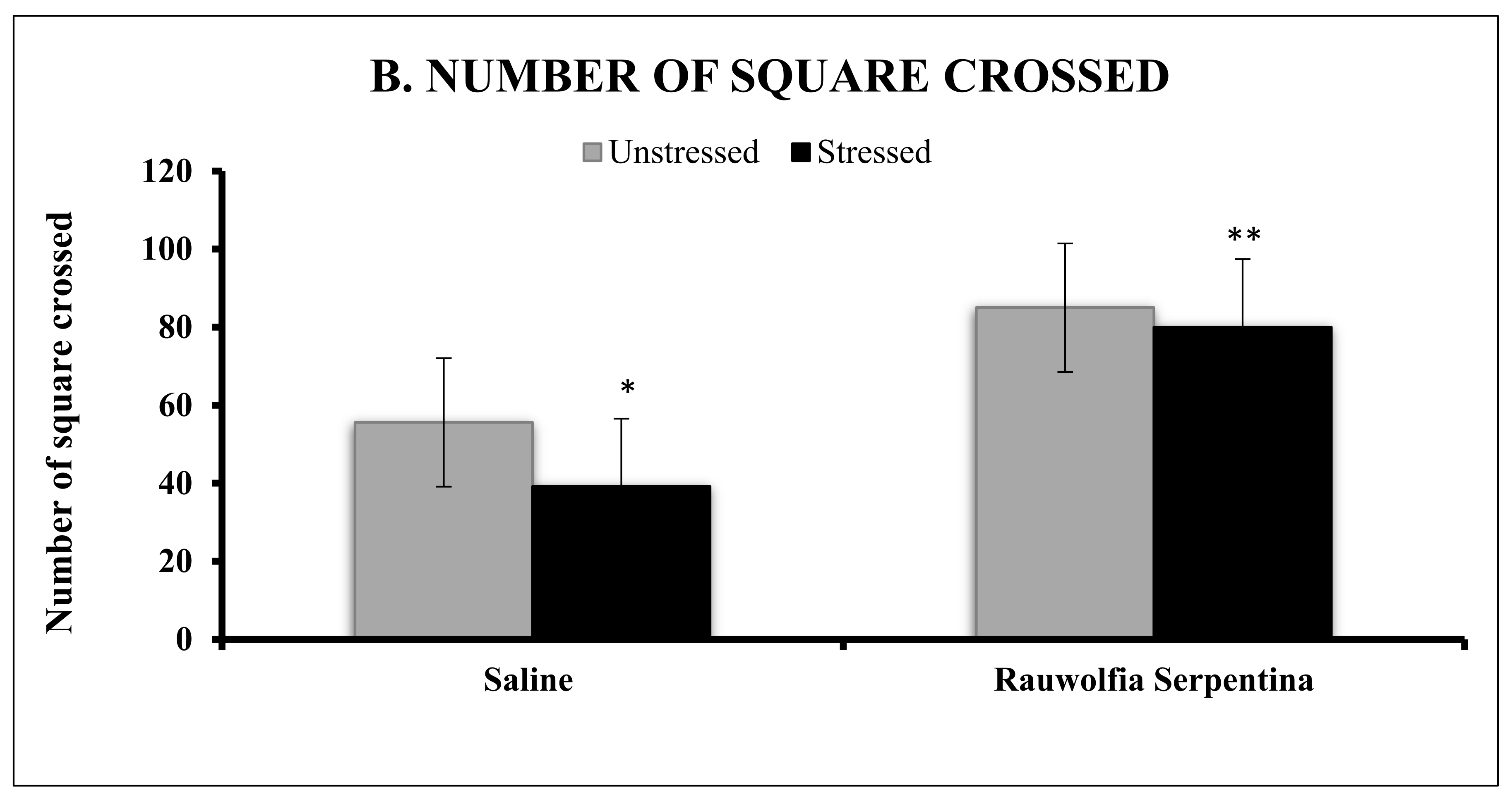
Figure 1B shows changes in motor activity in a novel environment in animals orally administered with Rauwolfia serpentina for 2 h before exposing the animal to acute immobilization stress for 2 h. Analysis of the data on the number of squares crossed (Figure 1B) showed significant effects of stress (F = 4.017 p < 0.05 df1,20) and Rauwolfia serpentina (F = 43.136 p < 0.01 df1,20). The interaction between the two factors was not significant (F = 1.143 N.S.).
Post hoc analysis by Newman–Keuls showed a decreased number of squares crossed by saline + stressed rats but not Rauwolfia serpentina + stressed rats. Rats treated with Rauwolfia serpentina alone showed increased locomotor activity in the open field. On the other hand, stress-induced decreases in locomotor activity were reversed in Rauwolfia serpentina-administered stressed rats.
Figure 2A shows changes in behavior in the light–dark transition test in animals orally administered with Rauwolfia serpentina for 2 h before exposing animals to acute immobilization stress for 2 h. Analysis of the data on entries in the light box (Figure 2A) showed significant effects of stress (F = 16.298 p < 0.01 df1,20)and an interaction between the two factors (F = 5.391 p < 0.01 df1,20). Effects of Rauwolfia serpentina were not significant (F = 1.589 N.S.).
Post hoc analysis by Newman–Keuls showed a decreased number of entries in the light–dark transition box in Rauwolfia serpentina + stressed and saline +stressed animals as compared to their respective controls. Rats treated with Rauwolfia serpentina alone showed an increased number of entries in the light–dark transition box. On the other hand, stress-induced decreases in the number of entries in the light–dark box were reversed in Rauwolfia serpentina-administered rats.
Figure 2B shows changes in behavior in the light–dark transition test in animals orally administered with Rauwolfia serpentina for2 h before exposing animals to acute immobilization stress for 2 h. Analysis of the data on time spent in the light box (Figure 2B) showed non-significant effects of stress (F = 1.146 N.S.) and significant effects of Rauwolfia serpentina (F = 20.861 p < 0.01 df1,20), as well as an interaction between the two factors (F = 7.740 p < 0.01 df2,20).
Post hoc analysis by Newman–Keuls showed decreased time spent in the light–dark transition box (sec) in saline +stressed rats but significantly increased in Rauwolfia serpentina +stressed rats. Rauwolfia serpentina alone did not increase locomotor activity in the light–dark transition box. On the other hand, the stress-induced decrease in locomotor activity was reversed in Rauwolfia serpentina-administered stressed rats.
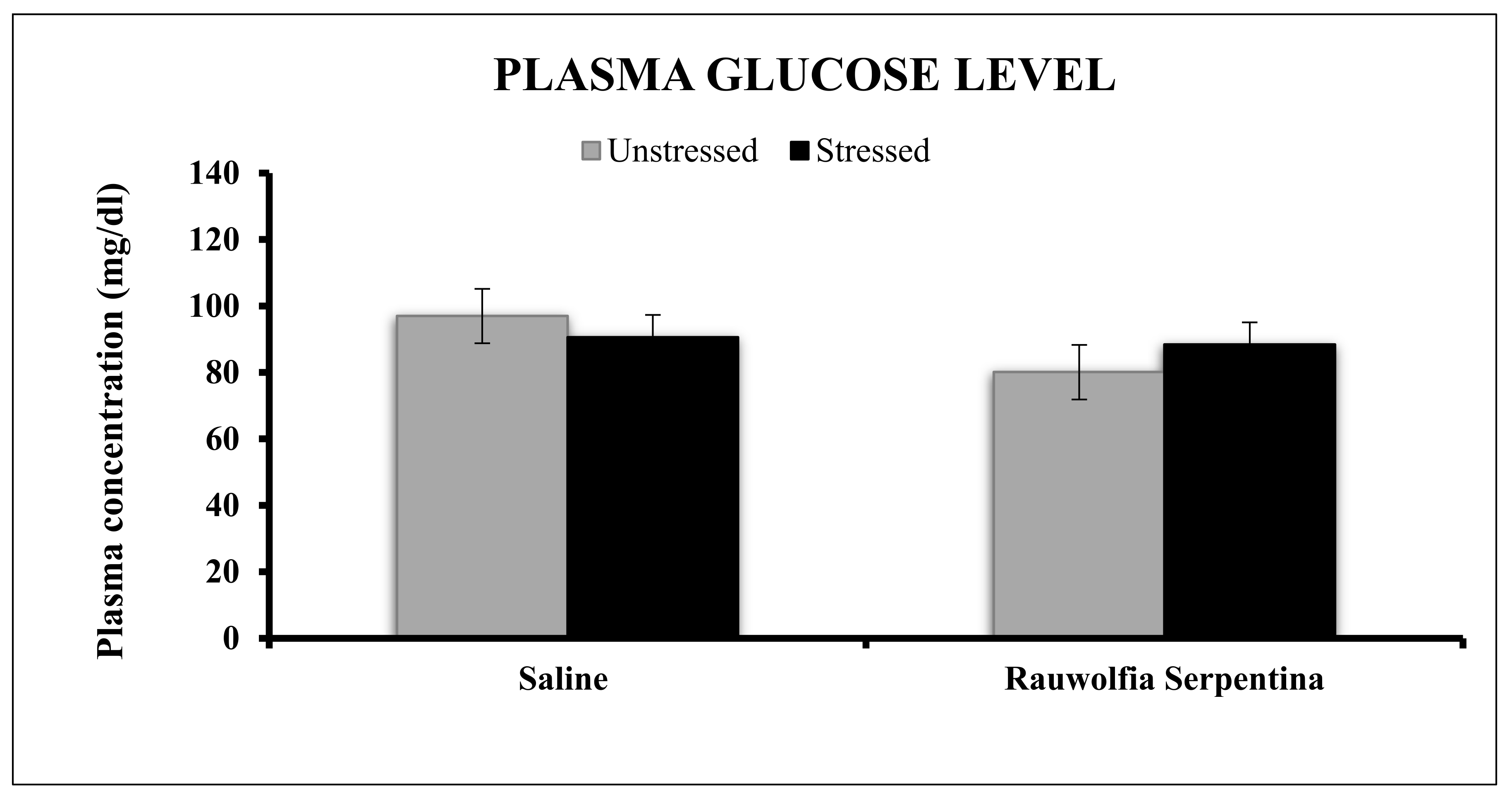
Figure 3 shows the effects of stress with the oral administration of Rauwolfia serpentina on the plasma glucose level. Analysis of the data on glucose level (Figure 3) showed non-significant effects of stress (F = 0.566 N.S.) and Rauwolfia serpentina (F = 2.144 N.S.), as well as the interaction between the two (F = 3.142 p < 0.005 df2,20).
Post hoc analysis by Newman–Keuls test revealed that the concentration of plasma glucose was not significant in all groups.
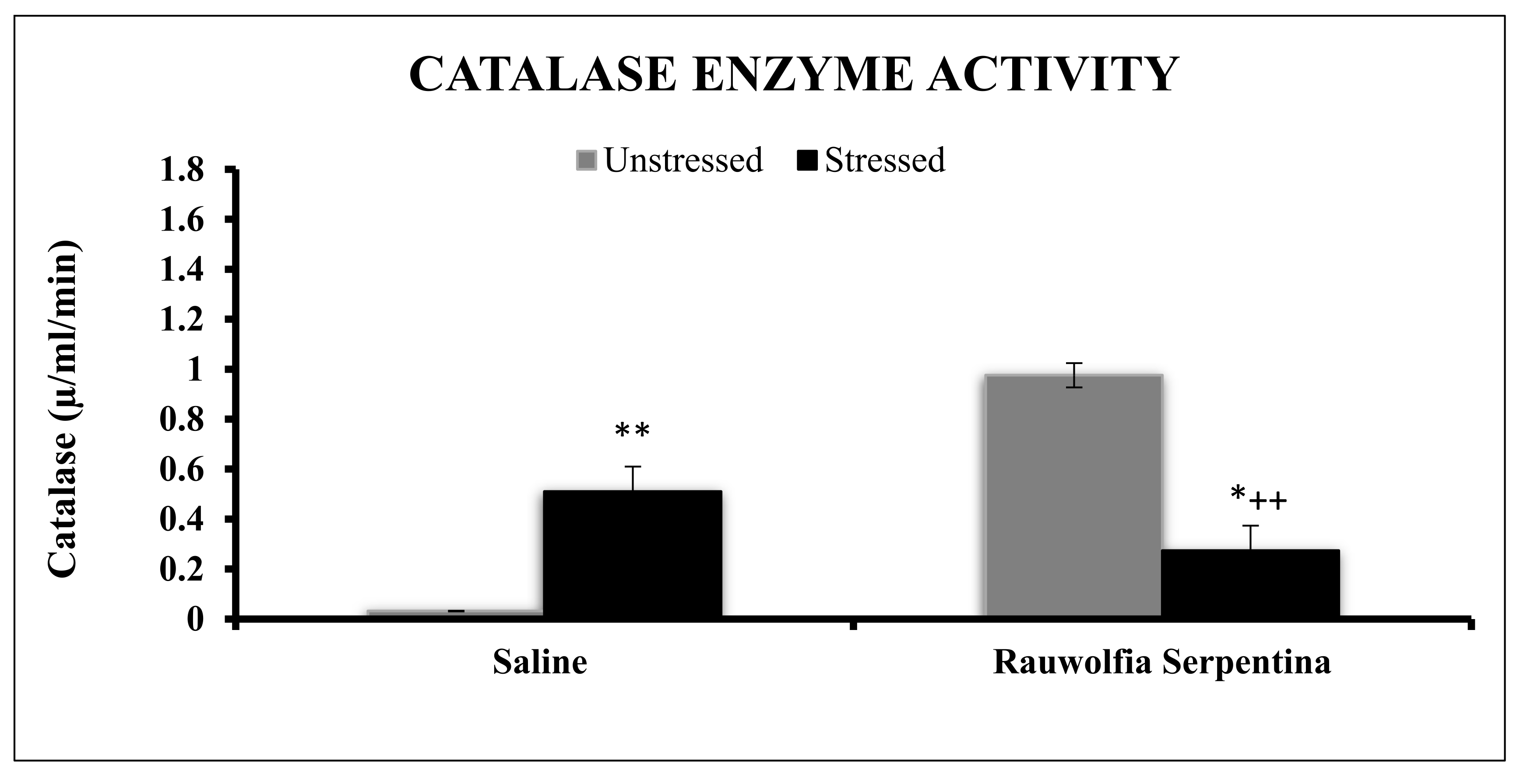
Figure 4 shows the effects of stress with the oral administration of Rauwolfia serpentina on plasma CAT activity. Analysis of the data on CAT activity (Figure 4) showed non-significant effects of stress (F = 0.508 N.S) and an interaction between the two factors (F = 2.802 N.S.). Effects of Rauwolfia serpentina were significant (F = 4.858 p < 0.05 df2,20).
Post hoc analysis by Newman–Keuls revealed that the activity of CAT was significantly increased in saline + stressed rats but significantly decreased in Rauwolfia serpentina + stressed rats. Rauwolfia serpentina administration alone increased CAT activity. On the other hand, the stress-induced increase in CAT activity was attenuated in Rauwolfia serpentina-administered stressed rats.
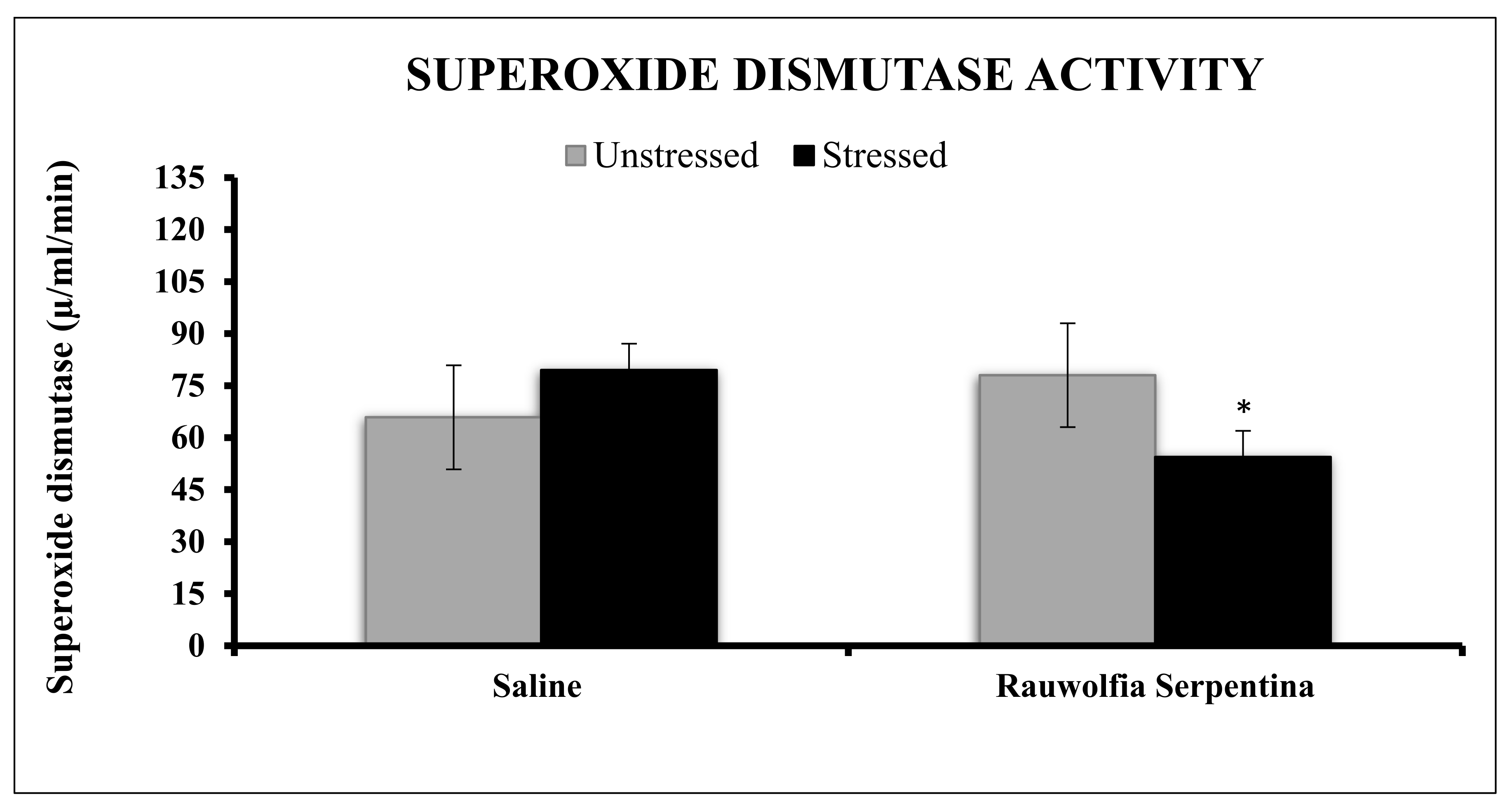
Figure 5 shows the effects of stress with the oral administration of Rauwolfia serpentina on plasma SOD activity. Analysis of the data on SOD activity (Figure 5) showed significant effects of stress (F = 3.282 p < 0.05 df1,20). Effects of Rauwolfia serpentina (F = 2.256 N.S) and the interaction between the two were not significant (F = 1.121 N.S.).
Post hoc analysis by Newman–Keuls showed that the activity of SOD was significantly decreased in Rauwolfia serpentina+ stressed rats. However, Rauwolfia serpentina alone did not alter the activity of SOD. On the other hand, the activity of SOD was not significant in other groups.
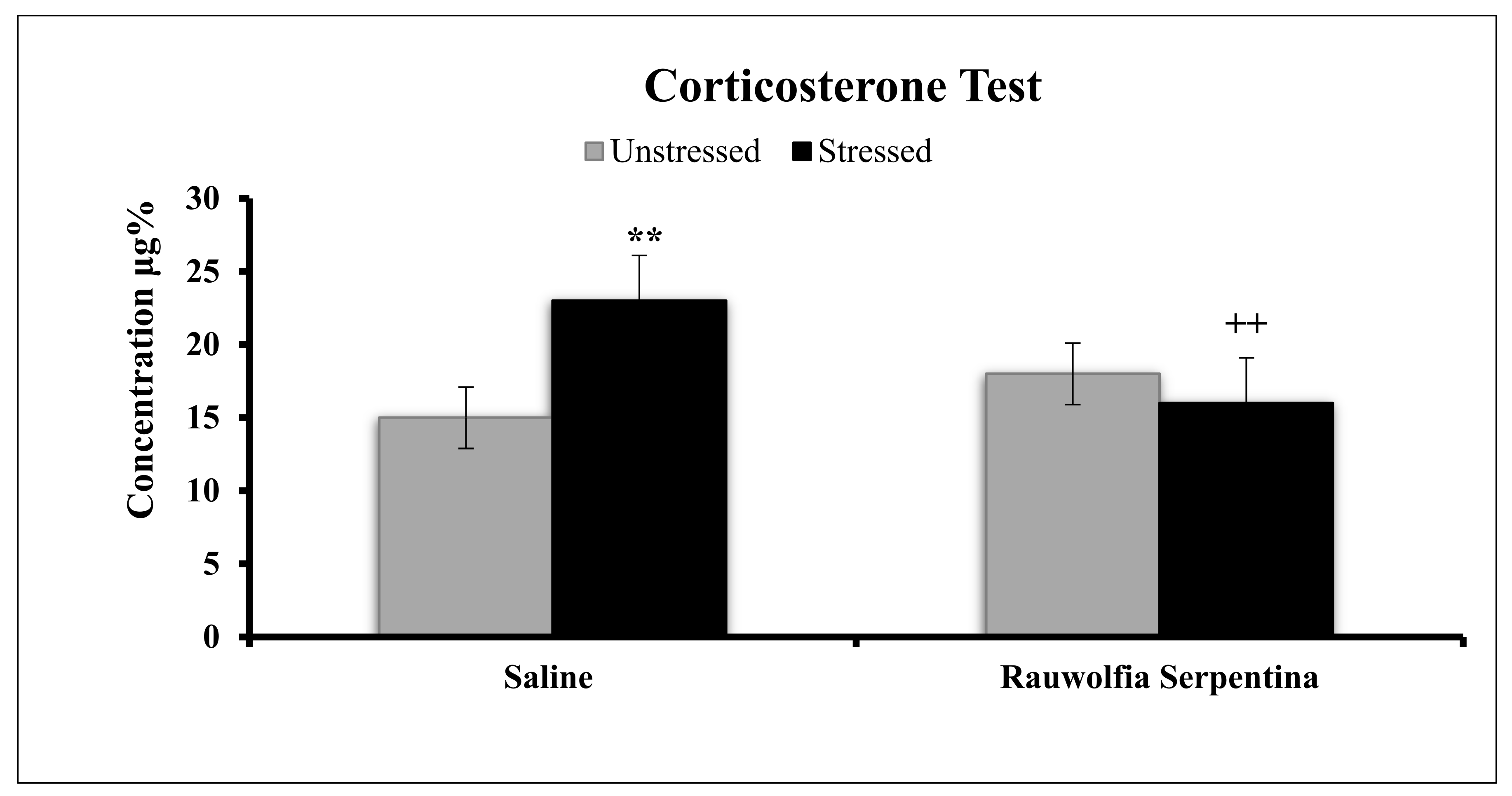
Figure 6 shows the effects of stress with the oral administration of Rauwolfia serpentina on the plasma corticosterone level. Analysis of the data on corticosterone levels (Figure 6) showed significant effects of stress (F = 9.0 df1,20 p < 0.01), Rauwolfia serpentina (F = 7.92 df2,20 p < 0.01), as well as the interaction between the two (F = 26.01 df1,20 p < 0.01).
Post hoc analysis by Newman–Keuls showed significantly increased levels of corticosterone in saline +stressed animals but decreased in Rauwolfia serpentina +stressed animals. On the other hand, an immobilization-stress-induced increase in corticosterone did not occur in single Rauwolfia serpentina administered animals.
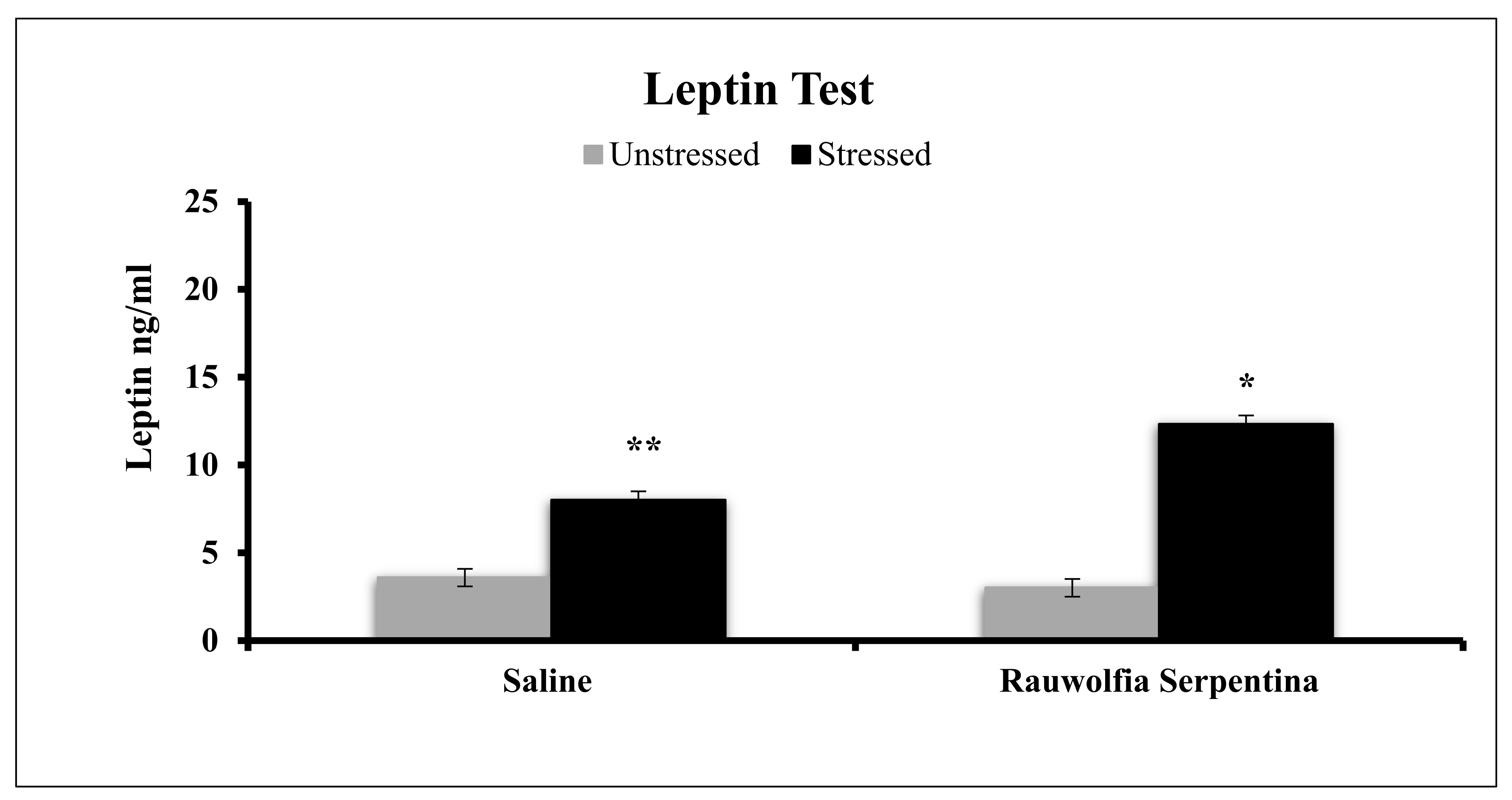
Figure 7 shows the effects of stress with the oral administration of Rauwolfia serpentina on plasma leptin levels. Analysis of the data on leptin levels (Figure 7) showed significant effects of stress (F = 9.0 df1,20 p < 0.01) and Rauwolfia serpentina (F = 7.92 df2,20 p < 0.05). A non-significant effect of the interaction between the two factors was noted (F = 26.01 N.S.).
Post hoc analysis by Newman–Keuls showed significant increases in both saline +stressed and Rauwolfia serpentina +stressed animals as compared to their unstressed control rats, respectively. Rauwolfia serpentina +stressed rats showed increase levels of leptin in comparison with saline + stressed rats.

5. Discussion

Experiencing stress is an inexorable part of everyday life and plays a significant role in shaping adaptive behavior [41]. Acute exposure to immobilization stress has been reported to impair motor activity, cause memory dysfunction, modulate anxiety [42] and pain perception [43] and elicit depression-like behaviors [44] in animals. The goal of the current study was to observe the neuroprotective effects of Rauwolfia serpentina on the behavioral activity of animals in a novel environment and light–dark transition box activity following acute exposure to 2 h immobilization stress in rats. Alterations in the levels of corticosterone, glucose and leptin were also measured to establish a link between oxidative stress and the HPA axis following administration of the plant extract. We also probed the concentrations of antioxidant enzymes such as catalase and superoxide dismutase to delineate the relationship of oxidative stress with behavioral deficits in rats. A consistent finding of the present study is that the oral administration of Rauwolfia serpentina plant extracts attenuated immobilization-stress-induced behavioral deficits and alterations in antioxidant enzymes levels in the rats. Moreover, plasma leptin and corticosterone were also mitigated in these rats, suggesting a role of the antioxidant components of the plant extract, which may elicit neuroprotective effects.
In the present study, we examined the effects of Rauwolfia serpentina on the modulation of immobilization-stress-induced behavioral deficits with two extensively used behavioral models of anxiety-like behavior, including the open field and light–dark transition test. These tests may be useful to examine anxiolytic-like or anxiogenic-like activity in mice [45]. The present results showed that 2 h immobilization exhibited a significant decrease in the number of squares crossed but not latency to move in the open field as compared to the unstressed animals (Figure 1). Our findings are consistent with previous studies that showed that acute exposure to (2 h) immobilization stress produces anxiety-like symptoms in rats, and the animals did not explore rapidly enough to find and enter the dark compartment; instead, they tended to freeze and remain immobile for the majority of the test session [46]. Therefore, immobilized stressed animals avoided exploring the new environment in the light–dark box as well as in the open field exploration test. Conversely, administration of the oral Rauwolfia serpentina extract alone increased the number of squares crossed in the open field in rats. On the other hand, oral administration of Rauwolfia serpentina extract attenuated 2 h immobilization-stress-induced decreases in locomotor activity in the open field. Similarly, a significant increase in the numbers of entries in the light box and time spent in the light compartment of the light–dark transition box were also observed in these animals, suggesting a reduction in novel-environment-induced anxiogenic effects (Figure 2). Therefore, this anxiolytic effect of Rauwolfia serpentina plant extract could be explainable in terms of the presence of numerous phytochemical compounds or secondary metabolites, such as alkaloids, carbohydrates, flavonoids, glycosides, phlobatannins, phenols, resins, saponins, sterols, tannins and terpenes in the plant extract [24,47,48]. The present results are therefore in agreement with previous findings that the phenolic antioxidants present in plant extracts could produce anxiolytic effects [49].
Oxidative stress has been implicated in the response to stress [50] and in the pathogenesis of neurologic and psychiatric diseases [51]. An antioxidant is a substance that is present at low concentrations and significantly inhibits or prevents the oxidation of the oxidizable substrate [52]. Endogenous antioxidants play a vital role in conserving optimal cellular functions. However, endogenous antioxidants may not be adequate under certain conditions that could promote oxidative stress [53,54], as observed in the current results (Figure 4 and Figure 5). Thus, elevated superoxide dismutase and catalase activities were found in rats immobilized for 2 h compared to control animals, signifying that acute exposure to stress can promote the formation of ROS and lead to oxidative stress. In such cases, dietary antioxidants should be supplied to maintain optimal cellular functions. Some antioxidants can interact with other antioxidants in order to regenerate their original properties. This process is referred to as the “antioxidant network” [55]. It has been suggested that a diet rich in antioxidants can bring health benefits [56] and a lot of interest is directed towards assessing the antioxidant capacity of natural products. In recent years, many studies evidenced that the majority of the antioxidant activity of plants may be from compounds such as phenolic acids, flavonoids and ascorbic acids, which can provide protection against ROS [57,58,59,60]. From this perspective, the plant extract containing flavonoids and the ascorbic acid content of Rauwolfia serpentina exhibit antioxidant capacity, which expands its nutraceutical value [61]. In the present study, oral administration of Rauwolfia serpentina (Figure 4 and Figure 5) attenuated the immobilization-induced increase in the antioxidant enzymes CAT and SOD’s activities, suggesting the antioxidant capacity of the plant extract components, particularly flavonoids and ascorbic acid. Conversely, we also observed that oral administration of Rauwolfia serpentina alone increases CAT but not SOD activity. It has been indicated that the balance between pro-oxidant and antioxidant compounds moderately favors pro-oxidants under physiological conditions. Consequently, it leads to slight oxidative stress and requires the intervention of the endogenous antioxidant systems of the organism [62]. It seems possible that the alkaloid and flavonoid components of Rauwolfia serpentina plant extract could contribute, along with the endogenous antioxidant system, to counteracting oxidative stress under basal conditions.
It is well recognized that exposure to acute stress causes the formation of free radicals, which may lead to oxidative damages [63]. The HPA axis is the neuroendocrine system that regulates responses to stress [64]. The production of high levels of free radicals in the glands that comprise the HPA axis is related to the activation of the stress response system [65,66,67]. In terms of the activity of the HPA axis, it is now eminent that neurons in the paraventricular nucleus (PVN) of the hypothalamus release corticotropin-releasing factor (CRF) to stimulate the synthesis and release of adrenocorticotropin (ACTH) from the anterior pituitary. ACTH then travels to the adrenal gland and induces the rapid [68] release of corticosteroids, which later activate various physiological processes to assist an organism in coping with a stressful situation and reinstate homeostasis under a potentially threatening condition [34,69,70]. The present investigation demonstrates that animals subjected to immobilization stress exhibit increased corticosterone levels (Figure 6). This is not unexpected, since it has been previously reported that acute restraint stress [71] and immobilization stress [72] increase corticosterone levels, and this is considered to be an important indicator of stress [73,74,75]. However, oral administration of Rauwolfia serpentina alone did not alter corticosterone levels as compared to saline plus unstressed animals. Conversely, immobilization-induced elevated levels of corticosterone were attenuated in Rauwolfia serpentina-treated animals (Figure 6). Previously, it was reported that the chronically immobilized [63] and restraint [76] stress-induced attenuation of corticosterone levels is explainable in terms of anti-stress activity. It is therefore interesting to relate the Rauwolfia serpentina-induced modulation of corticosterone levels in terms of suppressing HPA mobilization in response to stress by normalizing elevated plasma corticosterone levels back to baseline. Thus, oral administration of Rauwolfia Serpentina reduced the adverse effects of acute exposure to (2 h) immobilization stress and is thought to be beneficial for the body to prevent stress-induced damages.
As per clinical evidence, elevated levels of corticosterone in response to stress also increase the plasma glucose concentration [77,78]. From previous studies, it was reported that stress causes an increase in plasma glucocorticoid levels [79,80,81], which stimulates liver gluconeogenesis, which then leads to elevated blood glucose [82]. Regardless of the wide use of glucose as an indicator of stress, some authors [83,84] have emphasizedthatcarehastobetakenwhenusingplasmaglucoseastheonlyindicator. It has been reported that glucose measurements show many inconsistencies and should be a complement to stress tests rather than a main indicator [85]. In the present results, acute (2 h) exposure to immobilization stress was unable to alter the plasma glucose concentration. Previously, preclinical studies on the antidiabetic potential of the methanolic root extract of Rauwolfia serpentina have been reported. It was found to be effective in lowering blood glucose levels [31]. However, in our findings, oral administration of Rauwolfia serpentina did not show any significant decrease in the levels of glucose as compared to the saline plus unstressed rats (Figure 3). Similarly, treatment with Rauwolfia serpentina also did not alter stress-induced changes in glucose concentration in rats (Figure 3). It seems that 30 mg/kg of Rauwolfia serpentina was not sufficient to produce significant hypoglycemic effects in our present study paradigm. The reason for the variation between our observation and that in the mentioned study is unclear, but it may be due to the discrepancy in the nature of the stressful or ambient environments.
We report, for the first time, the potential therapeutic role of Rauwolfia serpentina in endogenous leptin and corticosterone levels. Leptin secretion is basically under the influence of neural and hormonal control [86,87,88]. The influence of leptin on the HPA axis is one of the mechanisms by which leptin can improve stress controllability to produce antidepressant and anxiolytic-like effects. Previously, preclinical studies reported that exposure to 1 h immobilization [89], 10 min forced swimming [90] and 120 dB noise [91] showed an increase in circulating levels of leptin. These studies are consistent with our present data where in exposure to acute (2 h) immobilization stress resulted in a significant increase in the circulating levels of leptin (Figure 7). As many components of the HPA axis contain leptin receptors, it seems promising that systemically circulating leptin can alter the stress response at every focal point of the axis [92]. On the other hand, stress-induced releases of corticosterone have the opposite influence on leptin expression in adipocytes and its secretion into the blood circulation. It has been reported that pretreatment with recombinant mouse leptin inhibited the stress-mediated stimulation of plasma ACTH and corticosterone in mice [93], and this inhibitory effect could be produced by receptors in the hypothalamus. The present results showed that oral administration of Rauwolfia serpentina significantly augments immobilization-stress-induced increases in plasma leptin levels (Figure 7) but inhibits corticosterone levels (Figure 6). It is therefore suggested that leptin could elicit a feedback effect over the activity of the HPA axis. Thus, the role of leptin in HPA axis functioning suggests that their relationship is bidirectional [92]. However, a role of leptin in alleviating stress perception is also apparent from studies reporting anxiolytic-like effects of pharmacological doses of exogenous leptin in rodent models of anxiety and an inhibition of stress-induced anxiety in these models [93]. It has also been reported that conventional potential anxiolytic compounds inhibited the corticosterone response to an acute stressor [94,95] and reversed stress-induced behavioral deficits [96,97]. Similarly, we found that oral administration of Rauwolfia serpentina reversed acute (2 h) immobilization-stress-induced behavioral deficits (Figure 1 and Figure 2). It is therefore suggested that the oral administration of the plant extract could possibly elicits an anxiolytic-like effect (Figure 1 and Figure 2) by modulating endogenous leptin levels and thus inhibiting the stress-induced activation of the HPA axis.
We suggest that Rauwolfia serpentina has potential to antagonize the adverse effects of acute (2 h) immobilization stress by reducing stress perception. Despite an apparently promising role in reducing stress perception, the molecular mechanism underlying the acute anxiolytic effects of the oral administration of Rauwolfia serpentina plant extract remains to be determined. Future studies are also needed to determine the effects of the oral administration of Rauwolfia serpentina plant extract before and after exposure to unpredictable stress perception to further evaluate its potential as an anxiolytic compound, and this may facilitate the development of alternative treatment strategies for stress-related disorders including anxiety and depression.

6. Conclusions

The present study concludes that Rauwolfia serpentina is an effective anxiolytic agent as it attenuates stress-induced behavioral deficits and improves locomotor activity. The majority of the components present in Rauwolfia serpentina are beneficial and provide positive outcomes regarding the antioxidant enzyme levels of restrained animals, but in the case of unstrained animals, it showed increased antioxidant enzyme levels that might be due to the presence of any alkaloid. On the other hand, our results showed that Rauwolfia serpentina was found to prevent the stress-induced increase in corticosterone. Moreover, an increase in the levels of endogenous leptin attenuates the stress-induced activity of the HPA axis and reverses the adverse effects of acute stress. It is also concluded that 30 mg/kg of Rauwolfia serpentina was not sufficient to produce hypoglycemic effects. However, more studies are recommended to explain the particular action by which Rauwolfia serpentina produces its effects.

Author Contributions

W.B.A. and E.S. designed the study. W.B.A., S.K., N.J., M.M. and N.M. conducted the laboratory experiments. W.B.A. and N.J. contributed to laboratory analyses. E.S. and W.B.A. analyzed the data. W.B.A. wrote the manuscript and E.S. and D.J.H. commented on the manuscript. All authors have read and agreed to the published version of the manuscript.

Funding

This research was funded by University of Karachi.

Institutional Review Board Statement

The study was designed and conducted in accordance with the National Institute of Health (NIH) guidelines for the Care and Use of Laboratory Animals and a protocol (ASP No.: 2018-0001) approved by the Institutional Committee for Animal Care and Use.

Conflicts of Interest

The authors declare no conflict of interest.

Abbreviations

CatalaseCAT
Superoxide dismutaseSOD
Oxidative stressOS
Reactive oxygen species ROS
Glutathione peroxidase GPx
Nigella sativa NS
Olea europaea OE
Methanolic root extract MREt
Paraventricular nucleus PVN
Corticotropin-releasing factor CRF
Adrenocorticotropin hormoneACTH

References

  1. Raio, C.M.; Phelps, E.A. The influence of acute stress on the regulation of conditioned fear. Neurobiol. Stress 2015, 1, 134–146. [Google Scholar] [CrossRef] [PubMed] [Green Version]
  2. Robbins, P.R. Coping with Stress: Commonsense Strategies; McFarland: Jefferson, NC, USA, 2007; ISBN 978-0-7864-2875-5. [Google Scholar]
  3. Dhir, A.; Padi, S.S.V.; Naidu, P.S.; Kulkarni, S.K. Protective effect of naproxen (non-selective COX-inhibitor) or rofecoxib (selective COX-2 inhibitor) on immobilization stress-induced behavioral and biochemical alterations in mice. Eur. J. Pharmacol. 2006, 535, 192–198. [Google Scholar] [CrossRef]
  4. Imbe, H.; Iwai-Liao, Y.; Senba, E. Stress-induced hyperalgesia: Animal models and putative mechanisms. Front. Biosci. 2006, 11, 2179–2192. [Google Scholar] [CrossRef] [PubMed] [Green Version]
  5. McEwen, B.S. The Ever-Changing Brain: Cellular and Molecular Mechanisms for the Effects of Stressful Experiences. Dev. Neurobiol. 2012, 72, 878–890. [Google Scholar] [CrossRef] [Green Version]
  6. Weiss, J.M.; Goodman, P.A.; Losito, B.G.; Corrigan, S.; Charry, J.M.; Bailey, W.H. Behavioral depression produced by an uncontrollable stressor: Relationship to norepinephrine, dopamine, and serotonin levels in various regions of rat brain. Brain Res. Rev. 1981, 3, 167–205. [Google Scholar] [CrossRef]
  7. Parveen, T.; Haider, S.; Mumtaz, W.; Razi, F.; Tabassum, S.; Haleem, D.J. Attenuation of stress-induced behavioral deficits by lithium administration via serotonin metabolism. Pharmacol. Rep. 2013, 65, 336–342. [Google Scholar] [CrossRef]
  8. Day, P. Killers in the Brain: Essays in Science and Technology from the Royal Institution; Oxford University Press: Oxford, UK, 1999; ISBN 978-0-19-850540-2. [Google Scholar]
  9. Doble, A. The role of excitotoxicity in neurodegenerative disease: Implications for therapy. Pharmacol. Ther. 1999, 81, 163–221. [Google Scholar] [CrossRef]
  10. Beal, M.F.; Hyman, B.T.; Koroshetz, W. Do defecs in mitochondrial energy metabolism underlie the pathology of neurodegenerative diseases? Trends Neurosci. 1993, 16, 125–131. [Google Scholar] [CrossRef]
  11. Beal, M.F. Mitochondrial dysfunction in neurodegenerative diseases. Biochim. Biophys. Acta (BBA)-Bioenerg. 1998, 1366, 211–223. [Google Scholar] [CrossRef] [Green Version]
  12. Barnham, K.J.; Masters, C.L.; Bush, A.I. Neurodegenerative diseases and oxidative stress. Nat. Rev. Drug Discov. 2004, 3, 205–214. [Google Scholar] [CrossRef]
  13. Ji, L.L. Exercise and oxidative stress: Role of the cellular antioxidant systems. Exerc. Sport Sci. Rev. 1995, 23, 135–166. [Google Scholar] [CrossRef] [PubMed]
  14. Niedzielska, E.; Smaga, I.; Gawlik, M.; Moniczewski, A.; Stankowicz, P.; Pera, J.; Filip, M. Oxidative Stress in Neurodegenerative Diseases. Mol. Neurobiol. 2016, 53, 4094–4125. [Google Scholar] [CrossRef] [PubMed] [Green Version]
  15. Schiavone, S.; Jaquet, V.; Trabace, L.; Krause, K.-H. Severe Life Stress and Oxidative Stress in the Brain: From Animal Models to Human Pathology. Antioxid. Redox Signal. 2013, 18, 1475–1490. [Google Scholar] [CrossRef] [PubMed] [Green Version]
  16. Poulsen, H.E.; Specht, E.; Broedbaek, K.; Henriksen, T.; Ellervik, C.; Mandrup-Poulsen, T.; Tonnesen, M.; Nielsen, P.E.; Andersen, H.U.; Weimann, A. RNA modifications by oxidation: A novel disease mechanism? Free Radic. Biol. Med. 2012, 52, 1353–1361. [Google Scholar] [CrossRef] [PubMed]
  17. Furukawa, S.; Fujita, T.; Shimabukuro, M.; Iwaki, M.; Yamada, Y.; Nakajima, Y.; Nakayama, O.; Makishima, M.; Matsuda, M.; Shimomura, I. Increased oxidative stress in obesity and its impact on metabolic syndrome. J. Clin. Investig. 2004, 114, 1752–1761. [Google Scholar] [CrossRef] [PubMed]
  18. Matés, J.M. Effects of antioxidant enzymes in the molecular control of reactive oxygen species toxicology. Toxicology 2000, 153, 83–104. [Google Scholar] [CrossRef]
  19. Tiwari, S. Plants: A rich source of herbal medicine. J. Nat. Prod. 2008, 1, 27–35. [Google Scholar]
  20. Kumari, R.; Rathi, B.; Rani, A.; Bhatnagar, S. Rauvolfia serpentina L. Benth. ex Kurz.: Phytochemical, Pharmacological and Therapeutic Aspects. Int. J. Pharm. Sci. Rev. Res. 2013, 23, 348–355. [Google Scholar]
  21. Ghani, A. Medicinal plants of Bangladesh: Chemical constituents and uses. In Medicinal Plants of Bangladesh: Chemical Constituents and Uses; Asiatic Society of Bangladesh: Nimtali, Bangladesh, 1998. [Google Scholar]
  22. Qureshi, S.A.; Udani, S.K. Hypolipidaemic activity of Rauwolfia serpentina benth. Pak. J. Nutr. 2009, 8, 1103–1106. [Google Scholar] [CrossRef] [Green Version]
  23. Itoh, A.; Kumashiro, T.; Yamaguchi, M.; Nagakura, N.; Mizushina, Y.; Nishi, T.; Tanahashi, T. Indole Alkaloids and Other Constituents of Rauwolfia serpentina. J. Nat. Prod. 2005, 68, 848–852. [Google Scholar] [CrossRef]
  24. Dey, A.; De, J.N. Ethnobotanical aspects of Rauvolfia serpentina (L). Benth. ex Kurz. in India, Nepal and Bangladesh. JMPR 2011, 5, 144–150. [Google Scholar]
  25. Tyler, V.E.; Robbers, J.E.; Brady, L.R. Pharmacognosy, 9th ed.; Lee & Febiger: Philadelphia, PA, USA, 1988; ISBN 978-0-8121-1071-5. [Google Scholar]
  26. Mehdi, B.J.; Tabassum, S.; Haider, S.; Perveen, T.; Nawaz, A.; Haleem, D.J. Nootropic and anti-stress effects of rice bran oil in male rats. J. Food Sci. Technol. 2015, 52, 4544–4550. [Google Scholar] [CrossRef] [PubMed] [Green Version]
  27. Perveen, T.; Hashmi, B.M.; Haider, S.; Tabassum, S.; Saleem, S.; Siddiqui, M.A. Role of Monoaminergic System in the Etiology of Olive Oil Induced Antidepressant and Anxiolytic Effects in Rats. Available online: https://www.hindawi.com/journals/isrn/2013/615685/ (accessed on 27 July 2018).
  28. Batool, F.; Shah, A.H.; Ahmed, S.D.; Saify, Z.S.; Haleem, D.J. Protective effects of aqueous fruit extract from Sea Buckthorn (Hippophae rhamnoides L. Spp. Turkestanica) on haloperidol-induced orofacial dyskinesia and neuronal alterations in the striatum. Med. Sci. Monit. 2010, 16, BR285–BR292. [Google Scholar] [PubMed]
  29. Naz, F.; Shireen, E. Suppression and treatment of Haloperidol induced extra-pyramidal side effects and anxiety syndrome by the coadministeration of red rice bran oil in rats. Int. J. Endors Health Sci. Res. 2014, 2, 82–92. [Google Scholar] [CrossRef]
  30. Cheema, M.A.R.; Nawaz, S.; Gul, S.; Salman, T.; Naqvi, S.; Dar, A.; Haleem, D.J. Neurochemical and behavioral effects of Nigella sativa and Olea europaea oil in rats. Nutr. Neurosci. 2018, 21, 185–194. [Google Scholar] [CrossRef]
  31. Azmi, M.B.; Qureshi, S.A. Methanolic Root Extract of Rauwolfia serpentina Benth Improves the Glycemic, Antiatherogenic, and Cardioprotective Indices in Alloxan-Induced Diabetic Mice. Available online: https://www.hindawi.com/journals/aps/2012/376429/ref/ (accessed on 5 July 2017).
  32. Haider, S.; Khaliq, S.; Ahmed, S.P.; Haleem, D.J. Long-term tryptophan administration enhances cognitive performance and increases 5HT metabolism in the hippocampus of female rats. Amino Acids 2006, 31, 421–425. [Google Scholar] [CrossRef]
  33. Haleem, D.J.; Parveen, T. Brain regional serotonin synthesis following adaptation to repeated restraint. Neuroreport 1994, 5, 1785–1788. [Google Scholar] [CrossRef]
  34. Haleem, D.J.; Kennett, G.; Curzon, G. Adaptation of female rats to stress: Shift to male pattern by inhibition of corticosterone synthesis. Brain Res. 1988, 458, 339–347. [Google Scholar] [CrossRef]
  35. Kennett, G.A.; Dickinson, S.L.; Curzon, G. Enhancement of some 5-HT-dependent behavioural responses following repeated immobilization in rats. Brain Res. 1985, 330, 253–263. [Google Scholar] [CrossRef]
  36. Haleem, D.J.; Naz, H.; Parveen, T.; Haider, S.; Ahmed, S.P.; Khan, N.H.; Haleem, M.A. Serotonin and serotonin 1-A receptors in the failure of ethanol-treated rats to adapt to a repeated stress schedule. J. Stud. Alcohol. 2002, 63, 389–396. [Google Scholar] [CrossRef]
  37. Haleem, D.J. Behavioral deficits and exaggerated feedback control over raphe-hippocampal serotonin neurotransmission in restrained rats. Pharmacol. Rep. 2011, 63, 888–897. [Google Scholar] [CrossRef]
  38. Patterson, B.D.; Payne, L.A.; Chen, Y.-Z.; Graham, D. An Inhibitor of Catalase Induced by Cold in Chilling-Sensitive Plants. Plant. Physiol. 1984, 76, 1014–1018. [Google Scholar] [CrossRef] [PubMed] [Green Version]
  39. Siddiqui, Z.S. Effects of double stress on antioxidant enzyme activity in Vigna radiata (L.) Wilczek. Acta Bot. Croat. 2013, 72, 145–156. [Google Scholar] [CrossRef] [Green Version]
  40. Beyer, W.F.; Fridovich, I. Assaying for superoxide dismutase activity: Some large consequences of minor changes in conditions. Anal. Biochem. 1987, 161, 559–566. [Google Scholar] [CrossRef]
  41. Gross, J.J.; Thompson, R.A. Emotion Regulation: Conceptual Foundations. In Handbook of Emotion Regulation; Guilford Press: New York, NY, USA, 2007; pp. 3–24. ISBN 978-1-59385-148-4. [Google Scholar]
  42. Esch, T.; Stefano, G.B.; Fricchione, G.L.; Benson, H. The role of stress in neurodegenerative diseases and mental disorders. Neuro Endocrinol. Lett. 2002, 23, 199–208. [Google Scholar]
  43. McEwen, B.S. Protection and damage from acute and chronic stress: Allostasis and allostatic overload and relevance to the pathophysiology of psychiatric disorders. Ann. N. Y. Acad. Sci. 2004, 1032, 1–7. [Google Scholar] [CrossRef]
  44. Poleszak, E.; Wlaź, P.; Kedzierska, E.; Nieoczym, D.; Wyska, E.; Szymura-Oleksiak, J.; Fidecka, S.; Radziwoń-Zaleska, M.; Nowak, G. Immobility stress induces depression-like behavior in the forced swim test in mice: Effect of magnesium and imipramine. Pharmacol. Rep. 2006, 58, 746–752. [Google Scholar]
  45. Belzung, C.; Misslin, R.; Vogel, E.; Dodd, R.H.; Chapouthier, G. Anxiogenic effects of methyl-β-carboline-3-carboxylate in a light/dark choice situation. Pharmacol. Biochem. Behav. 1987, 28, 29–33. [Google Scholar] [CrossRef]
  46. Ardayfio, P.; Kim, K.-S. Anxiogenic-like effect of chronic corticosterone in the light-dark emergence task in mice. Behav. Neurosci. 2006, 120, 249–256. [Google Scholar] [CrossRef] [Green Version]
  47. Sourabh, P. Ethnomedicinal uses and cultivation of Rauvolfia serpentina Benth.: A minireview. Recent Adv. Med. Plants Cultiv. 2012, 40, 153–159. [Google Scholar]
  48. Chaudhary, R.; Singh, B.; Chhillar, A.K. Ethanomedicinal Importances of Rauvolfia serpentine L. Benth. Ex Kurz in the Prevention and Treatment of Diseases. Available online: https://www.researchgate.net/publication/306358162_Ethanomedicinal_Importances_of_Rauvolfia_serpentine_L_Benth_Ex_Kurz_in_the_Prevention_and_Treatment_of_Diseases_PDF_created_with_pdfFactory_Pro_trial_version_wwwpdffactorycom (accessed on 1 March 2022).
  49. Bhardwaj, N.; Yadav, M. Evaluation of the chemical composition of Rauwolfia serpentina and Leucas aspera—A comparative study. World J. Pharm. Pharm. Sci. 2016, 5, 914–920. [Google Scholar]
  50. Rasheed, N.; Ahmad, A.; Al-Sheeha, M.; Alghasham, A.; Palit, G. Neuroprotective and anti-stress effect of A68930 in acute and chronic unpredictable stress model in rats. Neurosci. Lett. 2011, 504, 151–155. [Google Scholar] [CrossRef] [PubMed]
  51. Sorce, S.; Krause, K.-H. NOX enzymes in the central nervous system: From signaling to disease. Antioxid. Redox Signal. 2009, 11, 2481–2504. [Google Scholar] [CrossRef] [PubMed]
  52. Kohen, R.; Nyska, A. Oxidation of biological systems: Oxidative stress phenomena, antioxidants, redox reactions, and methods for their quantification. Toxicol. Pathol. 2002, 30, 620–650. [Google Scholar] [CrossRef] [PubMed] [Green Version]
  53. Nadeem, A.; Masood, A.; Masood, N.; Gilani, R.A.; Shah, Z.A. Immobilization stress causes extra-cellular oxidant-antioxidant imbalance in rats: Restoration by L-NAME and vitamin E. Eur. Neuropsychopharmacol. 2006, 16, 260–267. [Google Scholar] [CrossRef] [PubMed]
  54. Kamper, E.F.; Chatzigeorgiou, A.; Tsimpoukidi, O.; Kamper, M.; Dalla, C.; Pitychoutis, P.Μ.; Papadopoulou-Daifoti, Z. Sex differences in oxidant/antioxidant balance under a chronic mild stress regime. Physiol. Behav. 2009, 98, 215–222. [Google Scholar] [CrossRef]
  55. Sies, H.; Stahl, W.; Sevanian, A. Nutritional, dietary and postprandial oxidative stress. J. Nutr. 2005, 135, 969–972. [Google Scholar] [CrossRef]
  56. Gülçin, İ. Antioxidant activity of food constituents: An overview. Arch. Toxicol. 2012, 86, 345–391. [Google Scholar] [CrossRef]
  57. Urquiaga, I.; Leighton, F. Plant polyphenol antioxidants and oxidative stress. Biol. Res. 2000, 33, 55–64. [Google Scholar] [CrossRef]
  58. Sumazian, Y.; Syahida, A.; Hakiman, M.; Maziah, M. Antioxidant activities, flavonoids, ascorbic acid and phenolic contents of Malaysian vegetables. J. Med. Plants Res. 2010, 4, 881–890. [Google Scholar]
  59. Badami, S.; Channabasavaraj, K.P. In Vitro. Antioxidant Activity of Thirteen Medicinal Plants of India’s Western Ghats. Pharm. Biol. 2007, 45, 392–396. [Google Scholar] [CrossRef]
  60. Mittler, R. Oxidative stress, antioxidants and stress tolerance. Trends Plant Sci. 2002, 7, 405–410. [Google Scholar] [CrossRef]
  61. Harisaranraj, R.; Suresh, K.; Saravanababu, S. Evaluation of the Chemical Composition Rauwolfia serpentina and Ephedra vulgaris. Adv. Biol. Res. 2009, 3, 174–178. [Google Scholar]
  62. Dröge, W. Free radicals in the physiological control of cell function. Physiol. Rev. 2002, 82, 47–95. [Google Scholar] [CrossRef] [PubMed]
  63. Anusha, C.; Sarumathi, A.; Shanmugapriya, S.; Anbu, S.; Ahmad, R.S.; Saravanan, N. The effects of aqueous leaf extract of Aegle marmelos on immobilization-induced stress in male albino Wistar rats. Int. J. Nutr. Pharmacol. Neurol. Dis. 2013, 3, 11. [Google Scholar] [CrossRef]
  64. Dean, O.; Giorlando, F.; Berk, M. N-acetylcysteine in psychiatry: Current therapeutic evidence and potential mechanisms of action. J. Psychiatry Neurosci. 2011, 36, 78–86. [Google Scholar] [CrossRef] [Green Version]
  65. Seo, J.-S.; Park, J.-Y.; Choi, J.; Kim, T.-K.; Shin, J.-H.; Lee, J.-K.; Han, P.-L. NADPH oxidase mediates depressive behavior induced by chronic stress in mice. J. Neurosci. 2012, 32, 9690–9699. [Google Scholar] [CrossRef] [Green Version]
  66. Papadimitriou, A.; Priftis, K.N. Regulation of the Hypothalamic-Pituitary-Adrenal Axis. Neuroimmunomodulation 2009, 16, 265–271. [Google Scholar] [CrossRef]
  67. Kino, T. Stress, glucocorticoid hormones, and hippocampal neural progenitor cells: Implications to mood disorders. Front. Physiol. 2015, 6, 230. [Google Scholar] [CrossRef] [Green Version]
  68. Siswanto, H.; Hau, J.; Carlsson, H.-E.; Goldkuhl, R.; Abelson, K.S.P. Corticosterone concentrations in blood and excretion in faeces after ACTH administration in male Sprague-Dawley rats. In Vivo 2008, 22, 435–440. [Google Scholar]
  69. Haleem, D.J. Repeated corticosterone treatment attenuates behavioural and neuroendocrine responses to 8-hydroxy-2-(di-n-propylamino) tetralin in rats. Life Sci. 1992, 51, PL225–PL230. [Google Scholar] [CrossRef]
  70. Haleem, D.J.; Kennett, G.A.; Whitton, P.S.; Curzon, G. 8-OH-DPAT increases corticosterone but not other 5-HT1A receptor-dependent responses more in females. Eur. J. Pharmacol. 1989, 164, 435–443. [Google Scholar] [CrossRef]
  71. Hidalgo, J.; Armario, A.; Flos, R.; Dingman, A.; Garvey, J.S. The influence of restraint stress in rats on metallothionein production and corticosterone and glucagon secretion. Life Sci. 1986, 39, 611–616. [Google Scholar] [CrossRef]
  72. Ahn, T.; Bae, C.-S.; Yun, C.-H. Acute stress-induced changes in hormone and lipid levels in mouse plasma. Vet. Med. 2016, 61, 57–64. [Google Scholar] [CrossRef] [Green Version]
  73. Urhausen, A.; Gabriel, H.; Kindermann, W. Blood hormones as markers of training stress and overtraining. Sports Med. 1995, 20, 251–276. [Google Scholar] [CrossRef]
  74. Möstl, E.; Palme, R. Hormones as indicators of stress. Domest. Anim. Endocrinol. 2002, 23, 67–74. [Google Scholar] [CrossRef]
  75. Bauer, M.E.; Perks, P.; Lightman, S.L.; Shanks, N. Restraint stress is associated with changes in glucocorticoid immunoregulation. Physiol. Behav. 2001, 73, 525–532. [Google Scholar] [CrossRef]
  76. Ainsah, O.; Nabishah, B.; Osman, C.; Khalid, B. Naloxone and vitamin E block stress-induced reduction of locomotor activity and elevation of plasma corticosterone. Exp. Clin. Endocrinol. Diabetes 2009, 107, 462–467. [Google Scholar] [CrossRef]
  77. Bratusch-Marrain, P.R. Insulin-counteracting hormones: Their impact on glucose metabolism. Diabetologia 1983, 24, 74–79. [Google Scholar] [CrossRef] [Green Version]
  78. Yamada, F.; Inoue, S.; Saitoh, T.; Tanaka, K.; Satoh, S.; Takamura, Y. Glucoregulatory hormones in the immobilization stress-induced increase of plasma glucose in fasted and fed rats. Endocrinology 1993, 132, 2199–2205. [Google Scholar] [CrossRef]
  79. Munck, A.; Guyre, P.M.; Holbrook, N.J. Physiological functions of glucocorticoids in stress and their relation to pharmacological actions. Endocr. Rev. 1984, 5, 25–44. [Google Scholar] [CrossRef]
  80. Leung, K.; Munck, A. Peripheral actions of glucocorticoids. Annu. Rev. Physiol. 1975, 37, 245–272. [Google Scholar] [CrossRef] [PubMed]
  81. Droste, S.K.; de Groote, L.; Atkinson, H.C.; Lightman, S.L.; Reul, J.M.H.M.; Linthorst, A.C.E. Corticosterone Levels in the Brain Show a Distinct Ultradian Rhythm but a Delayed Response to Forced Swim Stress. Endocrinology 2008, 149, 3244–3253. [Google Scholar] [CrossRef] [PubMed] [Green Version]
  82. Donald, W.; Kufe, M.D.; Raphael, E.; Pollock, M.D.; Ralph, R.; Weichselbaum, M.D.; Robert, C. Holland-Frei Cancer Medicine, 6th ed.; BC Decker: Hamilton, ON, Canada, 2003; ISBN 978-1-55009-213-4. [Google Scholar]
  83. Mommsen, T.P.; Vijayan, M.M.; Moon, T.W. Cortisol in teleosts: Dynamics, mechanisms of action, and metabolic regulation. Rev. Fish. Biol. Fish. 1999, 9, 211–268. [Google Scholar] [CrossRef]
  84. Flodmark, L.E.W.; Urke, H.A.; Halleraker, J.H.; Arnekleiv, J.V.; Vøllestad, L.A.; Poléo, A.B.S. Cortisol and glucose responses in juvenile brown trout subjected to a fluctuating flow regime in an artificial stream. J. Fish. Biol. 2002, 60, 238–248. [Google Scholar] [CrossRef]
  85. Martínez, M.; Martínez, L.R. Cortisol and Glucose: Reliable indicators of fish stress? Pan-Am. J. Aquat. Sci. 2009, 14, 158–178. [Google Scholar]
  86. Slieker, L.J.; Sloop, K.W.; Surface, P.L.; Kriauciunas, A.; LaQuier, F.; Manetta, J.; Bue-Valleskey, J.; Stephens, T.W. Regulation of expression of ob mRNA and protein by glucocorticoids and cAMP. J. Biol. Chem. 1996, 271, 5301–5304. [Google Scholar] [CrossRef] [Green Version]
  87. Armario, A.; Castellanos, J.M.; Balasch, J. Effect of acute and chronic psychogenic stress on corticoadrenal and pituitary-thyroid hormones in male rats. Horm. Res. 1984, 20, 241–245. [Google Scholar] [CrossRef]
  88. Mantzoros, C.S.; Qu, D.; Frederich, R.C.; Susulic, V.S.; Lowell, B.B.; Maratos-Flier, E.; Flier, J.S. Activation of beta(3) adrenergic receptors suppresses leptin expression and mediates a leptin-independent inhibition of food intake in mice. Diabetes 1996, 45, 909–914. [Google Scholar] [CrossRef]
  89. Patterson-Buckendahl, P.; Pohorecky, L.A.; Kvetnansky, R. Differing effects of acute and chronic stressors on plasma osteocalcin and leptin in rats. Stress 2007, 10, 163–172. [Google Scholar] [CrossRef]
  90. Zareian, P.; Karimi, M.V.; Dorneyani, G. The comparison of the effects of acute swimming stress on plasma corticosterone and leptin concentration in male and female rats. Acta Med. Iran. 2011, 49, 284–287. [Google Scholar]
  91. Chandralekha, G.; Jeganathan, R.; Viswanathan; Charan, J.C. Serum Leptin and Corticosterone Levels after Exposure to Noise Stress in Rats. Malays. J. Med. Sci. 2005, 12, 51–56. [Google Scholar] [PubMed]
  92. Haleem, D.J. Investigations into the involvement of leptin in responses to stress. Behav. Pharmacol. 2014, 25, 384–397. [Google Scholar] [CrossRef] [PubMed]
  93. Heiman, M.L.; Ahima, R.S.; Craft, L.S.; Schoner, B.; Stephens, T.W.; Flier, J.S. Leptin Inhibition of the Hypothalamic-Pituitary-Adrenal Axis in Response to Stress. Endocrinology 1997, 138, 3859–3863. [Google Scholar] [CrossRef] [PubMed]
  94. Kehne, J.H.; Cain, C.K. Therapeutic utility of non-peptidic CRF1 receptor antagonists in anxiety, depression, and stress-related disorders: Evidence from animal models. Pharmacol. Ther. 2010, 128, 460–487. [Google Scholar] [CrossRef] [Green Version]
  95. Saiyudthong, S.; Marsden, C.A. Acute effects of bergamot oil on anxiety-related behaviour and corticosterone level in rats. Phytother. Res. 2011, 25, 858–862. [Google Scholar] [CrossRef] [PubMed]
  96. Haleem, D.J.; Jabeen, B.; Parveen, T. Inhibition of restraint-induced anorexia by injected tryptophan. Life Sci. 1998, 63, PL205–PL212. [Google Scholar] [CrossRef]
  97. Samad, N.; Perveen, T.; Haider, S.; Haleem, M.A.; Haleem, D.J. Inhibition of restraint-induced neuroendocrine and serotonergic responses by buspirone in rats. Pharmacol. Rep. 2006, 58, 636–642. [Google Scholar]
Figure 1. Changes in motor activity in a novel environment in animals orally administered with Rauwolfia serpentina exposed to 2 h acute immobilization stress. Values are means ± S.D. (n = 24). Significant differences evaluated by Newman–Keuls test. ** p < 0.01 and * p < 0.05 from similarly treated unstressed control animals. ++ p < 0.01 from respective (unstressed or stressed) animals.
Blsf 12 00032 g001aBlsf 12 00032 g001b
Figure 2. Changes in behavior in light–dark transition test in animals orally administered with Rauwolfia serpentina exposed to 2 h acute immobilization stress. Values are means ± S.D. (n = 24). Significant differences evaluated by Newman–Keuls test. ** p < 0.01 from similarly treated unstressed control animals. ++ p < 0.01 and from respective (unstressed or stressed) animals.
Blsf 12 00032 g002
Figure 3. Changes in the levels of glucose in animals orally administered with Rauwolfia serpentina exposed to 2 h acute immobilization stress. Values are means ± S.D. (n = 24).
Blsf 12 00032 g003
Figure 4. Changes in the levels of catalase activity in animals orally administered with Rauwolfia serpentina exposed to 2 h acute immobilization stress. Values are means ± S.D. (n = 24). Significant differences evaluated by Newman–Keuls test. ** p < 0.01 and * p < 0.05 from similarly treated unstressed control animals. ++ p < 0.01 and from respective (unstressed or stressed) animals.
Blsf 12 00032 g004
Figure 5. Changes in the levels of superoxide dismutase activity in animals orally administered with Rauwolfia serpentina exposed to 2 h acute immobilization stress. * p < 0.05.
Blsf 12 00032 g005
Figure 6. Changes in the levels of corticosterone in animals administered with Rauwolfia serpentina exposed to 2 h acute immobilization stress. Values are means ± S.D. (n = 24). Significant differences evaluated by Newman–Keuls test. ** p < 0.01 from similarly treated unstressed control animals. ++ p < 0.01 and from respective (unstressed or stressed) animals.
Blsf 12 00032 g006
Figure 7. Changes in the levels of leptin in animals orally administered with Rauwolfia serpentina exposed to 2 h acute immobilization stress. Values are means ± S.D. (n = 24). Significant differences evaluated by Newman–Keuls test. ** p < 0.01 and * p < 0.05 from similarly treated unstressed control animals.
Blsf 12 00032 g007
Publisher’s Note: MDPI stays neutral with regard to jurisdictional claims in published maps and institutional affiliations.