Proceeding Paper

Elucidation of the Radiosensitivity Level of Amorphophallus paeoniifolius (Dennst.) Nicolson Embryogenic Callus Induced by Gamma Ray Irradiation †

Research Center for Plant Conservation and Botanic Gardens, National Research and Innovation Agency of the Republic of Indonesia, Bogor City 16122, Indonesia
*
Author to whom correspondence should be addressed.
Presented at the 2nd International Electronic Conference on Plant Sciences—10th Anniversary of Journal Plants, 1–15 December 2021; Available online: https://iecps2021.sciforum.net/.
Biol. Life Sci. Forum 2022, 11(1), 93; https://doi.org/10.3390/IECPS2021-11951
Published: 21 June 2022

Abstract

:
The tuber of Amorphophallus paeoniifolius (Dennst.) Nicolson (Araceae) has been receiving attention as an alternative food source. However, the tuber contains oxalate compounds which reduce the utilization of this species as a food material. Inducing genetic mutations using gamma ray irradiation followed by a selection process can be used to increase genetic diversity and produce genetically improved cultivars of A. paeoniifolius. To achieve an effective mutation through gamma ray irradiation, the threshold of the sensitivity level of particular tissues is necessary to be elucidated in advance. Hence, the objective of the current study was to determine the level of radiosensitivity of in vitro-cultured A. paeoniifolius embryogenic callus to gamma rays. The main treatment factor in this experiment was different levels of gamma ray irradiation, including 0, 5, 10, 15, and 20 Gy. Plant growth parameters, such as the number of roots, shoots, and leaves, and also the height of plantlets arising from the callus, were decreased by applying gamma ray irradiation. Importantly, applying irradiation doses greater than 15 Gy significantly decreased the proportion of surviving embryogenic calluses. The lethal doses 20, 30, and 50 (LD20, LD30, and LD50) of calluses were 1.75, 5.44, and 12.84 Gy gamma irradiation, respectively. Since the high frequency of mutation was previously often found in around LD20–LD50 irradiated callus in other plants, this present study suggested that the effective gamma irradiation of A. paeoniifolius embryogenic callus was between 1.75 Gy until 12.84 Gy.

1. Introduction

Amorphophallus paeoniifolius (Dennst.) Nicolson belongs to Araceae and is widely distributed in Asia, Australia, and Africa. Within the Asian countries, the gene pool centers of this species are documented in Indonesia, Thailand, and India [1].
It has been previously reported that the tuber and other plant organs of A. paeoniifolius can be used as a source of medicine [2,3,4,5] or a raw food material [6,7,8,9,10,11,12]. Besides having a high potential utilization as both a medicine and food, the productivity of this crop is relatively high at approximately 50–80 ton ha−1 [13]. However, the presence of acridity and oxalate compounds contributes to the underutilization of this plant as an alternative food source [14,15].
Various approaches have been previously recognized to improve the usability of the plant, such as altering the structures and properties of the A. paeoniifolius tuber by gamma ray irradiation, directly using the starch as a downstream material [6]. In the more upstream step, Abraham et al. [16] recently reported their efforts to improve the A. paeoniifolius characters by genetic transformation. Induced mutations by ionizing radiation such as gamma rays have been also widely used as one of the plant breeding methods to increase genetic diversity and improve crop quantity and quality in numerous plants [17]. For example, the gamma ray irradiation technique was used for enhancing the genetic diversity and improving several crops, such as potato (Solanum tuberosum L.) [18], cassava (Manihot esculenta) [19], white taro (Xhanthosoma sagittifolium (L.) Schott) [20], Bogor taro (Colocasia esculenta (L.) Schott) [21], rodent tuber (Typhonium flagelliforme (Lodd.) Blume) [22], sweet potato (Ipomoea batatas (L.) Lam.) [23], and Amorphophallus muelleri Blume [24]. To achieve an effective mutation through gamma ray irradiation, the threshold of the sensitivity level of particular tissues is necessary to be elucidated in advance. Hence, the objective of the current study was to determine the level of radiosensitivity of in vitro-cultured A. paeoniifolius embryogenic callus to gamma ray irradiation.

2. Experiments

The explants were derived from the vigorous leaflets and petioles of A. paeoniifolius in vitro plantlets collection of the Plant Tissue Culture Laboratory, Research Center for Plant Conservation and Botanic Gardens, National Research and Innovation Agency of the Republic of Indonesia. Amorphophallus paeoniifolius embryogenic calluses were induced by using the modified full-strength in vitro culture Murashige and Skoog (MS) medium [25]. Tissue cultures were incubated at 23–25 °C room temperature with a 16/8 h light/dark cycle of 800–1000 lux in light.
The embryogenic calluses of A. paeoniifolius were exposed to a 60Co gamma ray irradiation (Gamma Chamber 4000A) at the Research and Technology Center for Application of Isotope and Radiation, National Research and Innovation Agency of the Republic of Indonesia. The calluses were treated with five irradiation levels, viz. 0, 5, 10, 15, and 20 Gy. For each treatment, fifty glass bottles containing a single embryogenic callus were irradiated. A completely randomized design was used for this experiment. The irradiated calluses were immediately transferred to the newly prepared modified MS medium. Afterwards, the subcultures were performed every month to keep up the nutrients available for the calluses. One month after irradiation, the calluses were both qualitatively and quantitatively observed. The survival rate of the irradiated callus was initially scored on a six-level callus color freshness scale (6 = green, 5 = light green, 4 = greenish white, 3 = brownish white, 2 = light brown, and 1 = brown). The survival percentage of callus and callus covered area was also analyzed. Importantly, the lethal dose level at 20, 30, and 50, or LD20, LD30, and LD50, respectively, of irradiated embryogenic calluses were quantitatively predicted by a linear curve estimation. Furthermore, the observation of numerous growth parameters of the plantlets arising from the embryogenic calluses were conducted two months after gamma ray irradiation treatment. Data were analyzed using analysis of variance by f-test at 5% significance level. Significantly different results were further analyzed by Duncan’s Multiple Range Test (DMRT) with a 5% level of significance. Microsoft Excel 2013 and Statistical Tool for Agricultural Research (STAR) for analyzing the data were used.

3. Results and Discussion

3.1. Embryogenic Callus Survival Rate

Embryogenic calluses of A. paeoniifolius were induced by tissue culture technique. The fresh and healthy embryogenic calluses were produced after six-months incubation (Figure 1a). The tissue culture method, as previously reported, could increase the plant genetic diversity through somaclonal variation [26,27,28]. Moreover, the combination of in vitro culture and exogenously applied mutations, such as gamma ray irradiation, could enhance the genetic diversity [17]. Hence, the calluses were exposed to five levels of gamma ray irradiation, namely 0, 5, 10, 15, and 20 Gy, as previously reported in another Amorphophallus species [24]. As shown in Figure 1b–f, the tissues of calluses were gradually damaged by increasing the level of gamma ray irradiation. The mechanism of tissue injury altered by external gamma ray irradiation has been comprehensively explained by Sparrow et al. [29].
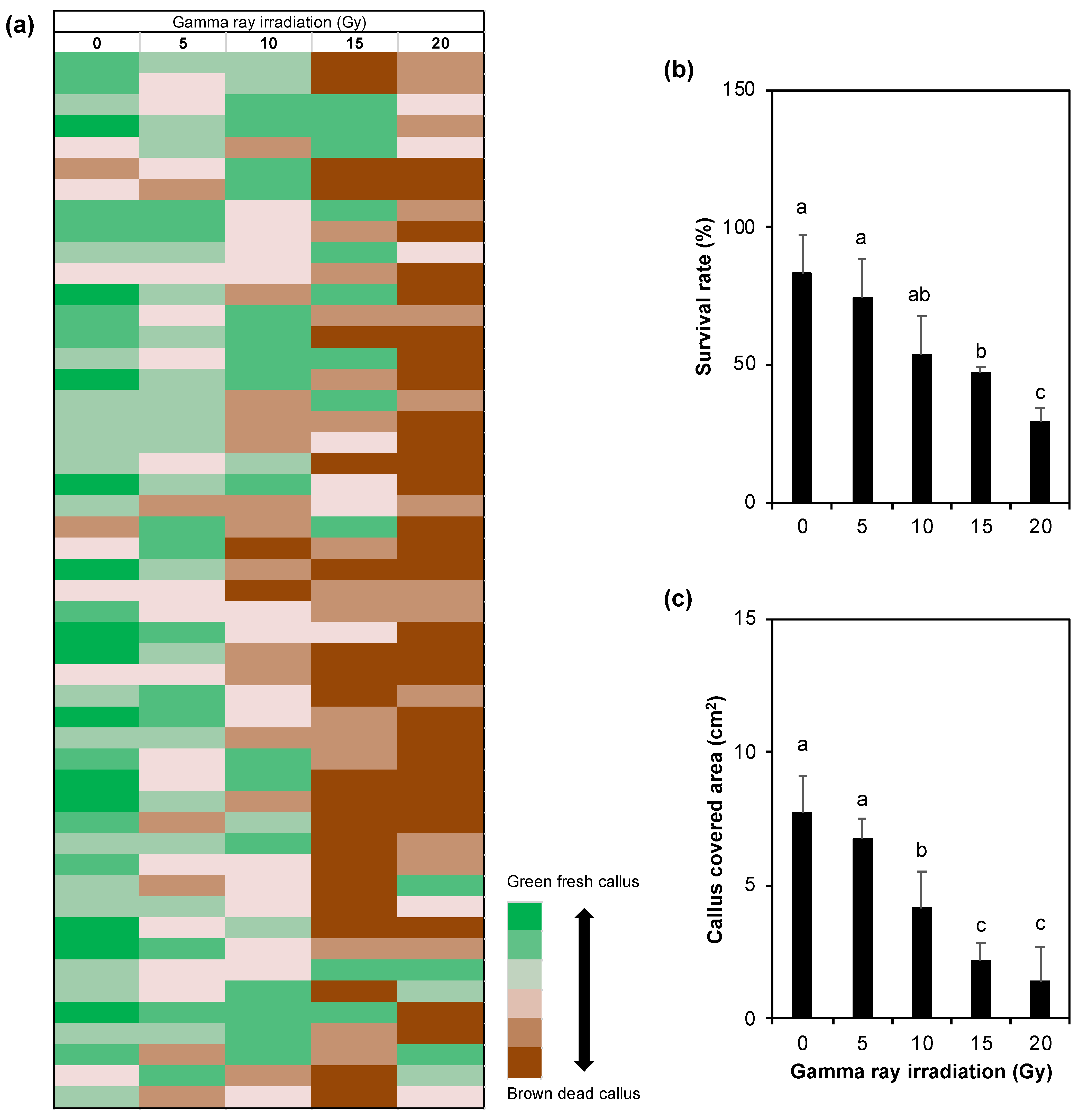
To quantitatively measure the tissue destruction affected by gamma ray irradiation, the survival rate of the irradiated callus was objectively observed by six-level freshness scale scoring (6 = green fresh callus to 1 = brown dead callus). The heatmap illustration of each single callus condition among the treatments (n = 50) is shown in Figure 2a. The proportion of survived callus was significantly decreased by applying 15 Gy gamma ray irradiation. In addition, application of 10 Gy substantially declined the callus covered area (Figure 2c). These results suggested that the 10–15 Gy was the critical irradiation level for A. paeoniifolius embryogenic callus survival rate.

3.2. Radiosensitivity Level of the Embryogenic Callus

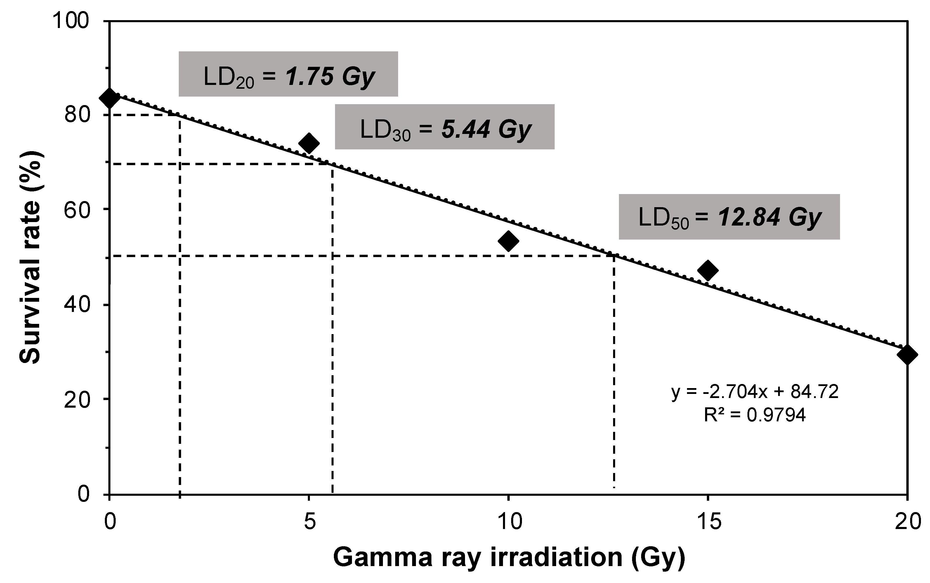
To further investigate the gamma ray irradiation-induced tissue destruction of A. paeoniifolius embryogenic calluses and to determine the LD20, LD30, and LD50 (exposures required to decrease survival by 20, 30, and 50%), the radiosensitivity level of the calluses were analyzed using linear curve estimation as previously reported [30]. As shown in Figure 3, an estimation of LD20, LD30, and LD50 of A. paeoniifolius embryogenic calluses from this study were 1.75 Gy, 5.44 Gy, and 12.84 Gy, respectively. In comparison with the previous studies, the estimated LD20, LD30, and LD50 of Celosia argentea L. in vitro plantlets were 35.65 Gy, 46.68 Gy, and 68.73 Gy, respectively [30]. Isnaini and Novitasari [31] predicted the LD50 of two variants of Nepenthes ampullaria Jack in vitro plantlets were around 31.0 Gy and 41.6 Gy. These findings suggested that the lethal dose level affected by gamma ray irradiation differs among plant species and highly depend on tissues examined and developmental stages. Since the high frequency of mutation was previously often found in around LD20–LD50 irradiated tissues in other herbaceous and crop species [30,32], this present study suggested that the effective gamma irradiation of A. paeoniifolius embryogenic callus was between 1.75–12.84 Gy.

3.3. Growth of Plantlets

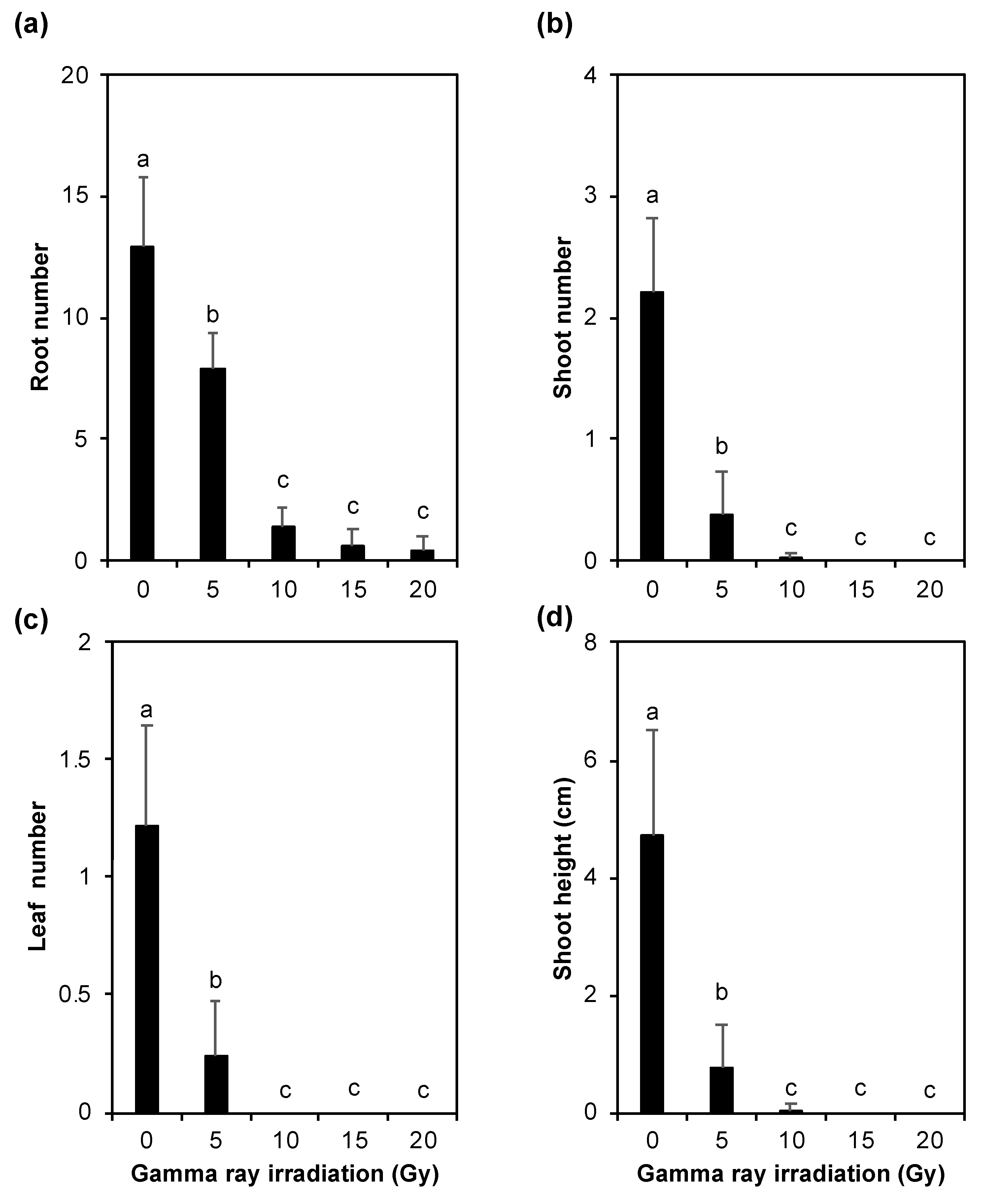
In general, the gamma ray irradiation inhibited the growth and development of plantlets arising from the embryogenic calluses of A. paeoniifolius (Figure 4). The number of roots, shoots, and leaves, and the shoot height were significantly reduced by 5 Gy gamma ray irradiation (Figure 4). The growth inhibition caused by gamma ray irradiation was more remarkedly found in 10–20 Gy treatments, suggesting that the A. paeoniifolius tissues from embryogenic calluses were highly sensitive to the gamma ray irradiation.

4. Conclusions

The exposures required from gamma ray irradiation to decrease the survival rate of A. paeoniifolius embryogenic calluses by 20, 30, and 50% were 1.75 Gy, 5.44 Gy, and 12.84 Gy, respectively. Thus, the findings indicated an effective range of irradiation level which possibly induce A. paeoniifolius embryogenic calluses mutation was around 1.75–12.84 Gy. In this study, the resulted potential mutant has not been recognized, and further study on that aspect is necessary in the near future.

Author Contributions

R.R.R. and Y. conceived the study. R.R.R. designed the experiments. R.R.R. and Y.I. performed the experiments. R.R.R. analyzed the data. R.R.R. wrote the manuscript with help from all the others. R.R.R. is a main contributor for this work. All authors have read and agreed to the published version of the manuscript.

Funding

DIPA fiscal year 2015 of the Research Center for Plant Conservation and Botanic Gardens, Indonesian Institute of Sciences (LIPI).

Institutional Review Board Statement

Not applicable.

Informed Consent Statement

Not applicable.

Data Availability Statement

All data necessary to evaluate the conclusions in this work are included in the published manuscript. Additional data, if required, will be made available by the corresponding author upon request.

Acknowledgments

The authors acknowledge Sutini and Suratmi (Plant Culture Laboratory, Research Center for Plant Conservation and Botanic Gardens, National Research and Innovation Agency of the Republic of Indonesia) for providing the in vitro culture medium in this study.

Conflicts of Interest

The authors declare no conflict of interest. The funders had no role in the design of the study; in the collection, analyses, or interpretation of data; in the writing of the manuscript, or in the decision to publish the results.

References

  1. Santosa, E.; Lian, C.L.; Sugiyama, N.; Misra, R.S.; Boonkorkaew, P.; Thanomchit, K. Population structure of elephant foot yams (Amorphophallus paeoniifolius (Dennst.) Nicolson) in Asia. PLoS ONE 2017, 12, e0180000. [Google Scholar] [CrossRef] [PubMed]
  2. Angayarkanni, J.; Ramkumar, K.M.; Priyadharshini, U.; Ravendran, P. Antioxidant potential of Amorphophallus paeoniifolius in relation to their phenolic content. Pharm. Biol. 2010, 48, 659–665. [Google Scholar] [CrossRef] [PubMed]
  3. Hurkadale, P.J.; Shelar, P.A.; Palled, S.G.; Mandavkar, Y.D.; Khedkar, A.S. Hepatoprotective activity of Amorphophallus paeoniifolius tubers against paracetamol-induced liver damage in rats. Asian. Pac. J. Trop. Biomed. 2012, 2, S238–S242. [Google Scholar] [CrossRef]
  4. Morvin, Y.J.E.; Prabhu, S.; Vijayakumar, S. An ethnobotanical study of medicinal plants used by traditional healers in silent valley of Kerala, India. J. Ethnopharmacol. 2014, 154, 774–789. [Google Scholar] [CrossRef] [PubMed]
  5. Dey, Y.N.; Wanjari, M.M.; Kumar, D.; Lomash, V.; Jadhav, A.D. Curative effect of Amorphophallus paeoniifolius tuber on experimental hemorrhoids in rats. J. Ethnopharmacol. 2016, 192, 183–191. [Google Scholar] [CrossRef]
  6. Reddy, C.K.; Suriya, M.; Vidya, P.V.; Vijina, K.; Haripriya, S. Effect of γ-irradiation on structure and physico-chemical properties of Amorphophallus paeoniifolius starch. Int. J. Biol. Macromol. 2015, 79, 309–315. [Google Scholar] [CrossRef]
  7. Sukhija, S.; Singh, S.; Riar, C.S. Effect of oxidation, cross-linking and dual modification on physicochemical, crystallinity, morphological, pasting and thermal characteristics of elephant foot yam (Amorphophallus paeoniifolius) starch. Food Hydrocoll. 2016, 55, 56–64. [Google Scholar] [CrossRef]
  8. Suriya, M.; Rajput, R.; Reddy, C.K.; Haripriya, S.; Bashir, M. Functional and physicochemical characteristics of cookies prepared from Amorphophallus paeoniifolius flour. J. Food Sci. Technol. 2017, 54, 2156–2165. [Google Scholar] [CrossRef]
  9. Suriya, M.; Reddy, C.K.; Haripriya, S.; Harsha, N. Influence of debranching and retrogradation time on behavior changes of Amorphophallus paeoniifolius nanostarch. Int. J. Biol. Macromol. 2018, 120, 230–236. [Google Scholar] [CrossRef]
  10. Yuzammi. The diversity of aroids (Araceae) in Bogor Botanic Gardens, Indonesia: Collection, conservation and utilization. Biodiversitas 2018, 19, 140–152. [Google Scholar] [CrossRef]
  11. Nagar, M.; Sharanagat, V.S.; Kumar, Y.; Singh, L.; Mani, S. Influence of xanthan and agar-agar on thermo-functional, morphological, pasting and rheological properties of elephant foot yam (Amorphophallus paeoniifolius) starch. Int. J. Biol. Macromol. 2019, 136, 831–838. [Google Scholar] [CrossRef] [PubMed]
  12. Mutaqin, A.Z.; Kurniadie, D.; Iskandar, J.; Nurzaman, M.; Partasasmita, R. Ethnobotany of suweg, Amorphophallus paeoniifolius: Utilization and cultivation in West Java, Indonesia. Biodiversitas 2020, 21, 1635–1644. [Google Scholar]
  13. Ravi, V.; Ravindran, C.; Suja, G. Growth and productivity of elephant foot yam (Amorphophallus paeoniifolius (Dennst.) Nicolson): An overview. J. Root Crop 2009, 35, 131–142. [Google Scholar]
  14. Bradbury, J.H.; Nixon, R.W. The acridity of raphides from the edible aroids. J. Sci. Food Agric. 1998, 76, 608–616. [Google Scholar] [CrossRef]
  15. Singh, A.K.; Chaurasiya, A.K.; Mitra, S. Oxalate content in elephant foot yam (Amorphophallus paeoniifolius Dennst.-Nicolson) dry and fry cubes. J. Pharma. Phytochem. 2018, 7, 2905–2909. [Google Scholar]
  16. Abraham, L.N.; Kamala, S.; Sreekumar, J.; Makeshkumar, T. Optimization of parameters to improve transformation efficiency of elephant foot yam (Amorphophallus paeoniifolius (Dennst.) Nicolson. 3 Biotech 2021, 11, 1–8. [Google Scholar] [CrossRef]
  17. Ahloowalia, B.S.; Maluszynski, M.; Nichterlein, K. Global impact of mutation-derived varieties. Euphytica 2004, 135, 187–204. [Google Scholar] [CrossRef]
  18. Cheng, L.; Yang, H.; Lin, B.; Wang, Y.; Li, W.; Wang, D.; Zhang, F. Effect of gamma-ray radiation on physiological, morphological characters and chromosome aberrations of minitubers in Solanum tuberosum L. Int. J. Radiat. Biol. 2010, 86, 791–799. [Google Scholar] [CrossRef]
  19. Khumaida, N.; Maharani, S.; Ardie, S.W. The leaf color performance on several lines of cassava and its relation with tuber yield as early reference. Procedia Environ. Sci. 2015, 24, 39–46. [Google Scholar] [CrossRef] [Green Version]
  20. Fadli, N.; Syarif, Z.; Satria, B.; Akhir, N. The effect of gamma Cobalt-60 ray irradiation on cultivar growth in taro white (Xhanthosoma sagittifolium L.). Int. J. Environ. Agric. Biotechnol. 2018, 3, 2020–2025. [Google Scholar] [CrossRef]
  21. Nurilmala, F.; Hutagaol, R.P.; Widhyastini, I.M.; Widyastuti, U.; Suharsono, S. Somaclonal variation induction of bogor taro (Colocasia esculenta) by gamma irradiation. Biodiversitas 2017, 18, 28–33. [Google Scholar] [CrossRef]
  22. Sianipar, N.F.; Maarisit, W. Detection of gamma-irradiated mutant of rodent tuber (Typhonium flagelliforme Lodd.) in vitro culture by RAPD molecular marker. Procedia Chem. 2015, 14, 285–294. [Google Scholar] [CrossRef] [Green Version]
  23. Kuranouchi, T.; Kumazaki, T.; Kumagai, T.; Nakatani, M. Breeding erect plant type sweetpotato lines using cross breeding and gamma-ray irradiation. Breed Sci. 2016, 66, 456–461. [Google Scholar] [CrossRef]
  24. Poerba, Y.S.; Imelda, M.; Wulansari, A.; Martanti, D. Induksi mutasi kultur in vitro Amorphophallus muelleri Blume dengan irradiasi gamma (in Indonesian). J. Tek. Ling. 2009, 10, 355–364. [Google Scholar]
  25. Murashige, T.; Skoog, F. A revised medium for rapid growth and bio assays with tobacco tissue cultures. Physiol. Plant 1962, 15, 473–497. [Google Scholar] [CrossRef]
  26. Karp, A. Mechanisms of somaclonal variation. Biotechnol. Biotechnol. Equip. 1993, 7, 20–25. [Google Scholar] [CrossRef]
  27. Hutami, S.; Mariska, I.; Supriati, Y. Peningkatan keragaman genetik tanaman melalui keragaman somaklonal (in Indonesian). J. AgroBiogen 2016, 2, 81. [Google Scholar] [CrossRef] [Green Version]
  28. Krishna, H.; Alizadeh, M.; Singh, D.; Singh, U.; Chauhan, N.; Eftekhari, M.; Sadh, R.K. Somaclonal variations and their applications in horticultural crops improvement. 3 Biotech 2016, 6, 54. [Google Scholar] [CrossRef] [Green Version]
  29. Sparrow, A.H.; Schwemmer, S.S.; Bottino, P.J. Effects of external gamma radiation from radioactive fallout on plants with special reference to crop production. Radiat. Bot. 1971, 11, 85–118. [Google Scholar] [CrossRef]
  30. Hayati, D.; Aisyah, S.I.; Krisantini. Radiosensitivity levels of in vitro cultured Celosia cristata planlets by γ—ray irradiation. J. Trop. Crop Sci. 2016, 3, 61–65. [Google Scholar] [CrossRef]
  31. Isnaini, Y.; Novitasari, Y. Penentuan kisaran dosis iradiasi gamma optimal dalam pemuliaan mutasi Nepenthes ampullaria Jack. secara in vitro (in Indonesian). J. Ilm. Apl. Isot. dan Radiasi 2020, 16, 15–22. [Google Scholar]
  32. Astuti, D.; Sulistyowati, Y.; Nugroho, S. Uji radiosensitivitas sinar gamma untuk menginduksi keragaman genetik sorgum berkadar lignin tinggi (in Indonesian). J. Ilm. Apl. Isot. dan Radiasi 2019, 15, 1–6. [Google Scholar]
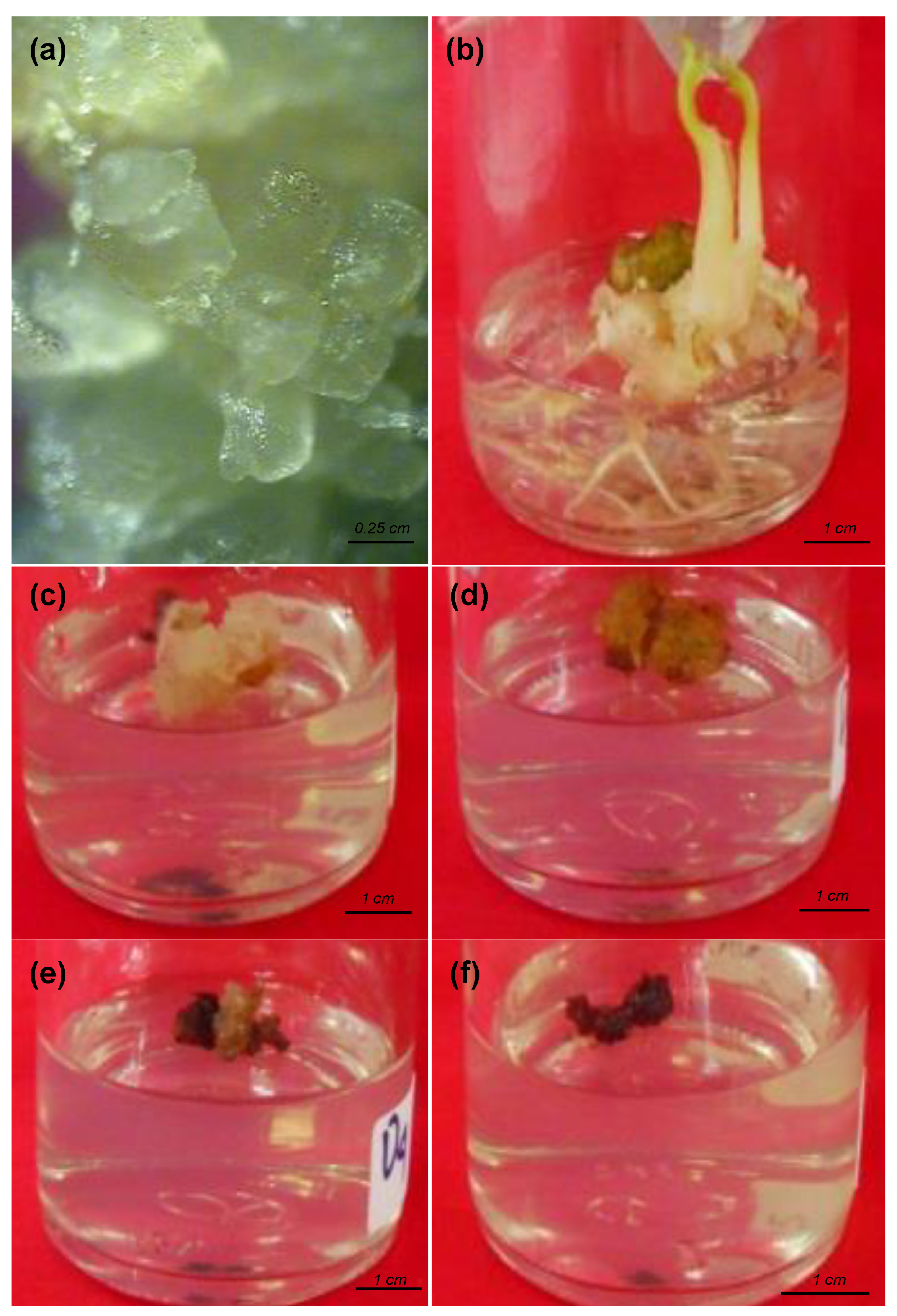
Figure 1. Embryogenic callus of Amorphophallus paeoniifolius. (a) Before gamma ray irradiation at six months after cultivation, and at (b) 0 Gy, (c) 5 Gy, (d) 10 Gy, (e) 15 Gy, and (f) 20 Gy after one month gamma ray irradiation (seven months after cultivation).
Blsf 11 00093 g001
Figure 2. Survival rate of Amorphophallus paeoniifolius embryogenic callus. (a) Heatmap illustration of the callus condition, (b) survival percentage of the callus, and (c) callus covered area after one-month gamma ray irradiation (seven months after cultivation). Values are means ± SD. Different letters mean significant difference. Duncan’s Multiple Range Test, p < 0.05 (n = 50).
Blsf 11 00093 g002
Figure 3. Estimated lethal dose (LD) values of Amorphophallus paeoniifolius calluses after one-month gamma ray irradiation (seven months after cultivation).
Blsf 11 00093 g003
Figure 4. Plantlet growth arising from the embryogenic callus of Amorphophallus paeoniifolius. (a) Root number, (b) shoot number, (c) leaf number, and (d) shoot length after two-months gamma ray irradiation (eight months after cultivation). Values are means ± SD. Different letters mean significant difference. Duncan’s Multiple Range Test, p < 0.05 (n = 50).
Blsf 11 00093 g004
Publisher’s Note: MDPI stays neutral with regard to jurisdictional claims in published maps and institutional affiliations.