Article

Altered Antimicrobial Activity and Selectivity of Dihydro-Protoberberines over Their Corresponding Protoberberines

by
Juan Ostos-Hernandez
1,
Hannah Bhakta
1,
Caleb VanArragon
2,
Lanna Sirhan
2,
Danielle Orozco-Nunnelly
2 and
Jeffrey Pruet
1,*
1
Department of Chemistry, Valparaiso University, 1710 Chapel Drive., Valparaiso, IN 46383, USA
2
Department of Biology, Valparaiso University, 1610 Campus Drive East, Valparaiso, IN 46383, USA
*
Author to whom correspondence should be addressed.
Future Pharmacol. 2025, 5(3), 53; https://doi.org/10.3390/futurepharmacol5030053
Submission received: 8 August 2025 / Revised: 10 September 2025 / Accepted: 13 September 2025 / Published: 17 September 2025

Abstract

Background/Objectives: The rise of multidrug-resistant bacteria and fungi, or “superbugs”, makes the development of new antimicrobial compounds of continued importance. In this context, we have explored structural variants of the plant-derived phytocompound berberine, seeking higher antimicrobial activity and selectivity. Our prior work prepared fourteen protoberberine variants (B1B14), and found that a partially reduced dihydro-protoberberine (B14) was significantly more active against Gram-positive bacteria. To further investigate this trend, we prepared a series of protoberberines and related dihydro-protoberberines, with the goal of better understanding the effects of the partial reduction of the protoberberine core. Methods: Protoberberines were prepared from a cyclization between glyoxal and substituted N-benzyl-phenethylamines, prepared by reductive amination. Dihydro-derivatives were obtained via NaBH4 reduction. Biological activity was assessed with a Kirby–Bauer assay to determine zones of inhibition against a panel of twelve microorganisms. Cytotoxicity was also assessed using an MTT assay against a T84 human colon carcinoma cell line. Results: The majority of the prepared compounds showed greater Gram-positive antibacterial activity compared to original berberine, and nearly all dihydro-protoberberines had improved Gram-positive antibacterial activity over their unreduced form. Additionally, the reduced variants were less active against fungi, indicating a step towards higher microbial selectivity. All variants showed greater potency against cancer cells. Conclusions: The present work highlights a significant improvement in antibacterial activity and selectivity for this set of dihydro-protoberberines over their unreduced counterparts.

Graphical Abstract

1. Introduction

With the increasing number of microorganisms that are resistant to multiple forms of antimicrobial treatments, the need for new antimicrobial agents has garnered significant medical attention [1,2]. Notably, these drug-resistant microbes can be quite prevalent in hospitals, where they pose one of the leading causes of hospital-acquired infection [3]. It is estimated that by 2050 these multidrug-resistant microbes will result in approximately 10 million deaths [2]. This threat expands beyond our terrestrial setting, as several opportunistic microbes have been observed on the international space station (ISS) and represent a threat to individuals involved in space travel [4]. Given this persistent increase in drug-resistant microbial threats, there is a constant need to explore new candidates for treatment.
Our group has been investigating the Argemone mexicana plant, which has ties to traditional medicine and has been studied for various medicinal applications [5,6]. We previously identified berberine (B), chelerythrine, and sanguinarine from the extracts of A. mexicana, and we found that these phytocompounds are the primary sources of the antimicrobial and anticancer properties of this plant [7]. Guided by this knowledge, we subsequently prepared fourteen structural variants of berberine (referred to as B1B14) and four chelerythrine variants (C1C4). From this panel of synthesized compounds, we found that the berberine variants showed the most promise when screened against twelve distinct microbes (five Gram-positive, four Gram-negative, and three fungi) of interest due to the presence of these same microbial species on the ISS [8]. The antimicrobial activity of berberine stems from numerous modes of action, such as DNA binding, interfering with protein biosynthesis, induced leakage of intercellular proteins, altering GTPase activity, and inhibition of the “filamenting temperature-sensitive mutant Z” (FtsZ) protein [9,10,11,12,13,14]. Some aspects of berberine therapy gaining attention are its synergistic effects with other antimicrobial agents, enhancing the activity of antibacterial and antifungal drugs, and its inhibitory effects on the biofilm formation of multidrug-resistant bacteria and yeast [15]. Additionally, berberine remains attractive because its wide range of biological activities have potential for treating numerous conditions, such as diabetes, hypertension, cancer, and Alzheimer’s [16]. Several of the protoberberines prepared in our previous report had notably improved activity against Gram-positive bacteria compared to berberine, while being slightly less active against fungi [8]. Of note, variants B1, B3, B5, and B14 showed the most consistent improvements in antibacterial activity (Figure 1).
Of special note was variant B14, the dihydro-derivative of B1, prepared by NaBH4 reduction of the cationic isoquinoline ring. This dihydro-protoberberine variant was between 30 and 100% more active against the various Gram-positive bacteria investigated compared to the unreduced B1 (and between 100 and 230% more potent than original berberine). At the same time, B14 was roughly 20–30% less active against fungi compared to B1 (and 40–50% less active compared to original berberine) [8]. This indicated that the reduction of the iminium group dramatically enhanced its antibacterial effects, while slightly increasing Gram-positive selectivity. Results from an alkaline phosphatase assay indicated a significant increase in the leakage of intracellular proteins when S. aureus was treated with B14, as compared to treatment with original berberine [8]. Intrigued by this result, we set out to further investigate and better understand the overall effect of this partial reduction of the protoberberines on their antimicrobial activity. As such, we report herein the preparation of additional berberine variants (B15 and B17) and their corresponding dihydro-berberine derivatives (B16 and B18), as well as conversion of two promising prior variants (B3 and B5) into their dihydro-derivatives (B19 and B20). This work highlights the utility of dihydro-protoberberines in the development of antimicrobial agents. Furthermore, we see the dihydro-protoberberines as a critical stepping stone in the direction of microbial selectivity. The four dihydro-protoberberines (B16, B18B20) represent completely new compounds. Compounds B15 and B17 were previously investigated for their use as low-density-lipoprotein receptor up-regulators [17] and retinoid X receptor α (RXRα) activators [18], but their antibacterial and antifungal properties have not yet been reported. Antimicrobial activity was assessed using the Kirby–Bauer assay to determine zones of inhibition, while the effects of these compounds on T84 colon cancer cells was explored using an MTT assay. The Kirby–Bauer disc diffusion assay has been viewed in the literature as an essential and acceptable tool in the determination of antimicrobial susceptibility, and it benefits from its reproducibility across labs, simplicity, and low cost [19]. This makes the assay perfectly suited to our primarily undergraduate institution.

2. Materials and Methods

2.1. General Information

The reagents and solvents used were purchased from Aldrich Chemical Co. (St. Louis, MO, USA) and used as provided by the company. 1H and 13C NMR spectra were recorded in either DMSO-d6, methanol-d4, or chloroform-d, with a Bruker spectrometer, using the solvent as the reference. All filtrations were performed under vacuum. A Büchi RE 121 rotary evaporator was used to aid the evaporation of solvents. A Waters UPLC with a QDa detector was used in the assessment of purity. Berberine chloride (B) was purchased from Aldrich Chemical Co. The synthesis of the protoberberine variants B1, B3, B5, and B14 was described in our prior publication [8].

2.2. Synthesis of Protoberberines (General Method A)

Protoberberines B15 and B17 were prepared using a method analogous to that reported for prior protoberberines (B1, B3, B5, etc.) [8]. Synthesis began with the preparation of the requisite secondary amine by refluxing equal molar quantities (10 mmol) of the appropriate substituted benzaldehyde with the appropriate substituted phenethylamine in 30 mL of dry dichloromethane (DCM) for 90 min, followed by removal of the solvent on the rotary evaporator and dissolution of the residue in 30 mL of dry methanol. This solution was cooled to 0 °C before the addition of 20 mmol NaBH4 portion-wise with stirring. This mixture was then stirred at ambient temperature for 1 h. Once the reduction was complete, the solvent was evaporated under vacuum, and the remaining gummy solid was partitioned between ethyl acetate (50 mL) and 0.1 M aqueous sodium carbonate (20 mL). After separation of the layers in the separatory funnel, the ethyl acetate layer was washed twice with saturated aqueous sodium chloride. Anhydrous sodium sulfate was added to dry the ethyl acetate layer, which was then filtered, and the solution was concentrated under vacuum. The resulting oil was treated with formic acid (30 mL) and 20 mmol of a 40% (w/w) glyoxal solution, and heated to 80 °C. Upon heating, 20 mmol anhydrous CuSO4 was added, and everything was stirred with continued heating for a total of 4 h. After cooling, an excess of CaO was added, which was then removed by filtration, and vacuum distillation was used for removal of formic acid. The resulting thick paste was mixed with a small volume of 3 M methanolic HCl and then cooled to 0 °C, producing a bright yellow solid. After collecting the solid by filtration, it was rinsed with cold methanol and dried in a vacuum desiccator to provide protoberberine chloride (Scheme 1). Compounds were identified based on NMR (1H and 13C) and mass spectrometry (Supplementary Materials).

2.3. Synthesis of Dihydro-Protoberberines (General Method B)

Dihydro-protoberberines B16, B18, B19, and B20 were prepared using a method analogous to that reported for the synthesis of B14 [8]. The corresponding protoberberine (1 mmol) was combined with 3 equivalents of K2CO3 in a round-bottom flask, with addition of 14 mL methanol. While this stirred at room temperature, 1 mmol NaBH4 was mixed with 0.5 mL of 1 M aqueous NaOH, and this solution was added via pipette to the solution of the protoberberine. After stirring at ambient temperature for 30 min, the resulting solid was recovered using vacuum filtration. The solid product was then rinsed using 15 mL DI water, as well as 5 mL of methanol, providing the dihydro-protoberberine after drying in a vacuum desiccator (Scheme 2). Compounds were identified based on NMR (1H and 13C) and mass spectrometry (Supplementary Materials).

2.4. Antimicrobial (Kirby–Bauer) Disc Diffusion Assay

An amount of 20 μL of a solution containing 6 mg/mL of each compound (in DMSO) was added to a sterile blank disc (Fisher Scientific, Pittsburgh, PA, USA) and allowed to evaporate, resulting in 0.12 mg of the compound per disc. The dry discs were then aseptically moved to a culture plate containing a lawn of either Bacillus cereus, Bacillus subtilis, Candida albicans, Corynebacterium pseudodiphtheriticum, Enterobacter aerogenes, Enterobacter cloacae, Escherichia coli, Penicillium chrysogenum, Proteus mirabilis, Saccharomyces cerevisiae, Staphylococcus aureus, or Staphylococcus epidermidis (Carolina Biological Supply Company, Burlington, NC, USA). Bacterial plates were incubated at 37 °C, and fungal plates were incubated at room temperature. Zones of inhibition were measured in millimeters after 2–5 days of growth, and at least five biological replicates were used for all means and error bars. Known antimicrobial agents (clotrimazole, streptomycin, vancomycin) served as positive controls, and the solvent alone (DMSO) was used for negative controls.

2.5. Cell Culture

The T84 human colon carcinoma cell line was a generous gift from Patrice Bouyer at Valparaiso University, obtained as originally described in [20] (University of California, San Diego, CA, USA) and later deposited at the ATCC (No. CCL-248; Gaithersburg, MD, USA). The cells were maintained with 5% CO2 at 37 °C (described in [21]) in advanced Dulbecco’s modified Eagle’s medium (ADMEM) (GIBCO-BRL, Grand Island, NY, USA) supplemented with 1% penicillin/streptomycin, 2% fetal bovine serum (FBS), 1% essential amino acids, and 1% glutamine (each obtained from Fisher Scientific, Pittsburgh, PA, USA). Adherent cells were washed using 2 mL of sterile 1× PBS (Fisher Scientific, Pittsburgh, PA, USA) for a 25 cm2 sized bottle or using 5 mL for a 75 cm2 sized bottle. For monolayer detachment of the cells, 1 mL of trypsin (Fisher Scientific, Pittsburgh, PA, USA) was used and allowed to incubate for 5–10 min at 37 °C. To neutralize the trypsin, a 1:1 ratio of trypsin/cell mixture–fresh media was used after transferring the solution to a sterile 15 mL falcon tube. Cells were then centrifuged at 22 °C for 5 min at 1000 rpm, and the supernatant was removed. Next, the cell pellet was resuspended in 2–4 mL of culture media via careful mixing. Finally, a pre-calculated volume of cells was added to a 25 cm2 bottle in 5 mL of culture media or to a 75 cm2 bottle in 10 mL of culture media. Cells were incubated with 5% CO2 at 37 °C until they reached approximately 90% confluency.

2.6. Variant Treatment and Viability Assay of Colon Cancer Cells

T84 cells were seeded in 24-well plates at a density of 30,000 cells per well. To compare the cytotoxic effects of the different structural variants, 10 μL of a 12 mg/mL solution of each compound (in DMSO) was added per well for a 1 h treatment. For the negative control wells, an equal volume of DMSO alone was added. To assess cell metabolic activity, the Vybrant® MTT Cell Proliferation Assay Kit was used (Molecular Probes, Eugene, OR, USA). Results were assessed at 570 nm via a plate reader, and the mean percentage of viable cells (normalized to the control) was determined from three biological replicates (each of which was the average of three technical replicates).

2.7. Statistical Analysis

Data were input and analyzed in Microsoft ® Excel for Mac Version 16.95.1 (Microsoft Corporation, Albuquerque, NM, USA). For all antimicrobial assays, mean zones of inhibition for each sample type are displayed with their associated standard error of the mean (SEM), where each mean represents five or more separate replicates. Positive antimicrobial controls were used for all tests, along with an appropriate negative control (disc with solvent alone). For each T84 MTT cell proliferation assay result, the mean percentage of viable cells normalized to the control is shown with the associated SEM. MTT viability means represent three biological replicates, each of which is the average of three technical replicates. Two-tailed t-tests were used for inferential statistical analyses, where unequal variances were assumed, and differences were considered statistically significant using a cutoff value of p ≤ 0.05.

3. Results

3.1. Antimicrobial Effects

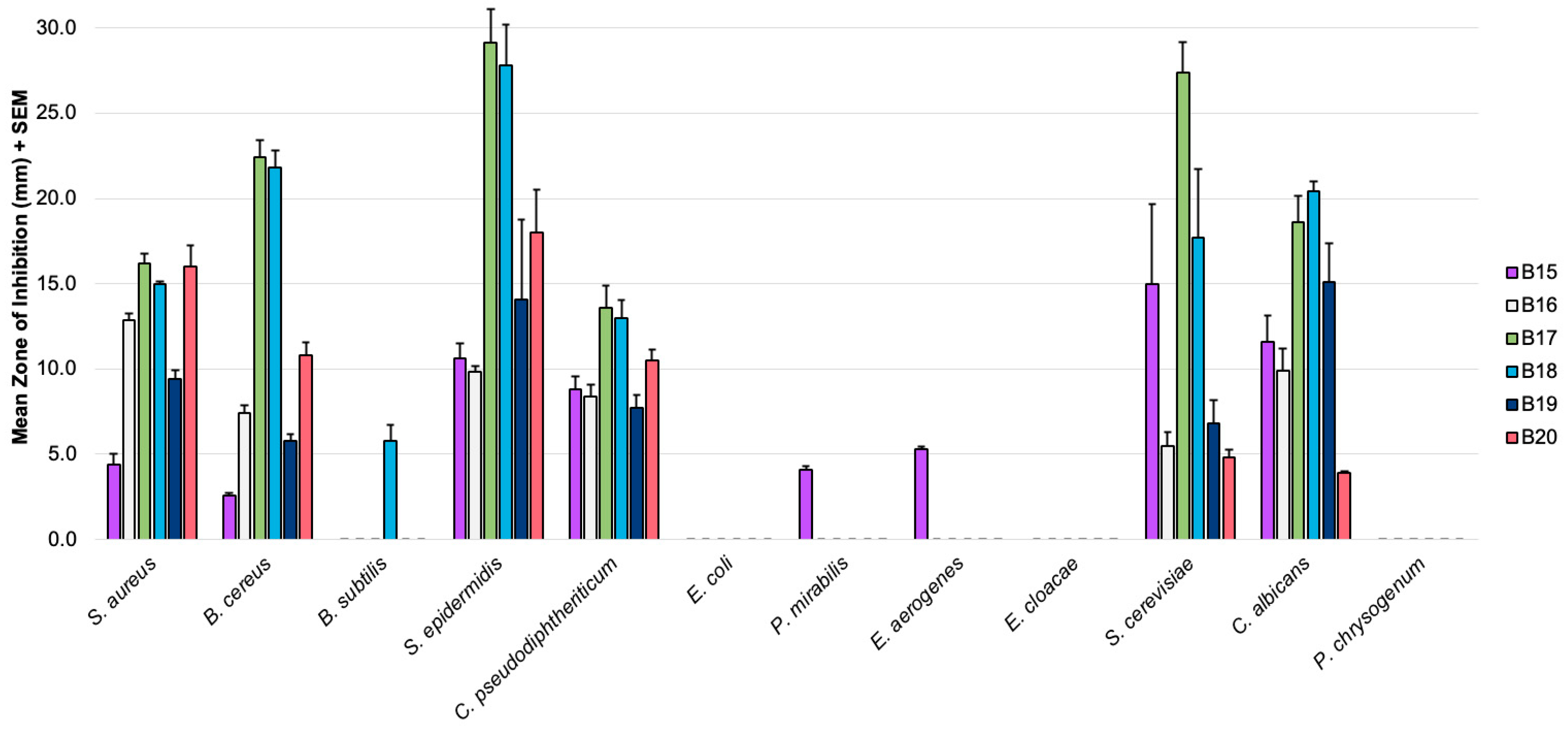
The mean zones of inhibition from the Kirby–Bauer assay for the new additions to our berberine variant library, along with the associated error (SEM), are summarized in Figure 2 below. Additionally, Table 1 presents these mean zones of inhibition for each of the twelve microbial lines investigated and highlights the comparisons between the fully aromatic protoberberines and the corresponding dihydro-protoberberine pairs. Mean zones of inhibition for key compounds disclosed in our prior report are also included in Table 1 to better illustrate these comparisons. A representative trial against S. aureus for all pairings can be found in the Supplementary Materials.

3.2. Anticancer Effects

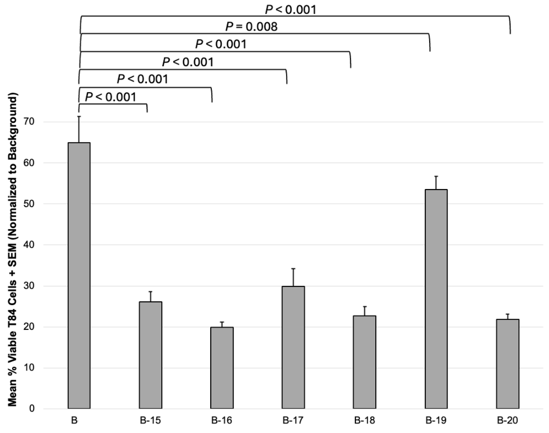
Given that berberine, and its derivatives, are also widely studied for their antitumor potential [12,22,23,24], we likewise wished to assess B15B20 for their effect on T84 colon cancer cell lines, with comparison to original berberine (B). Cell viability was determined using an MTT assay and represented as a percentage normalized to the 10 μL DMSO solvent control (Figure 3).

4. Discussion

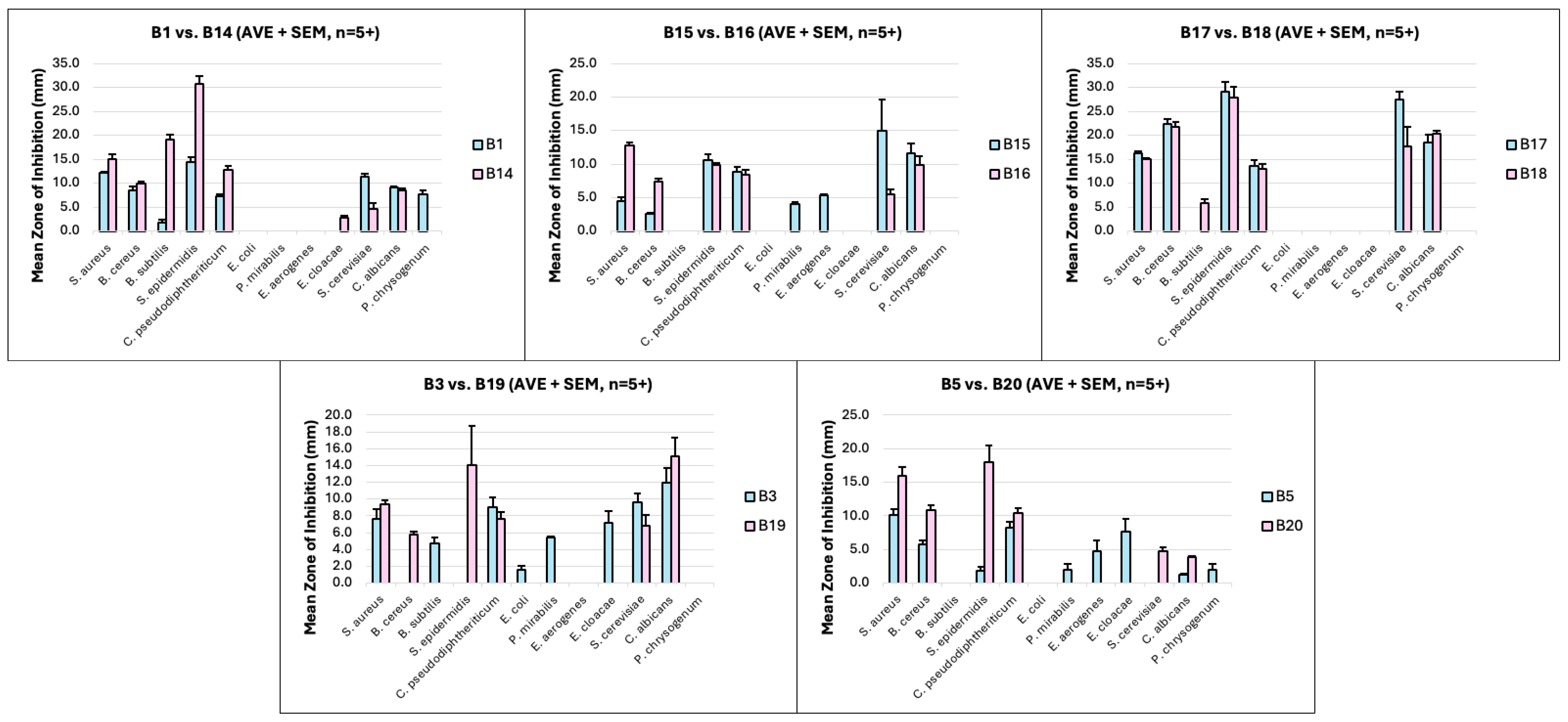
Consistent with our previous work, antimicrobial activity was mainly observed for the Gram-positive bacteria and the yeast cultures, with little to no activity seen for the Gram-negative bacterial lines (Figure 2; Table 1). As another means of making the comparisons of each pair as clear as possible, Figure 4 offers side-by-side comparisons of each unique protoberberine with its corresponding dihydro-protoberberine match.
Overall, the main trends observed for B1 versus B14 also appeared in the other pairings, although not always with perfect consistency. With some exception, it generally held that the partial reduction to the dihydro-derivatives enhanced antimicrobial effects against Gram-positive bacteria. The major exception to this was in B17/B18, where the mean zones of inhibition remained mostly identical. Another trend seen in B1/B14 that remained mostly consistent across all current pairs was the pronounced decrease in zones of inhibition against S. cerevisiae for the corresponding dihydro-derivatives; the main exception was the B5/B20 pair, which showed higher antifungal activity for B20 for both yeasts. Although most derivatives displayed little or no effect against Gram-negative bacteria, it is noteworthy that the three protoberberines that did show some Gram-negative activity (B15, B3, B5) had these effects abolished in their corresponding dihydro-form (B16, B19, B20).
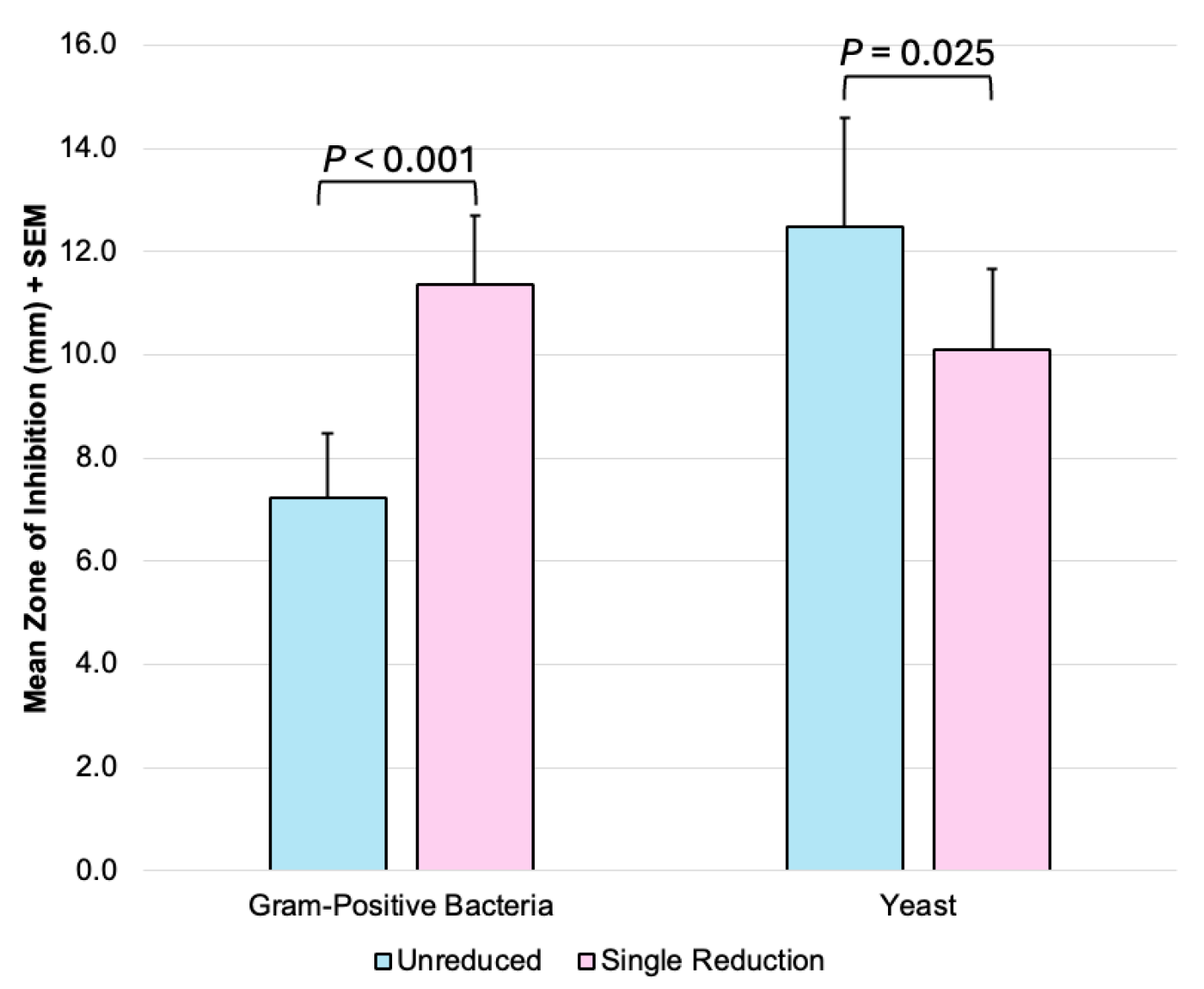
While no trend was universal across all pairings, the above data suggests that the partial reduction of protoberberines improves the antimicrobial effects against Gram-positive bacteria. There is also some apparent selectivity in how this reduction impacts antimicrobial effects (when comparing effects against Gram-positive bacteria versus yeast cells). In an effort to quantitatively assess this trend and its statistical significance, the mean zones of inhibition for all fully aromatic protoberberines were merged and compared to the merged mean zones of inhibition for all partially reduced dihydro-protoberberines, with Figure 5 summarizing the trends across organism type. Since there were minimal or no effects against Gram-negative bacteria and mold fungal organisms, these species were excluded from this analysis. As indicated in Figure 5, the net effect of the reduction of the isoquinoline ring was a roughly 50% increase in activity against Gram-positive bacteria, coupled with a 14% decrease in activity towards yeasts, and both of these changes were found to be of statistical significance. As such, we view this structural modification as being a worthy endeavor for those seeking enhanced agents to combat Gram-positive bacteria. Furthermore, we view this small structural modification as a potential stepping stone towards bacterial selectivity in future protoberberine medicinal applications. A potential explanation may be that reduction of the isoquinoline ring enhances binding events tied to berberine’s effects on the bacterial cell wall, while diminishing binding to biological targets associated with the more global antimicrobial effects of berberine [9,10,11,12,13,14]. As a representative example, the dihydro-protoberberines may serve as better ligands for the prokaryote-specific FtsZ protein. With the complex and multifaceted nature of the antimicrobial activity of berberine, these changes in activity may very well stem from numerous mechanisms of action.
With regard to structure–activity relationships, our prior publication containing 14 berberine variants had a larger pool of compounds, with a greater diversity in structure, giving greater confidence in the observed trends [8]. With the smaller pool of compounds in this study, it makes the definitive determination of structure–activity relationships less likely for this report. One observation is that B15/B17 and B16/B18 only differ in the location of the methylenedioxy group versus the methoxy substituent. From this small comparison, it appears clear that placement of the methylenedioxy group on the D-ring rather than the A-ring (see Figure 1) significantly increases antimicrobial effects on both Gram-positive bacteria and yeast.
The MTT assay results also show that B15B20 have pronounced inhibitory effects on the T84 colon cancer cell lines (Figure 3). Interestingly, all compounds were found to have a statistically stronger impact on colon cancer cell viability compared to original berberine. For additional context, our prior data for treatments with B3 and B5 showed roughly 47% and 85% cell viability [8]. This indicates that, with the exception of the B3/B19 pair, reduction of the isoquinoline ring also led to an increased cytotoxicity towards T84 colon cancer cells, with the most pronounced change coming in the B5 versus B20 pair. The exact cause for this change in cytotoxicity levels is still unclear. To better understand the potential anticancer activity of these compounds, future work may focus on determining IC50 values, testing a non-cancerous human colon cell line, and using RT-PCR to evaluate expression levels of colon cancer oncogenes and tumor suppressor genes, as we have done in the past for A. mexicana extracts [7].

5. Conclusions

This study was aimed at providing greater insight into the impact of small structural modifications on the antimicrobial properties of protoberberines, with specific emphasis on the effect of the conversion to the relevant dihydro-protoberberines. This work illustrates that the net effect of the reduction of the isoquinoline ring of protoberberines is an improvement in potency towards Gram-positive bacteria, as well as an increase in cytotoxicity towards tumor cells. While the current compounds may not display absolute microbial selectivity, the impact of this isoquinoline reduction appears greatest towards Gram-positive bacteria. Any small antibacterial effects against Gram-negative strains were removed in the reduced variants. Additionally, it was generally true that increases in antimicrobial effects towards Gram-positive bacteria were often met with subsequent decreases in antimicrobial effects on yeast. Therefore, dihydro-protoberberines can represent a significant step in the direction of a highly selective antibacterial agent. Notable exceptions to these effects were observed, so these changes may not apply to all protoberberines. Nevertheless, the strength of this study lies in its identification of dihydro-protoberberines as valuable compounds for the development of antibacterial drugs.

Supplementary Materials

The following supporting information can be downloaded at: https://www.mdpi.com/article/10.3390/futurepharmacol5030053/s1, Figure S1: Numbering system for protoberberines, Figure S2: Representative Kirby–Bauer assay against S. aureus, Figure S3: 1H-NMR spectra of B15 in CD3OD, Figure S4: 13C-NMR spectra of B15 in DMSO-d6, Figure S5: 1H-NMR spectra of B16 in CDCl3, Figure S6: 13C-NMR spectra of B16 in CDCl3, Figure S7: 1H-NMR spectra of B17 in DMSO-d6, Figure S8: 13C-NMR spectra of B17 in DMSO-d6, Figure S9: 1H-NMR spectra of B18 in CDCl3, Figure S10: 13C-NMR spectra of B18 in CDCl3, Figure S11: 1H-NMR spectra of B19 in CDCl3, Figure S12: 13C-NMR spectra of B19 in CDCl3, Figure S13: 1H-NMR spectra of B20 in CDCl3, Figure S14: 13C-NMR spectra of B20 in CDCl3.

Author Contributions

Conceptualization, J.P. and D.O.-N.; methodology, J.P., D.O.-N., and C.V.; investigation, J.O.-H., H.B., C.V., L.S., J.P., and D.O.-N.; formal analysis, J.O.-H., H.B., C.V., L.S., J.P., and D.O.-N.; validation, J.O.-H., H.B., C.V., L.S., J.P., and D.O.-N.; supervision, J.P. and D.O.-N.; writing—original draft preparation, review and editing, J.P., D.O.-N., and H.B.; funding acquisition, J.P. and D.O.-N. All authors have read and agreed to the published version of the manuscript.

Funding

This work was funded in part by the Indiana Space Grant Consortium and the Indiana Academy of Sciences Senior Research Grant.

Institutional Review Board Statement

Not applicable.

Informed Consent Statement

Not applicable.

Data Availability Statement

The data presented in this study is included in the article and the Supplementary Materials. Inquiries can be directed to the corresponding author upon request.

Acknowledgments

The authors gratefully acknowledge undergraduate researchers Brooke Ferkull, Abby Burton, Jenna Yehyawi, Nolan Brezina, Gracie Holt, and Katelynn Johnston for technical assistance with experiments, as well as Patrice Bouyer for his support with human cell culture at Valparaiso University.

Conflicts of Interest

The authors declare no conflicts of interest. The funders had no role in the design of the study; in the collection, analyses, or interpretation of data; in the writing of the manuscript; or in the decision to publish the results.

Abbreviations

The following abbreviations are used in this manuscript:
MTT3-(4,5-di methyl thiazol-2-yl)-2,5-diphenyltetrazolium bromide
FtsZFilamenting temperature-sensitive mutant Z
NMRNuclear magnetic resonance
DMSODimethyl sulfoxide
ISSInternational space station
UPLCUltra performance liquid chromatography
QDaQuadrupole Dalton
DCMDichloromethane
ADMEMAdvanced Dulbecco’s modified Eagle’s medium
FBSFetal bovine serum
PBSPhosphate-buffered saline
SEMStandard error of the mean

References

  1. Clatworthy, A.E.; Pierson, E.; Hung, D.T. Targeting virulence: A new paradigm for antimicrobial therapy. Nat. Chem. Biol. 2007, 3, 541–548. [Google Scholar] [CrossRef] [PubMed]
  2. O’Neill, J. Antimicrobial Resistance: Tackling a Crisis for the Health and Wealth of Nations. Review on Antimicrobial Resistance; Wellcome Trust: London, UK, 2014. [Google Scholar]
  3. Nordmann, P.; Naas, T.; Fortineau, N.; Poirel, L. Superbugs in the coming new decade; multidrug resistance and prospects for treatment of Staphylococcus aureus, Enterococcus spp. and Pseudomonas aeruginosa in 2010. Curr. Opin. Microbiol. 2007, 10, 436–440. [Google Scholar] [CrossRef]
  4. Checinska Sielaff, A.; Urbaniak, C.; Mohan, G.B.M.; Stepanov, V.G.; Tran, Q.; Wood, J.M.; Minich, J.; McDonald, D.; Mayer, T.; Knight, R.; et al. Characterization of the total and viable bacterial and fungal communities associated with the International Space Station surfaces. Microbiome 2019, 7, 50. [Google Scholar] [CrossRef]
  5. Brahmachari, G.; Gorai, D.; Roy, R. Argemone mexicana: Chemical and pharmacological aspects. Rev. Bras. Farmacogn. 2013, 23, 559–575. [Google Scholar] [CrossRef]
  6. Moore, M.; Kamp, M. Los Remedios: Traditional Herbal Remedies of the Southwest; Museum of New Mexico Press: Santa Fe, NM, USA, 2008. [Google Scholar]
  7. Orozco-Nunnelly, D.A.; Pruet, J.; Rios-Ibarra, C.P.; Bocangel Gamarra, E.L.; Lefeber, T.; Najdeska, T. Characterizing the cytotoxic effects and several antimicrobial phytocompounds of Argemone mexicana. PLoS ONE 2021, 16, e0249704. [Google Scholar] [CrossRef]
  8. Villegas, J.; Ball, B.C.; Shouse, K.M.; VanArragon, C.W.; Wasserman, A.N.; Bhakta, H.E.; Oliver, A.G.; Orozco-Nunnelly, D.A.; Pruet, J.M. Synthesis and biological evaluation of Argemone mexicana-inspired antimicrobials. Beilstein J. Org. Chem. 2023, 19, 1511–1524. [Google Scholar] [CrossRef]
  9. Schmeller, T.; Latz-Brüning, B.; Wink, M. Biochemical activities of berberine, palmatine and sanguinarine mediating chemical defence against microorganisms and herbivores. Phytochemistry 1997, 44, 257–266. [Google Scholar] [CrossRef]
  10. Čerňáková, M.; Košťálová, D. Antimicrobial activity of berberine—A constituent of Mahonia aquifolium. Folia Microbiol. 2002, 47, 375–378. [Google Scholar] [CrossRef] [PubMed]
  11. Grycová, L.; Dostál, J.; Marek, R. Quaternary protoberberine alkaloids. Phytochemistry 2007, 68, 150–175. [Google Scholar] [CrossRef] [PubMed]
  12. Kumar, A.; Ekavali; Chopra, K.; Mukherjee, M.; Pottabathini, R.; Dhull, D.K. Current knowledge and pharmacological profile of berberine: An update. Eur. J. Pharmacol. 2015, 761, 288–297. [Google Scholar] [CrossRef]
  13. Sun, N.; Chan, F.-Y.; Lu, Y.-J.; Neves, M.A.C.; Lui, H.-K.; Wang, Y.; Chow, K.-Y.; Chan, K.-F.; Yan, S.-C.; Leung, Y.-C.; et al. Rational Design of Berberine-Based FtsZ Inhibitors with Broad-Spectrum Antibacterial Activity. PLoS ONE 2014, 9, e97514. [Google Scholar] [CrossRef]
  14. Boberek, J.M.; Stach, J.; Good, L. Genetic Evidence for Inhibition of Bacterial Division Protein FtsZ by Berberine. PLoS ONE 2010, 5, e13745. [Google Scholar] [CrossRef]
  15. Kosalec, I.; Jembrek, M.J.; Vlainić, J. The Spectrum of Berberine Antibacterial and Antifungal Activities. In Promising Antimicrobials from Natural Products; Rai, M., Kosalec, I., Eds.; Springer: Cham, Switzerland, 2022. [Google Scholar] [CrossRef]
  16. Sun, P.; Wang, Z.; Ma, Y.; Liu, Y.; Xue, Y.; Li, Y.; Gao, X.; Wang, Y.; Chu, M. Advance in identified targets of berberine. Front. Pharmacol. 2025, 16, 1500511. [Google Scholar] [CrossRef] [PubMed]
  17. Yang, P.; Song, D.-Q.; Li, Y.-H.; Kong, W.-J.; Wang, Y.-X.; Gao, L.-M.; Liu, S.-Y.; Cao, R.-Q.; Jiang, J.-D. Synthesis and structure–Activity relationships of berberine analogues as a novel class of low-density-lipoprotein receptor up-regulators. Bioorg. Med. Chem. Lett. 2008, 18, 4675–4677. [Google Scholar] [CrossRef] [PubMed]
  18. Xu, B.; Jiang, X.; Xiong, J.; Lan, J.; Tian, Y.; Zhong, L.; Wang, X.; Xu, N.; Cao, H.; Zhang, W.; et al. Structure–Activity Relationship Study Enables the Discovery of a Novel Berberine Analogue as the RXRα Activator to Inhibit Colon Cancer. J. Med. Chem. 2020, 63, 5841–5855. [Google Scholar] [CrossRef]
  19. Salam, A.; Al-Amin, Y.; Pawar, J.S.; Akhter, N.; Lucy, I.B. Conventional methods and future trends in antimicrobial susceptibility testing. Saudi J. Biol. Sci. 2023, 30, 103582. [Google Scholar] [CrossRef]
  20. Dharmsathaphorn, K.; McRoberts, J.A.; Mandel, K.G.; Tisdale, L.D.; Masui, H. A human colonic tumor cell line that maintains vectorial electrolyte transport. Am. J. Physiol.-Gastrointest. Liver Physiol. 1984, 246, G204–G208. [Google Scholar] [CrossRef]
  21. Bouyer, P.G.; Tang, X.; Weber, C.R.; Shen, L.; Turner, J.R.; Matthews, J.B. Capsaicin induces NKCC1 internalization and inhibits chloride secretion in colonic epithelial cells independently of TRPV1. Am. J. Physiol.-Gastrointest. Liver Physiol. 2013, 304, G142–G156. [Google Scholar] [CrossRef] [PubMed]
  22. Kettmann, V.; Košt’álová, D.; Höltje, H.-D. Human topoisomerase I poisoning: Docking protoberberines into a structure-based binding site model. J. Comput.-Aided Mol. Des. 2004, 18, 785–796. [Google Scholar] [CrossRef]
  23. Li, T.-K.; Bathory, E.; LaVoie, E.J.; Srinivasan, A.R.; Olson, W.K.; Sauers, R.R.; Liu, L.F.; Pilch, D.S. Human Topoisomerase I Poisoning by Protoberberines: Potential Roles for Both Drug–DNA and Drug–Enzyme Interactions. Biochemistry 2000, 39, 7107–7116. [Google Scholar] [CrossRef]
  24. Haque, S.; Mathkor, D.M.; Bhat, S.A.; Musayev, A.; Khitoyva, L.; Ramniwas, S.; Phillips, E.; Swamy, N.; Kumar, S.; Yerer, M.B.; et al. A Comprehensive Review Highlighting the Prospects of Phytonutrient Berberine as an Anticancer Agent. J. Biochem. Mol. Toxicol. 2024, 39, e70073. [Google Scholar] [CrossRef] [PubMed]
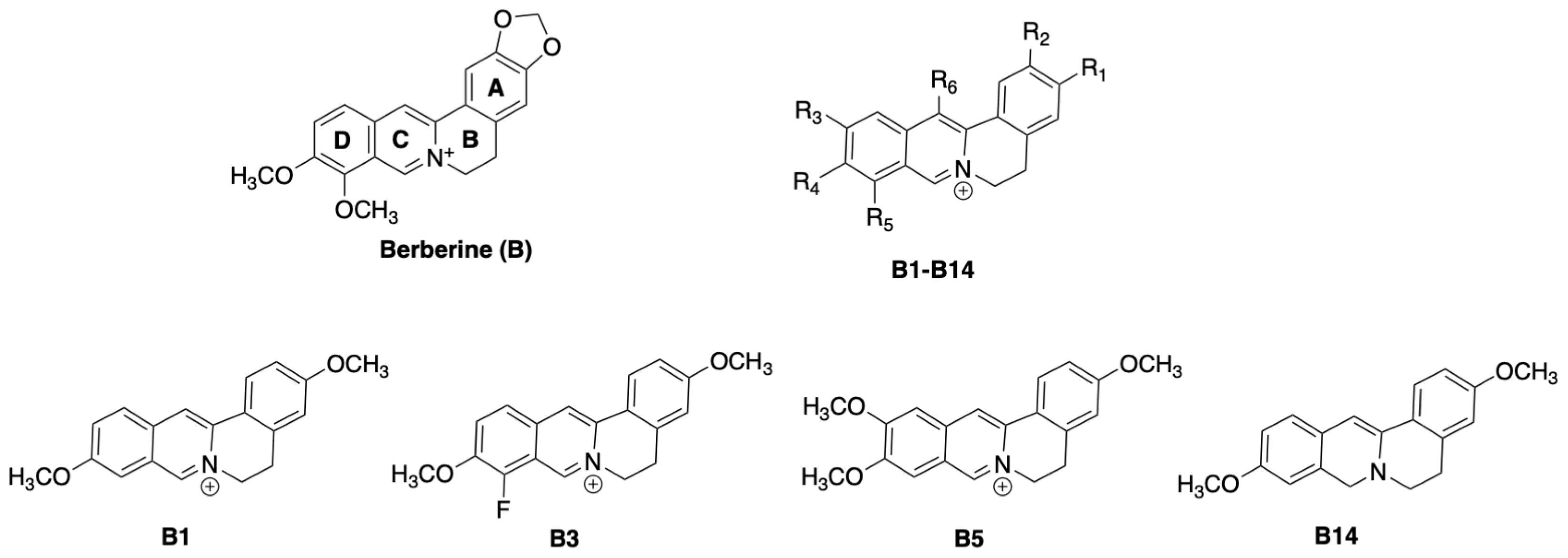
Figure 1. Structure of original berberine with ring designations indicated, along with the generic structure of the previously reported protoberberines B1B14, with the exact structures of four protoberberines of special note [8].
Futurepharmacol 05 00053 g001
Scheme 1. Synthesis of protoberberines B15 and B17.
Futurepharmacol 05 00053 sch001
Scheme 2. Synthesis of dihydro-protoberberines B16 and B18B20.
Futurepharmacol 05 00053 sch002
Figure 2. Antimicrobial disc diffusion assay results, showing mean zones of inhibition in millimeters for variants B15B20, with the associated error bars (mean + SEM). Positive controls were clotrimazole, streptomycin, or vancomycin, and DMSO was the negative control.
Futurepharmacol 05 00053 g002
Figure 3. Cytotoxic activities are shown for original berberine vs. variants B15B20 tested against a T84 human colonic carcinoma cell line. Cell metabolic activity after each treatment was determined using the MTT colorimetric assay. The mean percentage of viable cells normalized to DMSO alone (negative control) is displayed with the associated SEM (n = 9; 3 biological replicates with 3 technical replicates each). A two-tailed t-test analysis was used to determine statistical significance, where a p-value ≤ 0.05 was used as the cutoff.
Futurepharmacol 05 00053 g003
Figure 4. Side-by-side comparisons of the mean zones of inhibition for each protoberberine and dihydro-protoberberine pair.
Futurepharmacol 05 00053 g004
Figure 5. Trends across organism type (Gram-positive bacteria vs. yeast fungal organisms) after partial reduction of the isoquinoline ring (Gram-positive n = 30 + SEM; yeast n = 10 + SEM).
Futurepharmacol 05 00053 g005
Table 1. Mean Kirby–Bauer zones of inhibition for the protoberberines and the corresponding dihydro-protoberberine derivatives.
MicrobeMean Zones of Inhibition (mm) 1
Protoberberines 2Dihydro-Protoberberines 2
B1 [8]
(n = 5)
B15
(n = 6)
B17
(n = 5)
B3 [8]
(n = 5)
B5 [8]
(n = 5)
B14 [8]
(n = 5)
B16
(n = 6)
B18
(n = 5)
B19
(n = 5)
B20
(n = 5)
S. aureus12.24.416.27.610.215.112.815.09.416.0
B. cereus8.52.622.4-5.810.07.421.85.810.8
B. subtilis---4.7-19.2-5.8--
S. epidermidis14.410.629.1-1.830.89.927.814.118.0
C. pseudodiphtheriticum7.48.813.69.08.212.88.413.07.710.5
E. coli---2.7------
P. mirabilis-4.1-5.42.0-----
E. aerogenes-5.3--4.8-----
E. cloacae---7.27.62.9----
S. cerevisiae11.415.027.49.6-4.65.517.76.84.8
C. albicans9.111.618.612.01.38.69.920.415.13.9
P. chrysogenum7.8---2.0-----
1 Mean zones of inhibition in millimeters for 0.12 mg of each compound. A dash (-) indicates no measurable effect. Vancomycin, streptomycin, or clotrimazole were positive controls, and DMSO was the negative a control. 2 The relative order of protoberberines matches the respective order of the corresponding dihydro-protoberberines (B14 is derived from B1; B20 is derived from B5). Data for B1, B3, B5, and B14 Reproduced/Adapted from [8], Beilstein-Institut. 2023.
Disclaimer/Publisher’s Note: The statements, opinions and data contained in all publications are solely those of the individual author(s) and contributor(s) and not of MDPI and/or the editor(s). MDPI and/or the editor(s) disclaim responsibility for any injury to people or property resulting from any ideas, methods, instructions or products referred to in the content.