Review

On the Molecular Origin of the Toxicity of Erophaca baetica (L.) Boiss.

1
Molecular Chemistry, Materials and Catalysis Laboratory, Faculty of Sciences and Technologies, Sultan Moulay Slimane University, BP 523, Beni-Mellal 23000, Morocco
2
Institute of Pharmaceutical Chemistry Albert Lespagnol, Faculty of Pharmacy, University of Lille, 59290 Lille, France
*
Authors to whom correspondence should be addressed.
Future Pharmacol. 2025, 5(2), 28; https://doi.org/10.3390/futurepharmacol5020028
Submission received: 16 April 2025 / Revised: 5 June 2025 / Accepted: 11 June 2025 / Published: 12 June 2025

Abstract

:
Background/Objectives: The plant species Erophaca baetica (L.) Boiss. (synonym: Astragalus lusitanicus Lam.) is found essentially around the Mediterranean basin, with Morocco as its ancestral territory. The foliage of E. baetica is toxic to small ruminants, and for this reason the plant is often eliminated by farmers, despite its ecological and medicinal potential. The phytochemicals at the origin of the toxicity of E. baetica are not precisely known, but several potentially toxic products have been identified. In particular, aliphatic nitro compounds are present in the aerial parts of the plant, such as 3-nitro-propionic acid (NPA) and its precursor 3-nitro-propanol (NPOH) which are most likely at the origin of the plant toxicity. Results: The present review provides a detailed analysis of the nitrotoxins isolated from E. baetica and their mechanism of action. The covalent targeting of metabolic enzymes such as isocitrate lyase and succinate dehydrogenase by NPA is discussed. The mitochondrial chain blocker NPA is most likely responsible for the brain toxicity of E. baetica, but the presence of other potentially toxic chemicals—such as lusitoxamine and lusitoxamide—is also discussed. Conclusions: This review shed light on the widespread but little-known Mediterranean plant E. baetica and the phytochemicals responsible for the plant’s toxicity.

1. Introduction

In the Fabaceae (Leguminosae) family, the plant genus Erophaca is monophyletic, with a single species officially designated Erophaca baetica (L.) Boiss., also known as Astragalus lusitanicus Lam. and Colutea baetica (L.) Cav. It is an herbaceous perennial plant well distributed around the Mediterranean Sea (Figure 1). E. baetica is one of the earliest plants to bloom in the Mediterranean area from February until May, exhibiting racemes of creamy-green flowers. It is occasionally designated Iberian milkvetch, but the common name milkvetch is employed for all species of the Astragalus genus Populations of E. baetica are found in isolated patches across its distribution area, typically in cork oak forests from sea level to an altitude of about 1300 m. Most plants are approximately 0.7–1 m tall; however, they can grow up to 2 m in shaded areas.
There are two shrubby subspecies distributed at opposite ends of the Mediterranean region (Figure 1). E. baetica subsp. baetica is native to the southern half of the Iberian Peninsula (Portugal and Spain) and Northwest Africa (Morocco and Algeria). E. baetica subsp. orientalis is native to the Eastern Mediterranean (Greece, Turkey, Cyprus, and Lebanon). In Spain, notably in Sierra Morena, the plant is known as “el garbancillo” (little chickpea), but it should not be confused with the species Astragalus garbancillo Cav. Occasionally, the plant is also called “garbanzo del diablo” [1]. In Morocco, it is known as “fouila” (or fwila, or Katad al bortoghal) (small bean) in reference to the pods of the plant resembling beans in their long and swollen shape [2,3]. A morphological distinction between the two subspecies can be made based on leaflet hairiness, the teeth of the calyx, the color of their stipelles, and the fruits. For subsp. orientalis, appressed hairs, teeth of the calyx longer than half the calyx, purple stipelles and, usually, fruits smaller than 70 × 25 cm are present, while for subsp. baetica, glabrous leaflets, shorter teeth of the calyx, yellowish stipelles, and larger fruits can be found [4]. The fruit of the latter subspecies is known as ibaoune nil khmane (dromedary bean) due to its characteristic shape [5] (Figure 1c).
Interestingly, E. baetica presents an atypical breeding system, because male and bisexual (hermaphrodite) flowers can be found on the same plant [6]. This phenomenon, known as andromonoecy, is fairly rare in plants. It has been observed with a few other plants for which the seeds sired by male flowers perform better than those sired by hermaphroditic flowers, with a higher germination rate [7]. Andromonoecy is considered a morphological adaptation to enhance pollination success [8]. It is an important characteristic of E. baetica. The plant is essentially known for its toxicity, which is the main point discussed here.

2. Toxicity of Erophaca baetica

The toxicity of Astragalus species and E. baetica in particular has been recognized for a long time [1,9]. This plant is listed on the international poisonous plant checklist [10]. The foliage of E. baetica is very toxic to small ruminants such as sheep, goats, and cattle. Poisoned animals have revealed congestive lesions and edema in the brain and lungs. Both skeletal muscle and neurological effects have been observed [11]. Poisoned lambs showed alternated excitement and depression, with cardiac and respiratory disorders terminally. Microscopic examinations have shown vacuolar degeneration in neurons, hepatocytes, and in spleen and kidney cells [12]. In sheep, a major neurological disturbance has been underlined with the toxic signs persisting in poisoned animals even after cessation of plant consumption. Apparently, the plant consumption induces a persistent biochemical defect in neurons [13]. The nervous alterations induced upon ingestion of E. baetica are reminiscent of a form of locoism which is a neurovisceral disease observed in sheep, lambs, goats, and cattle upon the ingestion of certain toxins [14,15]. At the experimental level, hydro-alcoholic extracts of E. baetica were shown to be extremely toxic to mice, inducing both acute poisoning with convulsions and death in a few minutes, and a delayed poisoning, 10–15 days after a single intravenous administration of the plant extract to mice [16]. It is important to underline that toxicity assays in lambs have shown that E. baetica extracts prepared with organic solvents (petroleum ether, ether, ethyl acetate and 1-butanol) were found to be non-toxic. Conversely, aqueous extracts and plant decoctions were found to be extremely toxic. The lethal dose of the fresh plant is about 30 g/kg body weight (BW) compared to only 1.5 g/kg BW for the decoction. A fraction enriched in the toxic principle was found to be lethal at the dose of about 40 mg/kg [17]. However, seldom do studies address the question of what the toxic constituents of E. baetica are. The phytochemicals at the origin of the toxicity of E. baetica are not precisely known at present. The topic, which remains controverted, is discussed here.

3. Toxic Phytochemicals from E. baetica: The Nitrotoxin Hypothesis

The risk of intoxication with E. baetica is known for a while. In the late 1960s, it was already reported that the administration of aqueous extracts of A. lusitanicus (e.g., E. baetica) was toxic to mice within minutes and hydroalcoholic extracts were toxic in 48 h [18]. Different studies have confirmed the damages caused by an acute intoxication with a plant extract, notably severe cardiorespiratory alterations [19,20]. Early on, potentially toxic compounds such as cyanogenic glycosides and hemolytic saponins were identified in plant extracts but not in toxic concentrations [21,22,23]. Molecules such as pinitol, isormhamnetin glycosides, and saccharinic acid lactone (methyl-D-erythrono-1,4-lactone) were characterized, but the high toxicity could not be attributed to these compounds [16].
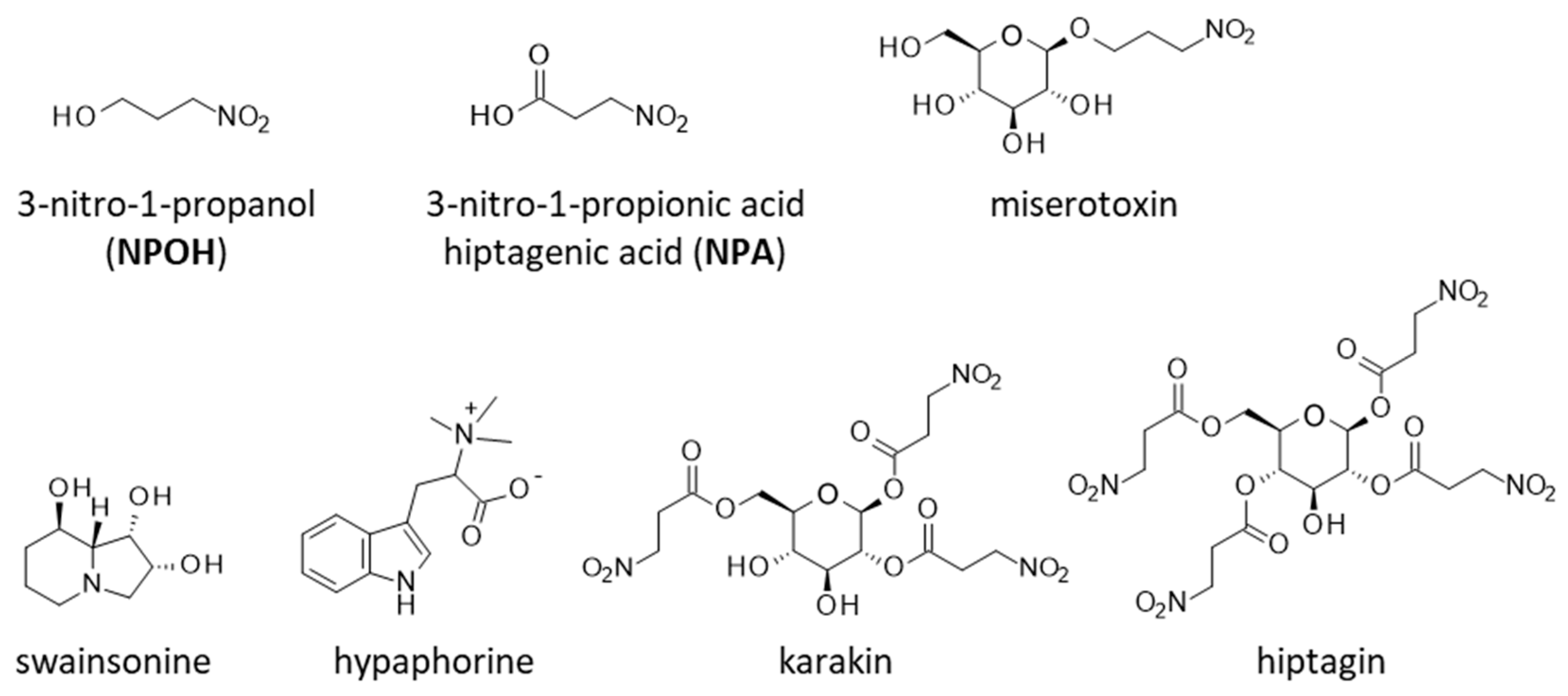
A major offending neurotoxin is the indolizidine alkaloid swainsonine (Figure 2), which has been detected in several Astragalus species, notably North American and South American Astragalus species. Swainsonine is a water-soluble natural product, rapidly absorbed and eliminated. It can cause severe toxicosis in livestock grazing swainsonine-containing plants [24]. This toxin is generally produced by an associated fungal symbiont [25,26,27]. But a chemical investigation has concluded that A. lusitanicus (i.e., E. baetica) does not contain swainsonine [12].
The indolic alkaloid hypaphorine (also known as lenticin or tryptophan betaine; see Figure 2) has been detected in E. baetica and considered as a potentially harmful compound [28]. But in fact, this alkaloid is not particularly toxic. Hypaphorine was shown to be non-toxic to goats even at a high dose of 2 g/kg by oral administration [29]. Another study also concluded that hypaphorine could not be held responsible for Astragalus poisonings in small ruminants [30]. It is rather viewed as an anti-inflammatory agent with hepatoprotective properties [31,32]. Hypaphorine derivatives acting as agonists of α7-nicotinic acetylcholine receptor (nAChR) have been designed and investigated as in anti-inflammatory regulators [33]. Neither swainsonine nor hypaphorine can be held responsible for the high toxicity observed upon the administration of E. baetica aqueous extracts.
Early on, the presence of potentially toxic aliphatic nitro compounds in the aerial parts of E. baetica was underlined, but the products were not fully characterized at this stage [34]. Diverse nitro compounds have been found in Astragalus species, with the most important ones being 3-nitropropionic acid (β-nitropropionic acid or 3-NPA, hereafter designated NPA) and its highly toxic precursor 3-nitropropanol (NPOH) [35] (Figure 2). More than forty years ago, Williams reported the identification of NPOH or NPA in 125 species of Astragalus, but the survey did not include A. lusitanicus [36]. Another study pointed out the presence of NPA in 20 Astragalus species, but again, this analysis did not include A. lusitanicus [37]. NPA has been identified in many Astragalus species, notably A. canadensis (Canadian milkvetch) [38], but its presence in A. lusitanicus remains to be confirmed. In fact, the presence of NPA in E. baetica is little documented. It is only mentioned in a PhD thesis [39], not in a referee-based journal. The doubt remains because, in parallel, a chemical investigation has concluded that E. baetica (A. lusitanicus) contained neither swainsonine nor miserotoxin, at least under the experimental conditions of this study [17]. Formally, the chemical responsible for the toxicity of E. baetica has not been identified, but NPA is highly suspected.
NPA is also known as hiptagenic acid, derived from the glycoside hiptagin isolated from the root bark of the tree Hiptage mandoblata [40]. It is a well-known toxin found in several plants such as Indigofera endecaphylla [41] and a metabolite of certain Penicillium spp. and Streptomyces spp. as well [42,43]. NPA (also named bovinocidin) has been isolated from a Streptomyces culture [44]. It is formed from 3-nitrosuccinate in Streptomyces [45]. The accumulation of NPA in certain plants may be produced by associated endophytic fungi [46]. In Penicillium atrovenetum, NPA is biosynthesized from L-aspartic acid (but not from the corresponding D-isomer) [47,48]. Recently, the biosynthetic pathway of NPA from koji mold Aspergillus oryzae has been uncovered [49]. Several molds (Aspergillus, Penicillium, and to a lesser extent Arthrinium) can produce NPA [50]. There are highly sensitive analytical methods, notably using HPLC-ESI-MS/MS (high-performance liquid chromatography–electrospray ionization–tandem quadrupole mass spectrometry), for the identification and quantitative analysis of NPA in Fabaceae species [51].
Astragalus species often contain NPA and/or the derivative NPOH, sometime jointly designated nitrotoxins. There are at least two naturally occurring glycosylated derivatives: (i) the glucose ester designated karakin (or carakine or endecaphillin A) isolated first from the karaka tree (Corynocarpus laevigatus) and found in different plant species [52]; (ii) 3-nitro-1-propyl-beta-D-glucopyranoside, better known as miserotoxin, which rapidly hydrolyzes to NPOH [53]. Miserotoxin was initially isolated from A. miser (timber milkvetch) but it can be found in other Astragalus species and certain legumes (Figure 2) [54].

4. Toxicity and Mechanism of Action NPA and NPOH

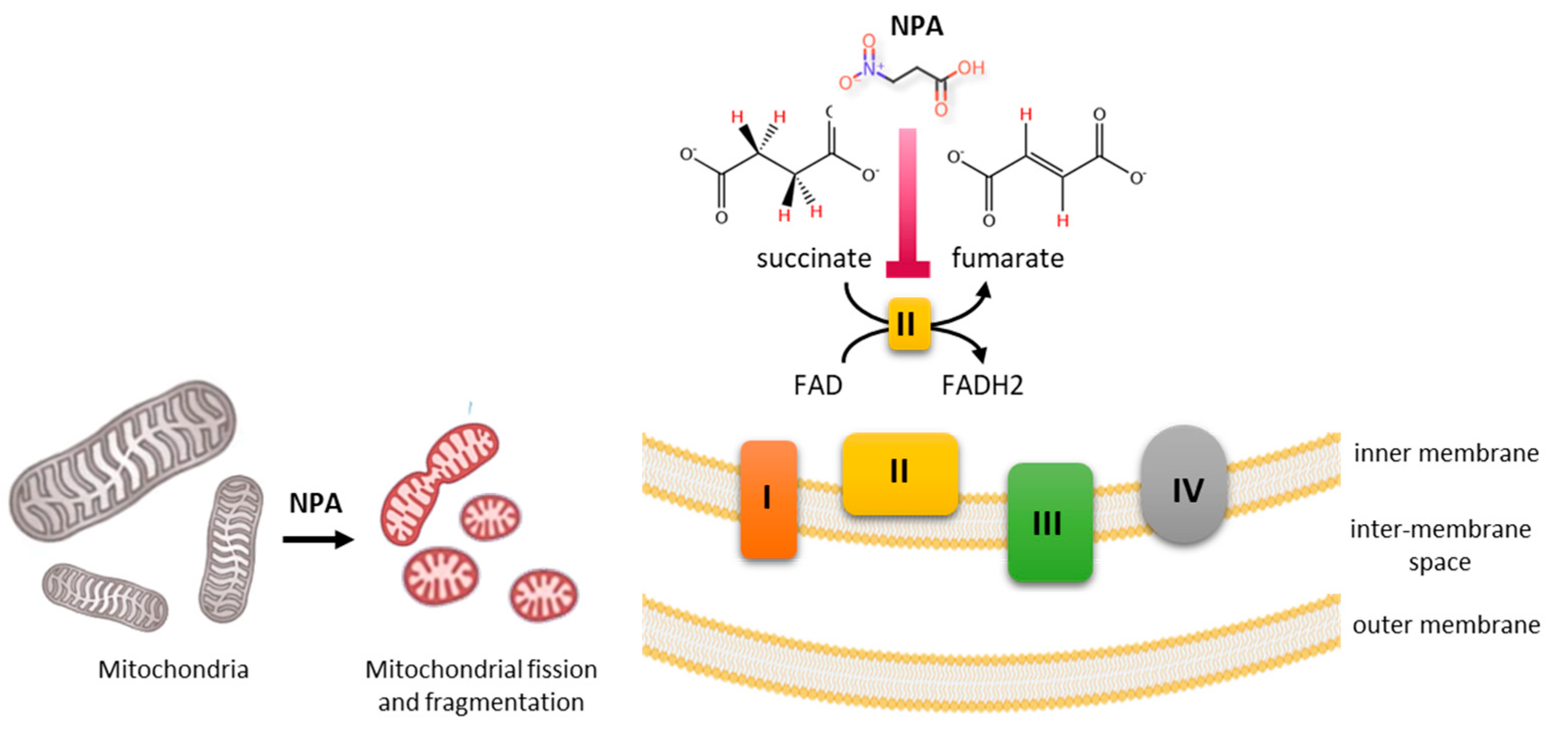
NPA, NPOH, and to a lesser extent its β-D-glucopyranoside miserotoxin are considered as plant and fungal toxins that interrupt mitochondrial electron transport, resulting in cellular energy deficit [55]. NPA functions as a suicide inhibitor of succinate dehydrogenase, an enzyme (complex II) participating in the citric acid cycle and the mitochondrial respiratory chain [56]. NPA binds covalently to the catalytic center of succinate dehydrogenase, causing the irreversible inhibition of mitochondrial respiration [57,58,59] (Figure 3). As such, it causes acute encephalopathy and late-onset dystonia [60]. In animal studies, the inhibition of succinate dehydrogenase by NPA triggers striatal lesions similar to those observed in Huntington’s disease [61,62]. In addition, NPA is used experimentally to induce Huntington’s disease-like pathogenic conditions in experimental models [63,64,65,66]. In rat striatum, NPA activates the calpain/cyclin-dependent kinase 5 (cdk5) pathway. The compound activates the calcium-dependent protease calpain to cleave the Cdk5 co-activator p35 into p25, thereby decreasing the phosphorylated form of transcription factor MEF2 (myocyte enhancer factor 2) which is involved in neuronal cell survival. The inactivation of MEF2 leads to neuronal cell death [67]. The toxin impairs the correct functioning of mitochondria and reduces ATP synthesis, leading to an excessive production of free radicals resulting in the degeneration of GABAergic neurons in the striatum (Figure 4) [63].
The water-soluble product 3-nitropropionate is slowly converted to the 3-NPA dianion (pK = 9.3) under physiological conditions. NPA is the metabolite of both NPOH and miserotoxin. When administered orally, miserotoxin is relatively innocuous (LD50 > 2.5 g/kg), but the metabolite NPOH (LD50 = 77 mg/kg) is considerably more toxic. The microbial hydrolysis of miserotoxin confers the toxicity (Figure 2) [69]. NPOH is then converted rapidly into the toxic metabolite NPA, in cattle and sheep [70]. It is interesting to note that there exist microorganisms capable of aiding their hosts with the detoxification of NPA. Bovine ruminal and equine cecal microbes metabolizing NPA have been identified, such as the ruminal bacterium Denitrobacterium detoxificans that respires on nitro compounds [71,72,73]. Insects can also contribute to the detoxification of nitroaliphatic toxins through their conjugation or degradation. This is the case, for example, for the microbiota associated with the invasive green bug Nezara viridula, which exerts a detoxifying activity toward NPA [74]. The gut microbial community of the insect harbors toxin-degrading bacteria which participate in the insect resistance to plant defense [75,76]. Other insects, such as Spodoptera littoralis caterpillars, can detoxify NPA through conjugation with amino acids within epithelial cells followed by the export of the nontoxic amino acid conjugates to the hemolymph [77].
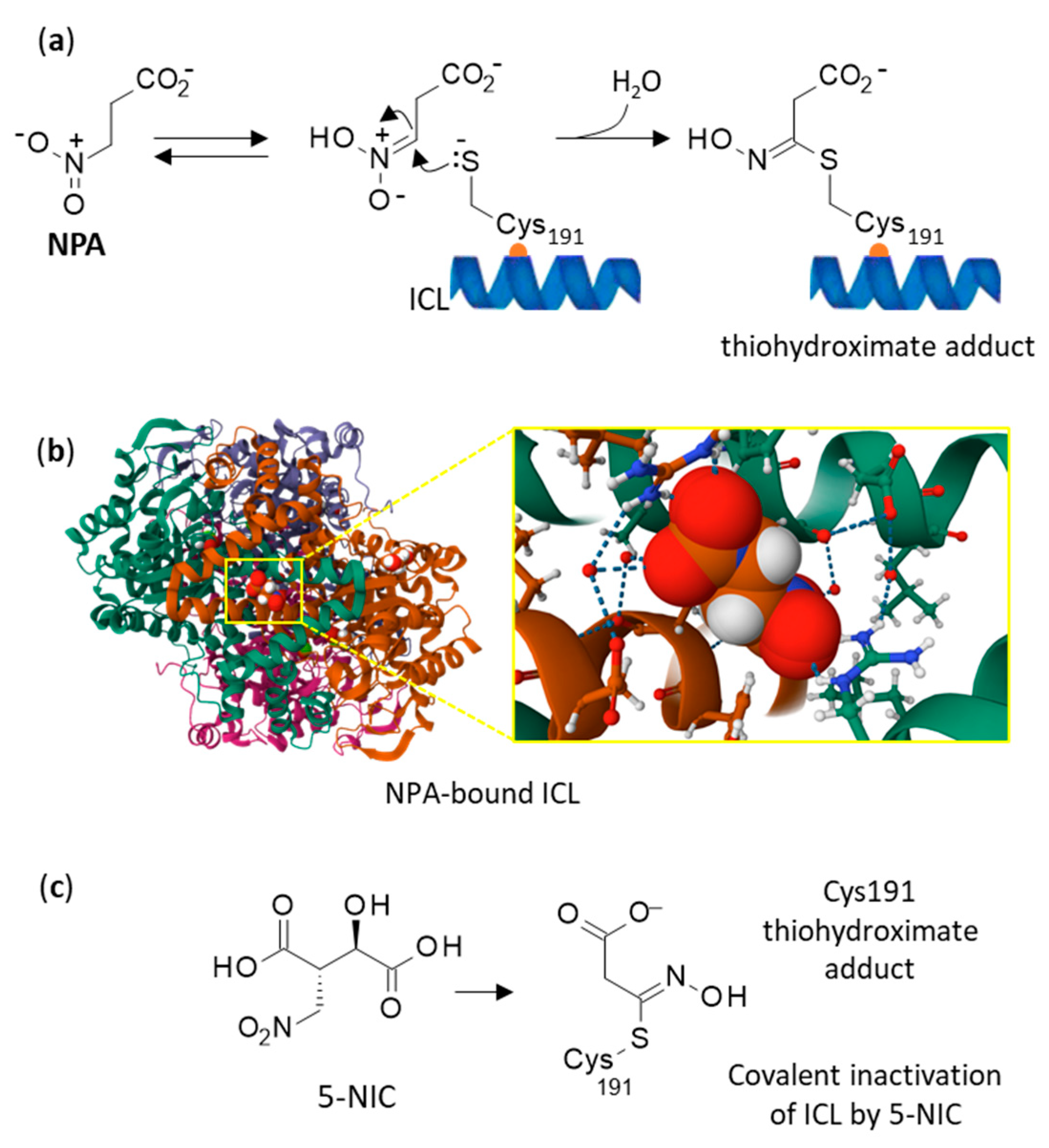
The covalent binding of NPA to proteins has been investigated in details. In M. tuberculosis, the nitro group of NPA has been shown to function as a masked electrophile, activated by conversion to its nitronic acid tautomer within the enzyme active site. It reacted with an active-site cysteine residue (C191) of isocitrate lyase (ICL) to yield a stable thiohydroximate adduct (Figure 5) [78]. ICL catalyzes the reversible cleavage of isocitrate into succinate and glyoxylate. This reaction mechanism has been exploited for the design of targeted covalent inhibitors, such as the pro-drug 5-NIC, which upon cleavage produces glyoxylate and NPA for the covalent inactivation of ICL from M. tuberculosis [79,80] (Figure 5c). Similarly, propionate-3-nitronate (P3N) is a conjugate base of NPA, an inhibitor of both ICL and mitochondrial succinate dehydrogenase [81,82]. NPA has inspired the design of different types of ICL inhibitors. NPA is highly reactive but also poorly selective, rendering it unsuitable for drug development. But other covalent inhibitors that operate through a mechanism similar to NPA have been designed, notably nitro-substituted 1,3-benzothiazinone derivatives [83].
NPA is a mitochondria-binding agent that mainly manifests its toxicity by interfering with mitochondrial bioenergetics. The compound displays also other effects. Notably, NPA has shown marked hypotensive effects in animal species. It decreased the blood pressure and provoked bradycardia. It is a toxic metabolite but it is not formally considered as being the cause of death in intoxication with Astragalus [9]. When NPA and NPOH were fed to sheep and cattle at doses which produced chronic or acute intoxication, the signs of intoxication—such as emphysema and difficulty in locomotion—were similar to those observed in poisoning that occurs under field conditions. NPA produced primary microscopic lesions in the CNS and lungs, including alveolar emphysema [84]. Intermittent exposure to NPA can cause also an activation of the dopaminergic system in the long term [85].
Several cases of NPA intoxication in humans have been reported [86]. For example, reports from China have documented the tragic consequences of NPA intoxication in humans consuming moldy sugarcane [37]. Recently, the first case of NPA intoxication was reported in Norway [87]. NPA is a neurotoxic agent, with the capacity to alter mitochondrial functions, oxidative stress, apoptosis, and to induce neuroinflammation. However, it is important to mention that in long-term studies, NPA did not exhibit carcinogenicity or chronic toxicity. The reported no observed adverse effect level (NOAEL) for NPA is 2.5 and 3.75 mg/kg/day for male and female rats, respectively. The acceptable daily intake (ADI) has been estimated to be 25 μg/kg/day or 1.75 mg/day for a 70 kg human [88].
NPA is a mitochondrial toxin capable of inducing brain injury, and the damage can be aggravated by other agents, such as lithium [89]. But here, again, there is no evidence for an accumulation of Li in E. baetica. The presence of selenium in the plant extracts was underlined initially [21], but a subsequent study concluded the opposite, that no selenium was found in Astragalus lusitanicus (i.e., E. baetica). Other elements such as iron, copper, zinc, molybdenum, chromium, and titanium have been quantified from root, stem, and leave extracts but in reasonable proportions (mostly in the roots) [90]. The observed severe intoxication upon the ingestion of E. baetica cannot be due to the presence of these trace elements or their combination with NPA.

5. Conclusions

Erophaca plants are sometimes cut down by shepherds and farmers to prevent them from spreading because they are considered as being toxic to ruminants. Consequently, a significant part of the plant population has been decimated, particularly in Morocco [4]. However, these plants can be useful for other purposes and should be preserved. A decoction made from the fresh roots of E. baetica can be used in traditional medicine for massages or cataplasms to treat knee and elbow diseases [91,92]. There are also useful insects feeding on E. baetica, notably with larvae and the attending ants located inside the fruit pods of the plant [93]. Moreover, the seeds of the plant may present a nutritional value owing to their high protein content (36% w/w). The antioxidant activity of polyphenol extracts from E. baetica was found to be higher than the activity of polyphenols extracted from other edible legume seeds [94]. For these reasons, E. baetica should be preserved. It is an old (Miocene) plant, largely present in Morocco, which represents an ancestral territory for the plant. A genetic analysis (based on the sequencing of the internal transcribed spacer region (ITS) of the nuclear ribosomal DNA (nrDNA) has revealed that the genetic diversity decreased with increasing distance from Morocco [4].
NPA and its precursor NPOH are responsible for the toxicity of Erophaca plants and E. baetica (L.) Boiss. is one of the most cited toxic plant species by individuals, implicated in major transhumance routes that cross Castilla La Mancha in Spain [1]. It is very likely that these two nitro-products are responsible for the toxicity of this species, although a formal proof is lacking at present. As a chemical product, NPA is commercially available (and relatively cheap), readily accessible via a two-step straightforward synthesis from acrolein [95]. The identification and quantitative assessment of the product from the aerial parts of E. baetica should not present difficulties. Efforts shall be dedicated to the dosage of NPA in E. baetica extracts, comparing plant samples collected in Morocco and other countries or regions. There is a need to compare the toxicity of Erophaca subspecies and to analyze the possible genetic variability among subspecies. These variations might influence toxicity. Similarly, it would be interesting to investigate the influence of environmental factors (like climate, soil composition, and symbiotic microbial communities) on the synthesis or accumulation of toxic compounds in E. baetica. Phytochemical analyses should focus on NPA/NPOH, without neglecting other potential toxic natural products.

6. Future Direction

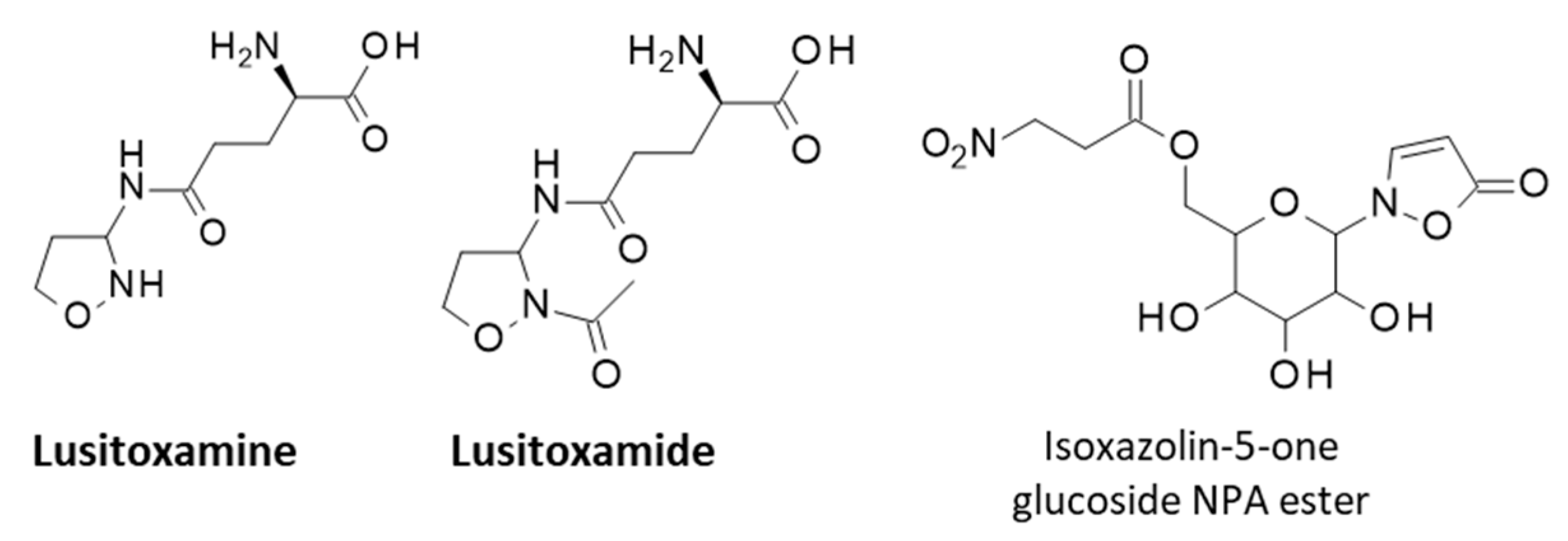
Over the course of our extensive analysis of the scientific literature pertaining to the plant E. baetica, a Ph.D. thesis on the phytochemical and toxicological analysis of Astragalus lusitanicus (i.e., E. baetica) [96] has been identified. The thesis referred to two unknown products isolated from this plant, named lusitoxamine and lusitoxamide, both bearing a 3-amino-isoxazolidine core linked to a γ-L-glutamyl moiety (Figure 6). Lusitoxamide is the N-acetyl analog of lusitoxamine. The thesis also mentioned the existence of two glycosyl derivatives designated lusitoxamidosides F and G (α-6-fructofuranosyl-lusitoxamine and α-1β-glucosipyranosyl-lusitoxamine, respectively). Unfortunately, apart from this PhD thesis, no other document has been identified to validate the existence of these natural products (no publication, no patent, no other thesis). There is no report of isoxazolidine derivatives isolated from Astragalus species. But there exist isoxazolinone derivatives, such as the isoxazolinone glycoside represented in Figure 6, initially isolated from some insects (chrysomelid beetles) [97] and later found in two Astragalus plant species: A. canadensis L. var. mortonii (Nutt) Wats. and A. collinus Dougl. ex Hook [98]. This natural product may play a role in the chemical defense of Astragalus plant species as it does in beetles. This type of 3-nitropropanoate ester with an isoxazolin-5-one glucoside moiety has been found in fungi, plants, and beetles [42,57,99,100]. Isoxazolinone glucosides esterified with 3-nitropropanoic acid (NPA ester) are viewed as antifeedants produced by plants to repel herbivores through toxicity, and also as pretoxins stored by beetles to protect themselves against predators [101,102,103].
NPA is a nitrotoxin used by diverse Astragalus species as a defense mechanism for protection against herbivores. It is most likely that E. baetica also uses NPA as a chemical defense. However, at this stage, the contribution of other phytochemicals like lusitoxamine cannot be excluded. The phytochemical content of the Moroccan species E. baetica warrants further investigation. With this review, it is expected to lay the groundwork for the future in answering these questions regarding the plant toxins. Additional research shall explore the further molecular origin of the toxicity and further therapeutic applications of Erophaca baetica (L.).

Author Contributions

Conceptualization, L.B. and C.B.; methodology, M.C., L.B. and C.B.; investigation, M.C., L.B. and C.B.; data curation, M.C., L.B. and C.B.; writing—original draft preparation, M.C., L.B. and C.B.; writing—review and editing, L.B. and C.B.; supervision, L.B. and C.B. All authors have read and agreed to the published version of the manuscript.

Funding

This research received no external funding.

Acknowledgments

The authors are thankful to Sultan Moulay Slimane University, Morocco, for partial support.

Conflicts of Interest

The authors declare no conflicts of interest.

Abbreviations

The following abbreviations are used in this manuscript:
ICLisocitrate lyase
NPA3-nitropropionic acid
NPOH3-nitropropanol

References

  1. Rivera, D.; Verde, A.; Fajardo Rodríguez, J.; Ríos, S.; Alcaraz, F.; Cárceles, C.; Ortíz, J.; Valdés, A.; Ruíz-Gallardo, J.R.; García-Flores, A.; et al. Ethnoveterinary Medicine and Ethnopharmacology in the Main Transhumance Areas of Castilla-La Mancha (Spain). Front. Vet. Sci. 2022, 9, 866132. [Google Scholar] [CrossRef] [PubMed]
  2. Najem, M.; Nassiri, L.; Ibijbijen, J. Vernacular names of toxic plants used as medicine in the central Middle Atlas-Morocco. Ethnobot. Res. Appl. 2020, 20, 48. [Google Scholar]
  3. Najem, M.; Nassiri, L.; Ibijbijen, J. Origin of vernacular names of plants: Case of toxic plants for medicinal use in the central Middle Atlas—Morocco. Interdiscip. Sci. Rev. 2021, 47, 40–61. [Google Scholar] [CrossRef]
  4. Casimiro-Soriguer, R.; Talavera, M.; Balao, F.; Terrab, A.; Herrera, J.; Talavera, S. Phylogeny and genetic structure of Erophaca (Leguminosae), an East-West Mediterranean disjunct genus from the Tertiary. Mol. Phylogenet. Evol. 2010, 56, 441–450. [Google Scholar] [CrossRef] [PubMed]
  5. Duval, J. Perceptions et Usages de la Diversité éCologique des Parcours Présahariens par les éLeveurs Camelins dans la Zone Rurale de M’Hamid El Ghizlane au Maroc. Mémoire de Fin d’éTudes. 2019. Available online: https://camed.cirad.fr/fr/content/download/4186/31380/version/1/file/Duval+2019+Perceptions+et+usages+diversit%C3%A9+%C3%A9colo+parcours+pr%C3%A9sahariens+par+%C3%A9leveurs+camelins+Zone+M%27Hamid+El+Ghizlane+Maroc+%28M%C3%A9moire%29.pdf (accessed on 15 April 2025).
  6. Casimiro-Soriguer, R.; Herrera, J.; Talavera, S. Andromonoecy in an Old World Papilionoid legume, Erophaca baetica. Plant Biol. 2013, 15, 353–359. [Google Scholar] [CrossRef]
  7. Lyu, S.T.; Zou, T.T.; Jiang, Q.L.; Wang, X.F. Maintenance of andromonoecy in an autogamous species: Superior male function in male flowers of the endangered Sagittaria guayanensis. Plant Divers. 2024, 46, 783–790. [Google Scholar] [CrossRef]
  8. Murakami, K.; Katsuhara, K.R.; Ushimaru, A. Intersexual flower differences in an andromonoecious species: Small pollen-rich staminate flowers under resource limitation. Plant Biol. 2022, 24, 259–265. [Google Scholar] [CrossRef]
  9. Rios, J.L.; Waterman, P.G. A Review of the Pharmacology and Toxicology of Astragalus. Phytother. Res. 1997, 11, 411–418. [Google Scholar] [CrossRef]
  10. Wagstaff, D.J. International Poisonous Plants Checklist. An Evidence-Based Reference; CRC Press: Boca Raton, FL, USA; London, UK; New York, NY, USA; Taylor & Francis Group: Boca Raton, FL, USA; London, UK; New York, NY, USA, 2008. [Google Scholar]
  11. Abdennebi, E.H.; el Ouazzani, N.; Lamnaouer, D. Clinical and analytical studies of sheep dosed with various preparations of Astragalus lusitanicus. Vet. Hum. Toxicol. 1998, 40, 327–331. [Google Scholar]
  12. Ouazzani, N.; Lamnaouer, D.; Abdennebi, E.H. Toxicology of Astragalus lusitanicus Lam. Therapie 1999, 54, 707–710. [Google Scholar]
  13. El-Hamidi, M.; Leipold, H.W. Poisoning of Sheep by Astragalus lusitanicus in Morocco: Field and Experimental Studies. J. Vet. Med. A 1989, 36, 115–121. [Google Scholar] [CrossRef] [PubMed]
  14. Marin, R.E.; Gardner, D.R.; Armien, A.G.; Fortunato, R.H.; Uzal, F.A. Intoxication of llamas by Astragalus punae in Argentina. J. Vet. Diagn. Investig. 2022, 34, 674–678. [Google Scholar] [CrossRef]
  15. Garcia Roman, A.; Moyano Salvago, M.R.; Soler Rodriguez, F.; Infante Miranda, F. Physiopathologic changes in lambs fed with Astragalus lusitanicus Lam. Vet. Hum. Toxicol. 1987, 29, 387–389. [Google Scholar] [PubMed]
  16. Tarazona, J.V.; Sanz, F. Toxicity of fractions obtained from the legume species Astragalus lusitanicus Lam. lusitanicus. Toxicon. 1990, 28, 235–237. [Google Scholar] [CrossRef]
  17. Bel-Kassaoui, H.; Lamnaouer, D.; Abdennebi, E.H.; Jossang, A. Experimental poisoning by extracts and fractions of Astragalus lusitanicus Lam. in sheep. Rev. Med. Vet. 2007, 158, 269–273. [Google Scholar]
  18. Poyato, J. Estudio fitoquimico, toxicologico y farmacologico del Astragalus lusitanicus. Rev. Patron. Biol. Anim. 1969, XII, 67–118. [Google Scholar]
  19. Gonzalez, J.M.; Tovar, P.; Castejon, F.J.; Gonzalez, M. Intoxicacion experimental por Astragalus lusitanicus Lam. I. Alteraciones cardio-respiratorias. Archos Zootec. 1980, 29, 23–30. [Google Scholar]
  20. Quintas, H.; Aguiar, C.; Ramos Antón, J.J.; Lacasta Lozano, D.; Ferrer Mayayo, L.M. Intoxicación por plantas en rumiantes: Bases para el diagnóstico clínico. In Agrárias: Pesquisa e Inovação nas Ciências que Alimentam o Mundo VII; Editora Artemis: Curitiba, Brazil, 2021; pp. 291–301. ISBN 978-65-87396-51-4. [Google Scholar]
  21. Pascual, J.; Hernandez, J.C.; Grande, M. Componentes de Astragalus lusitanicus. Lam. II. Flavonoides. An. Quim. 1979, 75, 1005–1007. [Google Scholar]
  22. Pascual, J.; Hernandez, J.C.; Sanchez, J.J. Componentes de Astragalus lusitanicus Lam. I. Selenio, pinitol y derivados nitrados. An. Quiet. 1979, 75, 775–777. [Google Scholar]
  23. Pascual, J.; Hernandez, J.C.; San Feliciano, A.; Miguel, J.M. Saccharinic acid lactone from Astragalus lusitanicus Lam. (-)-2-C-methyl-D-erythrono-1,4-lactone. Tetrahedron Lett. 1980, 21, 1359–1360. [Google Scholar]
  24. Cook, D.; Gardner, D.R.; Pfister, J.A. Swainsonine-containing plants and their relationship to endophytic fungi. J. Agric. Food Chem. 2014, 62, 7326–7334. [Google Scholar] [CrossRef] [PubMed]
  25. Cook, D.; Gardner, D.R.; Lee, S.T.; Pfister, J.A.; Stonecipher, C.A.; Welsh, S.L. A swainsonine survey of North American Astragalus and Oxytropis taxa implicated as locoweeds. Toxicon 2016, 118, 104–111. [Google Scholar] [CrossRef]
  26. Cook, D.; Gardner, D.R.; Martinez, A.; Robles, C.A.; Pfister, J.A. Screening for swainsonine among South American Astragalus species. Toxicon 2017, 139, 54–57. [Google Scholar] [CrossRef] [PubMed]
  27. Pistán, M.E.; Cook, D.; Gutiérrez, S.A.; Schnittger, L.; Gardner, D.R.; Cholich, L.A.; Gonzalez, A.M. Identification and distribution of a fungal endosymbiotic Alternaria species (Alternaria section Undifilum sp.) in Astragalus garbancillo tissues. Mycologia 2024, 116, 291–298. [Google Scholar] [CrossRef] [PubMed]
  28. Abdennebi, E.H.; Ouazzani, N.; Jossang, A.; Benkouka, F.; Lamnaouer, D. Inhibition of glycosidases by Astragalus lusitanicus and correlation with toxicity. Vet. Hum. Toxicol. 2001, 43, 266–269. [Google Scholar] [PubMed]
  29. Bel-Kassaoui, H.; Lamnaouer, D.; Jossang, A.; Abdennebi el, H.; Charrouf, Z.; Bodo, B. Role of hypaphorine in the toxicity of Astragalus lusitanicus. Nat. Prod. Res. 2008, 22, 453–457. [Google Scholar] [CrossRef]
  30. Mohamed, I.I.; Hassan, H.E.; Ahmed, S.A.; Bahaa, S.M. Alkaloids of Astragalus kahiricus DC. plant roots. Bull. Fac. Agric. Cairo Univ. 2009, 60, 366–370. [Google Scholar] [CrossRef]
  31. Hou, B.; Wen, Y.; Zhu, X.; Qi, M.; Cai, W.; Du, B.; Sun, H.; Qiu, L. Preparation and characterization of vaccarin, hypaphorine and chitosan nanoparticles and their promoting effects on chronic wounds healing. Int. J. Biol. Macromol. 2022, 221, 1580–1592. [Google Scholar] [CrossRef]
  32. Zhang, Y.; Huang, J.; Gan, L.; Wu, R.; Jin, J.; Wang, T.; Sun, S.; Zhang, Z.; Li, L.; Zheng, X.; et al. Hepatoprotective effects of Niudali (Callerya speciosa) root aqueous extracts against tetrachloromethane-induced acute liver injury and inflammation. Food Sci. Nutr. 2023, 11, 7026–7038. [Google Scholar] [CrossRef]
  33. Ivanov, I.A.; Siniavin, A.E.; Palikov, V.A.; Senko, D.A.; Shelukhina, I.V.; Epifanova, L.A.; Ojomoko, L.O.; Belukhina, S.Y.; Prokopev, N.A.; Landau, M.A.; et al. Analogs of 6-Bromohypaphorine with Increased Agonist Potency for α7 Nicotinic Receptor as Anti-Inflammatory Analgesic Agents. Mar. Drugs 2023, 21, 368. [Google Scholar] [CrossRef]
  34. Tarazona, J.V.; Sanz, F. Aliphatic nitro compounds in Astragalus lusitanicus Lam. Vet. Hum. Toxicol. 1987, 29, 437–439. [Google Scholar] [PubMed]
  35. Williams, M.C. Nitro compounds in foreign species of Astragalus. Weed Sci. 1981, 29, 261–269. [Google Scholar] [CrossRef]
  36. Williams, M.C. 3-Nitropropionic acid and 3-nitro-1-propanol in species of Astragalus. Can. J. Bot. 1982, 60, 1956–1963. [Google Scholar] [CrossRef]
  37. Hamilton, B.F.; Gould, D.H.; Gustine, D.L. History of 3-Nitropropionic Acid. Occurrence and Role in Human and Animal Disease. In Mitochondrial Inhibitors and Neurodegenerative Disorders. Contemporary Neuroscience; Sanberg, P.R., Nishino, H., Borlongan, C.V., Eds.; Humana Press: Totowa, NJ, USA, 2000; pp. 21–33. [Google Scholar] [CrossRef]
  38. Liu, H.; Shao, S.; Schellenberg, M. A Simple and Fast Procedure to Determine 3-Nitropropanoic Acid and 3-Nitropropanol in Freeze Dried Canadian Milkvetch (Astragalus canadensis). Toxins 2017, 9, 204. [Google Scholar] [CrossRef]
  39. Tarazona Lafarga, J.V. Origin of the Toxicicity of the Astrafalus lusitanicus Lam. and Its Relation with Acid 3-Nitropropanol and Other Extracts Spanish. Ph.D. Thesis, Universidad Complutense de Madrid, Madrid, Spain, 1988. Available online: https://dialnet.unirioja.es/servlet/tesis?codigo=198174 (accessed on 15 April 2025).
  40. Carter, C.L.; McChesney, W.J. Hiptagenic acid identified as beta-nitropropionic acid. Nature 1949, 164, 575. [Google Scholar] [CrossRef]
  41. Morris, M.P.; Pagán, C.; Warmke, H.E. Hiptagenic Acid, a Toxic Component of Indigofera endecaphylla. Science 1954, 119, 322–323. [Google Scholar] [CrossRef]
  42. Becker, T.; Pasteels, J.; Weigel, C.; Dahse, H.M.; Voigt, K.; Boland, W. A tale of four kingdoms—Isoxazolin-5-one- and 3-nitropropanoic acid-derived natural products. Nat. Prod. Rep. 2017, 34, 343–360. [Google Scholar] [CrossRef]
  43. Raistrick, H.; Stössl, A. Studies in the biochemistry of micro-organisms. 104. Metabolites of Penicillium atrovenetum G. Smith: β-nitropropionic acid, a major metabolite. Biochem. J. 1958, 68, 647–653. [Google Scholar]
  44. Anzai, K.; Suzuki, S. A New Antibiotic Bovinocidin, Identified as β-Nitropropionic Acid. J. Antibiot. 1960, 13, 133–136. [Google Scholar]
  45. Rotilio, L.; Boverio, A.; Nguyen, Q.T.; Mannucci, B.; Fraaije, M.W.; Mattevi, A. A biosynthetic aspartate N-hydroxylase performs successive oxidations by holding intermediates at a site away from the catalytic center. J. Biol. Chem. 2023, 299, 104904. [Google Scholar] [CrossRef]
  46. Chomcheon, P.; Wiyakrutta, S.; Sriubolmas, N.; Ngamrojanavanich, N.; Isarangkul, D.; Kittakoop, P. 3-Nitropropionic acid (3-NPA), a potent antimycobacterial agent from endophytic fungi: Is 3-NPA in some plants produced by endophytes? J. Nat. Prod. 2005, 68, 1103–1105. [Google Scholar] [CrossRef] [PubMed]
  47. Shaw, P.D.; McCloskey, J.A. Biosynthesis of nitro compounds. II. Studies on potential precursors for the nitro group of beta-nitropropionic acid. Biochemistry 1967, 6, 2247–2260. [Google Scholar] [CrossRef] [PubMed]
  48. Baxter, R.L.; Smith, S.L.; Martin, J.R.; Hanley, A.B. The fungal biosynthesis of 3-nitropropionic acid: Is the decarboxylation of L-nitrosuccinate an enzymatic reaction? J. Chem. Soc. Perkin Trans. 1 1994, 16, 2297–2299. [Google Scholar] [CrossRef]
  49. Johnson, C.W.; Ohashi, M.; Tang, Y. How Fungi Biosynthesize 3-Nitropropanoic Acid: The Simplest yet Lethal Mycotoxin. Org. Lett. 2024, 26, 3158–3163. [Google Scholar] [CrossRef]
  50. Francis, K.; Smitherman, C.; Nishino, S.F.; Spain, J.C.; Gadda, G. The biochemistry of the metabolic poison propionate 3-nitronate and its conjugate acid, 3-nitropropionate. IUBMB Life 2013, 65, 759–768. [Google Scholar] [CrossRef]
  51. Takács, O.; Nagyné Nedves, A.; Boldizsár, I.; Höhn, M.; Béni, S.; Gampe, N. Analysis of 3-nitropropionic acid in Fabaceae plants by HPLC-MS/MS. Phytochem. Anal. 2022, 33, 1205–1213. [Google Scholar] [CrossRef] [PubMed]
  52. MacAskill, J.J.; Manley-Harris, M.; Field, R.J. Quantification of nitropropanoyl glucosides in karaka nuts before and after treatment. Food Chem. 2015, 175, 543–548. [Google Scholar] [CrossRef]
  53. Johnson, D.L.; Majak, W.; Benn, M.H. Excretion of miserotoxin and detoxification of the aglycone by grasshoppers (Orthoptera: Acrididae). Phytochemistry 2001, 58, 739–742. [Google Scholar] [CrossRef]
  54. Stermitz, F.R.; Norris, F.A.; Williams, M.C. Miserotoxin, new naturally occurring nitro compound. J. Am. Chem. Soc. 1969, 91, 4599–4600. [Google Scholar] [CrossRef]
  55. Patocka, J.; Bielavský, J.; Cabal, J.; Fusek, J. 3-Nitropropionic acid and similar nitrotoxins. Acta Medica 2000, 43, 9–13. [Google Scholar] [CrossRef]
  56. Coles, C.J.; Edmondson, D.E.; Singer, T.P. Inactivation of succinate dehydrogenase by 3-nitropropionate. J. Biol. Chem. 1979, 254, 5161–5167. [Google Scholar] [CrossRef]
  57. Huang, L.S.; Sun, G.; Cobessi, D.; Wang, A.C.; Shen, J.T.; Tung, E.Y.; Anderson, V.E.; Berry, E.A. 3-nitropropionic acid is a suicide inhibitor of mitochondrial respiration that, upon oxidation by complex II, forms a covalent adduct with a catalytic base arginine in the active site of the enzyme. J. Biol. Chem. 2006, 281, 5965–5972. [Google Scholar] [CrossRef]
  58. Becker, T.; Ploss, K.; Boland, W. Biosynthesis of isoxazolin-5-one and 3-nitropropanoic acid containing glucosides in juvenile Chrysomelina. Org. Biomol. Chem. 2016, 14, 6274–6280. [Google Scholar] [CrossRef]
  59. Liot, G.; Bossy, B.; Lubitz, S.; Kushnareva, Y.; Sejbuk, N.; Bossy-Wetzel, E. Complex II inhibition by 3-NP causes mitochondrial fragmentation and neuronal cell death via an NMDA- and ROS-dependent pathway. Cell Death Differ. 2009, 16, 899–909. [Google Scholar] [CrossRef]
  60. He, F.; Zhang, S.; Qian, F.; Zhang, C. Delayed dystonia with striatal CT lucencies induced by a mycotoxin (3-nitropropionic acid). Neurology 1995, 45, 2178–2183. [Google Scholar] [CrossRef]
  61. Brouillet, E.; Guyot, M.C.; Mittoux, V.; Altairac, S.; Condé, F.; Palfi, S.; Hantraye, P. Partial inhibition of brain succinate dehydrogenase by 3-nitropropionic acid is sufficient to initiate striatal degeneration in rat. J. Neurochem. 1998, 70, 794–805. [Google Scholar] [CrossRef]
  62. Bouillaud, F. Inhibition of Succinate Dehydrogenase by Pesticides (SDHIs) and Energy Metabolism. Int. J. Mol. Sci. 2023, 24, 4045. [Google Scholar] [CrossRef]
  63. Upadhayay, S.; Yedke, N.G.; Rahi, V.; Singh, S.; Kumar, S.; Arora, A.; Chandolia, P.; Kaur, P.; Kumar, M.; Koshal, P.; et al. An Overview of the Pathophysiological Mechanisms of 3-Nitropropionic Acid (3-NPA) as a Neurotoxin in a Huntington’s Disease Model and Its Relevance to Drug Discovery and Development. Neurochem. Res. 2023, 48, 1631–1647. [Google Scholar] [CrossRef]
  64. Noureldeen, M.E.; Shahin, N.N.; Amin, H.A.A.; El-Sawalhi, M.M.; Ghaiad, H.R. Parthenolide ameliorates 3-nitropropionic acid-induced Huntington’s disease-like aberrations via modulating NLRP3 inflammasome, reducing microglial activation and inducing astrocyte shifting. Mol. Med. 2024, 30, 158. [Google Scholar] [CrossRef]
  65. Lum, P.T.; Sekar, M.; Seow, L.J.; Shaikh, M.F.; Arulsamy, A.; Retinasamy, T.; Gan, S.H.; Gnanaraj, C.; Esa, N.M.; Ramachawolran, G.; et al. Neuroprotective potency of mangiferin against 3-nitropropionic acid induced Huntington’s disease-like symptoms in rats: Possible antioxidant and anti-inflammatory mechanisms. Front. Pharmacol. 2023, 14, 1189957. [Google Scholar] [CrossRef]
  66. Túnez, I.; Tasset, I.; Pérez-De La Cruz, V.; Santamaría, A. 3-Nitropropionic acid as a tool to study the mechanisms involved in Huntington’s disease: Past, present and future. Molecules 2010, 15, 878–916. [Google Scholar] [CrossRef] [PubMed]
  67. Crespo-Biel, N.; Camins, A.; Pelegrí, C.; Vilaplana, J.; Pallàs, M.; Canudas, A.M. 3-Nitropropionic acid activates calpain/cdk5 pathway in rat striatum. Neurosci. Lett. 2007, 421, 77–81. [Google Scholar] [CrossRef]
  68. Sun, F.; Huo, X.; Zhai, Y.; Wang, A.; Xu, J.; Su, D.; Bartlam, M.; Rao, Z. Crystal structure of mitochondrial respiratory membrane protein complex II. Cell 2005, 121, 1043–1057. [Google Scholar] [CrossRef]
  69. Majak, W.; Pass, M.A.; Madryga, F.J. Toxicity of miserotoxin and its aglycone (3-nitropropanol) to rats. Toxicol. Lett. 1983, 19, 171–178. [Google Scholar] [CrossRef]
  70. Muir, A.D.; Majak, W.; Pass, M.A.; Yost, G.S. Conversion of 3-nitropropanol (miserotoxin aglycone) to 3-nitropropionic acid in cattle and sheep. Toxicol. Lett. 1984, 20, 137–141. [Google Scholar] [CrossRef]
  71. Teng, P.Y.; Kim, W.K. Roles of Nitrocompounds in Inhibition of Foodborne Bacteria, Parasites, and Methane Production in Economic Animals. Animals 2021, 11, 923. [Google Scholar] [CrossRef]
  72. Zhang, Y.; Long, R.; Warzecha, C.M.; Coverdale, J.A.; Latham, E.A.; Hume, M.E.; Callaway, T.R.; O’Neil, M.R.; Beier, R.C.; Anderson, R.C.; et al. Characterization of bovine ruminal and equine cecal microbial populations enriched for enhanced nitro-toxin metabolizing activity. Anaerobe 2014, 26, 7–13. [Google Scholar] [CrossRef] [PubMed]
  73. Anderson, R.C.; Rasmussen, M.A.; Jensen, N.S.; Allison, M.J. Denitrobacterium detoxificans gen. nov., sp. nov., a ruminal bacterium that respires on nitrocompounds. Int. J. Syst. Evol. Microbiol. 2000, 50, 633–638. [Google Scholar] [CrossRef] [PubMed]
  74. Coolen, S.; Rogowska-van der Molen, M.A.; Kwakernaak, I.; van Pelt, J.A.; Postma, J.L.; van Alen, T.; Jansen, R.S.; Welte, C.U. Microbiota of pest insect Nezara viridula mediate detoxification and plant defense repression. ISME J. 2024, 18, wrae097. [Google Scholar] [CrossRef]
  75. Rogowska-van der Molen, M.A.; Nagornîi, D.; Coolen, S.; de Graaf, R.M.; Berben, T.; van Alen, T.; Janssen, M.A.C.H.; Rutjes, F.P.J.T.; Jansen, R.S.; Welte, C.U. Insect Gut Isolate Pseudomonas sp. Strain Nvir Degrades the Toxic Plant Metabolite Nitropropionic Acid. Appl. Environ. Microbiol. 2022, 88, e0071922. [Google Scholar] [CrossRef]
  76. Rogowska-van der Molen, M.A.; Savova, H.V.; Janssen, E.A.T.; van Alen, T.; Coolen, S.; Jansen, R.S.; Welte, C.U. Unveiling Detoxifying Symbiosis and Dietary Influence on the Southern Green Shield Bug Microbiota. FEMS Microbiol. Ecol. 2024, 100, fiae150. [Google Scholar] [CrossRef] [PubMed]
  77. Novoselov, A.; Becker, T.; Pauls, G.; von Reuß, S.H.; Boland, W. Spodoptera littoralis detoxifies neurotoxic 3-nitropropanoic acid by conjugation with amino acids. Insect Biochem. Mol. Biol. 2015, 63, 97–103. [Google Scholar] [CrossRef]
  78. Ray, S.; Kreitler, D.F.; Gulick, A.M.; Murkin, A.S. The Nitro Group as a Masked Electrophile in Covalent Enzyme Inhibition. ACS Chem. Biol. 2018, 13, 1470–1473. [Google Scholar] [CrossRef] [PubMed]
  79. Mellott, D.M.; Torres, D.; Krieger, I.V.; Cameron, S.A.; Moghadamchargari, Z.; Laganowsky, A.; Sacchettini, J.C.; Meek, T.D.; Harris, L.D. Mechanism-Based Inactivation of Mycobacterium tuberculosis Isocitrate Lyase 1 by (2R,3S)-2-Hydroxy-3-(nitromethyl)succinic acid. J. Am. Chem. Soc. 2021, 143, 17666–17676. [Google Scholar] [CrossRef]
  80. Ray, S.; Murkin, A.S. New Electrophiles and Strategies for Mechanism-Based and Targeted Covalent Inhibitor Design. Biochemistry 2019, 58, 5234–5244. [Google Scholar] [CrossRef] [PubMed]
  81. Moynihan, M.M.; Murkin, A.S. Cysteine is the general base that serves in catalysis by isocitrate lyase and in mechanism-based inhibition by 3-nitropropionate. Biochemistry 2014, 53, 178–187. [Google Scholar] [CrossRef]
  82. Torres-Guzman, J.C.; Padilla-Guerrero, I.E.; Cervantes-Quintero, K.Y.; Martinez-Vazquez, A.; Ibarra-Guzman, M.; Gonzalez-Hernandez, G.A. Peculiarities of nitronate monooxygenases and perspectives for in vivo and in vitro applications. Appl. Microbiol. Biotechnol. 2021, 105, 8019–8032. [Google Scholar] [CrossRef]
  83. Richter, A.; Rudolph, I.; Möllmann, U.; Voigt, K.; Chung, C.W.; Singh, O.M.P.; Rees, M.; Mendoza-Losana, A.; Bates, R.; Ballell, L.; et al. Novel insight into the reaction of nitro, nitroso and hydroxylamino benzothiazinones and of benzoxacinones with Mycobacterium tuberculosis DprE1. Sci. Rep. 2018, 8, 13473. [Google Scholar] [CrossRef]
  84. James, L.F.; Hartley, W.J.; Williams, M.C.; Van Kampen, K.R. Field and experimental studies in cattle and sheep poisoned by nitro-bearing Astragalus or their toxins. Am. J. Vet. Res. 1980, 41, 377–382. [Google Scholar] [CrossRef]
  85. Johnson, J.R.; Robinson, B.L.; Ali, S.F.; Binienda, Z. Dopamine toxicity following long term exposure to low doses of 3-nitropropionic acid (3-NPA) in rats. Toxicol. Lett. 2000, 116, 113–118. [Google Scholar] [CrossRef]
  86. Birkelund, T.; Johansen, R.F.; Illum, D.G.; Dyrskog, S.E.; Østergaard, J.A.; Falconer, T.M.; Andersen, C.; Fridholm, H.; Overballe-Petersen, S.; Jensen, J.S. Fatal 3-Nitropropionic Acid Poisoning after Consuming Coconut Water. Emerg. Infect. Dis. 2021, 27, 278–280. [Google Scholar] [CrossRef] [PubMed]
  87. Bendiksen Skogvold, H.; Yazdani, M.; Sandås, E.M.; Østeby Vassli, A.; Kristensen, E.; Haarr, D.; Rootwelt, H.; Elgstøen, K.B.P. A pioneer study on human 3-nitropropionic acid intoxication: Contributions from metabolomics. J. Appl. Toxicol. 2022, 42, 818–829. [Google Scholar] [CrossRef]
  88. Burdock, G.A.; Carabin, I.G.; Soni, M.G. Safety assessment of β-nitropropionic acid: A monograph in support of an acceptable daily intake in humans. Food Chem. 2001, 75, 1–27. [Google Scholar] [CrossRef]
  89. Milutinović, A. Lithium chloride could aggravate brain injury caused by 3-nitropropionic acid. Bosn. J. Basic Med. Sci. 2016, 16, 261–267. [Google Scholar] [CrossRef]
  90. Somer, G.; Caliskan, A.C. Selenium and Trace Element Distribution in Astragalus Plants: Developing a Differential Pulse Polarographic Method for Their Determination. Turk. J. Chem. 2007, 31, 3. [Google Scholar]
  91. Lahsissene, H.; Kahouadji, A.; Tijane, M.; Hseine, S. Catalogue des plantes médicinales utilisées dans la région de Zaër (Maroc Oriental). Rev. Bot. 2009, 186, 2. [Google Scholar]
  92. Naciri, K.; Aboukhalaf, A.; Kalili, A.; Moujabbir, S.; Essaih, S.; Tbatou, M.; Belahyane, A.; Belahsen, R. Ethnobotanical knowledge of wild food plants in Khenifra, a province in the Middle Atlas region of Morocco. GSC Adv. Res. Rev. 2022, 13, 180–200. [Google Scholar] [CrossRef]
  93. Obregón, R.; Shaw, M.R.; Fernández-Haeger, J.; Jordano, D. Parasitoid and ant interactions of some Iberian butterflies (Insecta: Lepidoptera). SHILAP Rev. Lepidopterol. 2015, 43, 439–454. [Google Scholar]
  94. Cortés-Giraldo, I.; Alaiz, M.; Girón-Calle, J.; Megías, C.; Vioque, J. Nutritional and functional characteristics of Erophaca baetica seeds, a legume endemic to the Mediterranean region. Grasas y Aceites 2013, 64, 229–236. [Google Scholar] [CrossRef]
  95. Silva, P.C.; Costa, J.S.; Pereira, V.L.P. An expeditious synthesis of 3-nitropropionic acid and its methyl esters. Synth. Commun. 2001, 31, 595–600. [Google Scholar] [CrossRef]
  96. Bel Kassaoui, H. Etude phytochimique et toxicologique d’Astragalus lusitanicus Lam. et élucidation structurale d’un nouveau produit toxique pour les ruminants. Ph.D. Thesis, Université Mohammed V—Agdal, Faculté des Sciences, Maroc, Rabat, Morocco, 2007. [Google Scholar]
  97. Pasteels, J.M.; Braekman, J.C.; Daloze, D.; Ottinger, R. Chemical defence in Chrysomelid larvae and adults. Tetrahedron 1982, 38, 1891. [Google Scholar] [CrossRef]
  98. Benn, M.H.; Majak, W.; Aplin, R. A nitropropanoyl isoxazolinone derivative in two species of Astragalus. Biochem. System Ecol. 1997, 25, 467–468. [Google Scholar] [CrossRef]
  99. Becker, T.; Kartikeya, P.; Paetz, C.; von Reuss, S.H.; Boland, W. Synthesis and photosensitivity of isoxazolin-5-one glycosides. Org. Biomol. Chem. 2015, 13, 4025–4030. [Google Scholar] [CrossRef] [PubMed]
  100. Pauls, G.; Becker, T.; Rahfeld, P.; Gretscher, R.R.; Paetz, C.; Pasteels, J.; von Reuss, S.H.; Burse, A.; Boland, W. Two Defensive Lines in Juvenile Leaf Beetles; Esters of 3-nitropropionic Acid in the Hemolymph and Aposematic Warning. J. Chem. Ecol. 2016, 42, 240–248. [Google Scholar] [CrossRef]
  101. Tebayashi, S.; Moriyama, R.; Arakawa, R.; Sato, M. Induction of 2-cyanoethyl-isoxazolin-5-one as an antifeedant against the tobacco cutworm (Spodoptera litura) by jasmonic acid in sweet pea leaf. Biosci. Biotechnol. Biochem. 2020, 84, 1105–1112. [Google Scholar] [CrossRef]
  102. Fu, N.; Becker, T.; Brandt, W.; Kunert, M.; Burse, A.; Boland, W. Involvement of CYP347W1 in neurotoxin 3-nitropropionic acid-based chemical defense in mustard leaf beetle Phaedon cochleariae. Insect Sci. 2022, 29, 453–466. [Google Scholar] [CrossRef]
  103. Pasteéls, J.M.; Daloze, D.; de Biseau, J.C.; Termonia, A.; Windsor, D.M. Patterns in host-plant association and defensive toxins produced by neotropical chrysomeline beetles. In New Developments in the Biology of Chrysomelidae; Brill: Leiden, The Netherlands, 2004; pp. 669–676. [Google Scholar]
Figure 1. Erophaca baetica (L.) Boiss. (a) The plant and its white flowers (inset). (photos accessible from https://www.gbif.org/occurrence/4512232562, accessed on 14 April 2025). (b) Distribution map for E. baetica (L.) Boiss. (from https://www.worldfloraonline.org/taxon/wfo-0000190983, last accessed on 14 April 2025). (c) Details of the flowers and pods containing the seeds (from https://www.plantasyhongos.es/herbarium/htm/Erophaca_baetica.htm, last accessed on 14 April 2025).
Futurepharmacol 05 00028 g001
Figure 2. The structures of swainsonine, hypaphorine, and the nitrotoxins: miserotoxin (3-nitropropyl-beta-D-glucopyranoside), NPA (3-nitropropionic acid) and NPOH (3-nitropropanol). The related products karakin and hiptagin are also shown.
Futurepharmacol 05 00028 g002
Figure 3. (a) The crystal structure of mitochondrial respiratory complex II (from porcine heart) bound with inhibitor 3-nitropropionate (NPA). Complex II is a membrane protein complex in the tricarboxylic acid cycle (Krebs cycle) and the mitochondrial respiratory chain. (b,c) Detailed views of NPA bound to complex II (PDB: 1ZP0, last accessed on 14 April 2025) [68]. NPA interacts with the FAD-binding protein (in green), close to the FAD binding site.
Futurepharmacol 05 00028 g003
Figure 4. NPA functions as an irreversible inhibitor of the mitochondrial respiratory complex II and succinate dehydrogenase. The compound triggers a rapid ATP drop and increase in ROS. As such, it activates nitric oxide synthase (NOS) leading to the generation of nitric oxide (NO). NPA causes mitochondrial fragmentation and neuronal cell death [59].
Futurepharmacol 05 00028 g004
Figure 5. (a) The structure of NPA and the nitronic acid tautomer which can react with a cysteine residue (C191) of isocitrate lyase (ICL) to form a thiohydroximate adduct. (b) A molecular model of NPA-modified isocitrate lyase (ICL) from Mycobacterium tuberculosis (PDB: 6C4A, last accessed on 14 April 2025) [78]. (c) The structure of 5-NIC ((2R,3S)-2-hydroxy-3-(nitromethyl)succinic acid) and ICL1-thiohydroxamate adduct resulting from the reaction of 5-NIC with the C191 thiolate residue of ICL1 [79].
Futurepharmacol 05 00028 g005
Figure 6. The structure of lusitoxamine, lusitoxamide and an isoxazolinone glucoside NPA ester.
Futurepharmacol 05 00028 g006
Disclaimer/Publisher’s Note: The statements, opinions and data contained in all publications are solely those of the individual author(s) and contributor(s) and not of MDPI and/or the editor(s). MDPI and/or the editor(s) disclaim responsibility for any injury to people or property resulting from any ideas, methods, instructions or products referred to in the content.