Communication

Environmental DNA Detection in Marine Macrophyte Ecosystems as a Potential Blue Carbon Source in Sediments

by
Qikun Xing
1,*,
Samuel J. Kim
2 and
Charles Yarish
3
1
Key Laboratory of Marine Genetics and Breeding, Ministry of Education, College of Marine Life Science, Ocean University of China, Qingdao 266000, China
2
Shelton High School, Shelton, CT 06484, USA
3
Department of Ecology & Evolutionary Biology, University of Connecticut, Stamford, CT 06269, USA
*
Author to whom correspondence should be addressed.
Coasts 2024, 4(4), 687-696; https://doi.org/10.3390/coasts4040036
Submission received: 9 August 2024 / Revised: 16 October 2024 / Accepted: 22 October 2024 / Published: 20 November 2024

Abstract

:
“Blue carbon” refers to the carbon sequestered by the world’s oceanic and coastal ecosystems, particularly through coastal vegetation such as mangroves, salt marshes, seagrasses, and marine macroalgae. These ecosystems play a crucial role in the global carbon cycle by serving as significant carbon sinks, absorbing carbon dioxide from the atmosphere and storing it in biomass and sediments over long periods. This study explores the use of environmental DNA (eDNA) to detect marine macrophytes and microalgae assemblages contributing to blue carbon in sediments across various coastal ecosystems. The research addresses the challenges of traditional monitoring methods by utilizing high-throughput sequencing of the 18S-V9 region amplified using eDNA from sediment samples collected at eight locations in the United States and South Korea. The results reveal a diverse array of taxa, underscoring the variability in community composition across different conditions. Notably, sites with seagrass beds and Ulva blooms showed distinct patterns in microalgal community structure. This study underscores the potential of eDNA analysis in providing comprehensive insights into the biodiversity of marine macrophyte ecosystems, thus informing conservation efforts and enhancing the understanding of marine ecological dynamics.

1. Introduction

The global temperature has been increasing dramatically in the past century due to the CO2 emitted by human activities such as the burning of fossil fuels and widespread deforestation. These actions amplify the concentrations of greenhouse gases in the atmosphere, leading to altered climate systems and extreme weather events [1]. The IPCC forecasts that direct CO2 emissions from human activities could double or triple by 2050, potentially leading to an increase in global average surface temperatures between 3.7 °C and 4.8 °C by 2100, compared to pre-industrial levels [2]. In the face of this challenge, identifying natural systems that can capture and store carbon has become increasingly critical. Carbon absorption and sequestration are crucial steps toward achieving carbon neutrality. Recently, blue carbon ecosystems, including mangrove forests, tidal marshes, seagrass meadows, and kelp forests, have gained international recognition. These ecosystems play a significant role in mitigating and adapting to climate change. They sequester carbon in both their living biomass and their sediments [3,4]. Blue carbon ecosystems have great potential in carbon absorption and sequestration, which can store 8970–32,650 Tg carbon globally [5].
Moreover, blue carbon habitats offer additional ecological benefits, including enhancing biodiversity, supporting fisheries, and providing natural barriers against coastal erosion [6]. The conservation and restoration of these blue carbon ecosystems are vital, not only for carbon sequestration but also for the protection and sustainability of marine environments [7]. However, monitoring these ecosystems to understand their health and carbon storage capacities poses significant challenges. Traditional methods, which involve direct sampling, often fail to provide a comprehensive picture of these dynamic and sensitive environments.
This is where the use of environmental DNA (eDNA) becomes particularly instrumental. eDNA involves the collection and analysis of genetic material naturally excreted by organisms into their environment. This method allows for the detection and quantification of species without the need for physical specimen collection [8]. Traditional methods, such as direct observation, specimen collection, and morphological identification, are often labor-intensive, time-consuming, and limited in their ability to capture the full diversity of organisms, particularly in underwater or remote habitats. These approaches can also disturb sensitive ecosystems and may fail to detect elusive or rare species [9,10]. For blue carbon ecosystems, eDNA analysis offers a powerful tool for assessing biodiversity and monitoring ecosystem health [11]. Recent advances in eDNA methodologies have revolutionized ecological monitoring and conservation strategies. By extracting DNA from water and sediment samples, researchers can identify the species present in an area and gain insights into biological processes, including those related to carbon cycling [12]. Previous studies have conducted eDNA analysis to estimate the contribution of marine macrophytes to blue carbon in sediment [13,14,15]. Such information is crucial for informing conservation efforts and ensuring the effectiveness of blue carbon projects. To date, many marine ecosystems remain unexplored regarding the contribution of marine macrophytes and microalgae to blue carbon in the sediment.
In this study, we performed high-throughput sequencing on the 18S-V9 region of eDNA extracted from sediment samples at six locations. At each location, we estimated the abundance of marine macrophytes and microalgae contributing to blue carbon in the sediment.

2. Materials and Methods

2.1. Sampling Site and Sediment Collection

Field samples were collected from a variety of coastal sites, each characterized by distinct ecological conditions and target species. In the United States, samples were collected from New Haven, Connecticut (US1; 41°18′38.6″ N, 72°55′47.7″ W) and Avery Point, Groton, Connecticut (US2; 41°18′54.5″ N, 72°03′48.6″ W; Figure 1, Table 1) in June 2023. The New Haven site has muddy (fine) sediments characterized by seasonal Ulva spp. blooms. The Avery Point, Groton site is a sandy/rocky habitat with a diverse assemblage of seaweed species, with Ascophyllum nodosum and Fucus vesiculosus being the dominant taxa. A seagrass bed is also located nearby on the sandy sediment.
In South Korea, sampling was conducted at Dea-Isac Do and Yeoung Heung Do, Incheon, in April 2023. Both sites have thick, muddy sediments. At both sites, the samples were collected from two specific locations: the seagrass bed at Dae Isac Do (DI; 37°10′39.3″ N, 126°15′10.9″ E) and at Yeoung Heung Do (YH; 37°16′01.3″ N, 126°30′04.5″ E), and sites without vegetation at Dae Isac Do (DIC; 37°10′38.0″ N, 126°15′10.6″ E) and at Yeoung Heung Do (YHC; 37°15′59.3″ N, 126°29′52.1″ E) (Figure 1, Table 1).
These diverse locations were selected to encompass a range of ecological environments and target species, facilitating a comprehensive study of coastal ecosystems. Three to five sediment samples from the mid -intertidal and upper subtidal zones were collected at each location, and the top 2-cm surface sediments were collected with a polyethylene spatula. About 0.5 g of sediments was taken from each sample and placed in a Falcon tube. All samples were stored in a −80 °C refrigerator before eDNA extraction.

2.2. eDNA Extraction, PCR Amplification and Sequencing

The eDNA extraction was performed on a 0.5 g sediment sample from each replicate following the protocol of the DNeasy PowerLyzer PowerSoil Kit (Qiagen, Hilden, Germany). The quality and quantity of eDNA were checked using 1% agarose gel electrophoresis and a nanodrop™ Lite Spectrophotometer (Thermo Fisher Scientific, Waltham, MA, USA), respectively. PCR amplification of the V9 variable region of the 18S rDNA (18S-V9) was performed on all eDNA samples using the universal eukaryotic primers from the previous study [16]. The PCR mixture, with a final volume of 25 μL, contained 2.5 μL of Takara 10× PCR Buffer, 2 μL of dNTP Mixture, 0.125 μL of TaKaRa Taq™ DNA Polymerase, 1.25 μL of BSA, 0.5 μL of forward and reverse 18S-V9 primers, 2 μL of eDNA template, and 16.125 μL of PCR water. A sample without eDNA template was used as a negative control to avoid contamination and false positives. The PCR protocol began with an initial denaturation step at 95 °C for 5 min, followed by 30 cycles comprising denaturation at 95 °C for 30 s, annealing at 58 °C for 30 s, and extension at 72 °C for 40 s. Finally, there is a concluding extension step at 72 °C for 10 min. The PCR products were checked using 1% agarose gel electrophoresis. For samples from each location, multiple replicates were pooled together for sequencing, which was conducted using the MiSeq PE300 platform (Illumina, San Diego, CA, USA) by Macrogen Company, Seoul, Republic of Korea.

2.3. Data Analysis

The raw data were initially filtered and trimmed using Cutadapt (default settings, version 1.17) to remove adapters [17]. Then, the Fastq files were analyzed in Rstudio using the DADA2 package (V.1.26.0) [18]. Paired-end sequencing data were filtered, trimmed, dereplicated, and modeled using the DADA2 bioinformatics pipeline, followed by substitution error correction and chimera identification. After that, paired-end reads were merged for downstream analysis. Parameters in the pipeline were set to default except for one in the filter and trim step, where Trunclen = c(160,160). The assign Taxonomy algorithm in DADA2 was performed with default parameters and a custom reference database from SILVA 132 (SSURef_Nr99_tax, http://www.arb-silva.de, access on 15 August 2023; code available at https://github.com/ngeraldi/eDNA_DADA2_taxonomy_pipeline, access on 15 August 2023) for OTU annotation.

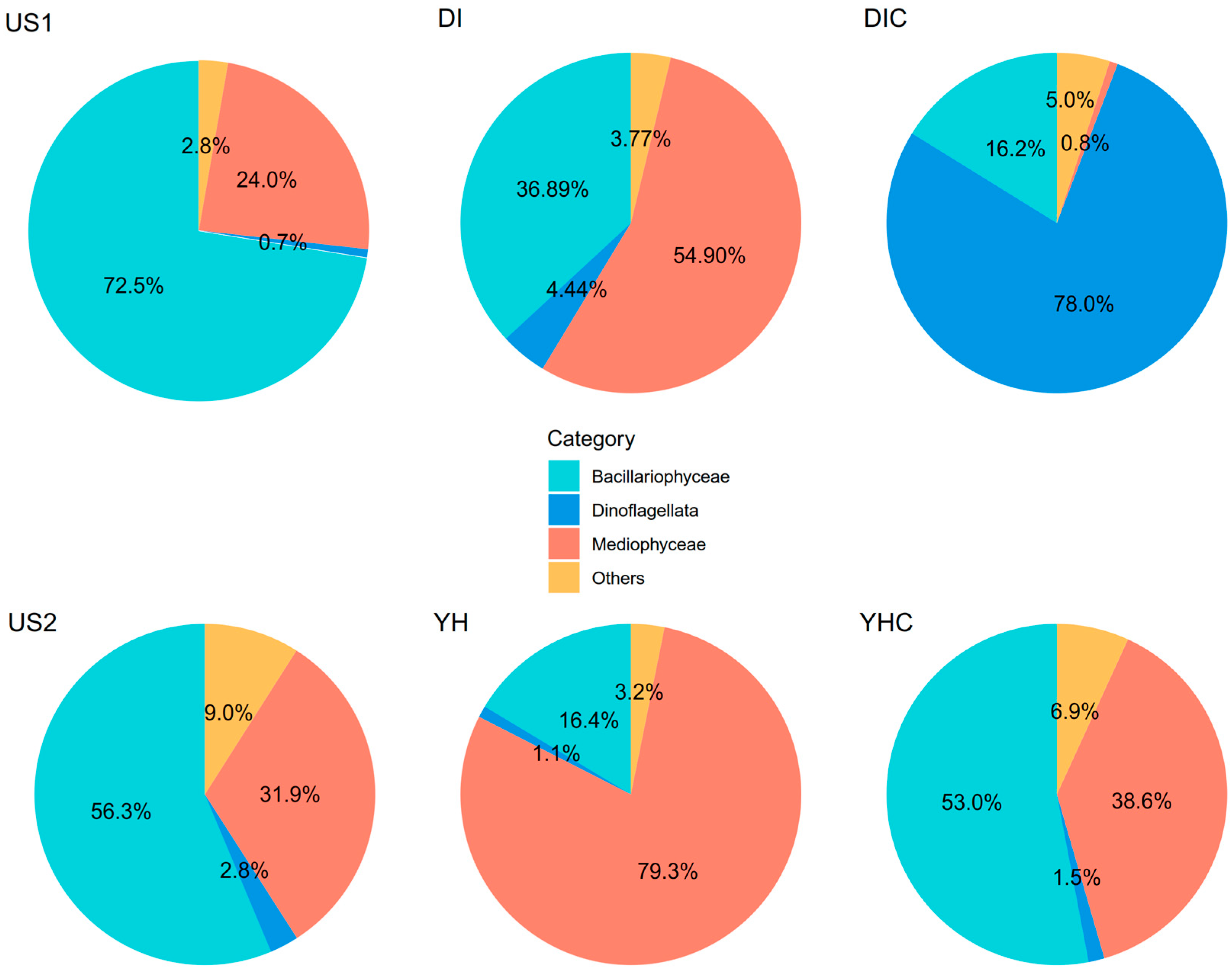
3. Results and Discussion

The eDNA analyses show a diverse range of taxa identified in different sediment samples. The samples from YH and YHC revealed a notable presence of Chloroplastida, indicating a significant presence of green algae and plants, as this group includes photosynthetic organisms such as green algae and land plants. In contrast, samples from YH, YHC, DI, and US1 exhibited a higher proportion of Stramenopiles, which encompass diatoms and brown algae—important contributors to primary production in aquatic ecosystems (Figure 2). Additionally, the US1, US2, and DIC samples demonstrated a diverse community composition, with significant representation from Alveolata, including protists such as ciliates, dinoflagellates, and apicomplexans, as well as Holozoa, a group that includes animals and their close unicellular relatives (Figure 2). This highlights the presence of various animals and related single-celled organisms, along with microalgae. In contrast, sediment samples collected from DI have a more balanced distribution of taxa at all six sites (Figure 2). Overall, the analyses illustrate the variability in community composition across different sediment samples, indicating that environmental conditions and ecological niches influence taxa diversity and abundance. These findings emphasize the importance of site-specific factors in shaping microbial communities in sediment environments and provide insights into the ecological dynamics and potential biogeochemical processes occurring in these areas [19].
The analysis of marine macrophyte DNA in sediment samples revealed significant diversity in macroalgal species detected across different sites. A minor presence of Chlorophyta taxa such as Blidingia dawsonii and Collinsiella tuberculata was observed only in samples US2 and YHC, respectively (Table 1). Ulva sp. showed higher abundance in US1 and US2, with values of 2.59% and 0.33%, respectively, but this alga was not detected in any Korean sites (Table 2). The US1 site had an Ulva bloom during the sampling period (Table 1), which could be the reason for the higher Ulva DNA in the sediment [20]. Rhodophyta taxa displayed variability, with Ceramium sp. notably abundant in YH and YHC at 2.01% and 0.22%, respectively, while C. diaphanum was present only in US1 and US2 (Table 2). Ceramium diaphanum is an epiphytic red alga that grows on other macroalgae or seagrass [21], which was not detected in sediment samples collected from Korea. Phaeophyceae showed a significant presence, with Sargassum sp. detected across all sites (Table 2). Ectocarpus siliculosus was exceptionally abundant in US2 at 4.25%. Scytosiphon lomentaria showed extreme abundance in YH and YHC, highlighting its potential dominance in these areas. Seagrass taxa like Zostera marina were present in lower proportions across multiple sites (Table 2). The seagrass beds at or near the sampling sites should influence these results [22,23]. Although no seagrass was observed at DIC and YHC, detached seagrass fragments can still be delivered to these two sites by the currents from the nearby seagrass bed. The results indicate varying ecological niches and potential environmental conditions favoring the deposition of specific taxa in different sediment samples. This diversity underscores the complex ecological dynamics within the study areas and suggests areas of high species richness and potential biodiversity hotspots [24].
Our results indicate that eDNA technology effectively discriminates between marine macrophyte taxa. However, several taxa that were abundant in situ were not detected in the eDNA pool, such as Ascophyllum nodosum and Fucus spp. at site US2 [25]. The absence of these species suggests that PCR bias and artifacts associated with the use of universal primers may be contributing factors. This issue could potentially be addressed by employing degenerated versions of the primers in the amplification process [13,26,27]. It can also be mitigated by using multiple primer sets.
The analysis focused on microalgae taxa indicates significant variation across different site types, including muddy bottoms with seagrass or Ulva blooms, or without vegetation, and seaweed and seagrass co-existing habitats. Figure 3 shows the relative abundance of DNA reads of microalgae in six sites. DI and YH (muddy bottoms with seagrass) and US1 (muddy with Ulva bloom) showed higher levels of microalgae DNA than DIC, YHC, and US2 (sandy/rocky) (Figure 3). The analysis of microalgae composition in eDNA samples from various sediment types reveals distinct patterns in the distribution of microalgal classes. Figure 4 shows the composition of microalgae DNA reads in six sites. In the muddy bottom sites in Korea, the microalgal compositions were significantly different depending on the presence of seagrass. For example, at DI and YH, Mediophyceae are dominant, followed by Bacillariophyceae and smaller contributions from Dinoflagellata and other classes (Figure 4). However, DIC is predominant with Dinoflagellata (78%), with a notable presence of Bacillariophyceae (16.2%), and YHC shows a mix of Bacillariophyceae (53%) and Mediophyceae (38.6%). Both US1 and US2 sites are mainly dominated by Bacillariophyceae (72.5% and 56.3%, respectively), followed by Mediophyceae (24% and 31.9%, respectively) (Figure 4). These findings indicate that environmental conditions, such as sediment and vegetation types, significantly influence the composition of microalgal communities [28,29]. Further investigations into the specific environmental parameters of each site are necessary to better understand the factors influencing microalgal distribution in these diverse sedimentary environments.
This study is among the growing body of research utilizing eDNA technology to identify marine macrophytes in sediments, which may contribute to blue carbon deposition [13]. However, identifying macroalgae through eDNA remains challenging and requires significant improvements in the existing barcode reference libraries [13,30,31]. Despite these limitations, our results suggest that coastal vegetative sediments can play a crucial role in carbon sequestration, and macroalgae constitute a significant portion of the eDNA pool in these habitats. This indicates that macroalgae should be considered in blue carbon assessments [32].

4. Conclusions

This study demonstrates the effectiveness of eDNA as a powerful tool for assessing the biodiversity of marine photosynthetic organisms within marine macrophyte ecosystems and associated sediments. By employing high-throughput sequencing of the 18S-V9 region from sediment samples across diverse coastal locations, we were able to uncover significant variability in taxa composition and community structures. The findings highlight the ecological diversity and distinct environmental conditions influencing the presence of green algae, diatoms, seagrasses, and other photosynthetic organisms in the sediments. The distinct patterns observed in the community composition across different sediment and vegetation types—such as muddy vs. sandy/rocky, and single species dominant (seagrass or Ulva) vs. diverse-species communities—emphasize the importance of habitat-specific factors in shaping microbial and photosynthetic organism distributions in the sediments. The high prevalence of microalgae in seagrass beds and Ulva bloom areas underscores the complex ecological interactions and biogeochemical processes within these habitats. Our results reinforce the potential of eDNA methodologies to revolutionize ecological monitoring, offering a non-invasive and comprehensive approach to understanding the health and functionality of marine macrophyte ecosystems. Future research should continue to refine eDNA techniques and expand the scope of studies to include broader geographical regions and more detailed environmental parameters, further enhancing our ability to protect and sustain these vital ecosystems. In the future, we also suggest incorporating eDNA technology alongside traditional methods, such as stable isotope analyses, to provide more accurate carbon assessments of blue carbon ecosystems in this project.

Author Contributions

Conceptualization, Q.X., S.J.K. and C.Y.; methodology, Q.X. and S.J.K.; formal analysis, Q.X. and S.J.K.; investigation, C.Y.; resources, C.Y.; data curation, Q.X.; writing—original draft preparation, Q.X. and S.J.K.; writing—review and editing, Q.X., S.J.K. and C.Y.; visualization, Q.X.; supervision, C.Y.; funding acquisition, C.Y. All authors have read and agreed to the published version of the manuscript.

Funding

This study was supported by the U.S. Department of Energy’s (DOE) ARPA-E MARINER program contract number DE-AR0000915.

Data Availability Statement

Data will be available upon request.

Acknowledgments

We acknowledge the support of the High-Performance Biological Supercomputing Center at the Ocean University of China for this research.

Conflicts of Interest

The authors declare no conflict of interest.

References

  1. Legg, S. IPCC, 2021: Climate Change 2021-the physical science basis. Interaction 2021, 49, 44–45. [Google Scholar]
  2. Grubb, M.; Okereke, C.; Arima, J.; Bosetti, V.; Chen, Y.; Edmonds, J.; Gupta, S.; Köberle, A.; Kverndokk, S.; Malik, A.; et al. Introduction and Framing. In Proceedings of the IPCC, 2022: Climate Change 2022: Mitigation of Climate Change. Contribution of Working Group III to the Sixth Assessment Report of the Intergovernmental Panel on Climate Change, IPCC, Sharm el-Sheikh, Egypt, 6–20 November 2022. [Google Scholar]
  3. Nellemann, C.; Corcoran, E.; Duarte, S.M.; Valdés, L.; DeYoung, C.; Fonseca, L.; Grimsditch, G. Blue Carbon: The Role of Healthy Oceans in Binding Carbon. A Rapid Response Assessment; UNEP/FAO/UNESCO/IUCN/CSIC; Birkeland Trykkeri AS: Aust-Agde, Norway, 2009.
  4. Herr, D.; Landis, E. Coastal Blue Carbon Ecosystems. Opportunities for Nationally Determined Contributions; Policy brief. IUCN; UN Environment Program: Nairobi, Kenya, 2016. [Google Scholar]
  5. Macreadie, P.I.; Costa, M.D.; Atwood, T.B.; Friess, D.A.; Kelleway, J.J.; Kennedy, H.; Lovelock, C.E.; Serrano, O.; Duarte, C.M. Blue carbon as a natural climate solution. Nat. Rev. Earth Environ. 2021, 2, 826–839. [Google Scholar] [CrossRef]
  6. Sánchez-Arcilla, A.; Cáceres, I.; Le Roux, X.; Hinkel, J.; Schuerch, M.; Nicholls, R.J.; Otero, D.M.; Staneva, J.; de Vries, M.; Pernice, U.; et al. Barriers and enablers for upscaling coastal restoration. Nat. Based Solut. 2022, 2, 100032. [Google Scholar] [CrossRef]
  7. Friess, D.A.; Shribman, Z.I.; Stankovic, M.; Iram, N.; Baustian, M.M.; Ewers Lewis, C.J. Restoring Blue carbon ecosystems. Camb. Prism. Coast. Futures 2024, 2, e9. [Google Scholar] [CrossRef]
  8. Taberlet, P.; Bonin, A.; Zinger, L.; Coissac, E. Environmental DNA: For Biodiversity Research and Monitoring; Oxford University Press: Oxford, UK, 2018. [Google Scholar]
  9. Valentini, A.; Taberlet, P.; Miaud, C.; Civade, R.; Herder, J.; Thomsen, P.F.; Bellemain, E.; Besnard, A.; Coissac, E.; Boyer, F.; et al. Next-generation monitoring of aquatic biodiversity using environmental DNA metabarcoding. Mol. Ecol. 2016, 25, 929–942. [Google Scholar] [CrossRef]
  10. Thomsen, P.F.; Willerslev, E. Environmental DNA—An emerging tool in conservation for monitoring past and present biodiversity. Biol. Conserv. 2015, 183, 4–18. [Google Scholar] [CrossRef]
  11. Schadewell, Y.; Adams, C.I.M. Forensics meets ecology—Environmental DNA offers new capabilities for marine ecosystem and fisheries research. Front. Mar. Sci. 2021, 8, 668822. [Google Scholar] [CrossRef]
  12. Ficetola, G.F.; Bonin, A.; Miaud, C. Population genetics reveals origin and number of founders in a biological invasion. Mol. Ecol. 2008, 17, 773–782. [Google Scholar] [CrossRef]
  13. Ortega, A.; Geraldi, N.R.; Duarte, C.M. Environmental DNA identifies marine macrophyte contributions to blue carbon sediments. Limnol. Oceanogr. 2020, 65, 3139–3149. [Google Scholar] [CrossRef]
  14. Hamaguchi, M.; Miyajima, T.; Shimabukuro, H.; Hori, M. Development of quantitative real-time PCR for detecting environmental DNA derived from marine macrophytes and its application to a field survey in Hiroshima Bay, Japan. Water 2022, 14, 827. [Google Scholar] [CrossRef]
  15. Zeng, Y.; Wang, X.; Liu, J.; Cao, J.; Sun, Y.; Zhao, S.; Chen, Z.; Kim, J.K.; Zhang, J.; He, P. Harnessing the power of eDNA technology for macroalgal ecological studies: Recent advances, challenges, and future perspectives. Algal Res. 2023, 77, 103340. [Google Scholar] [CrossRef]
  16. Amaral-Zettler, L.A.; McCliment, E.A.; Ducklow, H.W.; Huse, S.M. A method for studying protistan diversity using massively parallel sequencing of V9 hypervariable regions of small-subunit ribosomal RNA genes. PLoS ONE 2009, 4, e6372. [Google Scholar] [CrossRef]
  17. Martin, M. Cutadapt removes adapter sequences from high-throughput sequencing reads. EMBnet. J. 2022, 17, 10–12. [Google Scholar] [CrossRef]
  18. Callahan, B.J.; McMurdie, P.J.; Rosen, M.J.; Han, A.W.; Johnson, A.J.A.; Holmes, S.P. DADA2: High-resolution sample inference from Illumina amplicon data. Nat. Methods 2016, 13, 581–583. [Google Scholar] [CrossRef]
  19. Zhang, Z.; Zhang, Q.; Chen, B.; Yu, Y.; Wang, T.; Xu, N.; Fan, X.; Penuelas, J.; Fu, Z.; Deng, Y.; et al. Global biogeography of microbes driving ocean ecological status under climate change. Nat. Commun. 2024, 15, 4657. [Google Scholar] [CrossRef]
  20. Chávez-Sánchez, T.; Piñón-Gimate, A.; Serviere-Zaragoza, E.; Sánchez-González, A.; Hernández-Carmona, G.; Casas-Valdez, M. Recruitment in Ulva blooms in relation to temperature, salinity and nutrients in a subtropical bay of the Gulf of California. Bot. Mar. 2017, 60, 257–270. [Google Scholar] [CrossRef]
  21. Maggs, C.; Hommersand, M.H. Seaweeds of the British Isles; Volume 1 Rhodophyta, Part 3A Ceramiales; Pelagic Publishing: London, UK, 1993. [Google Scholar]
  22. Vaudrey, J.M.P.; Kremer, J.N.; Branco, B.F.; Short, F.T. Eelgrass recovery after nutrient enrichment reversal. Aquat. Bot. 2010, 93, 237–243. [Google Scholar] [CrossRef]
  23. Bae, J.I.; Shin, H.C.; Hwang, S.I.; Lee, J.H. Distribution of sedimentation environments and benthic macro-fauna communities in habitats and non-habitats of Zostera marina on the Yeongheung-do tidal flats, West Coast of Korea. Korean J. Environ. Biol. 2018, 36, 107–116. [Google Scholar] [CrossRef]
  24. Rozaimi, M.; Zainee, N.F.A.; Raynusha, C.; Arina, N.; Hidayah, N.; Hengjie, T.; Tangang, F. Carbon and Nitrogen Deposits of Macroalgal Origin on a Tropical Seagrass Meadow. Ecosyst. Health Sustain. 2024, 10, 0157. [Google Scholar] [CrossRef]
  25. van Patten, M.S.; Yarish, C. Seaweeds of Long Island Sound (2nd edition). In Connecticut Sea Grant College Program; NOAA: Silver Spring, MD, USA, 2009. Available online: https://repository.library.noaa.gov/view/noaa/45755 (accessed on 18 January 2024).
  26. Kanagawa, T. Bias and artifacts in multitemplate poly-merase chain reactions (PCR). J. Biosci. Bioeng. 2003, 96, 317–323. [Google Scholar] [CrossRef]
  27. Fuller, N.J.; Campbell, C.; Allen, D.J.; Pitt, F.D.; Le Gail, F.; Vaulot, D.; Scanlan, D.J. Analysis of photosynthetic picoeukaryote diversity at open ocean sites in the Arabian Sea using a PCR biased towards marine algal plastids. Aquat. Microb. Ecol. 2006, 43, 79–93. [Google Scholar] [CrossRef]
  28. Capriulo, G.M.; Smith, G.; Troy, R.; Wikfors, G.; Pellet, J.; Yarish, C. The Planktonic Food Web Structure of a Temperate Zone Estuary, and its Alteration Due to Eutrophication. Hydrobiologia 2002, 475/476, 263–333. [Google Scholar] [CrossRef]
  29. Lopez, G.; Carey, D.; Carlton, J.; Cerato, R.; Dam Guerrero, H.; Digiovanni, C.; Elphick, C.; Frisk, M.; Gobler, C.; Hice, L.; et al. Biology and Ecology of Long Island Sound, In Long Island Sound: Prospects for the Urban Sea; Latimer, J.S., Tedesco, M., Swanson, R.L., Yarish, C., Stacey, P., Garza, C., Eds.; Springer: New York, NY, USA, 2014; pp. 285–479. [Google Scholar]
  30. Saunders, G.W. Applying DNA barcoding to red macro-algae: A preliminary appraisal holds promise for future applications. Philos. Trans. R. Soc. 2005, 360, 1879–1888. [Google Scholar] [CrossRef] [PubMed]
  31. Saunders, G.W.; Kucera, H. An evaluation of rbcL, ufA, UPA, LSU and ITS as DNA barcode markers for the marine green macroalgae. Cryptogam. Algol. 2010, 31, 487. [Google Scholar]
  32. Krause-Jensen, D.; Lavery, P.; Serrano, O.; Marbà, N.; Masque, P.; Duarte, C.M. Sequestration of macroalgal carbon: The elephant in the Blue Carbon room. Biol. Lett. 2018, 14, 20180236. [Google Scholar] [CrossRef]
Figure 1. Map of sampling sites in the United States and South Korea.
Coasts 04 00036 g001
Figure 2. Composition of eukaryotic organisms’ DNA reads in six sites.
Coasts 04 00036 g002
Figure 3. Relative abundance of DNA reads of microalgae in six sites.
Coasts 04 00036 g003
Figure 4. Composition of microalgae’s DNA reads in six sites.
Coasts 04 00036 g004
Table 1. Substrate and vegetation types of sampling sites.
LocationSubstrateVegetation Type
Dae Isac Do, Incheon, Republic of Korea (DI)MuddySeagrass bed
Dae Isac Do, Incheon, Republic of Korea (DIC)MuddyNo vegetation
New Haven, CT, USA (US1)MuddyUlva blooms
Avery Point, CT, USA (US2)Sandy/RockyHighly diverse seaweed community (fucoid dominant)/Seagrass bed
Yeong Heung Do, Incheon, Republic of Korea (YH)MuddySeagrass bed
Yeong Heung Do, Incheon, Republic of Korea (YHC)MuddyNo vegetation
Table 2. List of marine macrophytes found in sediment of different locations. The number indicates percentage of the taxon among the total eDNA sequences of marine macrophytes.
LineageTaxaDIDICUS1US2YHYHC
ChlorophytaBlidingia dawsonii---0.32--
Collinsiella tuberculata-----0.31
Ruthnielsenia tenuis-----0.6
Ulva sp.--2.590.33--
RhodophytaCeramium diaphanum--0.910.23--
Ceramium sp.----2.010.22
Gracilaria sp.--0.49---
PhaeophytaCantina marsupialis----0.210.57
Sargassum sp.0.31.240.190.460.140.18
Ectocarpus siliculosus--0.364.25--
Scytosiphon lomentaria---0.8458.1419.55
Antarctosaccion applanatum0.11----0.41
SeagrassZostera marina-0.44-0.780.340.1
Disclaimer/Publisher’s Note: The statements, opinions and data contained in all publications are solely those of the individual author(s) and contributor(s) and not of MDPI and/or the editor(s). MDPI and/or the editor(s) disclaim responsibility for any injury to people or property resulting from any ideas, methods, instructions or products referred to in the content.

