Article

Recovery of Haemal Lordosis in European Seabass Dicentrarchus labrax (Linnaeus 1758) †

by
Stephanos Fragkoulis
1,‡,
Chara Kourkouta
1,‡,
George Geladakis
1,‡,
Alice Printzi
1,‡,§,
Alexis Glaropoulos
2 and
George Koumoundouros
1,*
1
Biology Department, University of Crete, 70013 Heraklion, Greece
2
AVRAMAR S.A., PEO Patron—Athinon 55, Agios Vasilios, 26500 Rio, Greece
*
Author to whom correspondence should be addressed.
This paper is dedicated to the memory of Dr. Pascal Divanach, an outstanding researcher and mentor who has made pioneering contributions to the science of Aquaculture. He was the first who conceived and tested the hypothesis of exercised-induced (haemal) lordosis in fish.
These authors contributed equally to this work.
§
Current address: IFREMER (Institut Français de Recherche pour l’Exploitation de la Mer), Marine & Coastal Sciences Doctoral School, University of Brest, CNRS, IRD, LEMAR, 29280 Brest, Plouzané, France.
Aquac. J. 2022, 2(1), 1-12; https://doi.org/10.3390/aquacj2010001
Submission received: 17 November 2021 / Revised: 11 December 2021 / Accepted: 24 December 2021 / Published: 4 January 2022

Abstract

:
Lordosis of the haemal part of the vertebral column is a frequent abnormality in reared fish. Haemal lordosis develops during the late larval and early juvenile life stages of fish, mainly due to the high swimming activity of the fish in the rearing tanks. In the present study, we have examined whether haemal lordosis recovers during the growth of European seabass. Furthermore, we aimed to develop simple morphometric indices (PrAn1 and PrAn2) that might link the severity of lordosis at the juvenile stage with fish morphological quality at harvesting. At 111 days post-hatching (dph, 53 ± 4 mm standard length, SL), 600 seabass juveniles with lordotic (L, 200 fish) or normal (N, 400 fish) external morphology were selected and introduced in a common tank. At 150 dph (75 ± 7 mm SL), 350 fish were randomly selected, pit-tagged and transferred to a sea cage for on-growing up to 502 dph (234 ± 16 mm SL). The morphological examination of the fish at 150 and 502 dph revealed that 60% (46 out of 77) of L juveniles turned into normal phenotype by the end of on-growing period. Interestingly, 56% of the fish with recovered external morphology (N-Rec) presented either a completely normal vertebral column (31%) or minor abnormalities of individual vertebrae (25%). Following the results of geometric morphometric analysis, the differences in the body shape between N-Rec and N fish were not statistically significant (p > 0.05, canonical variate analysis). The examined morphometric indices were effective in discriminating the normal fish from 58% (PrAn1) to 65% (PrAn2) of lordotic juveniles. Results are discussed with respect to the mechanism of lordosis recovery, and spotlights on their application for quality control and cull out of the abnormal fish in commercial hatcheries.

Graphical Abstract

1. Introduction

Skeletal abnormalities are an important issue of quality, health and welfare of reared finfish. They may develop on the skull, fins and vertebral column of the reared fish with a wide range of severity degrees. In marine teleosts, most skeletal abnormalities develop mainly during the embryonic and larval stages (i.e., the period where skeletal ontogeny takes place) via alterations of skeleton ontogenetic patterns. Typical examples of such ontogenetic alterations are the gill cover [1,2] and jaw abnormalities [3,4,5], the extra formation or partial lack of the caudal-fin rays [6,7], saddleback syndrome [8] or the abnormal patterning of vertebrae [9,10]. Known causative factors of skeletal abnormalities include abiotic parameters [7,10] and larval nutrition [3,11,12], as well as the genetic background of the fish [4,13]. In marine commercial hatcheries, coping with abnormalities involves not only the application of the appropriate rearing conditions, but also the standardization of quality control criteria on the basis of abnormalities’ effects on fish morphology at harvest [14].
The lordosis of haemal vertebrae (haemal lordosis) is a frequent axial abnormality that develops during the late metamorphosis and juvenile period. Unlike lordosis that develops in the pre-haemal part of the vertebral column, haemal lordosis is not associated with the non-inflation of swimbladder [15]. The high swimming activity of fish is suggested as the most important causative factor of haemal lordosis [15,16,17,18], followed by unfavorable water temperature [16,19] and larval nutrition [20]. In all species studied so far, the abnormality presents a remarkable variability with respect to the lordosis angle, the number of abnormal vertebrae, positioning across the vertebral column [16,18,21] and finally to the resulting effects on the external morphology of fish (insignificant to severe body-shape alterations). As it was shown in gilthead seabream, the lordotic phenotype may recover during fish growth in sea cages, through a process involving repairing of abnormal vertebrae [22]. In the same species, simple morphometrics were efficient in quantifying the severity of haemal lordosis at the juvenile stage, on the basis of its potential effects on fish phenotype at market size [23].
European seabass, Dicentrarchus labrax, is an important aquaculture species and the first in which haemal lordosis was thoroughly studied [15,16,24,25,26]. The recovery potential of haemal lordosis in this species is still unknown. Similarly, no scale exists for the assessment of lordosis severity on the body shape of seabass at harvest. Continuing our work on the recovery of haemal lordosis in gilthead seabream [22], in the present study we examined whether this kind of lordosis may also recover in European seabass; i.e., a fish species with comparatively more elongated body shape and thus different musculoskeletal anatomy. Moreover, we examined whether juvenile morphometric indices may predict the effects of lordosis on the body shape of seabass at harvest.

2. Materials and Methods

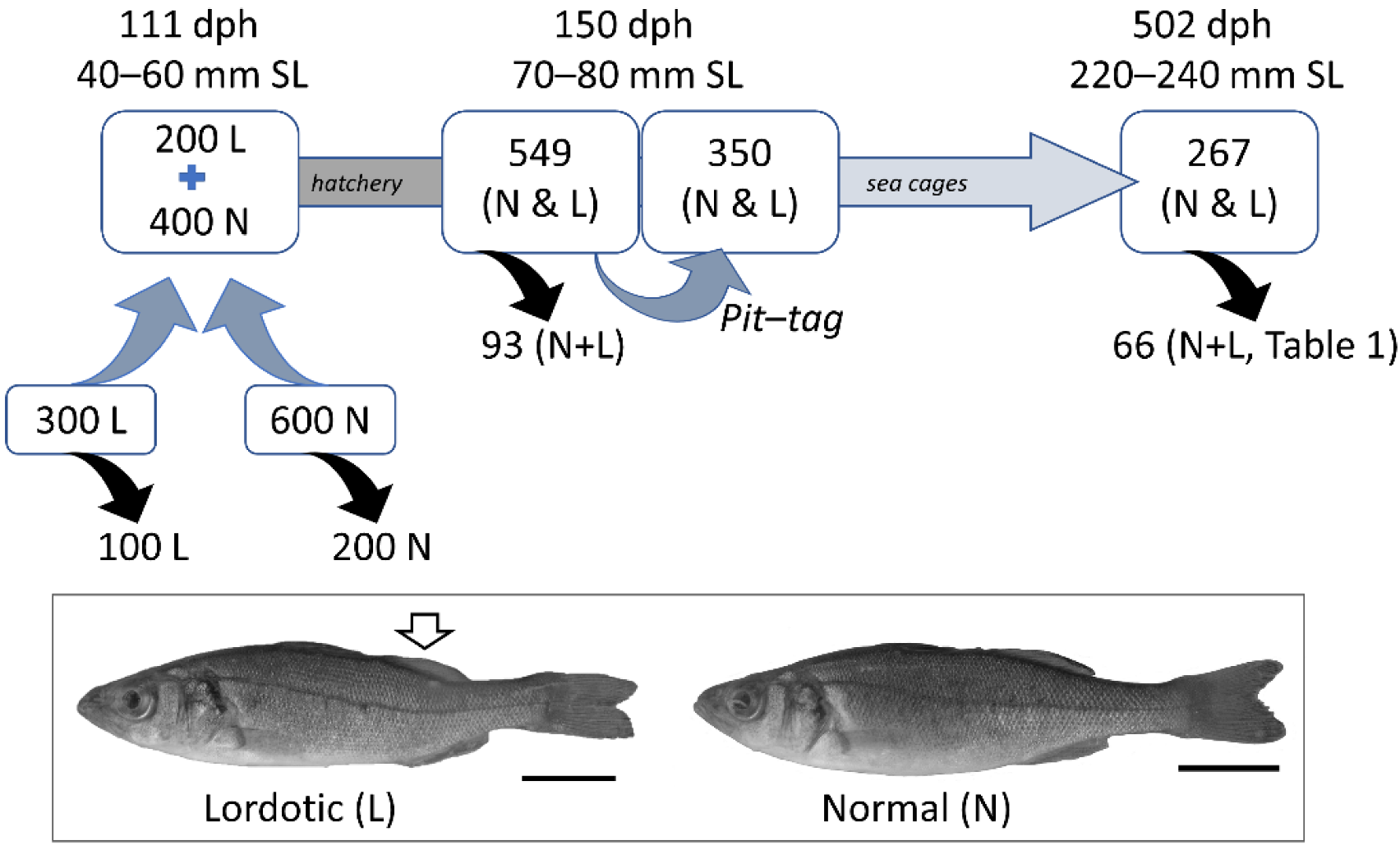
2.1. Experimental Design and Fish Origin

The group of fish examined originated from a common egg batch. When the population reached the age of 111 dph, fish were anaesthetized (ethyleneglycol–monophenyl ether, Merck, Darmstadt, Germany, 0.2–0.5 mL L−1) and examined for the dorsal shift of the caudal peduncle, typical characteristic of haemal lordosis [25] (Figure 1). Six hundred juveniles with normal (N) and 300 juveniles with lordotic (L) external morphology were finally selected and kept separately. For the radiographic validation of the morphological classification, a random sample of 100 and 200 fish was taken from the L and N group, respectively, anaesthetized and stored at −20 °C. The rest of the fish were placed together in a rearing tank until tagging (Figure 1). Fish radiography was performed by using General Electric Senograph 700T (26 KV, 25 mA s).
At 150 dph, 350 fish were randomly taken, anaesthetized, photographed on their left side, tagged electronically (FDX-B, Trovan Ltd., USA) and transferred into one sea cage. The on-growing period was terminated at 502 dph, when all fish were anaesthetized, photographed and scanned for ID recognition (Figure 1). All photographs were taken by a digital camera (Canon PowerShot G9), mounted on a tripod and positioned perpendicularly to the specimens. The significance of the difference in the rate of missing fish specimens (i.e., dead fish or fish with a lost tag) between L and N fish was tested by G-test [27].
The rearing of European seabass embryos, larvae and juveniles was performed by AVRAMAR S.A., according to the standard methodology followed by commercial marine hatcheries and cage farms for the species. In brief, larval rearing was performed with an initial stocking density of 50 individuals per L, in the presence of background phytoplankton (Chlorella sp., Pacific Trading Cο, Ltd., Fukuoka, Japan). Larvae were fed on rotifers Brachionus plicatilis (7–18 dph), Artemia sp. nauplii (14–45 dph, INVE Technologies, Dendermonde, Belgium), and finally on inert commercial diets (>35 dph, O.range, INVE Technologies, Dendermonde, Belgium). Water temperature was maintained in the range 16–20 °C during the hatchery period, whereas during on-growing period it followed the natural seasonal fluctuations (14–27 °C).

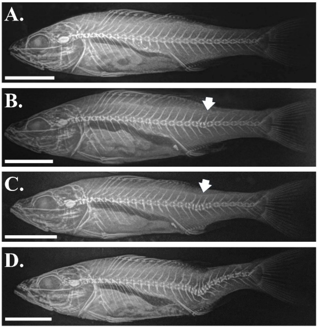
2.2. Categorization of the External Morphology during the On-Growing Period

To discriminate L fish from N fish at 150 and 502 dph, the photographs of pit-tagged individuals were examined by three independent observers. Discrimination was based on dorsally shifted caudal peduncle of the lordotic fish [25]. To validate the methodology of fish phenotypic categorization, at 150 dph a random sample of 93 juveniles was taken from the source population (Figure 1), anaesthetized, photographed and stored at −20 °C. Based on their external morphology, fish were scored as N or L. The results of phenotypic scoring were validated by the radiographic analysis of the fish (General Electric Proteus XR/e, 55 KV, 1.2 mA s) [22].
To validate any shift of lordotic to normal external morphology (lordosis recovery) during the on-growing period, 66 fish were selected at 502 dph on the basis of their initial (150 dph) and final (502 dph) phenotype (Figure 1, Table 1), and radiographically examined. All samples collected for X-ray analysis were euthanatized with an overdose of anesthetic (ethyleneglycol–monophenyl ether, Merck, Darmstadt, Germany). Fragkoulis et al. [22] was followed for fish radiography (General Electric Proteus XR/e, 55 KV, 1.2 mA s).

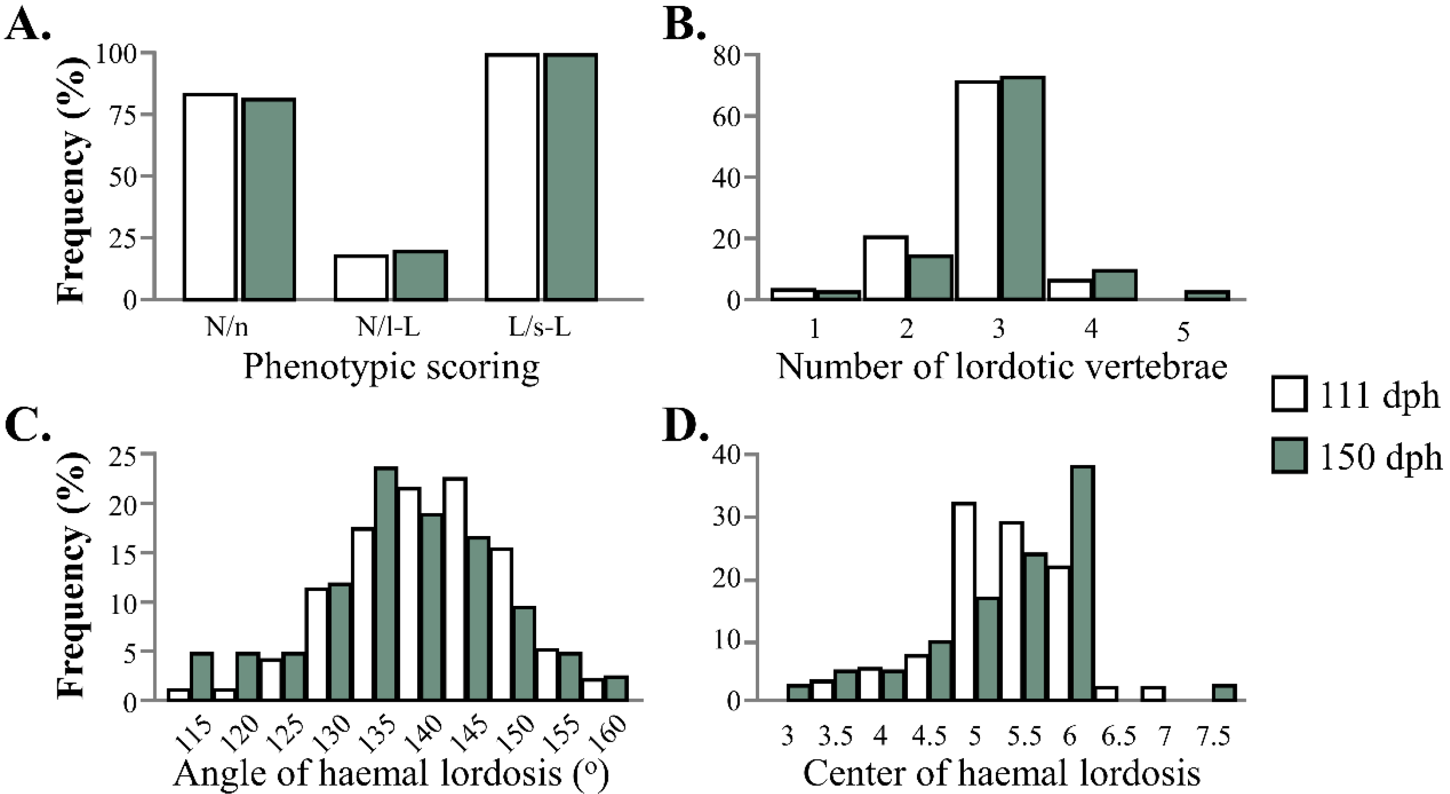
2.3. Geometric Morphometric and Meristic Analyses

Geometric morphometry was used to examine body-shape differences between juveniles with L and N external morphology at tagging (150 dph) and at the end of on-growing period (502 dph). At 150 dph, the analysis included a random sample of 60 N and 58 L juveniles. At 502 dph, the analysis included a random sample of 30 fish with normal and 20 with lordotic external phenotype since the beginning of on-growing (150 dph), and 32 initially lordotic fish with a recovered normal external morphology at the end of the trials (N-Rec, 502 dph).
In both analyses, thirteen landmark measurements were taken on the digital photographs of the examined fish (Figure 2A) by means of tpsDig2 software [28]. A generalized least square method was applied to adjust landmark configurations for centroid size and remove any effect irrelevant to shape (coordGen8 software, [29]). TpsRelw software [30] was used to calculate the weight matrix (partial warps, with uniform and nonuniform components of shape variation). Discriminant (150 dph) or canonical variate analysis (502 dph) was applied on the weight matrix to test the effect of lordosis on body shape (150, 502 dph), as well as the differences in body shape among N, L and N-Rec groups (502 dph). Spline diagrams were obtained after the regression of shape components on the canonical scores (tpsRegr) [31].
To examine whether haemal lordosis is further developing during the hatchery phase, the angle (tpsDig2) [28] and center of haemal lordosis, as well as the number of the abnormal vertebrae were scored on all the radiographed samples of 111 and 150 dph. The significance of the differences in these characters and in lordosis frequency between the two sampling ages was tested by means of Mann–Whitney U-test (SPSS v26) and G-test [27], respectively.

2.4. Simple Morphometric Descriptors of Lordosis-Induced Body-Shape Deviations

We examined whether simple morphometric indices may link juvenile lordotic phenotype (150 dph) with fish phenotype at the end of on-growing (502 dph), thus providing a predictor scale for lordosis severity. The first index tested, PrAn1 angle (Figure 2B), was suggested by Fragkoulis and Koumoundouros [23] as effective in quantifying the lordosis effects on the body shape in gilthead seabream. The second index tested, PrAn2 angle, was examined in an attempt to increase its discrimination efficiency by the evaluation of more morphometric data. PrAn2 was calculated as the summation of three angles (PrAn2 = An1 + An2 + (180° − PrAn1), Figure 2B), which may be affected by the lordosis-induced shifts of the caudal peduncle. Each angle was defined by a set of three landmark measurements previously used in the geometric morphometry (Figure 2B). Angles were calculated by the input of xy coordinates of these landmarks into the formula
tan ( p h i ) = | m 2 m 1 1 + ( m 2 m 1 ) |
where “phi” is the calculated angle (An1, An2 or PrAn1), and “m2” and “m1” are the slopes of the two segments of the “phi” angle (e.g., L10-L7 ( y 10 y 7 x 10 x 7 ) and L7-L4 ( y 7 y 4 x 7 x 4 ) segments for PrAn1 angle, [23]).
The seabass specimens examined were the 267 pit-tagged juveniles with scored external morphology at both the beginning (150 dph) and end (502 dph) of the on-growing period (Figure 1, Table 1).

3. Results

3.1. Validation of the Phenotypic Categorization by Means of External Phenotype

The radiographic examination of the fish samples at 111 dph verified the anatomical criteria used for the selection of the lordotic specimens. All fish with lordotic external morphology (L) presented a severe lordosis on the haemal part of the vertebral column (Figure 3D), whereas the majority (83%) of the fish with normal external morphology (N) presented a normal vertebral column (Figure 3A). The remainder 17% of N fish presented haemal lordosis of very light severity (Figure 3B,C). Similar results were found in samples taken at tagging (150 dph), with 19% (12 out of 62) of the N fish presenting light lordosis and all (31/31) the L fish presenting severe lordosis on the X-rays (Figure 4A). Overall, external phenotypic categorization was radiographically verified in 100% of the fish with lordotic (L) and 81–83% of the fish with normal (N) external morphology. The applied criteria failed to distinguish the externally normal fish with light vertebral abnormalities (N/l-L, Figure 4A).
No significant differences were present in the severity (number of affected vertebrae, angle, Figure 4B,C) or the position (Figure 4D) of lordosis between the first (111 dph) and the last (150 dph) hatchery sample (p > 0.05, Mann–Whitney U-test). On the basis of lordosis rates in the X-rayed samples of 111 and 150 dph, it is estimated that 44 % and 46% (Table S1), respectively, of the living fish were radiographically lordotic.

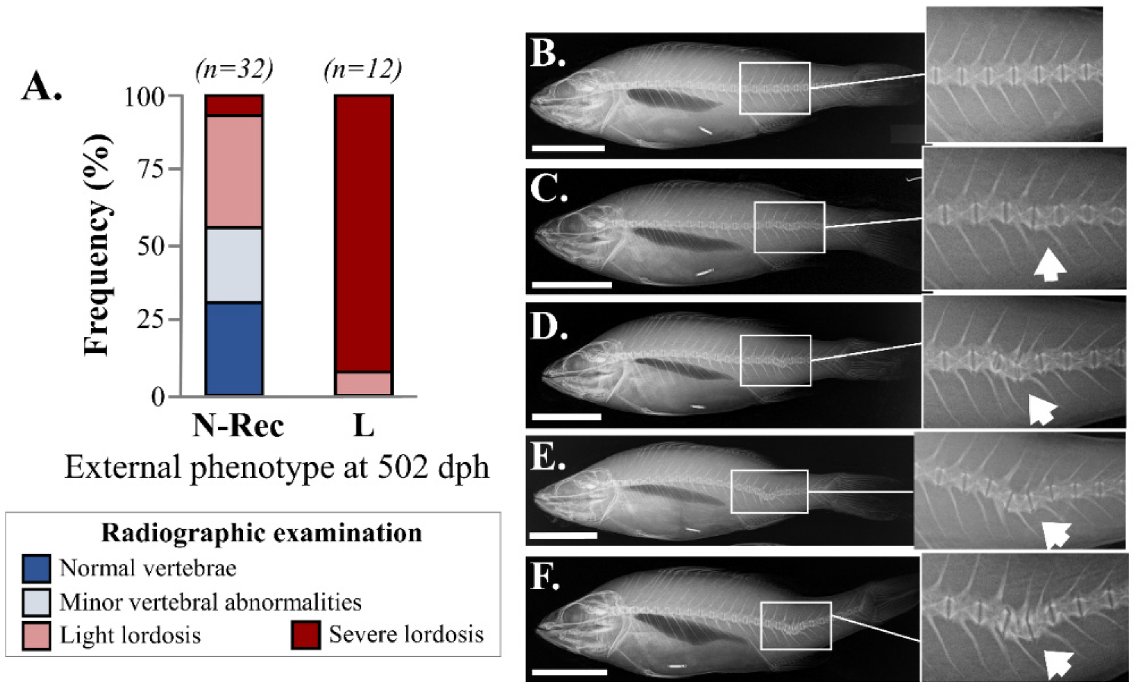
3.2. Recovery of Haemal Lordosis during the On-Growing Period (150–502 dph)

Comparison of the external morphology of each specimen between tagging (150 dph) and the end of on-growing period (502 dph) revealed that 60% (46 out of 77) of juveniles with an initially lordotic external morphology turned into normal phenotype. Lordosis recovery was verified by the radiographic examination of the fish at 502 dph. The 56% of the fish with recovered external morphology (N-Rec, 18 out of 32) presented either a completely normal vertebral column (31%, Figure 5A,B) or minor abnormalities of individual vertebrae (25%, Figure 5A,C). The rest of the N-Rec fish presented mainly haemal lordosis of light severity (38%, 12 out of 32 fish, Figure 5A,D) and to a less extent (6%, 2 out of 32), severe vertebral lordosis (Figure 5A,E). At 502 dph, the majority of specimens with lordotic external morphology presented a severe vertebral lordosis on the radiographies (92%, 11 out of 12 fish, Figure 5A,F). Finally, no vertebral abnormality was present on the radiographies of the fish with a normal external morphology at both 150 and 502 dph.
The 24% (83 out of 350) of the initially pit-tagged fish were not found at the end of the trials due to mortality or tag loss. The difference in tag recovery rate was not significantly different between the fish with N (190 out of 257, 74%) or L (77 out of 93, 83%) phenotype (G-test, p > 0.05).

3.3. Morphometric Analysis of Haemal Lordosis and Its Recovery

Discriminant analysis revealed that haemal lordosis significantly affected the body shape of seabass at 150 dph (Wilk’s λ = 0.333, p < 0.001) (Figure 6). Spline diagrams showed that lordotic fish were characterized by a ventral shift of the posterior dorsal-fin base (landmark 6) and the anterior anal-fin base (landmark 7), as well as by a dorsal-anterior shift of the caudal peduncle (landmarks 3–6). Similar effects of haemal lordosis on fish body shape were also found at the end of on-growing period (502 dph), with the first axis of canonical variate analysis (CV1) discriminating fish with a lordotic from those with a normal external phenotype (Wilk’s λ = 0.1142746, p < 0.001, Figure 7). Significant squared Mahalanobis distances were found between L and normal (N or N-Rec) fish, but not between N and N-Rec fish (Figure 7).
At 150 dph, the predictor angle PrAn1 was effective in discriminating the N (PrAn1 ≥ 145°) from 26% and 58% of the L juveniles (PrAn1 < 145°) with N-Rec or L final (502 dph) morphology, respectively, (Figure 8A, Table S2). At 502 dph, PrAn1 effectively discriminated 29% of the L fish (PrAn1 < 141°) from those of a normal (N or N-Rec) external phenotype (PrAn1 ≥ 141°, Figure 8A’). The inclusion of more landmarks in the calculation of the predictor angle (PrAn2) improved its discrimination efficiency at both 150 and 502 dph, with 7% more L fish outside the normal PrAn2 range (Figure 8B,B’). At 502 dph, all the fish with a recovered normal phenotype presented the same frequency distribution with the normal fish since the beginning of on-growing.

4. Discussion

The abnormalities of fish skeleton are generally thought as irreversible deviations from the normal phenotypic range of a species. In the last 15 years, however, a growing number of studies show that certain abnormality types may recover during fish growth. Specifically, in Atlantic salmon, fused vertebral centra may be remodeled into one normal centrum, but with multiple vertebral processes [32]. Gill cover abnormalities of light severity were shown to recover in gilthead seabream [33] and Atlantic salmon [34]. To our knowledge, haemal lordosis was the first severe axis abnormality in fish that was shown to recover, initially in gilthead seabream [22] and now in European seabass (present study). In 44–74% (seabream) [22] to 60% (seabass, present study) of the abnormal fish, the recovery of haemal lordosis was accompanied by a full recovery of the lordotic body shape.
Bone is subjected to lifelong reshaping and remodeling, triggered by organism developmental plan and allometric growth, mechanical loads or trauma [35]. Haemal lordosis has been suggested to develop as an adaptive response of the fish vertebral column to the increased mechanical loads of the muscles during intense swimming in rearing tanks [21,24]. In the present study, the radiographic analysis revealed that the recovery of lordotic external morphology was the result of a partial to complete repair of the abnormal vertebrae. In addition to this process, lordosis recovery in gilthead seabream was also linked (in 24% of the cases) with the development of a counteracting kyphosis-like bending of the vertebral column [22]. This species-specific difference in the presence of counteracting axial bending might be attributed to species-specific differences in the body shape (e.g., deeper body profile in seabream vs. more elongated shape in seabass) and/or muscle–bone interactions during swimming.
Fragkoulis et al. [22] suggested that the recovery of haemal lordosis in seabream might be triggered by the transport of the fish from the hatchery tanks to sea cages, and the subsequent adaptation of the growing vertebrae to a new environment of less intense water-current velocities. In the present study, to examine whether haemal lordosis may also recover during fish growth in the hatchery tanks, fish were radiographically examined at two different sampling ages (111 and 150 dph), before their transfer in the sea cage. Results demonstrated that lordosis rate and anatomy were not altered during this period (Figure 4), thus rejecting the hypothesis of lordosis recovery during the hatchery phase. Moreover, our results on lordosis recovery after fish transfer in sea cages supports the hypothesis of Fragkoulis et al. [22] on the link between recovery and fish growth under relatively smaller swimming intensity. In gilthead seabream, this process took place in the first 2–3 months of on-growing in cages [22], whereas in zebrafish within just one week of fish swimming in “static” water [36].
Lordotic vertebrae present trapezoidal centra, neural spines with inverted orientation of their distal part [15,18,24], associated with substantial histopathological alterations of bone, cartilage and notochord tissues [18,37]. The histological and molecular processes that take place during the remodeling of such phenotypes and the recovery of lordosis are unknown. In this research area, laboratory fish could be used not only to explore these mechanisms, but also to test the effects of nutrition and abiotic environment on the development and recovery of lordosis. In zebrafish, haemal lordosis can be quickly (within 4–7 days) and reliably induced in a swimming channel and at swimming speeds of 5–8 fish total length per second [18].
In marine pisciculture, the morpho-anatomical quality of the juveniles at the end of the hatchery phase has been considered as a valuable predictor of fish morphological quality at harvest. In the case of abnormalities which have a clear effect on fish phenotype (e.g., swimbladder non-inflation) [38], the presence or absence of the abnormal feature is adequate in predicting the final quality of fish. In the other cases, however, abnormalities may have a wide and continuous range of effects on fish external morphology (insignificant to severe) [22,25,39], as well a high recovery potential during fish growth [22,33]. In these cases, the use of juvenile predictor indices in predicting morphological quality at harvest needs the development of a quality scale to link the abnormalities severity with changes in fish external morphology [25,40]. Additionally, it needs the establishment of correlations between the phenotype of fish at the end of the hatchery period and at harvest [23]. In the present study, two simple morphometric indices (PrAn1, PrAn2) were effective in quantifying the lordosis effects on the body shape of juveniles and fish at harvest. Depending on the number of landmarks used, these indices discriminated the normal juveniles from 58% (three-landmarks index, PrAn1) to 65% (four-landmarks index, PrAn2) of the lordotic juveniles without recovery potential (Figure 8). Depending on the abnormality rate of each batch and the quality standards of the hatchery, these indices can be used for the cull out of a large proportion of lordotic fish, without affecting the normal fish or the lordotic fish that will recover during the on-growing period (Tables S2 and S3). Given that PrAn1 and PrAn2 indices use external landmark measurements, they can be easily collected on anaesthetized fish. In future, these indices can be used by computerized systems for the automatic quality grading of fish.

Supplementary Materials

The following are available online at https://www.mdpi.com/article/10.3390/aquacj2010001/s1, Table S1: Lordosis rates, sample and expected for the population, at 111 and 150 dph, Table S2: Cumulative frequency of fish with lordotic (L), recovered (N-Rec) and normal (N) external morphology based on the degrees of the PrAn1 index, Table S3: Cumulative frequency of fish with lordotic (L), recovered (N-Rec) and normal (N) external morphology based on the degrees of the PrAn2 index.

Author Contributions

Investigation, S.F., C.K., G.G., A.P. and A.G.; formal analysis and visualization, S.F. and G.K.; resources, A.G.; writing—original draft, G.K., S.F., C.K., G.G. and A.P.; conceptualization and supervision, G.K. All authors have read and agreed to the published version of the manuscript.

Funding

This study was financially supported by EU (European Maritime and Fisheries Fund, EMFF) and national (Greek) funds (NSRF 2014–2020, Novelty in Aquaculture, Project No. 5010952) of the Ministry of Rural Development and Food, Greece.

Institutional Review Board Statement

The rearing of European seabass embryos, larvae and juveniles was performed by AVRAMAR S.A., according to the standard methodology followed by commercial hatcheries and cage farms for the species. AVRAMAR S.A. is a company registered (registration numbers GGN 5200700699992) for aquaculture production in Greece and Spain. The company is certified by a GLOBAL G.A.P. quality certification, which requires a certified Veterinary Doctor to examine and verify the fish welfare and health quality. All animal samplings were collected by a qualified member of the staff, under routine procedures. All measures and legislations that were implemented by the commercial producer were in accordance with existing Greek (PD 56/2013) and EU (Directive 63/2010) legislation (protection of animals kept for farming). For the best optimization and avoidance of unnecessary suffering or injury, all the production and sampling were taken by experienced workers.

Informed Consent Statement

Not applicable.

Data Availability Statement

The data that support the findings of this study are included in the text and in the tables. Data of morphometric analyses are available from the corresponding author upon reasonable request.

Acknowledgments

We would like to thank K. Tzokas, P. Koutsopodiotis, D. Kerasonitis and G. Moukas for their contribution in fish husbandry and sampling, along with E. Karagiannakidis (MD radiologist) and M. Bastouna for their contribution in fish radiography.

Conflicts of Interest

The authors declare no conflict of interest.

References

  1. Beraldo, P.; Pinosa, M.; Tibaldi, E.; Canavese, B. Abnormalities of the operculum in Gilthead sea bream (Sparus aurata): Morphological description. Aquaculture 2003, 220, 89–99. [Google Scholar] [CrossRef]
  2. Koumoundouros, G.; Oran, G.; Divanach, P.; Stefanakis, S.; Kentouri, M. The Opercular complex deformity in intensive Gilthead sea bream (Sparus aurata L.) larviculture. Moment of apparition and description. Aquaculture 1997, 156, 165–177. [Google Scholar] [CrossRef]
  3. Fernández, I.; Hontoria, F.; Ortiz-Delgado, J.B.; Kotzamanis, Y.; Estévez, A.; Zambonino-Infante, J.L.; Gisbert, E. Larval performance and skeletal deformities in farmed Gilthead sea bream (Sparus aurata) fed with graded levels of vitamin A enriched rotifers (Brachionus plicatilis). Aquaculture 2008, 283, 102–115. [Google Scholar] [CrossRef] [Green Version]
  4. Fragkoulis, S.; Batargias, C.; Kolios, P.; Koumoundouros, G. Genetic parameters of the upper-jaw abnormalities in Gilthead seabream Sparus aurata. Aquaculture 2018, 497, 226–233. [Google Scholar] [CrossRef]
  5. Sawada, Y.; Sasaki, T.; Nishio, K.; Kurata, M.; Honryo, T.; Agawa, Y. Positive Phototaxis as the cause of jaw malformations in larval greater amberjack, Seriola dumerili (Risso, 1810): Mitigation by rearing in tanks with low-brightness walls. Aquac. Res. 2020, 51, 2261–2274. [Google Scholar] [CrossRef]
  6. Koumoundouros, G.; Gagliardi, F.; Divanach, P.; Boglione, C.; Cataudella, S.; Kentouri, M. Normal and abnormal osteological development of caudal fin in Sparus aurata L. fry. Aquaculture 1997, 149, 215–226. [Google Scholar] [CrossRef]
  7. Kourkouta, C.; Printzi, A.; Geladakis, G.; Mitrizakis, N.; Papandroulakis, N.; Koumoundouros, G. Long Lasting Effects of Early Temperature Exposure on the Swimming Performance and Skeleton Development of Metamorphosing Gilthead Seabream (Sparus Aurata L.) Larvae. Sci. Rep. 2021, 11, 8787. [Google Scholar] [CrossRef] [PubMed]
  8. Fragkoulis, S.; Paliogiannis, H.; Kokkinias, P.; Chiers, K.; Adriaens, D.; Koumoundouros, G. Saddleback syndrome in European sea bass Dicentrarchus labrax (Linnaeus, 1758): Anatomy, ontogeny and correlation with lateral-line, anal and pelvic fin abnormalities. J. Fish Dis. 2017, 40, 83–95. [Google Scholar] [CrossRef] [Green Version]
  9. Loizides, M.; Georgiou, A.N.; Somarakis, S.; Witten, P.E.; Koumoundouros, G. A new type of lordosis and vertebral body compression in gilthead sea bream, Sparus aurata L.: Aetiology, anatomy and consequences for survival. J. Fish Dis. 2014, 37, 949–957. [Google Scholar] [CrossRef] [Green Version]
  10. Sawada, Y.; Honryo, T.; Agawa, Y.; Kurata, M. Teratogenic Effects of isolated and combined short-term hypercapnia and hypoxia on red sea bream (Pagrus major) embryos. Aquac. Res. 2018, 49, 3176–3186. [Google Scholar] [CrossRef]
  11. Giebichenstein, J.; Giebichenstein, J.; Hasler, M.; Schulz, C.; Ueberschär, B. Comparing the performance of four commercial microdiets in an early weaning protocol for European seabass larvae (Dicentrarchus labrax). Aquac. Res. 2021. [Google Scholar] [CrossRef]
  12. Printzi, A.; Kourkouta, C.; Fragkoulis, S.; Dimitriadi, A.; Geladakis, G.; Orfanakis, M.; Mazurais, D.; Zambonino-Infante, J.L.; Koumoundouros, G. Balancing between Artemia and microdiet usage for normal skeletal development in zebrafish (Danio rerio). J. Fish Dis. 2021, 44, 1689–1696. [Google Scholar] [CrossRef] [PubMed]
  13. Fragkoulis, S.; Economou, I.; Moukas, G.; Koumoundouros, G.; Batargias, C. Caudal fin abnormalities in Gilthead seabream (Sparus aurata L.) have a strong genetic variance component. J. Fish Dis. 2020, 43, 825–828. [Google Scholar] [CrossRef] [PubMed]
  14. Boglione, C.; Gisbert, E.; Gavaia, P.; Witten, P.E.; Moren, M.; Fontagné, S.; Koumoundouros, G. Skeletal anomalies in reared European fish larvae and juveniles. Part 2: Main typologies, occurrences and causative factors. Rev. Aquac. 2013, 5, S121–S167. [Google Scholar] [CrossRef] [Green Version]
  15. Divanach, P.; Papandroulakis, N.; Anastasiadis, P.; Koumoundouros, G.; Kentouri, M. Effect of water currents on the development of skeletal deformities in sea bass (Dicentrarchus labrax L.) with functional swimbladder during postlarval and nursery phase. Aquaculture 1997, 156, 145–155. [Google Scholar] [CrossRef]
  16. Sfakianakis, D.G.; Georgakopoulou, E.; Papadakis, I.E.; Divanach, P.; Kentouri, M.; Koumoundouros, G. Environmental determinants of haemal lordosis in European sea bass, Dicentrarchus labrax (Linnaeus, 1758). Aquaculture 2006, 254, 54–64. [Google Scholar] [CrossRef]
  17. Palstra, A.P.; Roque, A.; Kruijt, L.; Jéhannet, P.; Pérez-Sánchez, J.; Dirks, R.P. Physiological effects of water flow induced swimming exercise in seabream Sparus aurata. Front. Physiol. 2020, 11, 1605. [Google Scholar] [CrossRef]
  18. Printzi, A.; Fragkoulis, S.; Dimitriadi, A.; Keklikoglou, K.; Arvanitidis, C.; Witten, P.E.; Koumoundouros, G. Exercise-induced lordosis in zebrafish Danio rerio (Hamilton, 1822). J. Fish Biol. 2021, 98, 987–994. [Google Scholar] [CrossRef]
  19. Georgakopoulou, E.; Katharios, P.; Divanach, P.; Koumoundouros, G. Effect of temperature on the development of skeletal deformities in Gilthead seabream (Sparus aurata Linnaeus, 1758). Aquaculture 2010, 308, 13–19. [Google Scholar] [CrossRef]
  20. Mazurais, D.; Glynatsi, N.; Darias, M.J.; Christodoulopoulou, S.; Cahu, C.L.; Zambonino-Infante, J.L.; Koumoundouros, G. Optimal levels of dietary vitamin A for reduced deformity incidence during development of European sea bass larvae (Dicentrarchus labrax) depend on malformation type. Aquaculture 2009, 294, 262–270. [Google Scholar] [CrossRef] [Green Version]
  21. Kihara, M.; Ogata, S.; Kawano, N.; Kubota, I.; Yamaguchi, R. Lordosis induction in juvenile red sea bream, Pagrus major, by high swimming activity. Aquaculture 2002, 1–4, 149–158. [Google Scholar] [CrossRef]
  22. Fragkoulis, S.; Printzi, A.; Geladakis, G.; Katribouzas, N.; Koumoundouros, G. Recovery of haemal lordosis in Gilthead seabream (Sparus aurata L.). Sci. Rep. 2019, 9, 9832. [Google Scholar] [CrossRef] [Green Version]
  23. Fragkoulis, S.; Koumoundouros, G. Simple morphometrics for predicting lordosis-induced deviations of body shape in reared Gilthead seabream (Sparus aurata L.). J. Fish Dis. 2021, 44, 1265–1267. [Google Scholar] [CrossRef] [PubMed]
  24. Kranenbarg, S.; Waarsing, J.H.; Muller, M.; Weinans, H.; van Leeuwen, J.L. Lordotic vertebrae in sea bass (Dicentrarchus labrax L.) are adapted to increased loads. J. Biomech. 2005, 38, 1239–1246. [Google Scholar] [CrossRef] [PubMed]
  25. Sfakianakis, D.G.; Georgakopoulou, E.; Kentouri, M.; Koumoundouros, G. Geometric quantification of lordosis effects on body shape in European sea bass, Dicentrarchus labrax (Linnaeus, 1758). Aquaculture 2006, 256, 27–33. [Google Scholar] [CrossRef]
  26. Bardon, A.; Vandeputte, M.; Dupont-Nivet, M.; Chavanne, H.; Haffray, P.; Vergnet, A.; Chatain, B. What is the heritable component of spinal deformities in the European sea bass (Dicentrarchus labrax)? Aquaculture 2009, 294, 194–201. [Google Scholar] [CrossRef] [Green Version]
  27. Sokal, R.R.; Rohlf, F.J. Biometry: The Principles and Practice of Statistics in Biological Research, 2nd ed.; W. H. Freeman and Company: San Francisco, CA, USA, 1981; 859p. [Google Scholar]
  28. Rohlf, F.J. tpsDig, Digitize landmarks and outlines, version 2.31; Department of Ecology and Evolution, State University of New York: Stony Brook, NY, USA, 2017. [Google Scholar]
  29. Sheets, H.D. Integrated Morphometrics Package (IMP) 8. 2014. Available online: https://www.animal-behaviour.de/imp/ (accessed on 10 May 2021).
  30. Rohlf, F.J. tpsRelw, Relative Warps Analysis, version 1.62; Department of Ecology and Evolution, State University of New York: Stony Brook, NY, USA, 2016. [Google Scholar]
  31. Rohlf, F.J. TpsRegr, Shape Regression, version 1.43; Department of Ecology and Evolution, State University of New York: Stony Brook, NY, USA, 2016. [Google Scholar]
  32. Witten, P.E.; Obach, A.; Huysseune, A.; Baeverfjord, G. Vertebrae fusion in Atlantic salmon (Salmo salar): Development, aggravation and pathways of containment. Aquaculture 2006, 258, 164–172. [Google Scholar] [CrossRef] [Green Version]
  33. Beraldo, P.; Canavese, B. Recovery of opercular anomalies in Gilthead sea bream, Sparus aurata L.: Morphological and morphometric analysis. J. Fish Dis. 2011, 34, 21–30. [Google Scholar] [CrossRef]
  34. Amoroso, G.; Cobcroft, J.M.; Adams, M.B.; Ventura, T.; Carter, C.G. Concurrence of lower jaw skeletal anomalies in triploid Atlantic salmon (Salmo salar L.) and the effect on growth in freshwater. J. Fish Dis. 2016, 39, 1509–1521. [Google Scholar] [CrossRef]
  35. Witten, P.E.; Huysseune, A. A comparative view on mechanisms and functions of skeletal remodelling in teleost fish, with special emphasis on osteoclasts and their function. Biol. Rev. 2009, 84, 315–346. [Google Scholar] [CrossRef]
  36. Printzi, A.; Mazurais, D.; Witten, P.E.; Zambonino-Infante, J.-L.; Koumoundouros, G. Recovery of haemal lordosis in juvenile zebrafish (Danio rerio). In Proceedings of the EAS 2021, Oceans of Opportunity, Madeira, Portugal, 4–7 October 2021; European Aquaculture Society: Madeira, Portugal, 2021; pp. 1028–1029. [Google Scholar]
  37. Ortiz-Delgado, J.B.; Fernández, I.; Sarasquete, C.; Gisbert, E. Normal and histopathological organization of the opercular bone and vertebrae in Gilthead sea bream Sparus aurata. Aquat. Biol. 2014, 21, 67–84. [Google Scholar] [CrossRef] [Green Version]
  38. Chatain, B. Abnormal swimbladder development and lordosis in sea bass (Dicentrarchus labrax) and sea bream (Sparus auratus). Aquaculture 1994, 119, 371–379. [Google Scholar] [CrossRef]
  39. de Azevedo, A.M.; Losada, A.P.; Barreiro, A.; Barreiro, J.D.; Ferreiro, I.; Riaza, A.; Vázquez, S.; Quiroga, M.I. Skeletal anomalies in reared Senegalese Sole Solea senegalensis Juveniles: A radiographic approach. Dis. Aquat. Org. 2017, 124, 117–129. [Google Scholar] [CrossRef] [PubMed] [Green Version]
  40. Loy, A.; Boglione, C.; Gagliardi, F.; Ferrucci, L.; Cataudella, S. Geometric morphometrics and internal anatomy in sea bass shape analysis (Dicentrarchus labrax L., Moronidae). Aquaculture 2000, 186, 33–44. [Google Scholar] [CrossRef]
Figure 1. Experimental design. At the beginning of the study (111 dph, days post-hatching), seabass juveniles with lordotic (L) or normal (N) external morphology were selected and introduced in a common tank. At 150 dph, 350 fish were randomly selected, pit-tagged and transferred to a sea cage for on-growing. At 502 dph, all fish with a recovered tag ID were individually photographed. At 111 and 150 dph, a random sample of fish (black arrows) was sacrificed and radiographically examined for the presence of vertebral lordosis. At 502 dph, fish sampling for radiographic examination was based on the phenotypic evolution of each specimen (see Table 1 for details). Categorization of external phenotype was based on the dorsal shift of the caudal peduncle in the lordotic fish. Scale bars are equal to 1 cm. SL, standard length.
Aquacj 02 00001 g001
Figure 2. (A) Landmark measurements taken for fish morphometry. 1, Anterior tip of the upper jaw; 2, posterior base of the dorsal fin; 3, dorsal tip of the caudal-fin base; 4, base of the central caudal lepidotrichium; 5, ventral tip of the caudal-fin base; 6, posterior anal-fin base; 7, anterior anal-fin base; 8, base of the pelvic fins; 9, ventral tip of the gill cover; 10, anterior eye margin; 11, posterior tip of the gill cover; 12, dorsal tip of the pectoral-fin base (left); 13, projection of the eighth landmark on the dorsal profile of the fish, perpendicularly to the axis that is defined by landmarks 4 and 10. (B) Predictor angles that were used to discriminate fish with normal, lordotic and recovered phenotype. All angle measurements were calculated from the landmark x–y coordinates.
Aquacj 02 00001 g002
Figure 3. Radiographic examination of the juveniles at 111 dph: (A) juvenile with normal external morphology and normal vertebral column (N/n); (B,C) juveniles with normal external morphology and minor vertebral abnormalities and light lordosis (arrows, N/l-L); (D) juvenile with lordotic external morphology and severe lordosis (L/s-L). Scale bars are equal to 1 cm.
Aquacj 02 00001 g003
Figure 4. Morphological and anatomical characters of the radiographed samples at 111 and 150 dph (days post-hatching). (A) Frequency distribution of juveniles with normal (N) or lordotic (L) external morphology in the X-rayed samples. After their phenotypic categorization by means of external morphology, all individuals were examined in association with the radiographic appearance of their vertebral column (n, normal; l-L, lordosis of light severity; s-L, severe lordosis). Frequency distribution of the number of abnormal vertebrae (B), angle (C) and center (D) of lordosis in the lordotic juveniles.
Aquacj 02 00001 g004
Figure 5. (A) Fish radiographic categorization at 502 dph. N-Rec, fish with a recovered normal external phenotype; L, fish with lordotic external phenotype. (BE) Representative cases of fish with normal vertebrae (B), minor vertebral abnormalities (C) and light (D) or severe vertebral lordosis (E,F). Scale bars are equal to 5 cm.
Aquacj 02 00001 g005
Figure 6. Distribution of the juveniles (150 dph) with normal (blue bars) and lordotic (red bars) external morphology along the canonical variate axis (CV). Spline diagrams demonstrate the components of shape change along the CV axis (X1).
Aquacj 02 00001 g006
Figure 7. Distribution of the different fish phenotypic categories (normal, recovered, lordotic) along the first two axes of canonical variate analysis (CV1, CV2) at 502 dph. Numbers in brackets are the percentage of shape variance, which is explained by each canonical axis. Spline diagrams demonstrate the components of shape changes that are relative to the extreme values (X1) of CV1. Squared Mahalanobis distances between the different groups (above diagonal) and the respective significance levels (below diagonal) are given in the table. ns, p > 0.05. *** p < 0.001. n, number of specimens for each group.
Aquacj 02 00001 g007
Figure 8. Cumulative frequency of the predictor angles (PrAn1, PrAn2) for the three phenotypic categories (N, normal; L, lordotic; N-Rec, recovered) at 150 (A,B) and 502 (A’,B’) dph.
Aquacj 02 00001 g008
Table 1. Number of fish that were radiographed at 502 days post-hatching (dph). Based on the evolution of fish external morphology between 150 and 502 dph, a representative number of fish was randomly taken from each group. N, fish with normal external morphology. L, fish with lordotic external morphology.
External Phenotype at Tagging (150 dph)Final External Phenotype (502 dph)TotalNumber of Radiographed Fish% of Fish Radiographed
NN1902212
L---
LN463270
L311239
Total2676625
Publisher’s Note: MDPI stays neutral with regard to jurisdictional claims in published maps and institutional affiliations.

