Article

Hematological Parameters and Iron Status in Adult Men and Women Using Altitude Adjusted and Unadjusted Hemoglobin Values for Anemia Diagnosis in Cusco, Peru (3400 MASL)

by
Dulce E. Alarcón-Yaquetto
1,2,
Ramón Figueroa-Mujica
3,
Valeria Valverde-Bruffau
1,
Cinthya Vásquez-Velásquez
1,
Juan José Sánchez-Huamán
3,
Luis Jimenez-Troncoso
4,
Rodrigo Rozas-Gamarra
3 and
Gustavo F. Gonzales
1,5,*
1
Laboratorio de Endocrinología y Reproducción, Laboratorio de Investigación y Desarrollo (LID), Departamento de Ciencias Biológicas y Fisiológicas, Facultad de Ciencias y Filosofía, Universidad Peruana Cayetano Heredia, Lima 15102, Peru
2
Unidad de Conocimiento y Evidencia (CONEVID), Universidad Peruana Cayetano Heredia, Lima 15102, Peru
3
Escuela Profesional de Medicina Humana, Facultad de Ciencias de la Salud, Universidad Nacional de San Antonio Abad del Cusco, Cusco 08000, Peru
4
Escuela Profesional de Ingeniería Electrónica, Facultad de Ingeniería Eléctrica, Electrónica, Informática y Mecánica, Universidad Nacional de San Antonio Abad Del Cusco, Cusco 08000, Peru
5
Instituto de investigaciones en Altura, Universidad Peruana Cayetano Heredia, Lima 15102, Peru
*
Author to whom correspondence should be addressed.
Physiologia 2022, 2(1), 1-19; https://doi.org/10.3390/physiologia2010001
Submission received: 11 October 2021 / Revised: 22 December 2021 / Accepted: 29 December 2021 / Published: 3 January 2022

Abstract

:
(1) Background: Current diagnosis of anemia in high altitude populations uses an adjustment of observed hemoglobin (Hb) values. Such an approach has been challenged by findings in different populations in Tibet, Ethiopia and the Andes as inappropriate, as it might incorrectly classify an individual with complete iron stores as anemic. We aimed to assess the suitability of this approach in adult men and women from Cusco, Peru (3400 m); (2) Methods: Complete blood count and iron status biomarkers were measured in 345 subjects (189 females and 156 males), iron status biomarkers were quantified with enzyme-linked immunoassays; (3) Results: Anemia prevalence was overestimated when the altitude-adjustment factor was applied. Hematological parameters were better correlated to iron status biomarkers in the non-adjusted anemia category. When stratified by sex, only women showed a significant association between Hb and other hematological parameters with iron storage and availability (Hepcidin and TFR-F); (4) Conclusion: The prevalence of anemia is overestimated with current guidelines. The rate of anemia using non-adjusted Hb values is more closely related to the rates of anemia or iron deficiency when used hematological parameters, markers of iron status, and measurements of hepcidin and erythropoietin. Sex differences related to iron status were observed, suggesting that men are at a higher risk of iron overload than women at high altitudes. It could be highlighted that a personalized approach is important when assessing a subject, taking in to account hematological parameters as well as origin (Southern Andean or other).

1. Introduction

Anemia is still considered a major burden on public health worldwide. It is estimated that around 1.96 to 2.2 billion people are diagnosed with anemia [1,2]. The marker to diagnose anemia is hemoglobin (Hb) measurement, but it is highly affected by several factors including sex, age, altitude of residence and pregnancy, among others. However, it has been suggested that whilst Hb alone cannot identify the underlying cause of anemia; in combination with other measurements, Hb concentration can provide important information about the severity of iron deficiency (ID) [3].
Automated red blood count (RBC) has been suggested as a better diagnostic method for anemia [4]; however, not all centers worldwide have this system available. Then, this technique is not suitable for point-of-care testing, particularly in low to middle-income countries. Automated RBC allows obtaining simultaneously different corpuscular indexes to be used for the assessment of anemia.
Subjects living at high altitudes show an increase in hemoglobin concentration as a compensatory mechanism to cope with low barometric pressure. Considering these elevated Hb levels in people living at different altitudes, the World Health Organization (WHO) has recommended adjusting the Hb cut-off value of 1000 m of altitude to define anemia [5]. With this correction, the prevalence of anemia increases substantially. Several authors have criticized this correction and some suggest changing the adjustment particularly at altitudes over 3000 m [6], whereas others suggest not adjusting Hb by altitude [7].
High Hb/hematocrit (Hct), as happens in excessive erythrocytosis, increases the risk of thrombosis [8] but also has an associated increased blood viscosity [9]. After Hb adjustment, its concentration in these cases is reduced but the effect on thrombosis and hyperviscosity is maintained intact but despite that, we are diagnosing after mathematical criteria that excessive erythrocytosis is lower than occurs.
No studies related to excessive erythrocytosis used the adjustment of Hb by altitude. A subject at 4500 m with Hb = 22 g/dL (Excessive erythrocytosis) after correction will have a corrected Hb of 17.5 g/dL (normal Hb). This subject with and without correction of Hb by altitude has hyperviscosity and high risk for disease. Both hyperviscosity and high Hb concentration contribute to reduced flow-mediated dilation in high altitude excessive erythrocytosis [10].
Another problem with the altitude adjustment is that it assumes that high altitude populations are similar in terms of adaptation. This is a major shortcoming of the WHO recommendation since it has been shown that populations living at the same high altitude (HA) but with different multi-generational times show different Hb values despite being exposed to the same degree of environmental hypoxia. The clearest example is the situation in Tibet in which the Aboriginal population living there for over 25,000 years has decreased Hb concentration in comparison to the Han ethnic population living in the same place for no more than 70 years since Tibet was annexed to China [11].
In the Andean region, it has been observed that populations living in the South, close to the Altiplano-Collao Plateau, have more antiquity living at high altitudes than those from the central Andes [12,13]. Hb concentrations are lower than in their counterparts in the Central Andes. In these populations, the adjustment of Hb by altitude results in the highest prevalence of anemia in children and pregnant women with values bordering 70% in children under 5 [7,14,15]. Meanwhile, there is a lack of anemia prevalence data for the adult male population because the Peruvian Demographic and Health Survey (DHS) does not address this population.
Most of the studies in the Southern Andes have been developed in Puno, Peru, and in the highlands of Bolivia [13,16,17,18]. Cusco is another population located in the Southern Andes; at 3400 m above sea level. The city of Cusco is the cradle of the Inca empire that developed at its best between the 14th and 16th centuries. Neonates from Cusco have a similar pulse oxygen saturation at the first minutes of birth than at sea level [19], whereas neonates from the central Andes have lower pulse oxygen saturation than at sea level [20]. Authors suggest that the population from Cusco is more adapted than those from the central Andes.
Sanger sequencing determined that single-nucleotide polymorphisms in endothelial PAS domain 1 (EPAS1) and egl nine homolog 1 (EGLN1), associated with lower hemoglobin in Tibetans, were not identified in Andeans. The non-G/G genotypes of SENP1 appeared to differentiate individuals with chronic mountain sickness (CMS) from healthy Andean highlanders [21].
Genes encoding for peroxisome proliferator-activated receptor (PPAR) subunits α and γ have been proposed as candidate genes for HA adaptation. N-acylethanolamides (NAEs) are endogenous fatty acid substances that bind to PPAR-α and -γ. Subjects with higher NAE values were those with higher Hb concentration and lower pulse oxygen saturation at HA [22]. It is probable that mutation in the genes encoding PPAR could be explaining low Hb concentrations observed at the Southern Andes in Peru compared to those measured at the highlands in the central Andes.
Sarna et al. showed that the altitude correction in adult populations from Ethiopia or Tibet increases the prevalence of anemia even when these subjects were iron sufficient, with normal levels of Vitamin B12 and folates and without any signs of inflammation [23,24].
We have observed in the Southern Andes that the use of uncorrected Hb has a better area under the receiver operating characteristic (ROC) curve (AUC) than using corrected Hb when ID was defined using serum ferritin [25] or total body iron (TBI) [26].
In addition, using corrected Hb, we showed in Puno in the Peruvian southern Andes that only 22% of the cases of anemia were attributable to ID and 27.8% to inflammatory causes. Then, 50.23% of the cases of anemia were attributable to other reasons, and/or that diagnosis was inaccurate [27]. No hereditary diseases have been described in the Peruvian highland population nor is the deficiency of the other micronutrients enough to increase the anemia prevalence by such magnitude.
However, to date, no study associating hematological markers of the automated red blood cells (RBCs) with iron status markers such as serum ferritin (SF), soluble transferrin receptor (sTfR), hepcidin, and erythropoiesis-regulating hormones such as erythropoietin (EPO) in adult populations living at HA with and without hemoglobin adjustment for altitude has been published.
This is important, since the programs to reduce anemia worldwide are based on iron supplementation. The measurement of only Hb, as occurs in almost all the countries, is insufficient to determine iron deficiency. Serum ferritin is the marker of the iron stored in the organism and low values are assessed to determine ID. When SF continues lowering, then anemia is also observed. As serum ferritin is an acute-phase protein, its serum levels are noticeably increased by iron overload and systemic inflammation [28]. The sTfR concentration is an indicator of functional ID that is not an acute-phase reactant.
The sTfR levels are decreased in situations characterized by diminished erythropoietic activity and are increased when erythropoiesis is stimulated by hemolysis or ineffective erythropoiesis. Elevated sTfR levels are also the characteristic feature of functional ID, a situation defined by tissue iron deficiency despite adequate iron stores but also indicate accelerated erythropoiesis due to elevated erythropoietin that occurs in an anemic individual [29]. However, it is also increased during continuous and intermittent exposure to hypoxia [30]. The situation in natives at high altitudes is unknown.
Iron status assessment in the National Health and Nutrition Examination Survey (NHANES) in the United States of North America has used the total body iron stores (TBI) model, in which the log ratio of sTfR to SF is assessed. Together, sTfR and SF concentrations cover the full range of iron status. The TBI model better predicts the absence of bone marrow iron than SF concentration alone and TBI can be analyzed as a continuous variable [31]. The iron content in hemoglobin is not included in the calculation of the TBI.
Nearly two-thirds of the body iron are found in the circulating red blood cells (RBCs) as a part of hemoglobin, with the remaining iron present in the storage form (bone marrow, liver, macrophages, etc.), myoglobin, and many enzymes involved in various physiological functions [32]. At HA, an increase in hemoglobin concentration is observed in response to the low environmental pressure. Previous data showed that TBI and serum hepcidin levels were similar at low altitudes (LA) and HA [33].
ID is defined when a deficit in circulating iron is observed. ID may be absolute (AID) when iron stores and circulating iron are reduced, or functional (FID) when circulating iron is reduced, while stores are preserved or increased [34]. The FID has been associated mainly with an elevation of inflammatory markers and anemia. Stores being increased may result in iron overload in the tissues, generating oxidative stress and disease [35].
Ideally, the gold standard for absolute iron deficiency is the measurement of bone marrow iron content. This method is invasive and rarely performed for population samples. For this reason, a full blood count can indicate anemia (based on Hb level), and the distribution of size (red cell distribution width, RDW), mean size (mean corpuscular volume, MCV), and Hb concentration (mean corpuscular hemoglobin, MCH) of red blood cells. In cases with low iron availability for erythropoiesis, RDW is increased and MCV and MCH are decreased. At the population level, high iron availability results in a slight increase in Hb, MCV, and MCH [36].
In a multivariable linear regression analysis, lower iron parameters remained independently associated with lower hemoglobin, MCV, MCH, and MCHC, and with higher RDW [37]. No studies associate these hematological markers with markers of iron status at HA.
Hepcidin, a hormone produced in the liver, is the main regulator of iron status. A suppressed hepcidin value is associated with ferritin levels, acute hypoxic exposure, and anemia [36].
Decreased levels of hepcidin are considered to cause iron overload [38]. Among the primary iron overload conditions, the most common is HFE-related hemochromatosis, which results from homozygosity for a mutation leading to the C282Y substitution in the HFE protein. In this disease, the expression of the iron regulatory hormone hepcidin is reduced, leading to increased dietary iron absorption and iron deposition in multiple tissues [39].
However, the situation in an HA-dwelling population is different since steady-state hypoxia in which erythropoietic drive is stable results in serum hepcidin levels similar to values observed at LA [33].
The present study attempts to determine if an altitude adjustment is needed for Hb by comparing groups diagnosed with anemia with and without said adjustment in adult men and women living in Cusco at 3400 m above sea level.

2. Materials and Methods

2.1. Setting and Design

This was a cross-sectional study carried out in Cusco city, Peru (3400 MASL). Recruitment took place between May 2019 and March 2020. Subjects were invited to participate through advertisements in public areas such as markets and main squares; senior individuals were recruited in elderly rest-homes. They were recruited if they fulfilled the following criteria: male or female over 18 years old, being born in the city of Cusco and living permanently there, and not having any chronic health conditions. Pregnant women were not recruited nor were participants taking iron supplementation.

2.2. Enrolment and Variables Measured

After signing the informed consent form, eligible participants were sized, weighted and their waist circumference was measured. Finger index pulse oxygen saturation (SpO2) and blood pressure was measured as well before blood sampling. The participants filled a sociodemographic questionnaire related to their occupation, mother tongue, education level, and civil status. The second questionnaire was the chronic mountain sickness (CMS) or Qinghai score [40], which records clinical signs of CMS such as paresthesia, cyanosis, tinnitus, headaches, and sleep disturbances. The questionnaire records their presence and severity with scores from zero to three for each clinical sign. Furthermore, the score considers the presence of excessive erythrocytosis (EE, Hb ≥ 21 g/dL in men and ≥19 g/dL in women) with a three-point score if it is present.

2.3. Sampling and Laboratory Analyses

Blood samples were drawn at fasting by trained personnel using the Vacutainer system. Two tubes were collected from each participant, one for the immediate measurement of hematological markers using an automated analyzer, and the other one was centrifuged; the serum was collected and stored at −40 °C for other biomarker measurements. The following hematological markers were included: red blood cell (RBC) counts, mean corpuscular volume (MCV), mean corpuscular hemoglobin (MCH), mean corpuscular hemoglobin concentration (MCHC), and red cell distribution width (RDW)-coefficient of variation (CV). Commercial enzyme-linked immunoassay kits (DRG, GmBh, Germany) were used to quantify hepcidin (ng/mL), ferritin (ng/mL), soluble transferrin receptor (ug/mL), erythropoietin (mU/mL) and interleukin-6 (pg/mL).
The Hb adjustment for altitude was performed using the following equation.
Hb adjustment (g/dL) = −0.032 × (altitude × 0.0032808) + 0.022 × (altitude × 0.0032808)2 [41]. For Cusco, the adjustment is 2.4 g/dl which should be subtracted from the measured value.
Mild anemia was defined as Hb = 11–12.9 g/dL and 10–11.9 g/dL, moderate anemia as Hb = 8–10.9 g/dL and 7–9.9 g/dL, and severe anemia as Hb = <8 g/dL and <7 g/dL in men and women respectively.
TFR-F was calculated as the ratio sTfR/log ferritin. TFR-F index < 1 excluded iron deficiency if serum ferritin is normal [42]. TFR-F index was not altered by inflammation in the adult population [43].
The sTFR-ferritin index is a more robust noninvasive index of systemic iron stores than serum ferritin, as it adjusts for the effects of inflammation [44].
IL-6 measured in healthy donors ranged from 0 to 43.5 pg/mL [45]. The same study showed that IL-6 increases with age. Another study considers as normal values from 0 to 40 pg/mL [46]. Age was also a factor in increasing IL-6 levels. In the present study, IL-6 levels were not correlated with the TFR-F index (p-value = 0.57) (Figure 1).

2.4. Statistical Analyses

Data are presented as mean ± standard deviation or percentage (%). Two-tailed, independent samples t-tests were compared biological characteristics, red blood cell and iron parameters between subjects diagnosed as adjusted anemia compared with those defined as iron sufficient without inflammation (TFR-F index < 1.0 and IL-6 < 50 pg/mL).
ANOVA was used when more than two groups were compared. A chi-square test was used to assess differences between categorical variables (proportion of IDA, low MCV, low MCH, elevated IL-6, anemia, and adjusted anemia). Anemia and its severity were defined according to the WHO guidelines (<12 g/dL for women and <13 g/dL for men) [5]. Low MCV was defined as values below 80 fL, while low MCH was defined when values were <27 pg/mL [47]. We further created a variable with the latter two variables to define subjects with both low MCV and low MCH. Total body iron was calculated using the following equation [18].
TBI (mg/kg) = −[log10 (sTfR × 1000/ferritin) − 2.8229] ÷ 0.1207.
Iron deficiency was defined using the soluble transferrin receptor-ferritin index with a cut-off value of >2 [42]. Correlation plots are presented with their correlation coefficient and p values. For comparison a subsample considered as healthy defined as iron sufficient (TFR-F index<1), IL-6 levels <50 pg/mL was assessed. The qualitative descriptor used to describe effect size was Cohen’s d. Differences between the two samples were defined as a small effect size with a value of 0.2–<0.5; a medium effect size when d = 0.5–<0.8 and a large effect size when d ≥ 0.8. For correlations, small effect size when r = (±) 0.1–<0.3; medium when 0.3–<0.5 and large when 0.5–1.0
A p-value of ≤0.05 denoted statistical significance.

3. Results

General characteristics of the total sample and grouped by sex are presented in Table 1.
In Table 2, data related to iron status markers and values of EPO are presented.
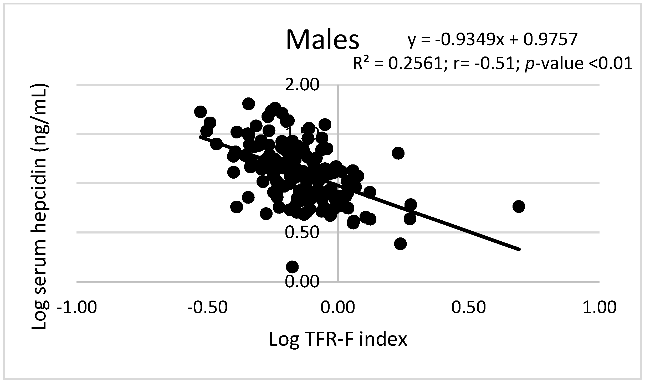
In high-altitude adult subjects, the correlations between hematological parameters and TFR-F Index were different between men and women. A negative correlation was observed between Hb, MCV, and MCH with TFR-F Index in women but not in men. RDW-CV correlated positively with TFR-F Index in women but not in men (Figure 2). Similarly, Hb, Htc, and MCV correlated with log hepcidin in women but not in men. However, the TFR-F index correlated significantly and negatively with log hepcidin in both men and women (Figure 2).
Of all the studied subjects, only two were diagnosed as anemic (0.87%). This value increased to 21 (9.17%) after altitude adjustment (10.5 times). The proportion of men and women with iron deficiency (TFR-F index > 2) were similar (0.68% in men and 2.39% in women; p > 0.05) but more men (84.45%) than women (73.05%) had normal iron defined as TFR-F index < 1 (p < 0.05). The rates of men and women with low MCV (0% and 3.17%), low MCH (0% and 3.17%), low MCV + MCH (0% and 2.38%) and low Hb + MCV + MCH (0% and 1.58%) for men and women respectively were low and similar between the sexes (p > 0.05) (Table 3).
Two cases with anemia were mild and one was moderate. After altitude adjustment, 19 cases with mild anemia were observed, one as moderate and one as severe anemia. Both subjects with anemia were female and, according to their corpuscular index, their condition could be classified as microcytic and hypochromic anemia without inflammation. After altitude adjustment, no subject had IDA measured by TBI, whereas 14.3% had a TFR-F index > 2 compatible with ID. Low MCV, MCH, and MCV+MCH were observed in 14.3%, 19%, and 14.3% of the cases, respectively. These values were significantly lower than in the group with anemia without Hb correction. Two out of 21 subjects with adjusted anemia had elevated IL-6 levels (Table 4).
In the iron sufficient subsample (TFR-F index < 1.0 and IL-6 < 50 pg/mL), only one out of 231 (0.4%; CI95%: 0.08%–2.41%) was anemic with unadjusted Hb, whereas 11 (4.76%; CI95%: 2.68%–8.32%) were diagnosed as anemic after Hb adjustment (p < 0.05). The characteristics of these subjects are presented in Table 5.
In the group with adjusted anemia only three out of 21 subjects (14.28%; CI95%: 4.98%–34.64%) had TFR-F values > 2 indicative of iron deficiency, whereas 13 subjects had TFR-F index < 1 (61.90%; CI95%: 40.88%–79.25%) and five with TFR-F index between 1 and 2 (23.80%; CI95%: 10.63%–45.09%). For analysis, the subjects with adjusted anemia were grouped according to TFR-F values (≥1 and <1) and compared with data obtained in healthy subjects (TFR-F < 1 and IL-6 < 50 pg/mL).
Comparing subjects with adjusted anemia and different TFR-F index, the MCV, MCH, hepcidin, and body iron content was higher but EPO level was lower in those with adjusted anemia and TFR-F < 1 than in those with adjusted anemia but TFR-F ≥ 1. Age, BMI, ASP, ADP, SpO2, MCV, MCH, RDW, hepcidin, TBI, and IL-6 levels were similar in subjects classified as having adjusted anemia with TRF-F < 1 and in healthy adult subjects (TFR-F < 1 and IL-6 < 50 pg/mL).
Hb, corrected Hb, RBC count, and MCHC were lower in the group with adjusted anemia and TFR-F < 1. The group with adjusted anemia and TFR-F ≥ 1 had lower age, Hb, corrected Hb, MCV, MCH, MCHC, hepcidin, and TBI than the group without anemia but TRF-F < 1 plus IL-6 < 50 pg/mL (healthy subjects) (Table 5).
The effect size assessment resulted in a large effect size for RBC count, MCV, MCH, serum hepcidin, log serum hepcidin, TBI, TFR-F index, and serum EPO levels (Table 5).
Hemoglobin (g/dL) correlated with TRF-F index in women (p < 0.01) but not in men (p > 0.05) (Figure 3). In the combined sample, Hb correlated inversely with the TRF-F index (Figure 3). Log TRF-F index correlated negatively with log serum hepcidin in men (p < 0.01) and women (p < 0.01) (Figure 4). TBI correlated positively (p < 0.001) with log serum hepcidin levels in men and women (Figure 5).
We have also assessed correlations comparing hemoglobin values with the serum ferritin, TBI, log serum hepcidin, and serum EPO. Hemoglobin concentration correlated with these variables in women but not in men (p > 0.05). Hb correlated directly with serum ferritin (p < 0.05), TBI (p < 0.01), and log hepcidin (p < 0.01) but inversely with serum EPO (p < 0.01) (Table 6).
In the logistic regression analysis, RDW-CV was the only variable directly associated with the TRF-F index ≥ 1 (p = 0.01). Age, RBC count, and MCV were not significantly associated with the TRF-F index ≥ 1 (Table 7).
Table 8 presents data of the logistic regression analysis to determine the association between adjusted anemia with the TRF-F index and hematological indices in men and women. The TFR-F index was not associated with adjusted anemia in men (p = 0.5699 nor in women (p = 0.302). Among hematological parameters, RBC count and MCV were significantly associated with adjusted anemia in men and RBC count with RDW-CV in women (Table 8).

4. Discussion

Different studies in children and pregnant women have suggested that recommendations to adjust hemoglobin (Hb) by altitude [5] overestimates the prevalence of anemia particularly in altitudes over 3000 m [41,48]. The WHO recommends daily iron supplementation for all women or children in areas where anemia prevalence is ≥40%, even though Hb concentration is generally considered to be a poor prognostic indicator of iron status. In the Peruvian highlands, only 25% of children aged 10–35 months that received iron supplementation, showed a reduction of anemia [49], and another study in the Southern Andes in children aged 6–59 months showed that 22% of the cases of anemia were attributable to iron deficiency [27]. In this latter population, the proportion of anemia attributable to inflammation was 27.8% suggesting that the remaining cases (50.23%) are due to other causes and/or due to a misdiagnosis of anemia as suggested in other studies [7,23,24,33].
In natives in HA, Hb concentration increases and it is not unusual to find values of Hb ≥ 21 g/dL in men and ≥ 19 g/dL in women considered as excessive erythrocytosis, a characteristic associated with chronic mountain sickness [40]. Unlike excessive erythrocytosis [50], the situation of anemia in the adult population living at high altitudes has been little studied. Two studies in adult populations, one in Tibet and the other in Ethiopia, showed that men and women with iron sufficiency and without inflammation were mistakenly identified as anemic after Hb adjustment [23,24].
The present study was designed to determine whether the adjustment of Hb values by the altitude of residence is also misdiagnosing anemia in adult men and women living in the south-east Peruvian Andes at 3400 m above sea level. Our results showed that the prevalence of anemia increased by 10.5 times after adjusting Hb levels by altitude. However, using hematological parameters (MCV and MCH), the prevalence of anemia ranged between 1.35% to 1.75%, and low values of Hb, MCV, and MCH—indicative of iron deficiency anemia (IDA)—were observed in only 0.87% of the studied subjects. Similarly, markers of iron status suggest that ID was observed in 0%–1.58% of the subjects. These figures are far from those observed when Hb adjustment is performed (9.17%).
Suitability of hemoglobin adjustment by altitude should show that the prevalence of anemia after adjustment is more related to ID rate than unadjusted Hb does [25]. However, current data suggest that, in the Peruvian Andes (America), as observed in the adult population in Ethiopia and Tibet (Africa and Asia), the adjustment of Hb by altitude is inappropriately increasing the prevalence of anemia. These results are following previous data in children from Puno, Peru at 3800 m also located in the Southern Andes [1].
Our data using serum hepcidin measurements also support the fact that Hb adjustment as recommended by the WHO is misleading the diagnosis of anemia. For instance, 13 out of 21 subjects diagnosed with adjusted anemia showed values of serum hepcidin similar to the control group classified as iron sufficient.
It is known that TBI, a marker of iron stores in the body, better predicts the absence of bone marrow iron than serum ferritin concentration alone, and TBI can be analyzed as a continuous variable [31]. In our sample, no subject showed TBI values <0 mg/kg indicative of iron deficiency.
During ID or after hemorrhage, hepcidin decreases to allow iron delivery to plasma through ferroportin, thus promoting compensatory erythropoiesis to revert IDA [51]. Hepcidin is also suppressed during acute exposure to altitude hypoxia due to accelerated erythropoiesis [52]. Reduction in hepcidin levels is secondary to the increased EPO and erythroferrone levels induced by the hypoxia exposure [53].
This is not the situation in populations natives at high altitudes in which serum hepcidin levels are similar or higher than at low altitudes as observed in subjects from Ethiopia [54]. Similarly, in Peru, serum hepcidin levels were similar between natives at low altitudes and natives at the highlands in the central Andean adult women and between adult men [33]. This means that, under steady-state hypoxia, hepcidin levels are not suppressed due to stable erythropoietic activity as observed after acute exposure to high altitudes in which an accelerated erythropoietic activity occurs [52].
One question raised by our results is why TBI increases alongside hepcidin in situations of normal hemoglobin values. Here, as well as in the cases of inflammation, the risk of iron overload seems to be increased. As the risk of inflammation in our sample is reduced, then, the elevated iron levels observed seem to be due to increased iron absorption at the duodenal level.
Although hemoglobin concentrations were comparable between anemia of chronic diseases (ACD) and IDA patients, the latter presented with significantly higher serum erythropoietin concentrations than ACD patients [55]. We have observed in HA adult subjects with adjusted anemia and a TFR-F index ≥ 1, a higher EPO concentration than in those with adjusted anemia with a TFR-F index <1 and in those classified as healthy subjects (TFR-F index < 1 and IL-6 < 50 pg/mL).
This group with a high EPO concentration represents 2.6% (CI95%: 1.2%–5.6%) of the studied sample. This value is lower than the rate of anemia after Hb correction (9.17%; CI95%: 6.1%–13.6%) confirming that Hb adjustment by altitude to define anemia at HA is inappropriate.
IL-6 levels were similar between subjects with adjusted anemia but different TFR-F index suggesting that these subjects with TFR-F < 1 were normal and did not belong to the ACD category.
Another important finding from our study is the different association of Hb, hepcidin, and the TFR-F index between men and women. We have observed that Hb correlated with the TFR-F index in women but not in men whereas the TFR-F index correlated negatively with hepcidin in both men and women. This means that in men a higher TFR-F was not associated with Hb but it was associated with low hepcidin levels, suggesting that men are exposed to a risk for increasing iron stores with a risk of iron overload that should be studied in further investigations. This was confirmed in the multivariate analysis after controlling for different variables, men were associated with high hepcidin levels and lower TFR-F index was associated with increased hepcidin levels.
When the TFR-F index increases, hepcidin levels decrease in both men and women, which implies that there is a greater capacity for iron absorption. This is a normal association observed in different studies [56]. However, the relationship between TFR-F index with Hb at HA seems to be different from that observed at LA, while in women, the TFR-F index increases depending on lower hemoglobin levels, which is a normal pattern of regulation observed at LA [57], in the case of men, the values of TFR-F index increase without changes in hemoglobin levels and these increases in the TFR-F index are associated with lower hepcidin values, which suggests that in men with normal or increased Hb values there are associated lower values of hepcidin which indicates greater availability of iron absorption, which in turn will enter the body, increasing the risk of iron overload. This is corroborated in the multivariate analysis, showing that Hb concentration levels do not correlate with the TFR-F index and that men were associated with a lower TFR-F index than women.
Further analysis with other hematological parameters shows the same sex difference. TFR-F index was associated with almost all hematological parameters assessed in women but not in men. The same was observed for the association of hematological parameters with serum hepcidin. According to the multivariate analysis, the most significant association with the TFR-F index was observed with RDW-CV. A previous study showed that elevated RDW-CV correlated highly with ID [58].
There are studies showing sex differences regarding iron status. Men accumulate more iron and have a higher incidence of liver injury [59]. The same sex-related variation in iron metabolism has been observed between male and female rats, in which iron is distributed differently in the tissues. This difference in iron distribution may be associated with the difference in the hepcidin level observed between men and women [60].
Lower serum hepcidin levels in healthy women have previously been reported, compared to values measured in men [61]. We have extended the observation to the HA population in which men also have more serum hepcidin levels than women as observed in a population at LA [62].
The level of serum hepcidin correlates with serum ferritin levels [63]. Then, it is postulated that both high hepcidin and ferritin levels are markers of iron overload [64]. It has been suggested that, in dysmetabolic iron overload syndrome (DIOS), increased hepcidin has a subtle impairment in the ability to restrain iron absorption following an iron challenge [65]. This may increase the availability of unneeded iron to the circulation and the tissues.
It is expected that, in subjects with normal body iron, the hepcidin secretion is increased, restricting iron absorption and avoiding the risk of iron overload. However, according to our data (particularly in men) and from others, this regulation does not occur. In our case, and in those such as DIOS and non-alcoholic fatty liver disease (NAFLD) patients who show elevated serum hepcidin levels, an elevated iron accumulation is observed. In these cases, it is assumed that the iron challenge does not restrain iron absorption despite adequate hepcidin production, suggesting an impaired hepcidin activity [66].
Results using hematological indices should be interpreted with caution. Microcytosis is a common red cell change seen in anemias of varied etiology. These include ID, thalassemia, chronic disease, and sideroblastic anemias [67]. Then, it is important to identify markers to discriminate the etiology associated with our findings. Firstly, thalassemia is a problem observed mainly in people of Mediterranean, Middle Eastern, and Asian descent [68]. Beta-thalassemia minor, also called carrier or trait, is the heterozygous state that is usually asymptomatic with mild anemia [69]. However, in Peru, the rate of thalassemia is very low and less in HA populations [70]. For these reasons, in our study it is more probable that low MCV and MCH are more related to ID than to heterozygous thalassemia.
RDW will increase in a patient moving from iron sufficient to iron deficiency but will be within normal values after long-lasting ID—and again, RDW will increase after iron supplementation, which is indicative of enhanced erythropoiesis [71].
In children under five years of age in Peru, vitamin B12 deficiency was present in 11% of Huancavelica anemics located at high altitudes and 29.7% of Coronel Portillo in the jungle; in Huancavelica, this deficiency exclusively represented 3.1% of anemia cases and in Coronel Portillo, 4.8% [72]. Then, it is possible to find people with both simultaneous iron deficiency and vitamin B12. We have not measured vitamin B12 and this is a limitation of our study. In these cases, is possible to find normal MCV and low MCHC.
Our data provide relevant information on iron regulation in the adult population at HA. However, some limitations need to be further considered. We have not assessed circulating iron nor non-transferrin-bound iron to evaluate the impact on functional ID. A second limitation is the lack of information about dietary iron intake. Finally, our study has a cross-sectional design and, as such, poses the great limitation of being unsuitable to draw cause–effect associations

5. Conclusions

We showed that the adjustment of Hb by altitude to define anemia in adult populations at HA inappropriately increases the rate of anemia by almost 10 times. The rate of anemia using the measured Hb value at HA is closer to the rates of anemia or ID when using hematological parameters, markers of iron status, and measurements of hepcidin and erythropoietin. The rates of low MCV, low MCH, low MCV+MCH were reduced after adjusted anemia. Only three subjects have both adjusted anemia and low iron status (TRF-F > 2), representing 0.87% of the total sample studied. Sex differences related to iron status suggest that men are at more risk of iron overload than women at HA. This work highlights that an individual approach must be taken. A patient with a given Hb should be assessed individually, taking in to account hematological parameters as well as origin (Southern Andean or other).

Author Contributions

Conceptualization, G.F.G., C.V.-V. and R.F.-M.; formal analysis, G.F.G., V.V.-B. and D.E.A.-Y.; investigation, D.E.A.-Y., J.J.S.-H., L.J.-T., R.R.-G., R.F.-M., G.F.G.; resources, R.F.-M., L.J.-T.; data curation, D.E.A.-Y., J.J.S.-H., L.J.-T., R.R.-G., G.F.G.; writing—original draft preparation, G.F.G.; writing—review and editing, D.E.A.-Y.; visualization, D.E.A.-Y., G.F.G.; supervision, R.F.-M., G.F.G..; project administration, R.F.-M., L.J.-T.; funding acquisition, G.F.G., C.V.-V., and R.F.-M. All authors have read and agreed to the published version of the manuscript.

Funding

This study was funded by the “Convenio UNSAAC-FONDECYT-CONCYTEC del Esquema Financiero E041-2017-UNSAAC-02, Proyectos de Investigación”.

Institutional Review Board Statement

The study was conducted according to the guidelines of the Declaration of Helsinki, and approved by the Institutional Review Board of Universidad Peruana Cayetano Heredia (SIDISI: 101555, 30 October 2018) and of Cusco’s Regional Health Direction (DIRESA, code 8878, 10 April 2019).

Informed Consent Statement

Informed consent was obtained from all subjects involved in the study. The subjects were given codes that were used throughout the project, including sample labeling, to safeguard their identities.

Data Availability Statement

Data generated in this study is available upon reasonable request to the corresponding author.

Conflicts of Interest

The authors declare no conflict of interest. The funding body did not have any role in the design of the study; in the collection, analyses, or interpretation of data; in the writing of the manuscript, or in the decision to publish the results.

References

  1. McLean, E.; Cogswell, M.; Egli, I.; Wojdyla, D.; de Benoist, B. Worldwide prevalence of anaemia, WHO Vitamin and Mineral Nutrition Information System, 1993–2005. Public Health Nutr. 2009, 12, 444. [Google Scholar] [CrossRef] [PubMed] [Green Version]
  2. Kassebaum, N.J. The Global Burden of Anemia. Hematol. Oncol. Clin. N. Am. 2016, 30, 247–308. [Google Scholar] [CrossRef] [PubMed] [Green Version]
  3. Lopes, K.d.S.; Yamaji, N.; Rahman, M.O.; Suto, M.; Takemoto, Y.; Garcia-Casal, M.N.; Ota, E. Nutrition-specific interventions for preventing and controlling anaemia throughout the life cycle: An overview of systematic reviews. Cochrane Database Syst Rev. 2021, 26, CD013092. [Google Scholar] [CrossRef]
  4. Toki, Y.; Ikuta, K.; Kawahara, Y.; Niizeki, N.; Kon, M.; Enomoto, M.; Tada, Y.; Hatayama, M.; Yamamoto, M.; Ito, S.; et al. Reticulocyte hemoglobin equivalent as a potential marker for diagnosis of iron deficiency. Int. J. Hematol. 2017, 106, 116–125. [Google Scholar] [CrossRef] [PubMed] [Green Version]
  5. World Health Organization. Haemoglobin Concentrations for the Diagnosis of Anaemia and Assessment of Severity. Geneva, Switzerland, 2011. Available online: https://apps.who.int/iris/handle/10665/85839 (accessed on 12 February 2021).
  6. Addo, O.Y.; Yu, E.X.; Williams, A.M.; Young, M.F.; Sharma, A.J.; Mei, Z.; Kassebaum, N.J.; Jefferds, M.E.D.; Suchdev, P.S. Evaluation of Hemoglobin Cutoff Levels to Define Anemia Among Healthy Individuals. JAMA Netw. Open 2021, 4, e2119123. [Google Scholar] [CrossRef]
  7. Gonzales, G.F.; Tapia, V.; Vásquez-Velásquez, C. Changes in hemoglobin levels with age and altitude in preschool-aged children in Peru: The assessment of two individual-based national databases. Ann. N. Y. Acad. Sci. 2021, 1488, 67–82. [Google Scholar] [CrossRef]
  8. Mellors, P.W.; Kourelis, T.; Go, R.S.; Muchtar, E.; Gertz, M.A.; Kumar, S.K.; Buadi, F.K.; Kapoor, P.; Lacy, M.Q.; Dingli, D.; et al. Characteristics and risk factors for thrombosis in POEMS syndrome: A retrospective evaluation of 230 patients. Am. J. Hematol. 2021, 11, 26422. [Google Scholar] [CrossRef]
  9. Mangin, O. High oxygen affinity hemoglobins. Rev. Med. Interne 2017, 38, 106–112. [Google Scholar] [CrossRef]
  10. Tremblay, J.C.; Hoiland, R.L.; Howe, C.A.; Coombs, G.B.; Vizcardo-Galindo, G.A.; Figueroa-Mujíca, R.J.; Bermudez, D.; Gibbons, T.D.; Stacey, B.S.; Bailey, D.M.; et al. Global REACH 2018: High Blood Viscosity and Hemoglobin Concentration Contribute to Reduced Flow-Mediated Dilation in High-Altitude Excessive Erythrocytosis. Hypertension 2019, 73, 1327–1335. [Google Scholar] [CrossRef]
  11. Wu, T.Y.; Wang, X.Q.; Wei, C.Y.; Cheng, H.; Wang, X.; Li, Y.; Dong, G.; Zhao, H.; Young, P.; Li, G.; et al. Hemoglobin levels in Tibet: Different effect of age and gender for Tibetans vs Han. Comp. Clin. Path. 2005, 14, 25–35. [Google Scholar] [CrossRef]
  12. Gonzales, G.F. Peruvian contributions to the study on human reproduction at high altitude: From the chronicles of the Spanish conquest to the present. Respir. Physiol. Neurobiol. 2007, 158, 172–179. [Google Scholar] [CrossRef]
  13. Stuber, T.; Scherrer, U. Circulatory Adaptation to Long-Term High Altitude Exposure in Aymaras and Caucasians. Prog. Cardiovasc. Dis. 2010, 52, 534–539. [Google Scholar] [CrossRef]
  14. Instituto de Estadistica e Informática I. Indicadores de Resultados de Programas Presupuestales 2015–2020. Encuesta Demográfica y de Salud Familiar; Lima, Peru, 2021. Available online: https://proyectos.inei.gob.pe/endes/2020/ppr/Indicadores_de_Resultados_de_los_Programas_Presupuestales_ENDES_2020.pdf (accessed on 29 December 2021).
  15. Gonzales, G.F.; Tapia, V.; Gasco, M.; Carrillo, C.E.; Fort, A.L. Association of hemoglobin values at booking with adverse maternal outcomes among Peruvian populations living at different altitudes. Int. J. Gynecol. Obstet. 2012, 117, 134–139. [Google Scholar] [CrossRef]
  16. Beall, C.M.; Brittenham, G.M.; Strohl, K.P.; Blangero, J.; Williams-Blangero, S.; Goldstein, M.C.; Decker, M.J.; Vargas, E.; Villena, M.; Soria, R.; et al. Hemoglobin concentration of high-altitude Tibetans and Bolivian Aymara. Am. J. Phys. Anthropol 1998, 106, 385–400. [Google Scholar] [CrossRef]
  17. Rothhammer, F.; Fuentes-Guajardo, M.; Chakraborty, R.; Lorenzo Bermejo, J.; Dittmar, M. Neonatal Variables, Altitude of Residence and Aymara Ancestry in Northern Chile. PLoS ONE 2015, 10, e0121834. [Google Scholar] [CrossRef] [Green Version]
  18. Cook, J.D.; Boy, E.; Flowers, C.; Mdel, C.D. The influence of high-altitude living on body iron. Blood 2005, 106, 1441–1446. [Google Scholar] [CrossRef] [PubMed] [Green Version]
  19. Hurtado, A.; Gonzales, G.F. Pulse oxygen saturation in healthy newborns at term in Cusco, Peru. Int. J. Gynecol. Obstet. 2006, 95, 155–156. [Google Scholar] [CrossRef] [PubMed]
  20. Gonzales, G.F.; Salirrosas, A. Arterial oxygen saturation in healthy newborns delivered at term in Cerro de Pasco (4340 m) and Lima (150 m). Reprod. Biol. Endocrinol. 2005, 3, 46. [Google Scholar] [CrossRef] [Green Version]
  21. Hsieh, M.M.; Callacondo, D.; Rojas-Camayo, J.; Quesada-Olarte, J.; Wang, X.; Uchida, N.; Maric, I.; Remaley, A.T.; Leon-Velarde, F.; Villafuerte, F.C.; et al. SENP1, but not fetal hemoglobin, differentiates Andean highlanders with chronic mountain sickness from healthy individuals among Andean highlanders. Exp. Hematol. 2016, 44, 483–490.e2. [Google Scholar] [CrossRef] [PubMed]
  22. Alarcón-Yaquetto, D.E.; Caballero, L.; Gonzales, G.F. Association between Plasma N-Acylethanolamides and High Hemoglobin Concentration in Southern Peruvian Highlanders. High. Alt. Med. Biol. 2017, 18, 322–329. [Google Scholar] [CrossRef]
  23. Sarna, K.; Gebremedin, A.; Brittenham, G.M.; Beall, C.M. WHO hemoglobin thresholds for altitude increase the prevalence of anemia among Ethiopian highlanders. Am. J. Hematol. 2018, 93, E229–E231. [Google Scholar] [CrossRef] [Green Version]
  24. Sarna, K.; Brittenham, G.M.; Beall, C.M. Current WHO hemoglobin thresholds for altitude and misdiagnosis of anemia among Tibetan highlanders. Am. J. Hematol. 2020, 95, E134–E136. [Google Scholar] [CrossRef]
  25. Gonzales, G.F.; Begazo, J.; Alarcón-Yaquetto, D.E. Suitability of Haemoglobin Adjustment to Define Anaemia at High Altitudes. Acta Haematol. 2020, 143, 511–512. [Google Scholar] [CrossRef] [PubMed]
  26. Choque-Quispe, B.M.; Alarcón-Yaquetto, D.E.; Paredes-Ugarte, W.; Zaira, A.; Ochoa, A.; Gonzales, G.F. Is the prevalence of anemia in children living at high altitudes real? An observational study in Peru. Ann. N. Y. Acad. Sci. 2020, 1473, 35–47. [Google Scholar] [CrossRef]
  27. Choque-Quispe, B.M.; Paz, V.; Gonzales, G.F. Proportion of anemia attributable to iron deficiency in high-altitude infant populations. Ann. Hematol. 2019, 98, 2601–2603. [Google Scholar] [CrossRef] [PubMed]
  28. Rujeerapaiboon, N.; Tantiworawit, A.; Piriyakhuntorn, P.; Rattanathammethee, T.; Hantrakool, S.; Chai-Adisaksopha, C.; Rattarittamrong, E.; Norasetthada, L.; Fanhchaksai, K.; Charoenkwan, P. Correlation Between Serum Ferritin and Viral Hepatitis in Thalassemia Patients. Hemoglobin 2021, 45, 175–179. [Google Scholar] [CrossRef]
  29. Beguin, Y. Soluble transferrin receptor for the evaluation of erythropoiesis and iron status. Clin. Chim. Acta 2003, 329, 9–22. [Google Scholar] [CrossRef] [Green Version]
  30. Koistinen, P.O.; Rusko, H.; Irjala, K.; Rajamäki, A.; Penttinen, K.; Sarparanta, V.P.; Karpakka, J.; Leppäluoto, J. EPO, red cells, and serum transferrin receptor in continuous and intermittent hypoxia. Med. Sci. Sports Exerc. 2000, 32, 800–804. [Google Scholar] [CrossRef]
  31. Pfeiffer, C.M.; Looker, A.C. Laboratory methodologies for indicators of iron status: Strengths, limitations, and analytical challenges. Am. J. Clin. Nutr. 2017, 106, 1606S–1614S. [Google Scholar] [CrossRef] [Green Version]
  32. Hanif, N.; Anwer, F. Chronic Iron Deficiency. 2021. Available online: http://www.ncbi.nlm.nih.gov/pubmed/32809711 (accessed on 29 December 2021).
  33. Gonzales, G.F.; Rubín de Celis, V.; Begazo, J.; Del Rosario Hinojosa, M.; Yucra, S.; Zevallos-Concha, A.; Tapia, V. Correcting the cut-off point of hemoglobin at high altitude favors misclassification of anemia, erythrocytosis and excessive erythrocytosis. Am. J. Hematol. 2018, 93, E12–E16. [Google Scholar] [CrossRef] [PubMed]
  34. Manckoundia, P.; Barben, J.; Asgassou, S.; Putot, A.; Konaté, A. Iron deficiency in adults: To understand what biological evaluation should be carried out. Rev. Med. Liege 2020, 75, 791–796. [Google Scholar]
  35. Manckoundia, P.; Konaté, A.; Hacquin, A.; Nuss, V.; Mihai, A.M.; Vovelle, J.; Dipanda, M.; Putot, S.; Barben, J.; Putot, A. Iron in the General Population and Specificities in Older Adults: Metabolism, Causes and Consequences of Decrease or Overload, and Biological Assessment. Clin. Interv. Aging 2020, 15, 1927–1938. [Google Scholar] [CrossRef]
  36. Donker, A.E.; van der Staaij, H.; Swinkels, D.W. The critical roles of iron during the journey from fetus to adolescent: Developmental aspects of iron homeostasis. Blood Rev. 2021, 50, 100866. [Google Scholar] [CrossRef] [PubMed]
  37. Tkaczyszyn, M.; Comín-Colet, J.; Voors, A.A.; van Veldhuisen, D.J.; Enjuanes, C.; Moliner-Borja, P.; Rozentryt, P.; Poloński, L.; Banasiak, W.; Ponikowski, P.; et al. Iron deficiency and red cell indices in patients with heart failure. Eur. J. Heart Fail. 2018, 20, 114–122. [Google Scholar] [CrossRef] [Green Version]
  38. Yu, L.N.; Wang, S.J.; Chen, C.; Rausch, V.; Elshaarawy, O.; Mueller, S. Direct modulation of hepatocyte hepcidin signaling by iron. World J. Hepatol. 2021, 13, 1378–1393. [Google Scholar] [CrossRef] [PubMed]
  39. Anderson, G.J.; Bardou-Jacquet, E. Revisiting hemochromatosis: Genetic vs. phenotypic manifestations. Ann. Transl. Med. 2021, 9, 731. [Google Scholar] [CrossRef] [PubMed]
  40. León-Velarde, F.; Maggiorini, M.; Reeves, J.T.; Aldashev, A.; Asmus, I.; Bernardi, L.; Ge, R.L.; Hackett, P.; Kobayashi, T.; Moore, L.G.; et al. Consensus Statement on Chronic and Subacute High Altitude Diseases. High. Alt. Med. Biol. 2005, 6, 147–157. [Google Scholar] [CrossRef] [PubMed]
  41. Sharma, A.J.; Addo, O.Y.; Mei, Z.; Suchdev, P.S. Reexamination of hemoglobin adjustments to define anemia: Altitude and smoking. Ann. N. Y. Acad. Sci. 2019, 1450, 190–203. [Google Scholar] [CrossRef] [Green Version]
  42. Abitbol, V.; Borderie, D.; Polin, V.; Maksimovic, F.; Sarfati, G.; Esch, A.; Tabouret, T.; Dhooge, M.; Dreanic, J.; Perkins, G.; et al. Diagnosis of iron deficiency in inflammatory bowel disease by transferrin receptor-ferritin index. Medicine 2015, 94, e1011. [Google Scholar] [CrossRef]
  43. Suega, K.; Kandarini, Y.; Tubung, J. Role of Soluble Transferrin Receptor and Transferrin Receptor-Ferritin Index to Detect Iron Deficiency Anemia in Regular Hemodialysis Patients. Open Access Maced. J. Med. Sci. 2019, 7, 97–102. [Google Scholar] [CrossRef] [PubMed] [Green Version]
  44. Kallianpur, A.R.; Wen, W.; Erwin, A.L.; Clifford, D.B.; Hulgan, T.; Robbins, G.K. Higher iron stores and the HFE 187C> G variant delay onset of peripheral neuropathy during combination antiretroviral therapy. PLoS ONE. 2020, 15, e0239758. [Google Scholar] [CrossRef]
  45. Said, E.A.; Al-Reesi, I.; Al-Shizawi, N.; Jaju, S.; Al-Balushi, M.S.; Koh, C.Y.; Al-Jabri, A.A.; Jeyaseelan, L. Defining IL-6 levels in healthy individuals: A meta-analysis. J. Med. Virol. 2021, 93, 3915–3924. [Google Scholar] [CrossRef]
  46. Alende-Castro, V.; Alonso-Sampedro, M.; Gude, F.; Gonzalez-Quintela, A. Serum Concentrations of Interleukin 6 in the General Adult Population: Possible Implications for Anti-IL-6 Therapy in SARS-Cov-2 Infection and IL-6-Related Diseases. J. Investig. Allergol. Clin. Immunol. 2021, 31, 75–78. [Google Scholar] [CrossRef] [PubMed]
  47. Sarma, P.R. Red Cell Indices. Butterworths; 1990. Available online: http://www.ncbi.nlm.nih.gov/pubmed/21250103 (accessed on 10 March 2021).
  48. Gonzales, G.F.; Steenland, K.; Tapia, V. Maternal hemoglobin level and fetal outcome at low and high altitudes. Am. J. Physiol. Integr. Comp. Physiol. 2009, 297, R1477–R1485. [Google Scholar] [CrossRef] [Green Version]
  49. Aparco Balboa, J.P.; Bullón, L.; Cusirramos, S. Impact of micronutrient powder on anemia in children aged 10-35 months in apurimac, Peru. Rev. Peru Med. Exp. Salud Publica 2019, 36, 17–25. [Google Scholar] [CrossRef] [Green Version]
  50. Richalet, J.-P. Adaptation à l’hypoxie chronique des populations de haute altitude. Rev. Mal. Respir. 2021, 38, 395–403. [Google Scholar] [CrossRef]
  51. Nemeth, E.; Ganz, T. Hepcidin-Ferroportin Interaction Controls Systemic Iron Homeostasis. Int. J. Mol. Sci. 2021, 22, 6493. [Google Scholar] [CrossRef]
  52. Breenfeldt Andersen, A.; Bonne, T.C.; Bejder, J.; Jung, G.; Ganz, T.; Nemeth, E.; Olsen, N.V.; Huertas, J.R.; Nordsborg, N.B. Effects of altitude and recombinant human erythropoietin on iron metabolism: A randomized controlled trial. Am. J. Physiol. Regul. Integr. Comp. Physiol. 2021, 321, R152–R161. [Google Scholar] [CrossRef] [PubMed]
  53. Emrich, I.E.; Scheuer, A.; Wagenpfeil, S.; Ganz, T.; Heine, G.H. Increase of plasma erythroferrone levels during high-altitude exposure: A sub-analysis of the TOP OF HOMe study. Am. J. Hematol. 2021, 96, E179–E181. [Google Scholar] [CrossRef]
  54. Lundgrin, E.L.; Janocha, A.J.; Koch, C.D.; Gebremedhin, A.; Di Rienzo, A.; Alkorta-Aranburu, G.; Brittenham, G.M.; Erzurum, S.C.; Beall, C.M. Plasma hepcidin of Ethiopian highlanders with steady-state hypoxia. Blood 2013, 122, 1989–1991. [Google Scholar] [CrossRef] [PubMed] [Green Version]
  55. Theurl, I.; Mattle, V.; Seifert, M.; Mariani, M.; Marth, C.; Weiss, G. Dysregulated monocyte iron homeostasis and erythropoietin formation in patients with anemia of chronic disease. Blood 2006, 107, 4142–4148. [Google Scholar] [CrossRef] [Green Version]
  56. Belo, L.; Rocha, S.; Valente, M.J.; Coimbra, S.; Catarino, C.; Bronze-da-Rocha, E.; Rocha-Pereira, P.; do Sameiro-Faria, M.; Oliveira, J.G.; Madureira, J.; et al. Hepcidin and diabetes are independently related with soluble transferrin receptor levels in chronic dialysis patients. Ren. Fail. 2019, 41, 662–672. [Google Scholar] [CrossRef] [Green Version]
  57. Krawiec, P.; Pac-Kożuchowska, E. Soluble transferrin receptor and soluble transferrin receptor/log ferritin index in diagnosis of iron deficiency anemia in pediatric inflammatory bowel disease. Dig. Liver. Dis. 2019, 51, 352–357. [Google Scholar] [CrossRef] [PubMed] [Green Version]
  58. Ritchie, B.; McNeil, Y.; Brewster, D.R. Soluble transferrin receptor in Aboriginal children with a high prevalence of iron deficiency and infection. Trop. Med. Int. Health. 2004, 9, 96–105. [Google Scholar] [CrossRef] [PubMed]
  59. Harrison-Findik, D.D. Gender-related variations in iron metabolism and liver diseases. World J. Hepatol 2010, 2, 302. [Google Scholar] [CrossRef]
  60. Kong, W.-N.; Niu, Q.-M.; Ge, L.; Zhang, N.; Yan, S.F.; Chen, W.B.; Chang, Y.Z.; Zhao, S.E. Sex Differences in Iron Status and Hepcidin Expression in Rats. Biol. Trace Elem. Res. 2014, 160, 258–267. [Google Scholar] [CrossRef] [PubMed]
  61. Ganz, T.; Olbina, G.; Girelli, D.; Nemeth, E.; Westerman, M. Immunoassay for human serum hepcidin. Blood 2008, 112, 4292–4297. [Google Scholar] [CrossRef] [Green Version]
  62. Cook, J.D.; Flowers, C.H.; Skikne, B.S. The quantitative assessment of body iron. Blood 2003, 101, 3359–3363. [Google Scholar] [CrossRef] [Green Version]
  63. Gromadzka, G.; Wierzbicka, D.; Litwin, T.; Przybyłkowski, A. Difference in iron metabolism may partly explain sex-related variability in the manifestation of Wilson’s disease. J. Trace Elem. Med. Biol. 2020, 62, 126637. [Google Scholar] [CrossRef]
  64. Rauber, M.R.; Pilger, D.A.; Cecconello, D.K.; Falcetta, F.S.; Marcondes, N.A.; Faulhaber, G.A.M. Hepcidin is a useful biomarker to evaluate hyperferritinemia associated with metabolic syndrome. An. Acad. Bras. Cienc. 2019, 91, e20180286. [Google Scholar] [CrossRef] [PubMed] [Green Version]
  65. Rametta, R.; Dongiovanni, P.; Pelusi, S.; Francione, P.; Iuculano, F.; Borroni, V.; Fatta, E.; Castagna, A.; Girelli, D.; Fargion, S.; et al. Hepcidin resistance in dysmetabolic iron overload. Liver Int. 2016, 36, 1540–1548. [Google Scholar] [CrossRef] [PubMed]
  66. Rametta, R.; Fracanzani, A.L.; Fargion, S.; Dongiovanni, P. Dysmetabolic Hyperferritinemia and Dysmetabolic Iron Overload Syndrome (DIOS): Two Related Conditions or Different Entities? Curr. Pharm. Des. 2020, 26, 1025–1035. [Google Scholar] [CrossRef] [PubMed]
  67. Kotwal, J.; Saxena, R.; Choudhry, V.P.; Dwivedi, S.N.; Bhargava, M. Erythrocyte indices for discriminating thalassaemic and non-thalassaemic microcytosis in Indians. Natl. Med. J. India 1999, 12, 266–267. [Google Scholar]
  68. Needs, T.; Gonzalez-Mosquera, L.F.; Lynch, D.T. Beta Thalassemia. In StatPearls; StatPearls Publishing: Treasure Island, FL, USA, 18 July 2021. [Google Scholar]
  69. Bajwa, H.; Basit, H. Thalassemia. In StatPearls; StatPearls Publishing: Treasure Island, FL, USA, 11 August 2021. [Google Scholar]
  70. Castillo, J. Hemoglobinas anormales en el Perú, su importancia genética y antropológica en nuestro mestizaje [Abnormal hemoglobins in Peru, their genetic and anthropological importance in our miscegenation]. An. Acad. Nac. Med. 2002, 1–15. Available online: https://anmperu.org.pe/anales/2002/VI_HEMOGLOBINASANORMALESENELPERU.pdf (accessed on 29 December 2021).
  71. Schoorl, M.; Schoorl, M.; van der Gaag, D.; Bartels, P.C. Effects of iron supplementation on red blood cell hemoglobin content in pregnancy. Hematol. Rep. 2012, 4, e24. [Google Scholar] [CrossRef]
  72. Gonzales, E.; Huamán-Espino, L.; Gutiérrez, C.; Aparco, J.P.; Pillaca, J. Caracterización de la anemia en niños menores de cinco años de zonas urbanas de Huancavelica y Ucayali en el Perú [Characterization of anemia in children under five years of age from urban areas of Huancavelica and Ucayali, Peru]. Rev. Peru. Med. Exp. Salud Publica 2015, 32, 431–439. [Google Scholar] [CrossRef] [PubMed]
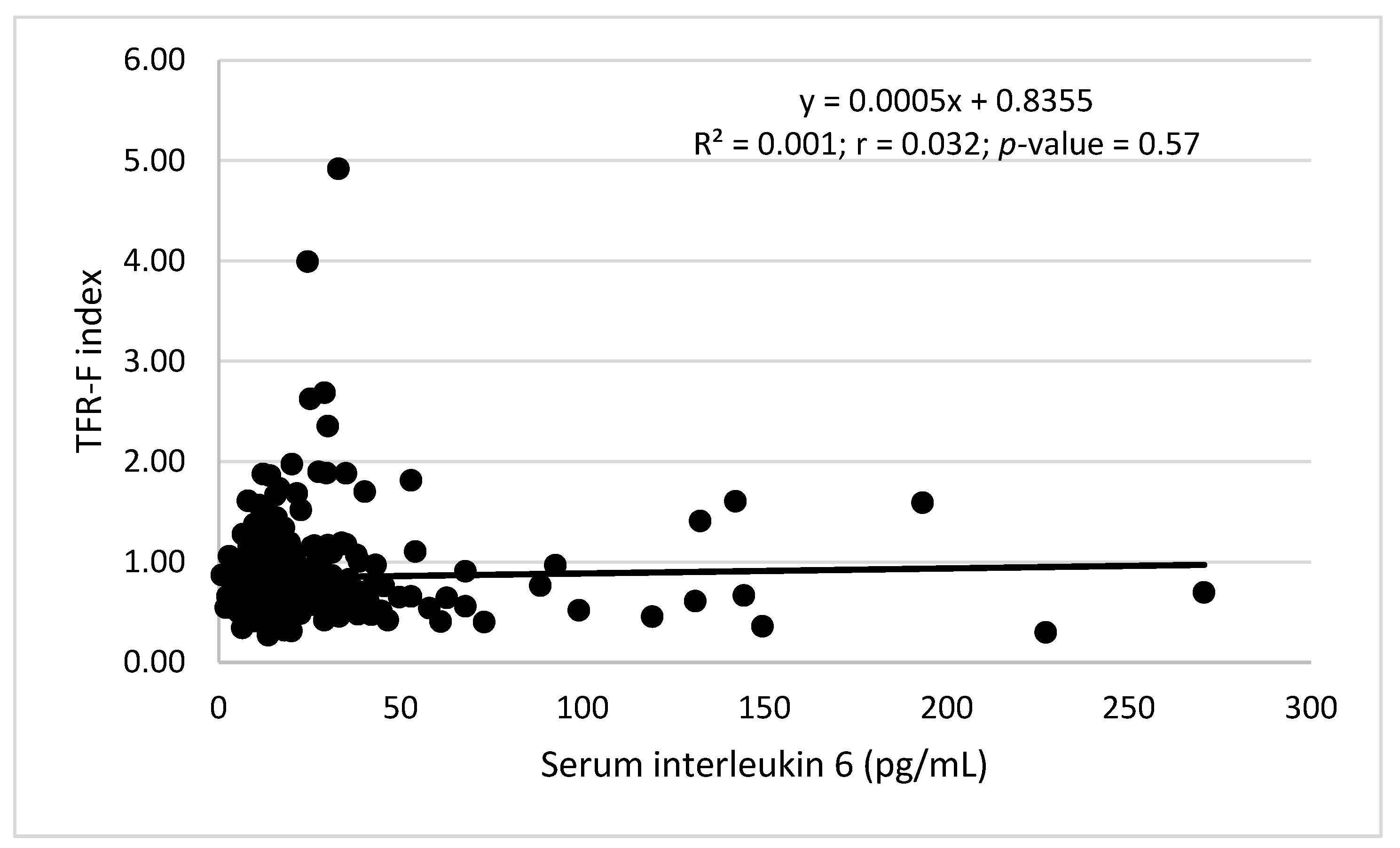
Figure 1. Correlation between serum interleukin-6 (IL-6) levels and Transferrin Receptor-Ferritin Index in adult subjects from Cusco, Peru at 3400 m above sea level.
Physiologia 02 00001 g001
Figure 2. Correlation matrix between hematological parameters and sTfR/Ferritin index and Log Hepcidin in (A) women and (B) men from Cusco (3400 masl). Values shown are the Pearson coefficient. * p < 0.05, ** p < 0.01, *** p < 0.001.
Physiologia 02 00001 g002
Figure 3. Correlation between hemoglobin (g/dL) and transferrin receptor (TfR-F) index in adult males (Upper), men (Middle), and males+females (Lower) from Cusco, Peru at 3400 m above sea level.
Physiologia 02 00001 g003aPhysiologia 02 00001 g003b
Figure 4. Correlation between log. transferrin receptor (TFR-F) index and log. hepcidin (ng/mL) in adult females (upper) and men (lower) from Cusco, Peru at 3400 m above sea level.
Physiologia 02 00001 g004aPhysiologia 02 00001 g004b
Figure 5. Correlation between Total Body Iron (TBI) (mg/Kg) and log. Serum hepcidin (ng/mL) in adult men (upper) and females (lower) from Cusco, Peru at 3400 m above sea level.
Physiologia 02 00001 g005
Table 1. Characteristics of the sample studied in Cusco (3400 m) by sex.
CharacteristicsMen (156)Women (189)Overall (345)
Age (years)46.23 [16.86]44.80 [14.42]45.42 [15.59]
BMI (kg/m2)26.27 [4.12]27.37 [4.95]26.91 [4.64]
SBP (mm Hg)118.40 [14.36]117.65 [14.97]117.97 [14.67]
DBP (mm Hg)75.76 [9.49]75.20 [12.09]75.44 [10.96]
SpO2 (%)91.51 [3.12]91.06 [3.16]91.25 [3.16]
Hb (g/dL)17.29 [0.99]15.35 [1.09]16.23 [1.48]
Adjusted Hb (g/dL)14.79 [0.99]12.85 [1.09]13.73 [1.48]
Hematocrit (%)50.21 [3.24]45.14 [2.88]47.42 [3.89]
RBC (*106)5.458 [0.362]4.948 [0.316]5.177 [0.427]
MCV (fL)91.93 [3.24]90.85 [6.32]91.34 [5.19]
MCH (pg)31.72 [1.15]31.14 [2.06]31.41 [1.67]
MCHC (g/dL)34.47 [0.62]34.29 [2.75]34.38 [2.04]
RDW-CV (%)12.09 [0.49]12.10 [0.69]12.09 [0.74]
Reticulocytes1.28 [0.37]1.30 [0.27]1.29 [0.37]
CMS score2.57 [2.62]3.84 [3.29]3.28 [2.97]
Data are mean [SD]. BMI: Body mass index, SBP: Systolic blood pressure, DBP: Diastolic blood pressure, SpO2: Pulse oxygen saturation, RBC: Red blood cell, MCV: Mean corpuscular volume, MCH: Mean corpuscular hemoglobin (pg), MCHC: Mean corpuscular hemoglobin concentration (g/dL), RDW: Red cell distribution width—CV (coefficient of variation). Hb: Hemoglobin. Bold values represent statistical significance.
Table 2. Iron status markers and erythropoietic hormones by sex.
Biological VariableMen (156)Women (189)Overall (345)
Ferritin (ng/mL)50.0 [45.71]24.43 [21.82]36.48 [37.32]
sTfR (ug/mL)1.13 [0.37]1.10 [0.41]1.11 [0.37]
TFR-F index0.77 [0.37]0.91 [0.41]0.85 [0.55]
Total Body Iron (mg/Kg)11.19 [2.99]9.05 [2.74]10.06 [2.97]
Hepcidin (ng/mL)16.41 [12.36]11.20 [8.65]13.65 [10.77]
Erythropoietin (mIU/mL)9.39 [5.87]10.53 [7.14]9.99 [6.49]
Data are mean [SD]. TFR-F index = sTfR/log. Ferritin.
Table 3. Proportion of subjects from Cusco (Peru) with anemia (<12 g/dL and <13 g/dL in women and men respectively), altitude adjusted anemia, different sTfR-F Index, low MCV, low MCH, low MCV + MCH, low Hb + MCV + MCH, and high serum IL-6 levels.
MenWomenTotal
Anemia0/103 (0%)2/126 (1.55%)2/229 (0.87%)
Adjusted Anemia4/103 (3.9%)17/126 (13.5%) *21/229 (9.17%)
TFR-F index
>21 (0.68%)4 (2.39%)5 (1.58%)
1–222 (14.86%)41 (24.55%) **63 (20%)
<1125 (84.45%)122 (73.05%) **247 (78.41%)
Low MCV (<80 fL)0/103 (0%)4/126 (3.17%)4/229 (1.75%)
Low MCH (<27 pg/mL)0/103 (0%)4/126 (3.17%)4/229 (1.75%)
Low MCV and MCH0/103 (0%)3/126 (2.38%)3/229 (1.31%)
Low Hb, MCV and MCH0/103 (0%)2/126 (1.58%)2/229 (0.87%)
Elevated IL-6 (>50 pg/mL)9/148 (0%)12/167 (7.18%)21/315 (8.25%)
TFR-F index = sTfR/log ferritin. MCV = Mean corpuscular volume. MCH = Mean corpuscular hemoglobin. IL-6 = interleukin 6. * p < 0.05, ** p < 0.01.
Table 4. Iron deficiency anemia, low MCV, MCH, MCV + MCH, and elevated serum IL-6 levels in adult subjects from Cusco (3400 m) classified with anemia or altitude-adjusted anemia.
Anemia (2)Adjusted Anemia (21)
IDA0/2 (0%)0/21 (0%)
TFR-F Index
<11/2 (50%)13/21 (61.9%)
1–21/2 (50%)5/21 (23.8%)
>20/2 (0%)3/21 (14.3%) *
Low MCV (<80 fL)2/2 (100%)3/21 (14.3%) *
Low MCH (<37 pg/mL)2/2 (100%)4/21 (19.0%) *
Low MCV and MCH2/2 (100%)3/21 (14.3%) *
Elevated IL-6 (>50 pg/mL)0/2 (0%)2/21 (9.5%)
Adjusted anemia was diagnosed after subtracting by 2.5 g/dL Hb in the adult population living in Cusco at 3400 m above sea level. ID was diagnosed when total body iron content (TBI) was <0 mg/kg. IDA when TBI was <0 mg/Kg and Hb < 12 g/dL in women and <13 g/dL in men. * p < 0.01 respect to values in subjects with anemia.
Table 5. Biological characteristics of subjects diagnosed as corrected anemia compared with those defined as iron sufficient without inflammation (TFR-F index < 1.0 and IL-6 < 50 pg/mL).
Healthy SubjectsAdjusted Anemia
TRF-F ≥ 1
Adjusted Anemia
TRF-F < 1
Size Effect
Cohen’s d
Male/female116/1150/84/9
Age45.85 [15.50]36.12 [10.32] *40.82 [11.34]0.43
BMI26.94 [5.77]26.97 [6.03]25.47 [2.73]0.32
ASP mm Hg117.24 [13.52]119.62 [19.03]110.41 [7.23]0.64
ADP mm Hg74.86 [11.09]76.12 [10.86]72.58 [6.93]0.39
SpO291.32 [3.19]92.12 [2.82]92.25 [1.59]0.05
CMS score3.35 [3.19]3.37 [3.46]2.69 [2.46]0.23
Hb16.38 [1.36 ]13.23 [0.98] *14.14 [1.29] *0.79
Corrected Hb13.88 [1.36]10.73 [0.98] *11.63 [1.29] *0.79
RBC count * 1065.18 [0.45]4.97 [0.39]4.68 [0.3] *0.83
MCV91.81 [5.31]81.58 [6.37] *90.0 [6.96] #1.26
MCH31.65 [1.36]26.81 [3.04] *30.2 [2.73] ##1.17
MCHC34.34 [0.75]32.75 [1.35] *33.49 [0.99] **0.62
RDW12.00 [0.61]13.47 [1.24] *12.62 [1.53]0.61
Hepcidin15.21 [11.09]4.31 [2.25] *13.22 [6.42] #1.84
Log hepcidin1.08 [0.30]0.59 [0.19] *1.03 [0.27] #1.88
TBI (mg/Kg)11.0 [16.86]4.54 [1.63] *10.56 [1.41] #3.95
TFR-F Index0.68 [0.15]1.90 [0.59] *0.62 [0.12] #3.00
TFR-F Index < 1231/231 (100%)0/8 (0%) *13/13 (100%) #
TFR-F Index > 20/231 (0%)3/8 (37.5%)0/13 (0%)
EPO8.92 [4.86]20.14 [10.85] *9.31 [6.18] ##1.22
IL-616.50 [9.27]22.12 [9.02]28.88 [27.18]0.33
Data are mean [SD]; * p < 0.01; ** p < 0.05 respect to healthy subjects; # p < 0.01; ## p < 0.05 respect to subjects with adjusted anemia and TFR-F ≥ 1.
Table 6. Correlation between hemoglobin concentration with serum ferritin, total body iron (TBI), log hepcidin, and serum erythropoietin (EPO) in men and women from Cusco at 3400 m above sea level.
Hemoglobin (g/dL)Men
Pearson Correlation, p
Women
Pearson Correlation, p
Serum Ferritin0.05, >0.050.21, <0.05
TBI−0.01, >0.050.28, <0.01
Log. Hepcidin0.07, >0.050.24, <0.01
Serum EPO−0.17, >0.05−0.32, <0.01
Table 7. Logistic regression analysis to determine the association of hematological parameters with an iron status marker.
TFR-F IndexOdds Ratio ± Std. ErrorzP95%C.I
Sex1.33 ± 0.490.770.440.642.75
Age (years)1.00 ± 0.010.220.830.981.02
RBC count1.68 ± 0.761.150.250.694.06
MCV (fL)1.01 ± 0.030.350.730.951.08
RDW-CV2.04 ± 0.582.490.011.163.56
Constant4.63 × 10 −6 ± 2.68 × 10⁻⁵−2.120.035.45 × 10 −110.39
0 = male; 1 = female; RBC = red blood cell; MCV = mean corpuscular volume; RDW-CV = Red cell distribution width-coefficient of variation; TFR-F index = soluble transferrin receptor/log ferritin (0 = TFR-F < 1 and 1 = TFR-F ≥ 1).
Table 8. Logistic regression analysis to determine the association of hematological parameters and iron status marker to anemia after hemoglobin correction (Adjusted anemia) in adult men and women from Cusco (3400 m).
Adjusted Anemia (Men)Odds Ratio ± Std. ErrorzP95%C.I
RBC count0.0004 ± 0.002−2.010.0451.87 × 10 −70.834
MCV (fL)0.56 ± 0.154−2.100.0350.3300.961
RDW-CV6.77 ± 13.1970.980.3270.148309.32
TFR-F Index0.068 ± 0.324−0.570.5696.86 × 10 −6691.21
Constant3.04 × 10 29 ± 1.36 × 10 311.520.1292.86 × 10−93.23 × 10 67
Adjusted anemia
(Women)
Odds Ratio ± Std. ErrorzP95%C.I
RBC count0.0004 ± 0.0009−3.540.0015.86 × 10−60.031
MCV (fL)0.96 ± 0.04−0.840.4010.8911.047
RDW-CV9.31 ± 6.583.160.0022.33137.214
TFR-F Index2.60 ± 2.411.030.3020.42315.988
Constant29,111.11 ± 272,116.31.100.2710.00032.63 × 1012
1 = With anemia (Hb < 15.5 g/dL in men and <14.5 g/dL in women) and 0 = Without anemia; MCH = mean corpuscular hemoglobin; TFR-F index = soluble transferrin receptor/log ferritin as dichotomic variable (0 = TFR-F < 1 and 1 = TFR-F ≥ 1).
Publisher’s Note: MDPI stays neutral with regard to jurisdictional claims in published maps and institutional affiliations.