Article

In Vitro Disinfection Efficacy Assay on Giardia duodenalis Cysts

Institute of Parasitology, Faculty of Veterinary Medicine, Leipzig University, 04103 Leipzig, Sachsen, Germany
*
Author to whom correspondence should be addressed.
Hygiene 2025, 5(4), 54; https://doi.org/10.3390/hygiene5040054
Submission received: 29 September 2025 / Revised: 7 November 2025 / Accepted: 15 November 2025 / Published: 25 November 2025
(This article belongs to the Section Veterinary, Livestock, and Biosafety)

Abstract

Background: The cysts of the protozoan parasite Giardia duodenalis, which targets a broad spectrum of hosts including humans, can withstand environmental conditions for months, making effective disinfectant measures crucial for minimizing the infection burden. Previous investigations concerning disinfection efficacy were based on cysts from fecal/water samples or animal models, which are either unfit for standardized procedures or related to ethical concerns. Methods: To perform standardized in vitro disinfectant testing, four different encystation protocols were compared firstly. The protocol with the highest efficacy in our hands (1.7 × 10⁵ cysts per tube) was used for the production of cysts to establish a disinfectant assay. Therefore, it was used for the production of cysts to establish a dis-infectant efficacy assay. After incubation with a commercial disinfectant (ViPiBaX Gi-ardien Ex®) or 30% hydrogen peroxide solution (H2O2) at 10 °C and room temperature, parasite cyst viability was evaluated by the yield of trophozoites obtained by the applied excystation protocol. Results: Only untreated Giardia cysts, which were used as a negative con-trol, released trophozoites. The protocol established for the evaluation of cyst viability delivered reproducible results and appeared suitable for testing the inactivation of cysts by chemical disinfection. Conclusions: Under the given conditions, the disinfectant ViPiBaX Giardien Ex® and H2O2 inactivated Giardia cysts.

1. Introduction

Giardia duodenalis (syn. G. intestinalis, G. lamblia) is a protozoan parasite that may infect a broad spectrum of hosts including humans. Hosts become infected by the oral uptake of cysts from the contaminated environment. Within the small intestine, trophozoites replicate after excystation. This may cause symptoms of, e.g., acute diarrhea, nausea and chronical disease [1,2], with the nature of symptoms largely depending on the balance of the host’s immune response [3,4]. Generally, cysts can withstand environmental conditions for months [5] and high incidence rates of Giardia infections have been reported in veterinary clinics and pet shops [6]. Effective cleaning and disinfection measures are, therefore, crucial for minimizing the infection burden in such environments. Biocidal compounds in surface disinfectants such as alcohol [7], vinegar [8] or hypochlorous acid solution [9] have shown a significant effect on Giardia cyst viability and may serve as an effective tool to limit pathogen transmission.
Giardia cyst viability after disinfection has been studied, whereby in most cases, cysts purified from fecal/water samples [8,9,10,11,12] or cysts received from animal models [7,13,14] were used. However, cysts from environmental or random fecal samples may be less suitable for standardized laboratory testing and the production of cysts via experimental animals is very resource-intensive and above all subject to ethical concerns [15]. At present, there is no method established as a standard procedure for the evaluation of disinfectant efficacy against Giardia cysts directly.
Considering the 3R principle, in vitro production of Giardia cysts is clearly preferable to establish a reproducible method for a standardized disinfectant testing protocol. In vitro protocols for the assessment of disinfectants against protozoa such as Toxoplasma gondii [16] and Cryptosporidium parvum [17,18,19] have already been reported. For Giardia, protocols were published for in vitro encystation [20,21,22,23] and in vitro excystation [22,24,25]. Based on published protocols, we here propose a method for the in vitro testing of the efficacy of chemical disinfectants on G. duodenalis cysts.

2. Materials and Methods

2.1. Axenic Culture of Giardia Trophozoites

G. duodenalis WB C6 trophozoites, assemblage A, a kind gift from Prof. Dr. Carmen Faso and Dr. Corina Wirdnam, Institute of Cell Biology, Faculty of Science, University of Bern, were used for all experiments. Culture conditions were as described earlier [26], with some modifications. The parasites were grown in an anaerobic environment using 3 mL cell culture tubes (NuncTM, ThermoFisher Scientific, Waltham, MA, USA) filled with modified TYI-S-33 medium (GM) containing 1.8 g casein peptone (Sigma-Aldrich, Steinheim, Germany), 1.0 g D(+)-Glucose (Glucose; Carl Roth, Karlsruhe, Germany), 0.9 g yeast extract (Sigma-Aldrich), 200 mg sodium chloride (NaCl; Carl Roth), 100 mg di-potassium hydrogen phosphate (K2HPO4; Carl Roth), 60 mg potassium di-hydrogen phosphate (KH2PO4; Carl Roth GmbH), 20 mg L(+)-ascorbic acid (AA; Carl Roth), 10 mL heat-inactivated fetal calf serum (FCS; PAN Biotech, Aidenbach, Germany), 200 mg L-Cysteine hydrochloride monohydrate (cysteine; Sigma-Aldrich), 52 mg dried, unfractionated bovine bile (BB; Sigma-Aldrich), 2.28 mg ammonium iron(III) citrate (AEC; Carl Roth) in 100 mL. The pH value of this medium was adjusted to 7.1 by adding 2 M sodium hydroxide (NaOH; Grüssing, Filsum, Germany). Afterwards, the medium was sterile filtered with Filtropur S 0.2 (Sarstedt, Nümbrecht, Germany) before use.
For the passage of trophozoites, we removed medium containing unattached and dead parasites from tubes with confluent cultures. Afterwards, the tubes were replenished with 6 mL freshly prepared medium and placed on ice for 15 min. Then, after a centrifugation step at 650× g for 10 min at 4 °C, the pellet was again dispersed in 1 mL of medium. A Neubauer chamber was used to count motile trophozoites at a magnification of 200×. Further on, a tube containing 10.5 mL of freshly prepared medium was inoculated with 104–105 trophozoites.

2.2. Comparison of Protocols for Encystation

We evaluated four protocols for encystation previously published by other authors for their efficacy in cyst production.
According to the protocol published by Davids and Gillin [22], we added 5.25 × 10⁴ trophozoites to a culture tube containing 10.5 mL freshly prepared pre-encystation medium containing GM without BB with a pH value of 7.1. After incubation at 37 °C for 72 h, pre-encystation medium and non-attached trophozoites were removed and replaced with 10.5 mL encystation medium#1 (Table A1). The tubes were incubated at 37 °C for 42 h, 45 h or 48 h.
The second protocol assessed was published by Einarsson et al. [23]. Culture tubes containing 10.5 mL GM were inoculated with 1 × 10⁵ trophozoites and incubated at 37 °C. After 24 h, medium with detached and dead trophozoites was replaced by 10.5 mL encystation medium#2 containing three different BB concentrations (Table A1). The parasites were incubated for 56 h at 37 °C.
The third protocol followed the instructions of Hausen et al. [21]. Firstly, we added 2.1 × 10⁶ trophozoites to a culture tube containing 10.5 mL of encystation medium#3 (Table A1) and incubated at 37 °C for 18 h. Then, culture tubes were placed on ice for 15 min, centrifuged at 500× g for 10 min at 4 °C and resuspended in 10.5 mL GM without BB (pH 7.0). Afterwards, the parasites were incubated for 7 h at 37 °C.
The fourth protocol was published by Kane et al. [20]. We filled culture tubes with 10.5 mL GM and inoculated them with 1 × 10⁵ trophozoites, followed by incubation for 48 h at 37 °C. Medium containing detached and dead cells was replaced by 10.5 mL encystation medium#4 (Table A1) and the parasites were further incubated for 24 h at 37 °C. Afterwards, the culture tubes were placed on ice for 15 min and a centrifugation step was conducted (500× g, 10 min, 4 °C). Pellets were resuspended in 1 mL GM and then culture tubes were filled up to 10.5 mL with GM and further incubated for 24 h at 37 °C.
We counted the cysts as following irrespective of the applied protocol. Cyst suspensions were incubated on ice for 15 min, followed by centrifugation at 1000× g for 10 min at 4 °C. Pellets were resuspended in 4 °C cold, sterile double-distilled water (ddH2O) and suspensions incubated for 30 min on ice again. After another centrifugation step under the same conditions, pelleted cysts, which were resistant against water lysis, were redispersed in 100 µL ddH2O and counted using a Neubauer chamber.
To ensure reproducibility, we performed experiments in triplicates and repeated them 3 times.

2.3. Comparison of Protocols for Excystation

We obtained cysts as previously described by Kane et al. [20], stored them in phosphate-buffered saline (PBS; Gibco, ThermoFisher Scientific) at 4 °C for 4–10 days (d) before use and two different protocols were conducted to induce excystation.
The first one was based on the three-step method published by Hausen et al. [21] (trypsin protocol).
Briefly, after centrifugation (900× g, 10 min, 4 °C), we resuspended 8 × 10⁴ cysts in 2 mL of freshly prepared solution 1 (68 mg L-Cysteine hydrochloride monohydrate (cysteine; Sigma-Aldrich), 68 mg reduced L-Glutathione (Carl Roth) and 52 mg sodium hydrogen carbonate (NaHCO3; Carl Roth) dissolved in 7 mL Hanks Balanced Salt Solution (HBSS; Gibco, ThermoFisher Scientific), supplemented with 15 mL double-distilled water (ddH2O) before the pH value was adjusted to 2.5 using 1 M hydrochloric acid (HCl; Carl Roth) and ddH2O added to a final volume of 25 mL) and the suspension incubated for 40 min at 37 °C. After centrifugation (200× g, 5 min, room temperature (RT)), cysts were incubated for 1 h at 37 °C in 2 mL of freshly prepared, sterile filtered solution 2 (20 mL Tyrode’s Salts with sodium bicarbonate (Sigma-Aldrich) supplemented with 200 mg lyophilized trypsin Type II-S (from porcine pancreas; Sigma-Aldrich) and an adjusted pH value to 8 using 2 M sodium hydroxide (NaOH; Grüssing)). During incubation, the cyst suspension was carefully shaken every 15 min. After centrifugation (200× g, 5 min, RT), pellets were resuspended in 1 mL modified TYI-S-33 (GM) containing 1.8 g casein peptone (Sigma-Aldrich), 1.0 g D(+)-Glucose (glucose; Carl Roth), 0.9 g yeast extract (Sigma-Aldrich), 200 mg sodium chloride (NaCl; Carl Roth), 100 mg di-potassium hydrogen phosphate (K2HPO4; Carl Roth), 60 mg potassium di-hydrogen phosphate (KH2PO4; Carl Roth), 20 mg L(+)-ascorbic acid (AA; Carl Roth), 10 mL heat-inactivated fetal calf serum (FCS; PAN Biotech), 200 mg L-Cysteine hydrochloride monohydrate (cysteine; Sigma-Aldrich), 52 mg dried, unfractionated bovine bile (BB; Sigma-Aldrich), 2.28 mg ammonium iron(III) citrate (AEC; Carl Roth) in 100 mL. The pH value of this medium was adjusted to 7.1 by adding 2 M sodium hydroxide (NaOH; Grüssing). Afterwards, the medium was sterile filtered with Filtropur S 0.2 (Sarstedt) before use. Then, the suspension was transferred to culture tubes filled with 9.5 mL GM and incubated at 37 °C for 1 h, 12 h, 24 h or 96 h.
The other protocol (pepsin protocol) was kindly provided by Prof. Dr. Carmen Faso and Dr. Corina Wirdnam, Institute of Cell Biology, Faculty of Science, University of Bern, and conducted as the following.
Firstly, we resuspended 8 × 10⁴ cysts in 2 mL sterile filtered excystation medium consisting of freshly prepared tyrode salt (8 g sodium chloride (NaCl; Carl Roth), 1 g D(+)-Glucose (Glucose; Carl Roth), 0.2 g calcium chloride (CaCl2; Carl Roth), 0.2 g potassium chloride (KCl; Carl Roth), 0.05 g sodium dihydrogen phosphate monohydrate (NaH2PO4; Carl Roth), 0.047 g magnesium chloride (MgCl2; Carl Roth) in 1 L supplemented with 50% 0.1 M NaHCO3 and 300 mg pepsin from porcine gastric mucosa (≥2500 units/mg protein; Sigma-Aldrich) in 100 mL with the pH value adjusted to 2 using HCl. An incubation step at 37 °C for 30 min was followed by centrifugation (200× g, 5 min, RT). Then, pellets were resuspended in 1 mL GM and transferred to culture tubes filled with 9.5 mL GM and incubated at 37 °C for 1 h, 12 h, 24 h or 96 h.
Afterwards, we placed the tubes on ice for 15 min and after centrifugation (200× g, 10 min, 4 °C), pellets were resuspended in 100 µL GM and trophozoites were counted using a Neubauer chamber.

2.4. Assessment of Inactivation by Disinfection of In Vitro Obtained Cysts

One million cysts were placed into each of 9 separate tubes (Greiner, Frickenhausen, Germany). After centrifugation (1000× g, 10 min, 4 °C), the cysts were resuspended in either 2 mL water of standardized hardness (WSH; negative control = NC) or 2 mL hydrogen peroxide (H2O2) solution (30% w/w in H2O; positive control = PC) or 2 mL of the disinfectant ViPiBaX Giardien Ex® (<0.25% activated Chlorine, Hannover, Germany) provided by ViPiBaX GmbH and incubated for 5 min at 10 °C. We stopped the disinfectant activity by adding 6 mL of 0.05 M sodium thiosulphate pentahydrate (Carl Roth) solved in WSH. We ensured that results are not biased by this mixture due to a modified pH value by adding 8 mL of a premixed solution containing the stopped disinfectant to the NC. The PC was filled up with WSH. Then, all cysts were washed with 15 mL WSH after centrifugation (1000× g, 10 min, 4 °C), followed by the initiation of excystation.
Excystation was induced following the pepsin protocol described above. Briefly, cysts were resuspended in 2 mL sterile filtered excystation medium followed by an incubation step at 37 °C for 30 min and centrifugation. Pellets were resuspended in 1 mL GM and transferred to a 24-well plate. The plate was sealed with a microseal® B adhesive seal (Bio-Rad Laboratories, Inc., Hertfordshire, UK) and incubated at 37 °C for 96 h. Afterwards, plates were placed for 30 min on ice and medium containing the excysted and detached trophozoites was transferred to a centrifuge tube. Adherent residual trophozoites were collected by 3 additional washing steps with cold phosphate-buffered saline (Gibco, ThermoFisher Scientific).
After centrifugation at 650× g for 10 min at 4 °C, pellets were resuspended in 100 µL and trophozoites were counted using a Neubauer chamber.
We ensured reproducibility by performing experiments in triplicates and repeated them 3 times.

2.5. Statistics

We performed statistical analysis using the software GraphpadPrism 10.3.1 (Graphpad Software, San Diego, CA, USA). The Shapiro–Wilk test and the Kolmogrov–Smirnov test were applied to evaluate for normal distribution of all data.
Since the data was not normally distributed, the Kruskal–Wallis test was applied followed by Dunn’s post hoc test.
p values < 0.05 were considered statistically significant. Figures display the exact p values.

3. Results and Discussion

The aim of this study was the establishment of a reproducible in vitro Giardia spp. disinfectant assay using a standardized lab strain. Therefore, we compared four encystation protocols at first.
By conducting the encystation protocol based on the publication of Kane et al. [20], we produced a significantly higher number of cysts resistant against water lysis per tube (p < 0.05; p < 0.01; p < 0.001; p < 0.0001) compared to all other protocols (Figure 1).
Other scientists [27,28,29] used the two-step encystation method described by Gillin et al. [30] and, in more detail, by Davids and Gillin [22]. Einarsson et al. [23] described the production of a higher yield of mature cysts, comparing their protocol to the protocol mentioned above. While Einarsson et al. [23] and Gillin et al. [30] conducted the harvest of cysts immediately after incubation in encystation medium, Kane et al. [20] stated that only the incubation in GM after inducing encystation yields viable cysts. Based on these findings, a modified protocol described by Hausen et al. [21] combined the findings of Gillin et al. [30] and Kane et al. [20].
The yield of cysts was lower than reported in former publications [20,23,27,30]. Clones and strains of Giardia may display significant differences in encystation efficiency [20,22], which may explain this observation. For instance, some of the former studies did not use the clone C6 of the WB strain like we did [20,21,27] and deviations of reagents used or experimental settings may contribute to the observed discrepancies in cyst yield.
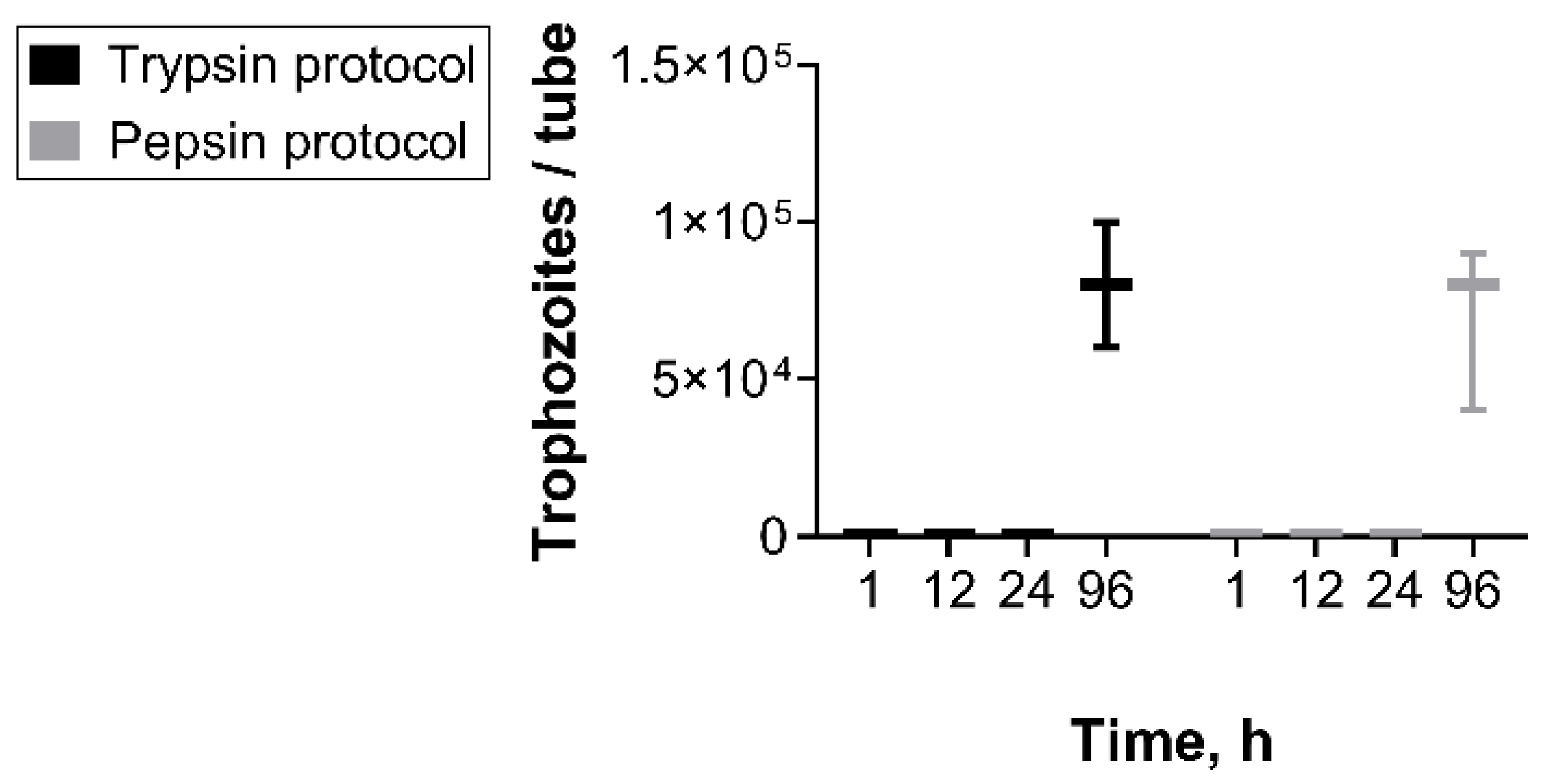
Former studies have proven that in vitro excystation of Giardia cysts is possible by using different protocols [24,25,31,32,33]. In our hands, the amount of trophozoites was first measurable 96 h after initiating excystation. We observed no significant differences between both protocols (Figure 2).
In most former studies, excystation was observed within one hour after completion of the initiating protocol [25,31,32,33], whereas in the current study, trophozoites were first detected 24 h post-initiation. Again, these differences may be related to the variability in Giardia strains utilized for the experimental procedures. Moreover, our cysts were obtained in vitro and not isolated from feces, as in previous studies [7,8,9,10,13,14], which may contribute to the variation in excystation efficacy. However, for a standardized protocol to assess cyst inactivation, we believe that a uniform source of cysts, i.e., cysts produced in vitro, is preferable. The storage of cysts can also affect excystation efficiency. Bertrand et al. [34] found no viable Giardia cysts 35 d after storage at 4 °C. On the other hand, storage is necessary for maturation of the cysts, resulting in a higher excystation rate [32,35,36]. We found that 10 d at 4 °C are suitable to promote excystation in our protocol. In contrast, other authors showed no need for maturation [24,37,38].
In further experiments, the protocol by Kane et al. [20] was selected for the evaluation of cyst inactivation by chemical disinfection, due to its highest efficiency in our hands. For enzymatic digestion, pepsin was used, similar to the protocol of Buchel et al. [37], since this method appeared very time-efficient, delivering equal numbers of trophozoites as trypsin digestion.
Giardia cysts can survive over months in the environment [5] and have even been found in conventionally treated drinking water [39,40,41]. While water chlorination is used to inactivate Giardia cysts, a high concentration, an adequate contact time and water temperatures around 25 °C are necessary for effective treatment [10,11]. Therefore, former authors focused on alternatives for water disinfection and showed that ultraviolet irradiation [42,43] or ozonation [12,44] are also capable techniques to prevent waterborne disease transmission. To our knowledge, little is known about the efficiency of chemical surface disinfectants against Giardia cysts. Chatterjee et al. [7] investigated the disinfecting efficacy of alcohol in hand sanitizers, concluding that the risk of spreading the disease may be minimized after use. Two studies investigated the effectiveness of vinegar [8] or a hypochlorous acid solution [9] to eliminate Giardia cysts in the context of sanitizing vegetables. Costa et al. [8] found 4% acetic acid at 21 °C to completely inactivate cysts within 60 min. The investigations of El Zawawy et al. [9] revealed a statistically significant reduction in Giardia cyst viability and infectivity after incubation in a sodium dichloroisocyanurate solution (NaDCC) for over 1 h.
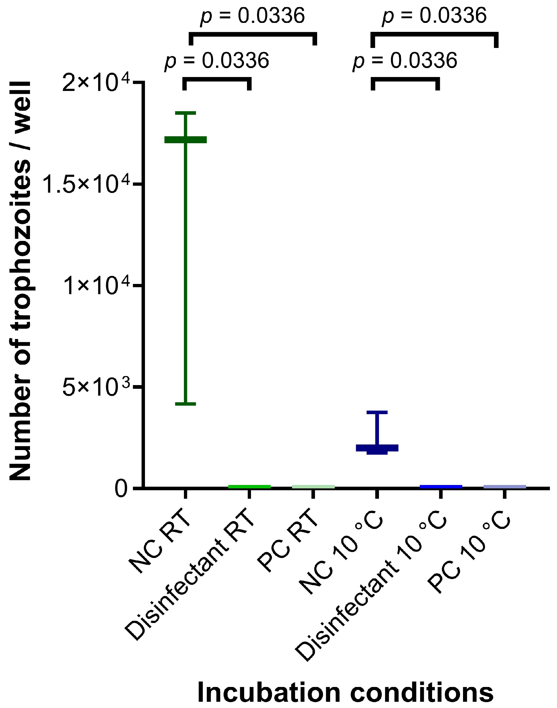
Here, we conducted the disinfectant test at RT and 10 °C. In the NC, the lower temperature resulted in clearly reduced trophozoite excystation as compared to the NC incubated at RT, although no significant difference could be calculated. H2O2 appeared suitable for application in the PC since we could not observe relevant trophozoite excystation in the PC irrespective of the incubation temperature. In contrast, the untreated cysts were able to excyst at a significant level in all replicates and experiments (p < 0.05; Figure 3). The commercial disinfectant included in the current inactivation assay successfully inactivated in vitro generated cysts and no trophozoites were found in the respective tubes.
Utilizing excystation for the assessment of Giardia cyst viability is an effective and reliable method to determine infectivity [13]. Despite the simplicity of conducting vital staining, it is prone to the overestimation of viability in comparison to excystation [34].
Silva and Sabogal-Paz [45] discussed various methods for the assessment of Giardia cyst viability, highlighting the risks of overestimation and false positives associated with vital staining and emphasizing that excystation techniques deliver a real-time assessment of cyst viability. Rousseau et al. [46] also reviewed different techniques to assess cyst viability and pointed out that an underestimation of inactivation efficacy, often related to viability assays, could be useful to ensure the safety of inactivation procedures. They also mentioned RT-qPCR-based assays that may be applied additionally to determine inactivation levels, which could be a perspective for improvement in the current method.
Due to their high resistance against chemical disinfectants, Cryptosporidium parvum oocysts [47] and Eimeria tenella oocysts [48,49] are established test organisms used to evaluate disinfectant effectiveness against protozoa. It appears that the resistance against chemical disinfectants of Giardia cysts is lower in comparison to C. parvum oocysts [50]. For instance, Adeyemo et al. [11] demonstrated that more Giardia cysts became non-viable at exposure to lower chlorine concentrations compared to Cryptosporidium oocysts and El Zawawy et al. [9] found Giardia cysts to be more susceptible to NaDCC than Cryptosporidium.

4. Conclusions

Although our results open perspectives for the development of a standardized inactivation procedure, further studies using other Giardia assemblages could widen the knowledge and lead to the optimization of cyst production and excystation. Refining the test readout could enhance the current protocol regarding sensitivity and test duration. Ultimately, such a disinfectant assay may facilitate the identification of chemicals that are suitable for eliminating cysts from contaminated surfaces, thus reducing the risk of disease transmission.

Author Contributions

Conceptualization, A.D.; methodology, M.K. and C.D.; validation, C.D.; formal analysis, M.K.; investigation, M.K. and C.D.; resources, A.D.; writing—original draft preparation, M.K.; writing—review and editing, C.D. and A.D.; visualization, M.K.; supervision, C.D. and A.D.; project administration, A.D.; funding acquisition, A.D. All authors have read and agreed to the published version of the manuscript.

Funding

This research was partly funded by ViPiBaX GmbH. Open Access publication was supported by the Open Access Publishing Fund of Leipzig University (Ticket#2025100110001056).

Institutional Review Board Statement

Not applicable.

Informed Consent Statement

Not applicable.

Data Availability Statement

The original contributions presented in the study are included in the article, further inquiries can be directed to the corresponding author.

Acknowledgments

The authors are most grateful for the support of Carmen Faso, Corina Wirdnam and the Institute of Cell Biology, Faculty of Science, University of Bern. We would also like to thank Uwe Müller, Institute of Immunology, Faculty of Veterinary Medicine, Leipzig University, and Malte Regelin for their support. Furthermore, we acknowledge the financial support of the Open Access Publishing Fund of Leipzig University.

Conflicts of Interest

Funding was provided in part by ViPiBaX GmbH. The funders had no role in the collection, analyses or interpretation of data; in the writing of the manuscript; or in the decision to publish the results.

Abbreviations

The following abbreviations are used in this manuscript:
AAL(+)-ascorbic acid
AECammonium iron(III) citrate
BBbovine bile
BSheat-inactivated donor bovine serum
cysteineL-Cysteine hydrochloride monohydrate
ddays
ddH2Odouble-distilled water
FCSheat-inactivated fetal calf serum
glucoseD(+)-Glucose
GMmodified TYI-S-33 medium
H2O230% hydrogen peroxide solution
KH2PO4potassium di-hydrogen phosphate
K2HPO4di-potassium hydrogen phosphate
LAL(+)-Lactic acid calcium salt hydrate
NaClsodium chloride
NaDCCsodium dichloroisocyanurate solution
NaOHsodium hydroxide
NCnegative control
PBbile extract porcine
PBSphosphate-buffered saline
PCpositive control
RTroom temperature
RT-qPCRreal-time quantitative polymerase chain reaction
WSHwater of standardized hardness

Appendix A

Table A1. Ingredients and pH values of encystation media in accordance with specific encystation protocols used in 100 mL.
IngredientsMedium#1Medium#2Medium#3Medium#4
Casein peptone, g1.81.81.81.8
D(+)-Glucose, g1.01.01.01.0
Yeast extract, g0.90.90.90.9
NaCl, mg200200200200
Cysteine, mg200200200200
K2HPO4, mg100100100100
KH2PO4, mg60606060
AA, mg20202020
AEC, mg2.282.282.282.28
FCS, mL10-1010
BS, mL-10--
BB, mg-2503755005001000
PB, mg250---
LA, mg55-0.55-
pH value7.87.87.87.8
AA = L(+)-ascorbic acid; AEC = ammonium iron(III) citrate; BB = bovine bile; BS = heat-inactivated donor bovine serum (Gibco, ThermoFisher Scientific); Cysteine = L-Cysteine hydrochloride monohydrate; FCS = heat-inactivated fetal calf serum; KH2PO4 = potassium di-hydrogen phosphate; K2HPO4 = di-potassium hydrogen phosphate; LA = L(+)-Lactic acid calcium salt hydrate (Carl Roth); NaCl = sodium chloride; PB = bile extract porcine (Sigma-Aldrich).

References

  1. Adam, R.D. Giardia duodenalis: Biology and Pathogenesis. Clin. Microbiol. Rev. 2021, 34, e0002419. [Google Scholar] [CrossRef]
  2. Fink, M.Y.; Singer, S.M. The Intersection of Immune Responses, Microbiota and Pathogenesis in Giardiasis. Trends Parasitol. 2017, 33, 901–913. [Google Scholar] [CrossRef]
  3. Robuffo, I.; Barassi, G.; Cordas, D. Inflammatory protein caspase-1 plays a crucial role in the immune response during microbial infections. Int. J. Infect. 2025, 9, 45–49. [Google Scholar]
  4. Sardinha-Silva, A.; Alves-Ferreira, E.V.C.; Grigg, M.E. Intestinal immune responses to commensal and pathogenic protozoa. Front. Immunol. 2022, 13, 963723. [Google Scholar] [CrossRef] [PubMed]
  5. Olson, M.E.; Goh, J.; Phillips, M.; Guselle, N.; McAllister, T.A. Giardia Cyst and Cryptosporidium Oocyst Survival in Water, Soil, and Cattle Feces. J. Environ. Qual. 1999, 28, 1991–1996. [Google Scholar] [CrossRef]
  6. Xu, H.; Jin, Y.; Wu, W.; Li, P.; Wang, L.; Li, N.; Feng, Y.; Xiao, L. Genotypes of Cryptosporidium spp., Enterocytozoon bieneusi and Giardia duodenalis in dogs and cats in Shanghai, China. Parasit. Vectors 2016, 9, 121. [Google Scholar] [CrossRef] [PubMed]
  7. Chatterjee, A.; Bandini, G.; Motari, E.; Samuelson, J. Ethanol and isopropanol in concentrations present in hand sanitizers sharply reduce excystation of Giardia and Entamoeba and eliminate oral infectivity of Giardia cysts in gerbils. Antimicrob. Agents Chemother. 2015, 59, 6749–6754. [Google Scholar] [CrossRef] [PubMed]
  8. Costa, A.O.; Thomaz-Soccol, V.; Paulino, R.C.; Alcântara de Castro, E. Effect of vinegar on the viability of Giardia duodenalis cysts. Int. J. Food Microbiol. 2009, 128, 510–512. [Google Scholar] [CrossRef]
  9. El Zawawy, L.A.; El-Said, D.; Ali, S.M.; Fathy, F.M. Disinfection efficacy of sodium dichloroisocyanurate (NADCC) against common food-borne intestinal protozoa. J. Egypt. Soc. Parasitol. 2010, 40, 165–185. [Google Scholar]
  10. Jarroll, E.L.; Bingham, A.K.; Meyer, E.A. Effect of chlorine on Giardia lamblia cyst viability. Appl. Environ. Microbiol. 1981, 41, 483–487. [Google Scholar] [CrossRef]
  11. Adeyemo, F.E.; Singh, G.; Reddy, P.; Bux, F.; Stenström, T.A. Efficiency of chlorine and UV in the inactivation of Cryptosporidium and Giardia in wastewater. PLoS ONE 2019, 14, e0216040. [Google Scholar] [CrossRef] [PubMed]
  12. Kondo Nakada, L.Y.; Urbano Dos Santos, L.; Guimarães, J.R. Pre-ozonation of surface water: An effective water treatment process to reduce the risk of infection by Giardia in drinking water. Environ. Pollut. 2020, 266, 115144. [Google Scholar] [CrossRef] [PubMed]
  13. Hoff, J.C.; Rice, E.W.; Schaefer, F.W. Comparison of animal infectivity and excystation as measures of Giardia muris cyst inactivation by chlorine. Appl. Environ. Microbiol. 1985, 50, 1115–1117. [Google Scholar] [CrossRef]
  14. Marchin, G.L.; Fina, L.R.; Lambert, J.L.; Fina, G.T. Effect of resin disinfectants-I3 and -I5 on Giardia muris and Giardia lamblia. Appl. Environ. Microbiol. 1983, 46, 965–969. [Google Scholar] [CrossRef]
  15. Feng, Y.; Xiao, L. Zoonotic potential and molecular epidemiology of Giardia species and giardiasis. Clin. Microbiol. Rev. 2011, 24, 110–140. [Google Scholar] [CrossRef]
  16. Ito, S.; Tsunoda, K.; Shimada, K.; Taki, T.; Matsui, T. Disinfectant effects of several chemicals against Toxoplasma oocysts. Nihon Juigaku Zasshi 1975, 37, 229–234. [Google Scholar] [CrossRef] [PubMed]
  17. Shahiduzzaman, M.; Dyachenko, V.; Keidel, J.; Schmäschke, R.; Daugschies, A. Combination of cell culture and quantitative PCR (cc-qPCR) to assess disinfectants efficacy on Cryptosporidium oocysts under standardized conditions. Vet. Parasitol. 2010, 167, 43–49. [Google Scholar] [CrossRef]
  18. Dresely, I.; Daugschies, A.; Lendner, M. Establishment of a germ carrier assay to assess disinfectant efficacy against oocysts of coccidian parasites. Parasitol. Res. 2015, 114, 273–281. [Google Scholar] [CrossRef]
  19. Delling, C.; Lendner, M.; Müller, U.; Daugschies, A. Improvement of in vitro evaluation of chemical disinfectants for efficacy on Cryptosporidium parvum oocysts. Vet. Parasitol. 2017, 245, 5–13. [Google Scholar] [CrossRef]
  20. Kane, A.V.; Ward, H.D.; Keusch, G.T.; Pereira, M.E. In vitro encystation of Giardia lamblia: Large-scale production of in vitro cysts and strain and clone differences in encystation efficiency. J. Parasitol. 1991, 77, 974–981. [Google Scholar] [CrossRef]
  21. Hausen, M.A.; Freitas, J.C.M.; Monteiro-Leal, L.H. The effects of metronidazole and furazolidone during Giardia differentiation into cysts. Exp. Parasitol. 2006, 113, 135–141. [Google Scholar] [CrossRef] [PubMed]
  22. Davids, B.J.; Gillin, F.D. Methods for Giardia Culture, Cryopreservation, Encystation, and Excystation In Vitro. In Giardia; Luján, H.D., Svärd, S., Eds.; Springer Vienna: Vienna, Austria, 2011; pp. 381–394. ISBN 978-3-7091-0197-1. [Google Scholar]
  23. Einarsson, E.; Troell, K.; Hoeppner, M.P.; Grabherr, M.; Ribacke, U.; Svärd, S.G. Coordinated Changes in Gene Expression Throughout Encystation of Giardia intestinalis. PLoS Negl. Trop. Dis. 2016, 10, e0004571. [Google Scholar] [CrossRef] [PubMed]
  24. Hausen, M.A.; Oliveira, R.P.d.; Gadelha, A.P.R.; Campanati, L.; de Carvalho, J.J.; de Carvalho, L.; Barbosa, H.S. Giardia lamblia: A report of drug effects under cell differentiation. Parasitol. Res. 2009, 105, 789–796. [Google Scholar] [CrossRef]
  25. Bingham, A.K.; Meyer, E.A. Giardia excystation can be induced in vitro in acidic solutions. Nature 1979, 277, 301–302. [Google Scholar] [CrossRef]
  26. Keister, D.B. Axenic culture of Giardia lamblia in TYI-S-33 medium supplemented with bile. Trans. R. Soc. Trop. Med. Hyg. 1983, 77, 487–488. [Google Scholar] [CrossRef]
  27. Touz, M.C.; Nores, M.J.; Slavin, I.; Piacenza, L.; Acosta, D.; Carmona, C.; Luján, H.D. Membrane-associated dipeptidyl peptidase IV is involved in encystation-specific gene expression during Giardia differentiation. Biochem. J. 2002, 364, 703–710. [Google Scholar] [CrossRef]
  28. Faso, C.; Bischof, S.; Hehl, A.B. The proteome landscape of Giardia lamblia encystation. PLoS ONE 2013, 8, e83207. [Google Scholar] [CrossRef]
  29. Hehl, A.B.; Marti, M.; Köhler, P. Stage-Specific Expression and Targeting of Cyst Wall Protein–Green Fluorescent Protein Chimeras in Giardia. Mol. Biol. Cell 2000, 11, 1789–1800. [Google Scholar] [CrossRef]
  30. Gillin, F.D.; Boucher, S.E.; Rossi, S.S.; Reiner, D.S. Giardia lamblia: The roles of bile, lactic acid, and pH in the completion of the life cycle in vitro. Exp. Parasitol. 1989, 69, 164–174. [Google Scholar] [CrossRef]
  31. Boucher, S.E.; Gillin, F.D. Excystation of in vitro-derived Giardia lamblia cysts. Infect. Immun. 1990, 58, 3516–3522. [Google Scholar] [CrossRef] [PubMed]
  32. Rice, E.W.; Schaefer, F.W. Improved in vitro excystation procedure for Giardia lamblia cysts. J. Clin. Microbiol. 1981, 14, 709–710. [Google Scholar] [CrossRef]
  33. Feely, D.E.; Gardner, M.D.; Hardin, E.L. Excystation of Giardia muris induced by a phosphate-bicarbonate medium: Localization of acid phosphatase. J. Parasitol. 1991, 77, 441–448. [Google Scholar] [CrossRef] [PubMed]
  34. Bertrand, I.; Maux, M.; Helmi, K.; Hoffmann, L.; Schwartzbrod, J.; Cauchie, H.-M. Quantification of Giardia transcripts during in vitro excystation: Interest for the estimation of cyst viability. Water Res. 2009, 43, 2728–2738. [Google Scholar] [CrossRef]
  35. Bingham, A.K.; Jarroll, E.L.; Meyer, E.A.; Radulescu, S. Giardia sp.: Physical factors of excystation in vitro, and excystation vs eosin exclusion as determinants of viability. Exp. Parasitol. 1979, 47, 284–291. [Google Scholar] [CrossRef]
  36. Hautus, M.A.; Kortbeek, L.M.; Vetter, J.C.; Laarman, J.J. In vitro excystation and subsequent axenic growth of Giardia lamblia. Trans. R. Soc. Trop. Med. Hyg. 1988, 82, 858–861. [Google Scholar] [CrossRef]
  37. Buchel, L.-A.; Gorenflot, A.; Chochillon, C.; Savel, J.; Gobert, J.-G. In vitro Excystation of Giardia from Humans: A Scanning Electron Microscopy Study. J. Parasitol. 1987, 73, 487. [Google Scholar] [CrossRef]
  38. Smith, A.J.; Lauwaet, T.; Davids, B.J.; Gillin, F.D. Giardia lamblia Nek1 and Nek2 kinases affect mitosis and excystation. Int. J. Parasitol. 2012, 42, 411–419. [Google Scholar] [CrossRef] [PubMed]
  39. Betancourt, W.Q.; Mena, K.D. Assessment of waterborne protozoan passage through conventional drinking water treatment process in Venezuela. J. Water Health 2012, 10, 324–336. [Google Scholar] [CrossRef]
  40. Castro-Hermida, J.A.; González-Warleta, M.; Mezo, M. Cryptosporidium spp. and Giardia duodenalis as pathogenic contaminants of water in Galicia, Spain: The need for safe drinking water. Int. J. Hyg. Environ. Health 2015, 218, 132–138. [Google Scholar] [CrossRef]
  41. Ramo, A.; Del Cacho, E.; Sánchez-Acedo, C.; Quílez, J. Occurrence of Cryptosporidium and Giardia in raw and finished drinking water in north-eastern Spain. Sci. Total Environ. 2017, 580, 1007–1013. [Google Scholar] [CrossRef] [PubMed]
  42. Shin, G.-A.; Linden, K.G.; Faubert, G. Inactivation of Giardia lamblia cysts by polychromatic UV. Lett. Appl. Microbiol. 2009, 48, 790–792. [Google Scholar] [CrossRef]
  43. Linden, K.G.; Shin, G.-A.; Faubert, G.; Cairns, W.; Sobsey, M.D. UV disinfection of Giardia lamblia cysts in water. Environ. Sci. Technol. 2002, 36, 2519–2522. [Google Scholar] [CrossRef] [PubMed]
  44. Li, Y.; Smith, D.W.; Belosevic, M. Morphological changes of Giardia lamblia cysts after treatment with ozone and chlorine. J. Environ. Eng. Sci. 2004, 3, 495–506. [Google Scholar] [CrossRef]
  45. Sammarro Silva, K.J.; Sabogal-Paz, L.P. Cryptosporidium spp. and Giardia spp. (oo)cysts as target-organisms in sanitation and environmental monitoring: A review in microscopy-based viability assays. Water Res. 2021, 189, 116590. [Google Scholar] [CrossRef]
  46. Rousseau, A.; La Carbona, S.; Dumètre, A.; Robertson, L.J.; Gargala, G.; Escotte-Binet, S.; Favennec, L.; Villena, I.; Gérard, C.; Aubert, D. Assessing viability and infectivity of foodborne and waterborne stages (cysts/oocysts) of Giardia duodenalis, Cryptosporidium spp., and Toxoplasma gondii: A review of methods. Parasite 2018, 25, 14. [Google Scholar] [CrossRef] [PubMed]
  47. Delling, C.; Holzhausen, I.; Daugschies, A.; Lendner, M. Inactivation of Cryptosporidium parvum under laboratory conditions. Parasitol. Res. 2016, 115, 863–866. [Google Scholar] [CrossRef] [PubMed]
  48. Blake, D.P.; Tomley, F.M. Securing poultry production from the ever-present Eimeria challenge. Trends Parasitol. 2014, 30, 12–19. [Google Scholar] [CrossRef] [PubMed]
  49. You, M.-J. Suppression of Eimeria tenella sporulation by disinfectants. Korean J. Parasitol. 2014, 52, 435–438. [Google Scholar] [CrossRef]
  50. Korich, D.G.; Mead, J.R.; Madore, M.S.; Sinclair, N.A.; Sterling, C.R. Effects of ozone, chlorine dioxide, chlorine, and monochloramine on Cryptosporidium parvum oocyst viability. Appl. Environ. Microbiol. 1990, 56, 1423–1428. [Google Scholar] [CrossRef]
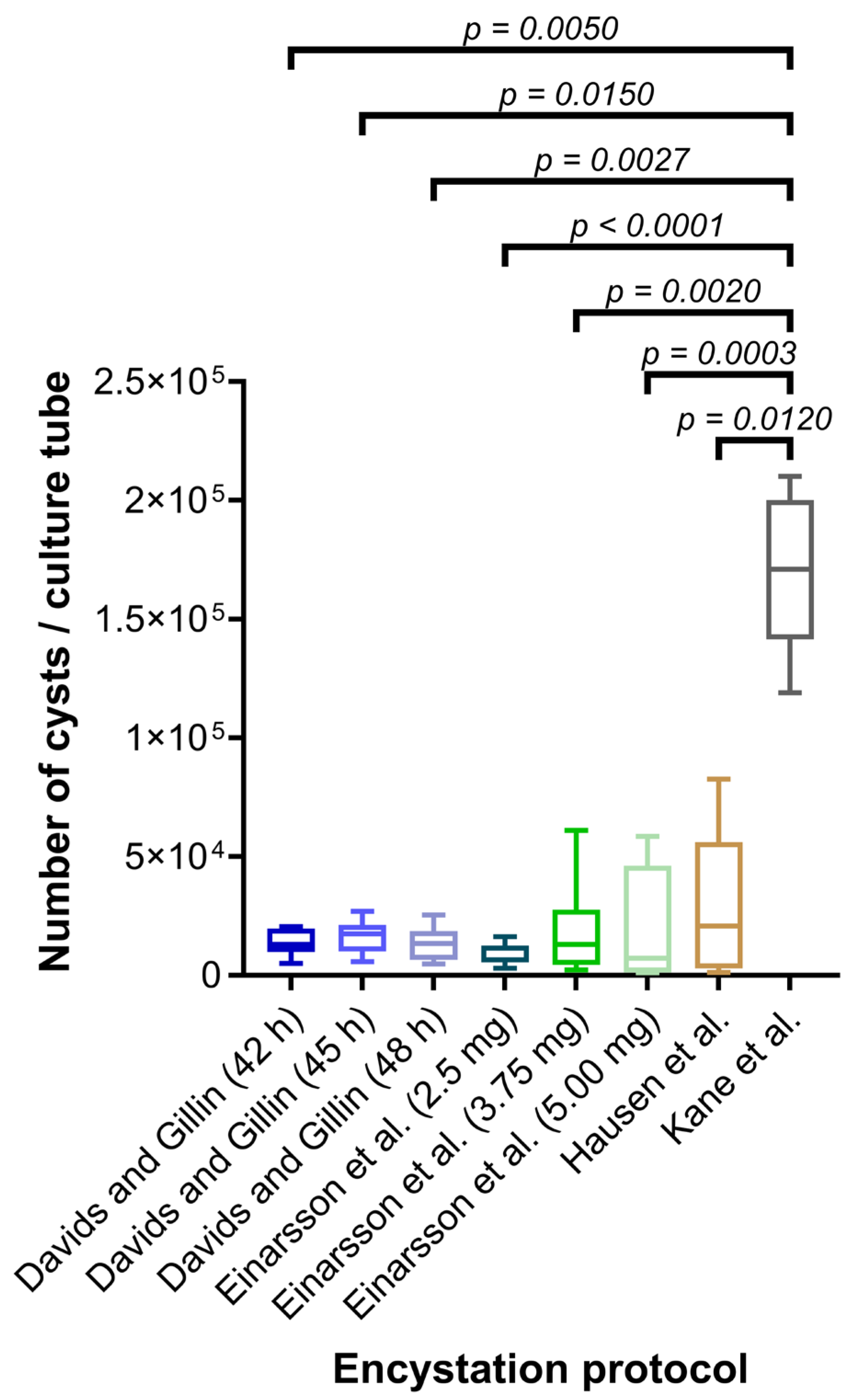
Figure 1. Number of Giardia cysts per culture tube after conducting different encystation protocols. Davids and Gillin [22]; Einarsson et al. [23]; Hausen et al. [21]; Kane et al. [20].
Hygiene 05 00054 g001
Figure 2. Total number of Giardia trophozoites per tube at several measuring points after stimulation of 8 × 10⁴ Giardia cysts for excystation conducting two protocols.
Hygiene 05 00054 g002
Figure 3. Number of Giardia trophozoites per well after disinfection; NC = negative control, cysts in WSH; PC = positive control, cysts treated with 30% hydrogen peroxide solution; RT = room temperature.
Hygiene 05 00054 g003
Disclaimer/Publisher’s Note: The statements, opinions and data contained in all publications are solely those of the individual author(s) and contributor(s) and not of MDPI and/or the editor(s). MDPI and/or the editor(s) disclaim responsibility for any injury to people or property resulting from any ideas, methods, instructions or products referred to in the content.