Review

Macroalgal Diseases: Exploring Biology, Pathogenesis, and Management Strategies

by
Damiano Spagnuolo
* and
Giuseppa Genovese
Phycological Lab, Department of Chemical, Biological, Pharmaceutical and Environmental Sciences, University of Messina, Salita Sperone 31, 98166 Messina, Italy
*
Author to whom correspondence should be addressed.
Phycology 2024, 4(3), 450-464; https://doi.org/10.3390/phycology4030026
Submission received: 29 July 2024 / Revised: 3 September 2024 / Accepted: 6 September 2024 / Published: 10 September 2024

Abstract

:
The global seaweed market is expected to reach USD 17.8 billion by 2032, fuelled by growing demand for sustainable and healthy food solutions and expanding applications in agriculture and aquaculture. However, this rapid growth poses significant challenges, particularly in managing diseases that often establish themselves in intensive macroalgal culture facilities. Red rot disease, Olpidiopsis, and green spot disease often affect marine macroalgae species of high commercial interest, as seen in Pyropia/Porphyra as has already happened for “ice-ice” malaise on Kappaphycus, causing huge economic losses. These diseases are caused by infectious agents that find their place in extreme environmental conditions, such as those characterized by sudden changes in temperature and pollution. Despite technological advances aimed at monitoring the well-being of cultivated seaweed, discrepancies between regions’ technological capabilities and species vulnerability exacerbate management difficulties. This review provides an overview of diseases prevalent among marine algae, their impact on aquaculture, and the effectiveness of currently adopted treatments. This study highlights the need to improve disease management strategies and highlights the importance of understanding host–pathogen interactions in order to mitigate future epidemics.

1. Introduction

The global seaweed market is expected to reach USD 17.8 billion by 2032, driven by growing demand for healthy and sustainable foods, expanding applications in the food and beverage industry and growing interest in agriculture and sustainable aquaculture [1,2]. The search for sustainable foods will determine the increase in the intensive cultivation of macroalgae, and consequently, the problems linked to the management of the plants and, above all, the pathologies that settle there will also increase. In fact, in intensive macroalgae cultivation, the costs required for the prevention and treatment of diseases are estimated to take up 30% to 50% of a company’s budget [3], and up to half of the biomass produced can be lost [4]. Currently, advanced quality control technologies are often used for cultivation [5], but there is often a discrepancy in the technological progress of nations and the cultivated species that is not closely related to the market price of algae or derived products [6].
Cultured macroalgae are frequently affected by numerous parasites that cause diseases that alter the morphology of the thallus, but despite this, this topic is largely under-documented. Parasites are increasingly considered key players in natural ecosystems, but they are also one of the most serious economic and environmental threats to macroalgae aquaculture.
There are two types of diseases in algae: infectious and non-infectious diseases. The former type involves a transmissible infectious agent (bacteria, fungi, viruses, etc.), while the latter type is induced by physiological factors such as extreme temperatures, salinity, light intensity, or pollution [7]. Non-infectious diseases are caused by unfavourable environmental conditions; anthropogenic activities such as heavy metal pollution can also cause diseases in marine algae. The symptoms caused by non-infectious diseases are numerous: high temperature and light intensity, for example, can lead to whitening and hardening of the thallus. But above all, algae stressed and weakened by unfavourable environmental conditions are more susceptible to infectious diseases. Very often, there is a synergy between the infectious agent and environmental conditions [8]; in algae, in fact, the pathogen–disease concept is not always appropriate, and pathologies often worsen on farms, where they become real technopathies. Added to this are grazing and nutrient deficiencies, which too often are accompanied by other disorders.
The marine environment is rich in life, and the microbial part has long been underestimated [9], even if some of this is pathogenic and very diversified, sometimes playing a fundamental role in natural ecosystems [10]. Research aimed at pathogens is driven by the impact they have on human health; therefore, the interest in algal diseases and their economic impact is becoming an important driver for research, especially in Europe, where interest in macroalgae is growing exponentially [11].
The biological, physical, and chemical properties of the surface of macroalgae certainly play a role in structuring the associated microbial community and its metabolic activity, subsequently influencing its state of health and the attack of pathogens. Several factors that influence the surface environment of macroalgae include algal metabolites, the resident microbial community and their secondary metabolites, and the physicochemical conditions that the surface of the thallus is subjected to, which can include carbon dioxide and oxygen, which can influence the surface pH and the overall microbial community [12]. Many of these parameters are subject to daily and seasonal modulations; cultivated algae may have altered parameters and therefore altered holobionts, and this could be one of the factors triggering diseases in cultivation. Of note, bacteria that enter into a stable association with a macroalgal host must therefore possess adaptive traits that reflect these niche conditions [12].
Macroalgae are under constant colonization pressure from billions of microorganisms present in the surrounding seawater, some of which are potential pathogens [10]. To defend themselves from harmful colonizers, macroalgae require general or specific strategies to control microbial growth. Macroalgae lack a cellular adaptive immune response but have defensive capabilities that fall into two broad categories: constitutive ones, those which are always expressed and do not depend on the qualitative and quantitative presence of the microbial community, and regulated ones, which are activated from “tissue” damage and cause oxidative bursts or hypersensitive responses [13]. Macroalgae diseases have been linked to bacteria, viruses, fungi, and other eukaryotes. However, the specific role of bacterial or fungal pathogens in these diseases remains largely unclear. This lack of understanding is partly due to the difficulty in distinguishing true pathogens, especially from saprophytes or other secondary colonizers that benefit from damaging macroalgae. All these diseases are exacerbated by global warming and intensive high-density biomass farming (e.g., Ulva spp. in Europe and Eucheuma spp. in Africa, especially in Tanzania), and the expansion of aquaculture increases the impact of these diseases, potentially leading to economic losses in several areas.
This review aims to illustrate an updated overview of the most known pathologies and pathogens related to macroalgae cultivation to improve crop management strategies.

2. Pyropia/Porphyra Species

Genera Pyropia/Porphyra (Bangiales) is among the most valuable aquaculture algae in the world and has been cultivated in Japan, China, and Korea for thousands of years and, recently, also in many other countries of the world. In Japan, it is most often used as nori (known in China as “zicai” and in Korea as “gim”), an ingredient of sushi. In Wales and England, it is used in a traditional dish, laver. In the last decade, many studies have been conducted on its nutritional value and pharmaceutical properties; for these reasons, these cultivations represent one of the most advanced algal industries, with a market value of over EUR 2 billion per year (EUR 2.5 billion in 2017). Nori farms, in terms of appearance and commercial approach, look more like crop agriculture areas rather than aquaculture areas, and nori was one of the first real macroalgae studied in “Phyconomist”, after Eucheuma/Kappaphycus [14]. This economic interest shifted the attention to production, and various pathologies have therefore been discovered. About more than ten/fifteen different diseases attack nori farms, including bacteria, viruses, and fungal-like organisms [3,14,15]. Very often, some pathologies can be seen to be “overlapping”; up to a few decades ago, various causes were attributed to the same pathology. With the advent of molecular analysis, these evaluation errors have been reduced, and such analysis techniques have been used to clarify the three major diseases listed above. Frequently, the pathologies are caused by Pythium sp., Olpidiopsis sp. (Oomycetes), and the virus “PyroV1” (green rot disease). Among bacterial agents, there are Flavobacterium spp. In Korea, where Pyropia/Porphyra cultivation has recently rapidly expanded, new disease outbreaks are reported every year, and they have reduced the crop output by around 20% in certain areas, causing a general decrease in product quality and considerably lowering the market value of harvested Pyropia blades [3,8].

2.1. Red Rot Disease (RRD)

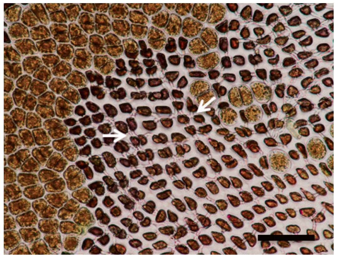
Red rot disease (RRD) is primarily caused by the necrotrophic oomycete Pythium porphyrae [3,8,16] and is the most widely studied disease of the gametophytic generation of Porphyra spp. Like the systematics of Bangiales [17], the systematics of fungi are also constantly evolving [18]. Red rot disease was first reported in Pyropia tenera in Japan by Arasaki [19], and its pathogenesis was characterized in Pyropia spp. [20]. The first symptoms that can be recognized are characterized by small red patches or bleached parts (a few micrometres in diameter); between 2 and 3 days after symptom onset, their natural reddish-brown colour becomes violet-red, before they turn green and, in the end, colourless, and the blades degenerate completely (Figure 1, Figure 2 and Figure 3). Mycelium of Pythium colonize the host–cell intracellularly, killing them and progressively forming the distinct patches described before. The infection spreads quickly to other areas on the blade, mainly via cell-to-cell spreading, and dead host tissue deteriorates, forming numerous small holes; the holes could “merge” into bigger holes, ultimately disintegrating the entire blade. To date, Pythium porphyrae and Pythium chondricola have been reported as the main causative agents of RRD [21,22], although some studies suggest that a fungus of the genus Alternaria is another causative agent of this disease [23]. In Japan, around 20% of biomass is lost due to RRD [24]. Pathogen zoospores’ adherence to the thalli is promoted by conditions characterized by a high temperature, low salinity, and the absence of free-change tide [25], and pathogen zoospores infect the thallus. A very modern approach was suggested in a recent study about exploiting the biocontrol of two strains of Pseudoalteromonas piscicida to fight against red rot disease in Pyropia yezoensis. Both strains inhibited the growth of the pathogen Pythium porphyrae without harming the algae. P6, combined with air drying, showed significant disease inhibition, suggesting that it is an effective method for controlling red rot in Pyropia [26].

2.2. Olpidiopsis Disease (OD), Chytrid Disease (CD), Olpidiopsis-Blight (O-B)

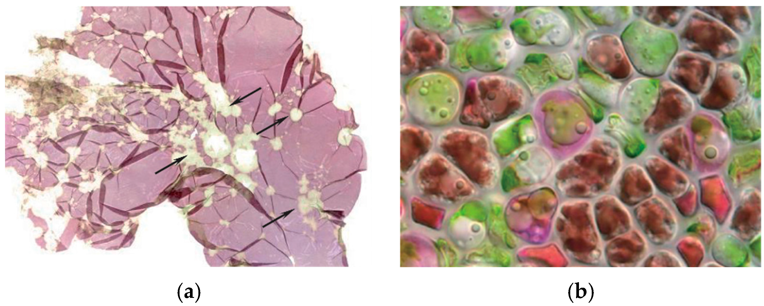
Infections caused by organisms of the Olpidiopsis genus are more aggressive than RRD [28]. Olpidiopsis Disease (OD) (often call Chytrid Disease or Olpidiopsis-Blight) OD is caused by the attack of obligate endoparasitic oomycetes and has been reported in China, Korea, and Japan [29]. In the last decade, it has also been reported in Europe, and the classification of the genus has been revised [28,30]. Symptoms initially manifest as distinct blanched areas on the blades, which progress to greenish lesions as the disease spreads. The infection route is like that of Pythium, but the spread seems more disordered on the surface of the thallus. The infection process begins when encysted Olpidiopsis zoospores attach to the surface of Pyropia/Porphyra and produce thin germ tubes that penetrate the host’s cell walls. Subsequently, the parasite forms multinucleated spherical thalli, which, after 2–3 days, develop into zoosporangia, which release the zoospores. The rotting of the “tissue” in the infected areas promotes the death of the entire blade (Figure 4). Many strategies have been adopted to control this pathology, but their effectiveness is not always certain [31]. A recent study explored non-acidic alternatives to control these pathogens. Among the calcium salts tested, calcium propionate emerged as the most effective. When Pyropia blades were briefly immersed in calcium propionate solutions, both the infection rate and the spread of oomycetes were significantly reduced [32].

2.3. Green Spot Disease

Green spot disease of the genera Pyropia/Porphyra was reported more than twenty years ago in Korea. However, to date, there have been no detailed studies describing its specific symptoms and infective agents. Green spot disease in Pyropia is identified by the presence of small, distinct lesions on the blade, characterized by broad green borders. These lesions can appear anywhere on the blade and are often accompanied by severe bacterial contamination. As the lesions grow and coalesce, slimy rot occurs as the host tissue breaks down (Figure 5). Subsequently, many types of Gram-negative bacteria attach to the surface of the thallus, such as Flavo-bacterium sp., Pseudoalteromonas sp., and Vibrio sp.; this has historically led to the incorrect attribution of this disease to bacterial agents [3]. Current understanding suggests that bacterial invasion should be considered secondary to the viral infection, as these bacteria act as opportunistic pathogens. PyroV1 is considered responsible for green spot disease [33].

2.4. Cyanobacteria Felt

Green spot disease and cyanobacterial infestations often occur at the same time on Pyropia, causing their symptoms to overlap. The cell wall structure of Pyropia cells loosens and degenerates in the presence of abundant bacteria and cyanobacteria on the surface, forming a long distinct “bristle” visible on the thallus [15]. Both single cells and large colonies of mucilage-secreting coccoid cyanobacteria are observable with TEM.

2.5. Diatom Felt

“Diatom felt” on Pyropia/Porphyra appears as a distinct brown fringe across the entire surface of the blade. When touched with the fingers, detached diatoms can be seen, so this disease can be easily recognized by non-professionals. Pyropia growth can be seriously affected by epiphytic diatoms, as they shade light, compete for nutrients, and cause bleaching of macroalgae thalli [3]. Infected Pyropia has a distinct, unpleasant, earthy odour. This disease does not cause serious production losses but directly affects the price of raw materials, creating concern among farmers.

2.6. Genetic Toolkits against Common Diseases in Porphyra/Pyropia Species

In recent years, we have introduced the diseases with the highest incidence into Porphyra farms, and efforts have been made to develop toolkits, including molecular ones, for faster identification to prevent disease progression (Table 1) [33]. The rapid expansion of seaweed aquaculture has resulted in sudden ecological consequences, including epidemics, the introduction of non-indigenous pathogens, and a reduction in the genetic diversity of native seaweed stocks. Thus, to overcome these challenges, it is crucial to understand the molecular basis of host–pathogen interactions and possible resistance. Porphyra/Pyropia have a large pool of defence genes, and the expression of these genes is differentially regulated during infection in response to pathogen types [34].

3. Kappaphycus sp. and Eucheuma sp.

3.1. “Ice-Ice” Malaise

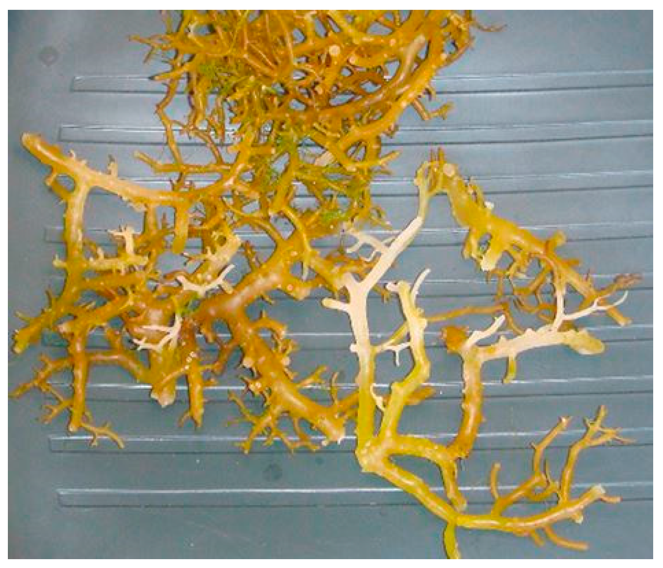
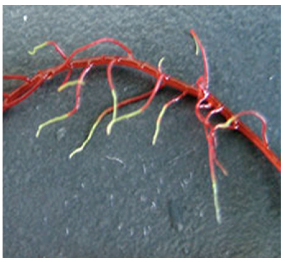
Kappaphycus alvarezii cultivations in the Philippines have always highlighted a phenomenon known as “Ice-ice” [4]. The whitening effect (Figure 6) is mainly caused by a defence response mechanism of the algae, triggered by the presence of halogenated volatile organic compounds which cause an oxidative burst. Stressors release H2O2 (hydrogen peroxide), which bleaches thalli after prolonged exposure [4,6]. Members of the Cytophaga-Flavobacterium-Bacteroides group and various genera of marine fungi, such as Aspergillus sp. and Phoma sp., were isolated from affected thalli. However, their main role is to exert a secondary effect, which occurs after the macroalgae have been weakened by physiological stresses. This secondary infection manifests itself in complete necrosis of the thalli, caused by both bacteria and fungi present in the affected algae. These microbial agents decompose the fibrillar component of the cell wall and use the amorphous part as the primary carbon source [4].

3.2. Goose Bumps Disease

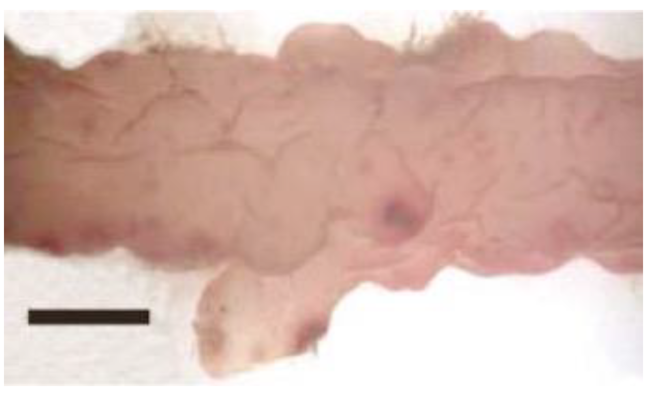
The disease ‘Goose bumps’, unlike “ice-ice”, is properly classified as a pathology. The initial obvious symptom is some “black pimples” appearing on the surface of the thallus of Kappaphycus/Eucheuma (Rhodophyta) and on the sites of sedimentation of the spores of the filamentous red alga Neosiphonia spp. (Figure 7). Germinating spores penetrate the cortical and medullary layers of host algae and develop into an endophytic filamentous algae (EFA) infestation [4]. Infestations by Kappaphycus spp. cultivation sites were first recorded in 1994 in the Philippines. The disease was initially erroneously ascribed to a red algae Polysiphonia sp. epiphytic outbreak (due to incorrect identification), only later being confirmed as Neosiphonia apiculata. Other Neosiphonia species have since been implicated; this genus is a genus with common epiphytic (but necessarily endophytic) species of brown algae Sargassum spp. [30] and may have been transferred by drift Sargassum being involved in the cultivation of Kappaphycus by epiphyte transfer [39,40,41], where it was very successful in carrying out attachment and subsequent reproduction through copious spore production under favourable conditions [42].

4. Gracilaria sp.

Like Kappaphycus cultivation companies, Gracilaria production also suffers from the attack of epiphytes on the thalli, which hinders productivity and reduces the market value of the crop. Overall, most of these epiphytes also belong to the order of red algae Ceramiales, including Ceramium spp. and Polysiphonia spp. [43]. The diseases affecting Gracilaria spp. are not clearly defined, and no attention has been paid to specific symptoms. However, the correlation between pathogens and mortality rate of Gracilaria gracilis in production facilities have led to the establishment of positive correlations between disease symptoms (“white tip” and “rotten thallus” syndromes, Figure 8) [44,45] and the presence of epiphytic agarolithic [46]. Among these, marine bacteria species of genus Pseudoalteromonas are particularly widespread and are the cause of “whitening stripe disease” in Gracilaria cordicata. Recently, many studies have provided a list of confirmed and presumed pathogenic bacteria of macroalgae, some of which attack Gracilaria thalli (Table 2). The disease hinders or stunts growth, shortens shelf life, and causes morphological deformities, making it difficult to market affected plants. Infected thalli show unusual lesions or small bump-like structures (galls) on the surface, and the thallus appears to have “witch’s broom”-like branches at the end.

5. Laminaria sp., Saccharina sp., and Undaria sp.

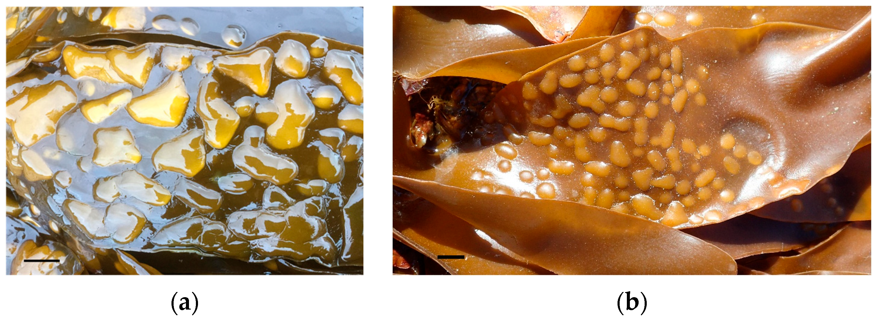
Brown algae are include several edible species, some of which also represent an important source of alginates. Species belonging to the genera Saccharina, Laminaria, and Undaria are commonly used for human nutrition and therefore subject to massive cultivation. The growth conditions to which they are subjected frequently expose them to alterations attributable to different causes. “Technopathologies” closely related to cultivation conditions are often found [48]. An example is the blister disease caused by a decrease in salinity (Figure 9). As regards real diseases, i.e., those caused by a pathogen, research on them, as is also the case for other groups of algae, has still made few steps. Among the presumed pathogens are some endophytic filamentous algae that, in addition to fungi, bacteria, and viruses, can have a negative impact on algal growth. For example, the most frequent endophytes are the genera Ochrophyta, Laminariocolax, and Laminarionema, which could be more widespread than believed among cultivated populations. Preliminary investigations have led to the conclusion that these endophytes invade the algal thallus early, causing significant disruptions to morphogenesis [49].

6. Ulva sp.

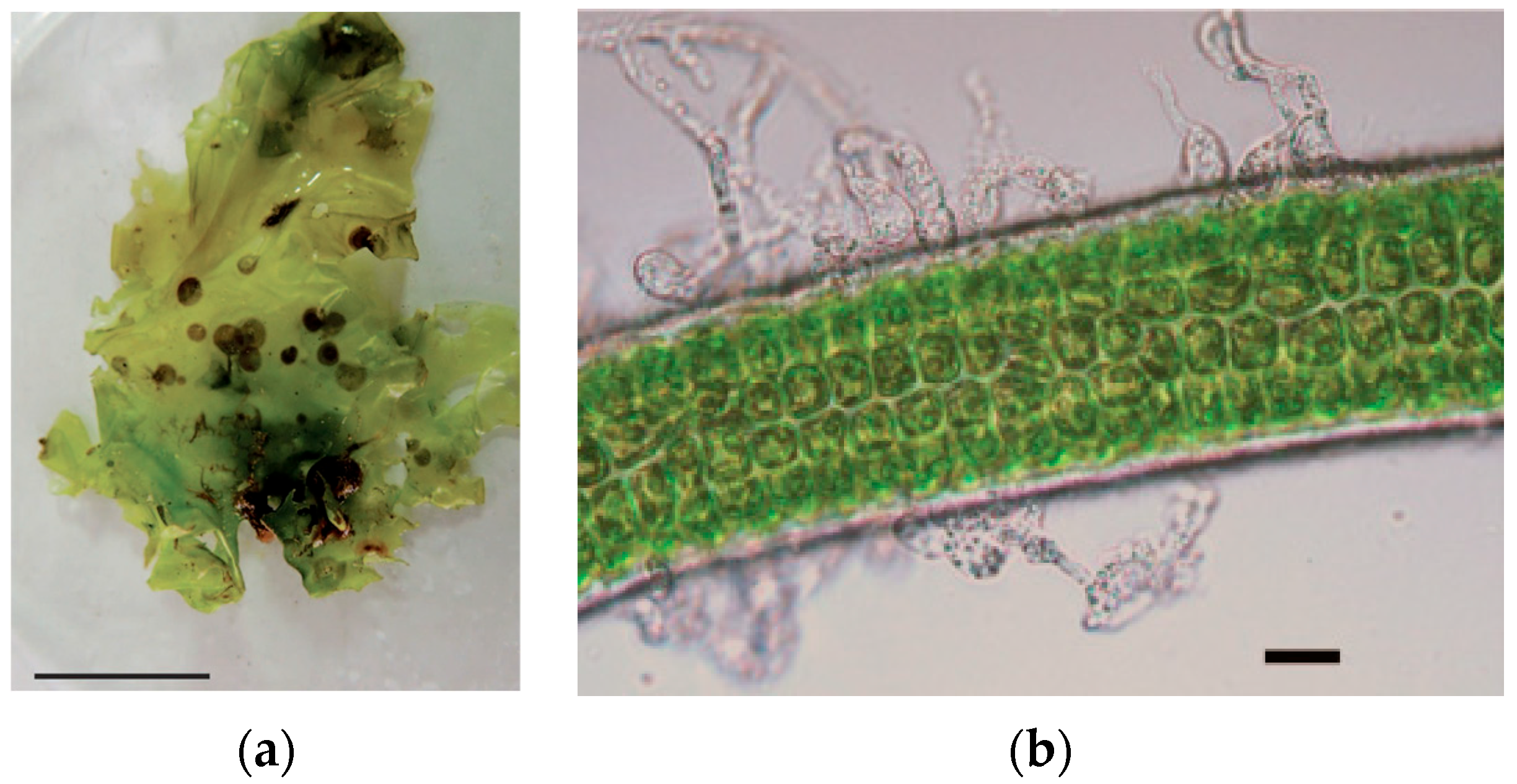
The genus Ulva includes green macroalgae widely used for human consumption. They are harvested worldwide from both natural populations and mass cultivation systems. Research on natural populations has revealed that species belonging to the genus Myrionema (Phaeophyceae, Ochrophyta) can be common epiphytes on Ulva thalli [50,51]. Their presence causes the onset of brown spots on the blade that can extend over the entire surface (Figure 10a). The presence of epiphytes leads to a reduction in the growth rates of green algae, probably also due to competition for nutrients. This leads to a significant reduction in the market value of the blade, both due to the appearance of the thalli and their size, consequently causing concern for commercial producers. Another pathogen found on Ulva spp. thalli is the mycelium of Pythium sp. This pathogenic parasite, being also present on Porphyra/Pyropia thalli and on terrestrial plants, is certainly a little more well studied, even if the mechanism of action remains uncertain (Figure 10b) [52].

7. A Case Study on Kappaphycus: What We Can Learn from Past Mistakes?

Regarding the diseases and their influence on cultivation, we need to take into consideration the worldwide production of Kappaphycus. This type of cultivation is spread worldwide, especially in many developing countries, wherein some regional cultivation and harvesting areas represent a substantial part of the economy (Table 3). In these developing countries, after a grace period of a few years, diseases typically worsen due to intensification of cultivation, sometimes leading to the collapse of the local industry (like the shrimp industry 20 years ago in South America). All of this happens for several reasons:
  • The main one is the neglect of cultivation rules (which are much more careful for the cultivation of terrestrial plants, agronomy, etc.), which occurs very often because you do not have the knowledge of certain pathologies or do not know the symptoms [53].
  • Another reason is the low genetic variation and loss of strain vigour, which has further ramifications in that the biomass becomes susceptible to pathogens, diseases, and epi- or endophyte infestations [6].
  • A third reason is lack of development in commercial utilization of local seaweed biodiversity leading to seemingly unnecessary introductions of non-indigenous Eucheumatoids and their unfettered expansion into new farming areas. Some of these introductions have caused serious environmental issues, such as an increased prevalence of invasive organisms. Also, a lot of the time, it is difficult to establish correlations with pathogens [54].
  • The final reason is the failure to innovate new techniques for Eucheumatoid farming, and the indigenous utilization of raw materials merely fuels the expansion of commercial operations through the unregulated transfer of seedlings to new farming areas to meet increasing global demands [14].
The new industries, for example, those in Europe, must take care of all of these things just to not commit the same errors. Environmental and cultivation policies must be carefully monitored, implementing insights from the study of alien species (not only those cultivated), attempting to promote a common policy, as has been seen in fields like agronomy. As such, the new term “Phyconomy” [14,54,55,56] is hereby coined to describe a general concept that embraces large-scale, sustainable seaweed farming for economic benefit in coastal waters. Phyconomic lessons learned from the successful/unsuccessful mass cultivation of red seaweeds are guidelines which can be applied to technology transfer and capacity building for other forms of commercial marine macroalgal production.

8. Conclusions

The study of diseases affecting seaweeds reveals important insights and poses challenges that operators in the sector must address. Cultivated species are an integral part of an industry that can have a high economic value, and maintaining high-quality system productivity is the goal for the future in this field. The limited research conducted so far has highlighted that some pathogens can significantly impact production by affecting particularly susceptible or high-commercial-value algal species (e.g., Pyropia affected by red rot disease). Effective monitoring and timely interventions could be essential to mitigating the impact on the entire production chain. Sustainable cultivation protocols and genetic resistance programmes could be essential strategies for combatting these problems. It is necessary to avoid the introduction of non-native species whose expansion could become uncontrollable, as this would cause serious problems, ranging from the alteration of the ecological balance to the emergence of new pathogens. To develop better management practices, it is important to understand host–pathogen interactions and the environmental factors triggering epidemics. Addressing the need for an integrated approach in disease management, combining traditional methods with modern biotechnological advances, is certainly the way forward. Environmental and cultivation policies must be carefully monitored, implementing insights from the study of alien species (not only cultivated ones), trying to promote a common policy like for agronomy. For this reason, we can use the newly coined term “Phyconomy” to describe a general concept that encompasses sustainable large-scale seaweed agriculture for economic benefit in coastal waters. Regular monitoring, early detection, and the development of resistant strains are key components of a robust defence strategy against algal diseases. Research focusing on the molecular mechanisms of disease resistance and pathogen virulence will provide the information needed to implement effective algal disease management strategies. Finally, it will be necessary to promote the implementation of environmentally sustainable cultivation practices, such as polyculture systems and the use of biocontrol agents, to reduce dependence on chemical treatments and produce healthier algal populations.
Strengthening collaboration between researchers, industry stakeholders, and policy makers will facilitate the dissemination of knowledge and the adoption of best practices in marine biotechnology. By improving our understanding and the response to these challenges, we can ensure the long-term viability and productivity of the increasingly essential marine resources.

Funding

This research was funded by FFABR2021-UniMe (Italian Ministry of University and Research).

Acknowledgments

The authors would like to thank Derek Mayes for providing photos and the two anonymous referees for their constructive comments and suggestions.

Conflicts of Interest

The authors declare no conflicts of interest.

References

  1. Van Oort, P.A.J.; Verhagen, A.; Van Der Werf, A.K. Can Seaweeds Feed the World? Modelling World Offshore Seaweed Production Potential. Ecol. Model. 2023, 484, 110486. [Google Scholar] [CrossRef]
  2. Liu, Y.; Cao, L.; Cheung, W.W.L.; Sumaila, U.R. Global Estimates of Suitable Areas for Marine Algae Farming. Environ. Res. Lett. 2023, 18, 064028. [Google Scholar] [CrossRef]
  3. Kim, G.H.; Moon, K.-H.; Kim, J.-Y.; Shim, J.; Klochkova, T.A. A Revaluation of Algal Diseases in Korean Pyropia (Porphyra) Sea Farms and Their Economic Impact. Algae 2014, 29, 249–265. [Google Scholar] [CrossRef]
  4. Loureiro, R.R.; Hurtado, A.Q.; Critchley, A.T. Impacts of AMPEP on Epiphytes and Diseases in Kappaphycus and Eucheuma Cultivation. In Tropical Seaweed Farming Trends, Problems and Opportunities; Hurtado, A.Q., Critchley, A.T., Neish, I.C., Eds.; Springer International Publishing: Cham, Switzerland, 2017; pp. 111–119. ISBN 978-3-319-63497-5. [Google Scholar]
  5. Che, S.; Du, G.; Wang, N.; He, K.; Mo, Z.; Sun, B.; Chen, Y.; Cao, Y.; Wang, J.; Mao, Y. Biomass Estimation of Cultivated Red Algae Pyropia Using Unmanned Aerial Platform Based Multispectral Imaging. Plant Methods 2021, 17, 12. [Google Scholar] [CrossRef] [PubMed]
  6. Hurtado, A.Q.; Critchley, A.T.; Neish, I. (Eds.) Tropical Seaweed Farming Trends, Problems and Opportunities: Focus on Kappaphycus and Eucheuma of Commerce. In Developments in Applied Phycology; Springer International Publishing: Cham, Switzerland, 2017; ISBN 978-3-319-63498-2. [Google Scholar]
  7. Largo, D.B. Recent Developments in Seaweed Diseases. In Proceedings of the National Seaweed Planning Workshop, Iloilo, Philippines, 2–3 August 2001. [Google Scholar]
  8. Gachon, C.M.M.; Sime-Ngando, T.; Strittmatter, M.; Chambouvet, A.; Kim, G.H. Algal Diseases: Spot-light on a Black Box. Trends Plant Sci. 2010, 15, 633–640. [Google Scholar] [CrossRef]
  9. Kirchman, D.L. (Ed.) Microbial Ecology of the Oceans, 2nd ed.; Thoroughly rev.; Wiley-Blackwell: Hoboken, NJ, USA, 2008; ISBN 978-0-470-04344-8. [Google Scholar]
  10. Quéré, G.; Nugues, M.M. Coralline Algae Disease Reduces Survival and Settlement Success of Coral Planulae in Laboratory Experiments. Coral Reefs 2015, 34, 863–870. [Google Scholar] [CrossRef]
  11. Araújo, R. Algae Biomass Status in Europe. In Proceedings of the EABA Webinar Seaweed Valorization, Webinar, Online, 7 April 2020. [Google Scholar]
  12. Egan, S.; Harder, T.; Burke, C.; Steinberg, P.; Kjelleberg, S.; Thomas, T. The Seaweed Holobiont: Understanding Seaweed–Bacteria Interactions. FEMS Microbiol. Rev. 2013, 37, 462–476. [Google Scholar] [CrossRef]
  13. Egan, S.; Fernandes, N.D.; Kumar, V.; Gardiner, M.; Thomas, T. Bacterial Pathogens, Virulence Mechanism and Host Defence in Marine Macroalgae: Bacterial Pathogens of Macroalgae. Environ. Microbiol. 2014, 16, 925–938. [Google Scholar] [CrossRef]
  14. Hurtado, A.Q.; Neish, I.C.; Critchley, A.T. Phyconomy: The Extensive Cultivation of Seaweeds, Their Sustainability and Economic Value, with Particular Reference to Important Lessons to Be Learned and Transferred from the Practice of Eucheumatoid Farming. Phycologia 2019, 58, 472–483. [Google Scholar] [CrossRef]
  15. Ward, G.M.; Faisan, J.P.; Cottier-Cook, E.J.; Gachon, C.; Hurtado, A.Q.; Lim, P.E.; Matoju, I.; Msuya, F.E.; Bass, D.; Brodie, J. A Review of Reported Seaweed Diseases and Pests in Aquaculture in Asia. J. World Aquac. Soc. 2019, 51, 815–828. [Google Scholar] [CrossRef]
  16. Badis, Y.; Han, J.W.; Klochkova, T.A.; Gachon, C.M.M.; Kim, G.H. The Gene Repertoire of Pythium porphyrae (Oomycota) Suggests an Adapted Plant Pathogen Tackling Red Algae. Algae 2020, 35, 133–144. [Google Scholar] [CrossRef]
  17. Yang, L.-E.; Deng, Y.-Y.; Xu, G.-P.; Russell, S.; Lu, Q.-Q.; Brodie, J. Redefining Pyropia (Bangiales, Rhodophyta): Four New Genera, Resurrection of Porphyrella and Description of Calidia pseudolobata sp. nov. from China. J. Phycol. 2020, 56, 862–879. [Google Scholar] [CrossRef] [PubMed]
  18. Rai, M.; Abd-Elsalam, K.; Ingle, A.P. (Eds.) Pythium: Diagnosis, Diseases and Management; CRC Press: Boca Raton, FL, USA, 2019; ISBN 978-0-367-25941-9. [Google Scholar]
  19. Arasaki, S. Studies on the Rot of Porphyra tenera by Pythium. Nippon Suisan Gakkaishi 1947, 13, 74–90. [Google Scholar] [CrossRef]
  20. Takahashi, M. Pythium porphyrae Takahashi & Sasaki, sp. nov. Causing Red Rot of Marine Red Algae Porphyra spp. Trans. Mycol. Soc. Jpn. 1977, 18, 279–285. [Google Scholar]
  21. Qiu, L.; Mao, Y.; Tang, L.; Tang, X.; Mo, Z. Characterization of Pythium chondricola Associated with Red Rot Disease of Pyropia yezoensis (Ueda) (Bangiales, Rhodophyta) from Lianyungang, China. J. Oceanol. Limnol. 2019, 37, 1102–1112. [Google Scholar] [CrossRef]
  22. Diehl, N.; Kim, G.H.; Zuccarello, G.C. A Pathogen of New Zealand Pyropia plicata (Bangiales, Rhodophyta), Pythium porphyrae (Oomycota). Algae 2017, 32, 29–39. [Google Scholar] [CrossRef]
  23. Mo, Z.; Li, S.; Kong, F.; Tang, X.; Mao, Y. Characterization of a Novel Fungal Disease That Infects the Gametophyte of Pyropia yezoensis (Bangiales, Rhodophyta). J. Appl. Phycol. 2016, 28, 395–404. [Google Scholar] [CrossRef]
  24. Kawamura, Y.; Yokoo, K.; Tojo, M.; Hishiike, M. Distribution of Pythium porphyrae, the Causal Agent of Red Rot Disease of Porphyra spp., in the Ariake Sea, Japan. Plant Dis. 2005, 89, 1041–1047. [Google Scholar] [CrossRef]
  25. Ding, H.; Ma, J. Simultaneous Infection by Red Rot and Chytrid Diseases in Porphyra yezoensis Ueda. J. Appl. Phycol. 2005, 17, 51–56. [Google Scholar] [CrossRef]
  26. Weng, P.; Yang, H.; Mo, Z.; Zhang, W.; Yan, Y.; Rong, X.; Li, J. Application and Evaluation of Probiotics against Red Rot Disease in Pyropia. Aquaculture 2024, 578, 740050. [Google Scholar] [CrossRef]
  27. Lee, S.J.; Jee, B.Y.; Son, M.-H.; Lee, S.-R. Infection and Cox2 Sequence of Pythium chondricola (Oomycetes) Causing Red Rot Disease in Pyropia yezoensis (Rhodophyta) in Korea. Algae 2017, 32, 155–160. [Google Scholar] [CrossRef]
  28. Badis, Y.; Klochkova, T.A.; Brakel, J.; Arce, P.; Ostrowski, M.; Tringe, S.G.; Kim, G.H.; Gachon, C.M.M. Hidden Diversity in the Oomycete Genus Olpidiopsis is a Potential Hazard to Red Algal Cultivation and Conservation Worldwide. Eur. J. Phycol. 2019, 55, 162–171. [Google Scholar] [CrossRef]
  29. Sekimoto, S.; Yokoo, K.; Kawamura, Y.; Honda, D. Taxonomy, Molecular Phylogeny, and Ultrastructural Morphology of Olpidiopsis porphyrae sp. nov. (Oomycetes, Straminipiles), a Unicellular Obligate Endoparasite of Bangia and Porphyra spp. (Bangiales, Rhodophyta). Mycol. Res. 2008, 112, 361–374. [Google Scholar] [CrossRef] [PubMed]
  30. Klochkova, T.A.; Shin, Y.J.; Moon, K.-H.; Motomura, T.; Kim, G.H. New Species of Unicellular Obligate Parasite, Olpidiopsis pyropiae sp. nov., That Plagues Pyropia Sea Farms in Korea. J. Appl. Phycol. 2016, 28, 73–83. [Google Scholar] [CrossRef]
  31. Wen, X.; Zuccarello, G.C.; Klochkova, T.A.; Kim, G.H. Oomycete Pathogens, Red Algal Defense Mechanisms and Control Measures. Algae 2023, 38, 203–215. [Google Scholar] [CrossRef]
  32. Kim, Y.T.; Kim, R.; Shim, E.; Park, H.; Klochkova, T.A.; Kim, G.H. Control of Oomycete Pathogens during Pyropia Farming and Processing Using Calcium Propionate. Algae 2023, 38, 71–80. [Google Scholar] [CrossRef]
  33. Im, S.H.; Klochkova, T.A.; Lee, D.J.; Gachon, C.M.M.; Kim, G.H. Genetic Toolkits of the Red Alga Pyropia tenera against the Three Most Common Diseases in Pyropia Farms. J. Phycol. 2019, 55, 801–815. [Google Scholar] [CrossRef]
  34. Tang, L.; Qiu, L.; Liu, C.; Du, G.; Mo, Z.; Tang, X.; Mao, Y. Transcriptomic Insights into Innate Immunity Responding to Red Rot Disease in Red Alga Pyropia yezoensis. Int. J. Mol. Sci. 2019, 20, 5970. [Google Scholar] [CrossRef]
  35. Mine, T.; Tanaka, S.; Kawamura, Y.; Kobayashi, G.; Kanda, K. Isolation and Application of Bacteriophages to Suminori Disease Control. Aquac. Sci. 2010, 58, 211–217. [Google Scholar]
  36. Guan, X.; Li, J.; Zhang, Z.; Li, F.; Yang, R.; Jiang, P.; Qin, S. Characterizing the Microbial Culprit of White Spot Disease of the Conchocelis Stage of Porphyra yezoensis (Bangiales, Rhodophyta). J. Appl. Phycol. 2013, 25, 1341–1348. [Google Scholar] [CrossRef]
  37. Liu, Q.; Zhi, Y.; He, Y.; Ren, Z.; Chen, H.; Yang, R. Changes in Phycospheric and Environmental Microbes Associated with an Outbreak of Yellow Spot Disease on Pyropia yezoensis. Aquaculture 2020, 529, 735651. [Google Scholar] [CrossRef]
  38. Azanza, R.V.; Escalona, K.S.; Largo, D.B. A sustainable and inclusive blue economy for the Philippine archipelago. Trans. NAST PHL 2022, 44, 1–11. [Google Scholar] [CrossRef]
  39. Yamamoto, K.; Endo, H.; Yoshikawa, S.; Ohki, K.; Kamiya, M. Various Defense Ability of Four Sargassacean Algae against the Red Algal Epiphyte Neosiphonia harveyi in Wakasa Bay, Japan. Aquat. Bot. 2013, 105, 11–17. [Google Scholar] [CrossRef]
  40. Borlongan, I.A.G.; Luhan, M.R.J.; Padilla, P.I.P.; Hurtado, A.Q. Photosynthetic Responses of ‘Neosiphonia sp. Epiphyte-Infected’ and Healthy Kappaphycus alvarezii (Rhodophyta) to Irradiance, Salinity and pH Variations. J. Appl. Phycol. 2016, 28, 2891–2902. [Google Scholar] [CrossRef]
  41. Ali, M.K.M.; Yasir, S.M.; Critchley, A.T.; Hurtado, A.Q. Impacts of Ascophyllum Marine Plant Extract Powder (AMPEP) on the Growth, Incidence of the Endophyte Neosiphonia apiculata and Associated Carrageenan Quality of Three Commercial Cultivars of Kappaphycus. J. Appl. Phycol. 2018, 30, 1185–1195. [Google Scholar] [CrossRef]
  42. Vairappan, C.S.; Chung, C.S.; Hurtado, A.Q.; Soya, F.E.; Lhonneur, G.B.; Critchley, A. Distribution and Symptoms of Epiphyte Infection in Major Carrageenophyte-Producing Farms. J. Appl. Phycol. 2008, 20, 477–483. [Google Scholar] [CrossRef]
  43. Muñoz, J.; Fotedar, R. Epiphytism of Gracilaria cliftonii (Withell, Millar & Kraft) from Western Australia. J. Appl. Phycol. 2010, 22, 371–379. [Google Scholar] [CrossRef]
  44. Lavilla-Pitogo, C.R. Agar-Digesting Bacteria Associated with ‘Rotten Thallus Syndrome’ of Gracilaria sp. Aquaculture 1992, 102, 1–7. [Google Scholar] [CrossRef]
  45. Sun, X.; He, Y.; Xu, N.; Xia, Y.; Liu, Z. Isolation and Identification of Two Strains of Pathogenic Bacteria and Their Effects on the Volatile Metabolites of Gracilariopsis lemaneiformis (Rhodophyta). J. Appl. Phycol. 2012, 24, 277–284. [Google Scholar] [CrossRef]
  46. Schroeder, D.C.; Jaffer, M.A.; Coyne, V.E. Investigation of the Role of a β(1–4) Agarase Produced by Pseudoalteromonas gracilis B9 in Eliciting Disease Symptoms in the Red Alga Gracilaria gracilis. Microbiology 2003, 149, 2919–2929. [Google Scholar] [CrossRef]
  47. Weinberger, F.; Friedlander, M.; Gunkel, W. A Bacterial Facultative Parasite of Gracilaria conferta. Dis. Aquat. Org. 1994, 18, 135–141. [Google Scholar] [CrossRef]
  48. Vettori, D.; Nikora, V.; Biggs, H. Implications of Hyposaline Stress for Seaweed Morphology and Biomechanics. Aquat. Bot. 2020, 162, 103188. [Google Scholar] [CrossRef]
  49. Bernard, M.S.; Strittmatter, M.; Murúa, P.; Heesch, S.; Cho, G.Y.; Leblanc, C.; Peters, A.F. Diversity, Biogeography and Host Specificity of Kelp Endophytes with a Focus on the Genera Laminarionema and Laminariocolax (Ectocarpales, Phaeophyceae). Eur. J. Phycol. 2019, 54, 39–51. [Google Scholar] [CrossRef]
  50. Gauna, M.C.; Escobar, J.F.; Odorisio, M.; Cáceres, E.J.; Parodi, E.R. Spatial and Temporal Variation in Algal Epiphyte Distribution on Ulva sp. (Ulvales, Chlorophyta) from Northern Patagonia in Argentina. Phycologia 2017, 56, 125–135. [Google Scholar] [CrossRef]
  51. Siniscalchi, A.G.; Gauna, M.C.; Cáceres, E.J.; Parodi, E.R. Myrionema strangulans (Chordariales, Phaeophyceae) Epiphyte on Ulva spp. (Ulvophyceae) from Patagonian Atlantic Coasts. J. Appl. Phycol. 2012, 24, 475–486. [Google Scholar] [CrossRef]
  52. Herrero, M.-L.; Brurberg, M.B.; Ojeda, D.I.; Roleda, M.Y. Occurrence and Pathogenicity of Pythium (Oomycota) on Ulva Species (Chlorophyta) at Different Salinities. Algae 2020, 35, 79–89. [Google Scholar] [CrossRef]
  53. Mateo, J.P.; Campbell, I.; Cottier-Cook, E.J.; Luhan, M.R.J.; Ferriols, V.M.E.N.; Hurtado, A.Q. Analysis of Biosecurity-Related Policies Governing the Seaweed Industry of the Philippines. J. Appl. Phycol. 2020, 32, 2009–2022. [Google Scholar] [CrossRef]
  54. Hayashi, L.; Hurtado, A.Q.; Msuya, F.E.; Bleicher-Lhonneur, G.; Critchley, A.T. A Review of Kappaphycus Farming: Prospects and Constraints. In Seaweeds and their Role in Globally Changing Environments; Seckbach, J., Einav, R., Israel, A., Eds.; Cellular Origin, Life in Extreme Habitats and Astrobiology; Springer: Dordrecht, The Netherlands, 2010; Volume 15, pp. 251–283. ISBN 978-90-481-8568-9. [Google Scholar]
  55. Hurtado, A.Q.; Critchley, A.T. Time for Applications of Biostimulants in Phyconomy: Seaweed Extracts for Enhanced Cultivation of Seaweeds (SEECS). In Sustainable Seaweed Technologies; Elsevier: Amsterdam, The Netherlands, 2020; pp. 103–127. ISBN 978-0-12-817943-7. [Google Scholar]
  56. Neish, I.C. Adaptive Phyconomy for Sustainable Management of Coastal Ecoscapes in Indonesia. IOP Conf. Ser. Earth Environ. Sci. 2021, 763, 012009. [Google Scholar] [CrossRef]
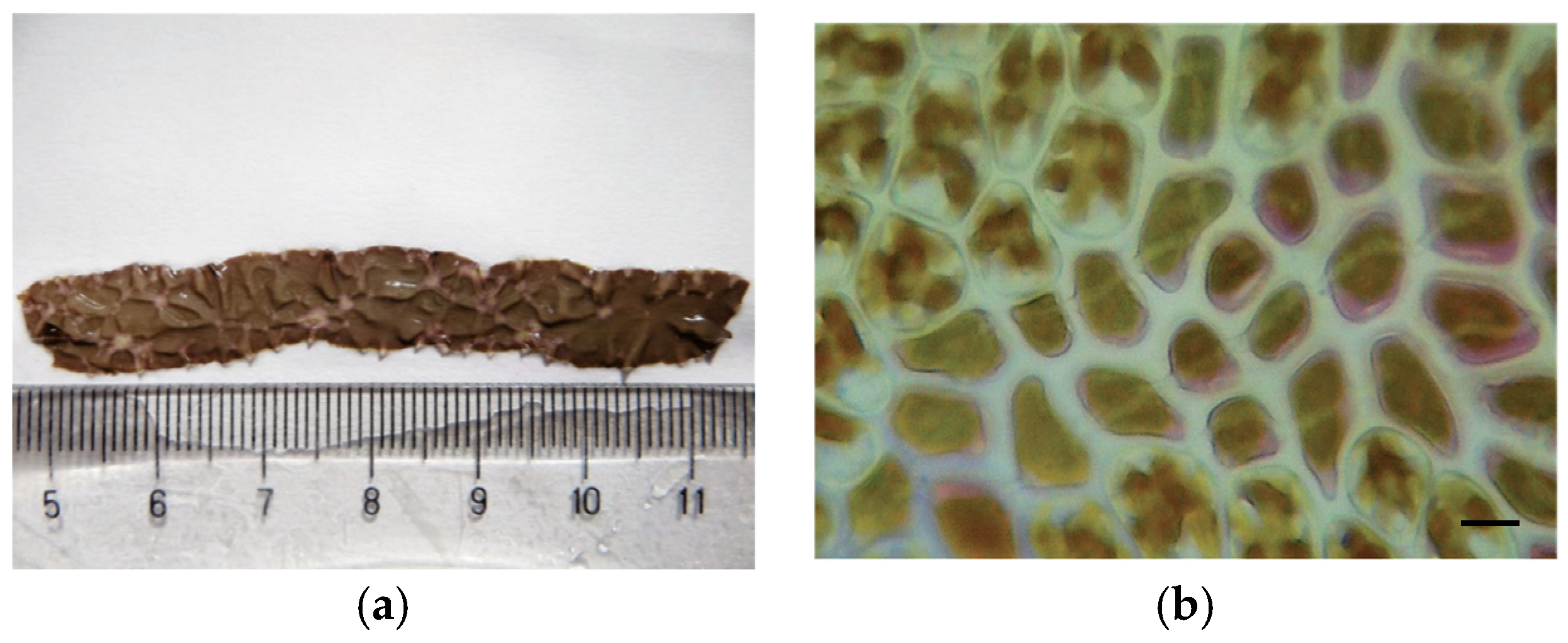
Figure 1. Clinical symptoms of Pyropia yezoensis red rot disease: (a) Macroscopic symptoms evident in infected thallus; (b) histopathology of the lesion area, presenting abnormal cells being penetrated by fungal mycelia, with an accumulation of released phycoerythrobilin-like material. Scale bar represents 10 μm [21].
Phycology 04 00026 g001
Figure 2. Mycelia of Pythium chondricola formed over the lesioned area in Pyropia yezoensis (arrows). Scale bars represents 50 μm [27].
Phycology 04 00026 g002
Figure 3. Infected cells of Pyropia plicata after 1 day (a), 3 days (b), and 9 days (c). Dark cells are newly infected cells, while light cells are older infected cells. After 9 days, almost all cells were dead. Pythium porphyrae hyphae were visible between cells. Scale bar represents 50 μm [22].
Phycology 04 00026 g003
Figure 4. Macroscopic and microscopic symptoms of olpidiopsis disease observed on Pyropia blades: (a) Arrows show decaying greenish areas (b); each green cell contains one Olpidiopsis thallus. Scale bar represents 50 μm [3].
Phycology 04 00026 g004
Figure 5. Typical symptoms of green spot disease infection in Pyropia sp.: (a) Infected blade with numerous lesions that look like bullet holes; (b) upon progression of infection, a chain of pinkish cells develops, encircling the green lesion. Scale bar represents 50 μm [3].
Phycology 04 00026 g005
Figure 6. “Ice-ice” infected Kappaphycus alvarezii [38].
Phycology 04 00026 g006
Figure 7. Host thallus of Kappaphycus sp. with “goose-bump”-like symptoms at the end of the epiphyte infection phase. Scale bar represents 300 μm [42].
Phycology 04 00026 g007
Figure 8. Tip whitening of Gracilaria lemaneiformis [45].
Phycology 04 00026 g008
Figure 9. The clinical symptoms of a technopathology, namely blister disease, caused by a sudden decrease in salinity due to the mixing of rainwater with seawater: (a) Laminaria hyperborea; (b) Laminaria digitata. Scale bar represents 1 cm (photo courtesy of Derek Mayes).
Phycology 04 00026 g009
Figure 10. Brown spot disease caused by Phaeophyceae species and fungal infestation on Ulva species: (a) Epiphytic brown algal genus Myrionema, cause of brown spot disease; (b) Pythium on the surface of Ulva intestinalis after inoculation. Scale bar in (a) represents 3 cm, and in (b), it represents 20 μm [50,52].
Phycology 04 00026 g010
Table 1. Table summarizing Porphyra/Pyropia diseases, with the pathology names, pathogens, symptoms, current treatments, treatment effectiveness, and severity as the average between mortality, incidence, and treatment effectiveness. Grey colour: no sufficient data.
Disease NameCausative Organism/TaxonomySymptomsCurrent TreatmentEffectiveness of
Treatment
Severity
  
References
Red rot diseasePythium porphyrae, Pythium chondricola/Oomycete
Alternaria sp./Ascomycota
Red patches on the blade; blade’s colour changes from natural brownish-red to violet-red; formation of numerous holes, followed by disintegration of the bladeExposure of culture nets to air; acid washPartially effectiveHigh[3,8,16,17,18,19,20,21,22,23,24,25,26,27]
Olpidiopsis
disease
Olpidiopsis porphyrae, Olpidiopsis pyropiae, Olpidiopsis sp./OomyceteBleached portion on the blades; appearance of greenish lesions; formation of numerous holes, followed by disintegration of the entire bladeExposure of culture nets to air; decrease in density of culture nets; acid wash; calcium propionateNoHigh[3,28,29,30,31,32]
Green-spot
disease
Primary: PyroV1/Virus
Secondary: Flavobacterium sp., Pseudoalteromonas sp., Vibrio sp./Gram-negative bacteria
Lesions with wide green borders; slimy rots and holes in the bladeExposure of culture nets to air; acid washNoHigh[3,33]
“Cyanobacteria felt”Filamentous and coccoid blue-green algae/CyanobacteriaDirty blade surface; lesions and holes in the bladeDrying of culture nets; acid washPartially effectiveMedium[15]
“Diatom felt”Fregellaria sp., Licmopohora flabellata, Melosira sp., Navicula sp./BacillariophyceaeDirty blade surface; blade bleaching; rust-coloured powderDrying of culture nets; acid washPartially effectiveMedium[3]
White blight
disease
?Random bleached areas on the blade; cell lysisNo treatmentNoLow[15]
White rot
disease
Vibrio sp./Gram-negative bacteriaRandom circular bleached areas of thallusNo treatmentNoLow[15]
“Suminori”
disease
Gaetbulibacter saemankumensis, Arthrobacter tumbae, Flavobacterium spp., Vibrio spp./Gram-negative bacteriaBlack lustreless colour of blade; plasmoptysis of blade cellsExposure of culture nets to air; acid washPartially effectiveMedium[35]
“Anaaki” disease (often associated with green spot)Flavobacterium sp., Pseudoalteromonas sp., Vibrio sp./Gram-negative bacteriaRandom holes on the blade; fast degradation of the bladeExposure of culture nets to air; acid washPartially effectiveMedium[3]
Unnamed
disease
“Pseudomonas-like” bacteria/Gram-negative bacteriaSimilarity to white rot diseaseNo treatmentNoLow[15]
White spot
disease
Phoma sp./CoelomyceteBleaching of oyster shell with shell-boring conchocelisDiscarding infected oyster shellsYesLow[36]
Yellow spot
disease
Vibrio mediterranei 117-T6/Gram-negative bacteriaYellow spots gradually spread around and form lesions of different sizes//n/a[37]
Table 2. Summary of Gracilaria spp. diseases, with the pathology names, pathogens, symptoms, current treatments, treatment effectiveness, and severity as the average between mortality, incidence, and treatment effectiveness. Grey colour: no sufficient data.
Disease NameCausative Organism
/Taxonomy
SymptomsCurrent TreatmentEffectiveness of TreatmentSeverity
  
References
EpiphytesCeramium minuta, Polysiphonia forfex, Hypnea spp., and more species/RhodophytaGenerally, epiphytes are attached superficially to the surface of the host; however, genera such as Polysiphonia spp. and Ceramium spp. can penetrate the host tissue, affecting its growth and, hence, its productivityControl of nutrients; move and shift growing structuresPartially effectiveMedium[44]
Rotten thallus syndrome or “Thalluswhitening”Vibrioparahaemolyticus, Vibrio spp., Thalassospira spp./Gram-negative bacteria (agarolytic)Slow growth, whitening of axesand branches, increased thallusfragilityTransfer to areas with slightly greater water currentPartially effectiveMedium[45,46]
Bleaching Stripe Disease or “Cell-wall degradation”Pseudoalteromonas spp./Gram-negative bacteria (agarolytic)Cell wall degradation//n/a[46]
White-tip diseaseBacterial strain OR-I1?Fast development of white necrotic tissues, followed by thallus fragmentation//n/a[44]
Brown points diseaseBacterial strain OR-I1?“Tumour-like” growth, leading to proliferations of nearly1 mm diameter//n/a[47]
Gracilaria Gall syndromeBacterial?Small bump-like structures//Medium[44]
GrazingFishes and invertebratesLoss of biomassFloating culture; control grazingYesMedium-
Table 3. Countries where a crisis in the cultivation of Kappaphycus occurred and where it is expected.
Country/Region/StateStart Massive CultivationFirst Report of DiseaseCollapseRecovered
Philippines19691975 (“ice-ice”)20022005/2008
Indonesia19752000--
Malaysia1978-20122019/2022
Tanzania199019952006Arguably never
South America20002010--
Disclaimer/Publisher’s Note: The statements, opinions and data contained in all publications are solely those of the individual author(s) and contributor(s) and not of MDPI and/or the editor(s). MDPI and/or the editor(s) disclaim responsibility for any injury to people or property resulting from any ideas, methods, instructions or products referred to in the content.