Review

Advances in Ophthalmic Organ-on-a-Chip Models: Bridging Translational Gaps in Disease Modeling and Drug Screening

Nancy E. and Peter C. Meinig School of Biomedical Engineering, Cornell University, Ithaca, NY 14853, USA
Int. J. Transl. Med. 2024, 4(4), 710-725; https://doi.org/10.3390/ijtm4040049
Submission received: 1 November 2024 / Revised: 27 November 2024 / Accepted: 30 November 2024 / Published: 4 December 2024

Abstract

:
Background: Organ-on-a-chip models have emerged as transformative tools in ophthalmology, offering physiologically relevant platforms for studying ocular diseases and testing therapeutic interventions. These microfluidic devices replicate human eye tissue architecture, addressing limitations of traditional in vitro and animal models. Methods: A narrative review of recent advancements in organ-on-a-chip technology was conducted, focusing on models simulating ocular structures like the retina and cornea and their applications in studying diseases such as dry eye disease (DED), age-related macular degeneration (AMD), and glaucoma. Results: Advanced organ-on-a-chip models successfully mimic key ocular features, providing insights into disease mechanisms and therapeutic responses. Innovations in microengineering and cellular integration have enhanced these platforms’ translational potential, though challenges like scalability and regulatory validation persist. Conclusions: Organ-on-a-chip models are poised to enhance preclinical research and clinical applications in ophthalmology. Addressing scalability and regulatory hurdles will be key to unlocking their full potential in drug discovery and disease modeling.

1. Introduction

The human eye is a highly specialized organ comprising several interconnected structures, each essential for vision. The cornea, the eye’s transparent outer layer, refracts light to focus images onto the lens, which further focuses the light onto the retina. The retina is a layered structure comprised of a complex arrangement of neurons and photoreceptor cells responsible for detecting light and converting it into electrical signals. These signals are processed and transmitted to the brain via the optic nerve [1]. A barrier known as the blood–retinal barrier (BRB) protects the retina from harmful substances circulating in the blood, ensuring only essential nutrients and oxygen reach the retinal cells [2].
Diseases that affect these structures, such as glaucoma, age-related macular degeneration (AMD), and diabetic retinopathy, are leading causes of visual impairment and blindness worldwide. Glaucoma, for example, is characterized by increased intraocular pressure (IOP), which damages the optic nerve, often leading to irreversible vision loss [3,4]. AMD, on the other hand, primarily affects the macula—the central part of the retina responsible for sharp vision—resulting in gradual central vision loss [5,6]. Diabetic retinopathy involves damage to the blood vessels in the retina, causing leakage and retinal edema, which eventually leads to vision loss [7]. Despite advancements in treatment, many patients with these conditions face limited therapeutic options due to gaps in understanding the diseases’ underlying mechanisms and challenges in developing effective long-term therapies.
Traditionally, researchers have relied on two-dimensional (2D) cell cultures and animal models to study these ocular diseases [8,9]. While 2D cell cultures provide valuable insights into cellular behavior, they lack the three-dimensional (3D) structure and functional complexity of the actual human eye tissues, which makes it difficult to replicate the intricate interactions between different cell types and the dynamic environment of the eye. For example, 2D cultures fail to mimic the mechanical forces exerted on the trabecular meshwork in glaucoma or the complex retinal layering crucial for vision [10]. While animal models contribute to understanding basic biological processes, they often fail to accurately predict clinical outcomes in humans due to species-specific differences in physiology, particularly in ocular structure and function [11,12,13]. These limitations have underscored the need for advanced in vitro models that more closely mimic the human eye.
Organ-on-a-chip technology represents a significant advancement in biomedical research, designed to address the shortcomings of traditional in vitro and in vivo models [14]. By integrating advanced biotechnologies, including soft lithography [15,16], 3D bioprinting [17,18,19], and iPSC-derived organoid [20,21], these microfluidic devices can replicate the structural and functional complexity of human organs by using human cells to recreate the tissue-specific microenvironments. Organ-on-a-chip models mimic the cellular interactions, mechanical forces, and biochemical gradients present in human organs, providing a more physiologically relevant platform for studying disease mechanisms and testing therapeutic interventions [22].
In ophthalmology, organ-on-a-chip models have shown great promise for simulating key aspects of ocular physiology and disease, including tear film regulation [23] and the blood–retinal barrier’s protective functions [24]. These models are particularly valuable for studying diseases like dry eye disease (DED) and AMD, where understanding the interplay between mechanical forces and cellular behavior is crucial [25,26]. Another key advantage of organ-on-a-chip models is their ability to incorporate human cells, making them more representative of human physiology than animal models. This is particularly important in ophthalmology, where species-specific differences can lead to discrepancies in drug efficacy and toxicity. For instance, cells from patients with retinitis pigmentosa have been used to model the disease in vitro, providing insights into disease progression and facilitating the development of targeted therapies [27]. This personalized approach is especially critical for rare genetic diseases, where traditional drug development pipelines may be less effective due to limited patient populations.
This review aims to provide an in-depth overview of the current state of organ-on-a-chip technology as it applies to ophthalmology, with the major focus on cornea and retina organ-on-a-chip platforms. We will explore the design and structure of organ-on-a-chip devices specific to the eye and how these devices replicate the microenvironment and functional aspects of ocular tissues. Additionally, the review will examine how these models are being utilized in translational applications, including drug screening, disease modeling, and personalized medicine. By highlighting notable examples of organ-on-a-chip devices, this paper will demonstrate how these platforms are helping to bridge the gap between laboratory research and clinical practice. The review will also address the technical limitations and translational hurdles that must be overcome to realize the full potential of organ-on-a-chip models in clinical applications.

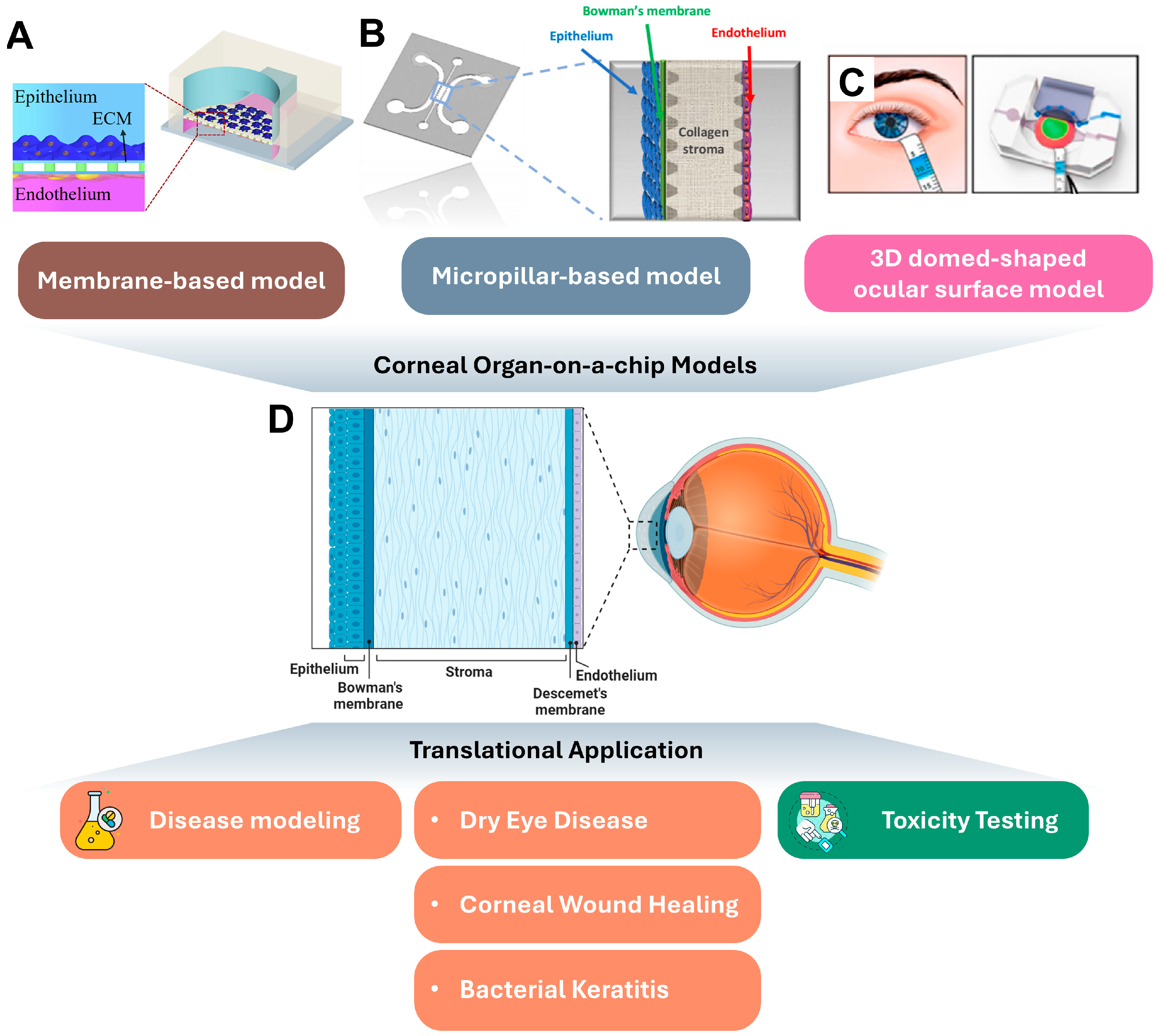
2. Corneal Organ-on-a-Chip Platforms and Their Translational Applications

2.1. Cornea Autonomy

The cornea is a transparent, avascular structure at the front of the eye, contributing more than two-thirds of the eye’s total refractive power [28]. It is composed of corneal epithelium (50 μm), Bowman’s membrane (12 μm), the stroma (480–500 μm), Descemet’s membrane (8–10 μm), and the endothelium (5 μm) [29]. The epithelium, consisting of about five to seven cell layers that regenerate regularly, serves as the primary protective barrier. Beneath the epithelium lies the stroma, which makes up about 90% of the cornea’s thickness and is composed of collagen fibrils and specialized cells called keratocytes. Keratocytes are responsible for maintaining the stroma’s transparency and structure by synthesizing collagen and extracellular matrix components. The innermost endothelium layer maintains corneal transparency by regulating the water content of the corneal stroma [30]. Notably, the cornea is among the most highly innervated tissues in the body, with 300 times more nerve endings than the skin, and these nerves are essential for maintaining epithelial health and promoting wound healing [31].
The corneal epithelium is covered by tear film, which is a thin, multi-layered liquid that lubricates the ocular surface and delivers oxygen and nutrients to the avascular cornea. The tear film comprises three layers: an outer lipid layer, an intermediate aqueous layer, and the inner mucin layer. Abnormalities in the tear film can lead to ocular diseases, such as DED [32].

2.2. Corneal Organ-on-a-Chips

The development of corneal organ-on-a-chip platforms began with Puleo et al. in 2009, who co-cultured corneal epithelial cells and keratocytes on opposite sides of a vitrified collagen layer within a polydimethylsiloxane (PDMS) microfluidic device. Unlike conventional co-culture models using transwells or porous polymer membranes, the vitrified collagen layer used here, which is prepared through a vitrification process, converted a conventional collagen gel into a rigid, glass-like membrane by drying at 37 °C for two weeks, and then it was rehydrated within the PDMS-based device, forming a gel-like substrate suitable for cellular growth. Additionally, sacrificial enzymatic etching with collagenase provides a method to remove the CV, creating a purely cellular bilayer structure while preserving tissue integrity [33]. One of the limitations of this study is the monolayer of corneal epithelial cells, which does not replicate the cornea’s multilayered epithelial structure.
Since the emergence of organ-on-a-chip techniques since the 2010s, two general types of organ-on-a-chip platforms have been widely used in modelling various tissues and disease: barrier tissue chips, where cells are cultures on permeable barriers such as porous membranes (Figure 1A); and solid organ chips, such as micropillar or microwell based devices where cells are cultured in 3D conditions (Figure 1B) [22]. In 2018, Bennet et al. utilized barrier chips with a porous polycarbonate (PC) membrane to create multilayered corneal epithelial structures through sequential cell-seeding techniques. The fabrication of this chip involved oxygen plasma treatment and APTES coating to bond the PC membrane with PDMS-based microfluidic channels. The PC membrane, functionalized with fibronectin via NHS/EDC chemistry and UV cross-linking, mimicked the topography and stiffness of the corneal basement membrane. This functionalization has been shown to be key in promoting epithelial layer formation while maintaining the mechanical properties akin to Bowman’s layer [34]. Alternatively, Bai et al. developed a corneal-on-chip model using a micropillar-based 3-channel microfluidic device, which incorporated PDMS microchannels fabricated through soft lithography and bonded to glass coverslips after plasma activation. The central channel is filled with a collagen matrix to mimic the corneal stroma and adjacent channels, simulating the epithelium and endothelium, respectively. A condensed collagen layer, representing Bowman’s layer, was created using viscous finger patterning, which involves the displacement of a more viscous fluid by a less viscous one to form a thin, uniform collagen structure. (Figure 1B) [35].
In 2019, Seo et al. introduced a specialized organ-on-a-chip model for the cornea, marking a significant advancement in the field. Their model featured a reverse-engineered human ocular surface with a dome-shaped polystyrene cell culture substrate attached to a perfusion chamber, a tear channel, and an artificial eyelid capable of mimicking blinking (Figure 1C). The fabrication of this model involved designing a multilayered elastomeric device using PDMS, bonded to a dome-shaped polystyrene scaffold that was molded using soft lithography techniques. The scaffold was functionalized with an extracellular matrix (ECM) hydrogel to embed keratocytes and support epithelial cell adhesion and growth. The perfusion chamber incorporated a programmable syringe pump to deliver controlled fluid flow, while the artificial eyelid, made of biocompatible hydrogel, was electromechanically actuated to simulate natural blinking dynamics. This was also the first cornea-on-chip model to incorporate an air–liquid interface (ALI), in which the basal surface of the scaffold was perfused with media at a controlled flow rate, while the apical epithelial cells were exposed to air to mimic in vivo conditions. ALI has been shown to enhance epithelial differentiation and stratification by upregulating basal cell markers (p63), tight junction proteins (occludin), and corneal epithelial markers (cytokeratin-3 and -12) [23]. ALI has since become a common feature in subsequent corneal organ-on-a-chip models. Simpler, porous membrane-based corneal models have been used by Yu et al. for epithelial wound healing studies [36,37] and by Deng et al. to investigate bacterial keratitis [38], both incorporating ALI into their designs.
One limitation of Seo’s model is the absence of neurons, which play a critical role in corneal homeostasis [39]. To address this, Bonneau et al. developed a compartmentalized organ-on-a-chip model that allows interaction between sensory neurons from the trigeminal ganglion and corneal epithelial cells. The microfluidic chip was fabricated from PDMS molded using soft lithography to create two macrochambers connected by arrays of straight microchannels. A compartmentalized culture protocol was employed: trigeminal neurons were seeded into the proximal compartment with neurotrophic factors such as NGF and GDNF to stimulate axonal growth, while corneal epithelial cells were seeded in the distal compartment after axonal networks were established. The microchannels facilitated selective interaction by allowing only axons, not neuronal soma, to extend into the epithelial compartment, thereby mimicking in vivo physiological conditions. Using this setup, the authors demonstrated axonal toxicity and stress responses induced by benzalkonium chloride (BAC), a common ophthalmic preservative, and highlighted its effects on epithelial integrity and neuronal stress markers (Figure 1D) [40].
High-throughput capabilities are another advantage of organ-on-a-chip platforms compared to traditional models. In 2024, Cho et al. developed a high-throughput microfluidic platform for evaluating ocular toxicity. Their platform integrates gradient concentration generation, micropumps, and microvalves, controlled by an Arduino-based system to precisely regulate drug concentrations and exposure times, effectively simulating drug delivery and blinking processes [41].
Microfabrication techniques also allow for the creation of extracellular matrix (ECM) topologies, which have been shown to influence cellular behaviors [42]. Öncel et al. developed a biomimetic platform using PDMS substrates with topography resembling white rose petals, which mimics the structural and biochemical properties of Descemet’s membrane. This design enhances corneal endothelial cell proliferation and maintains their phenotype in vitro [43]. Similarly, Lam et al. used microfluidic patterning to create substrates with aligned collagen fibrils for culturing keratocytes, replicating the native corneal stroma structure. By treating substrates with fibronectin and platelet-derived growth factor (PDGF), they simulated a wound-healing biochemical environment and observed significant changes in keratocyte morphology and stress fiber formation [44].

2.3. Translational Application of Corneal Organ-on-a-Chips

2.3.1. Disease Modeling and Drug Screening

Corneal barrier function and pharmacokinetics are among the first translational applications of corneal organ-on-a-chip platforms. In pioneering study of Puleo et al., fluorescein was used to assess the permeability of engineered corneal models with varying layers (epithelium, keratocytes, or bilayer). They also simulated epithelial damage using NaOH, investigating its impact on barrier function [33]. Similarly, Bai et al. tested dextran with different molecular weights to study the size-specific transport profile of the engineered cornea model [35]. Bennet et al. further investigated the pharmacokinetics of two ocular drugs, Pred Forte and Zaditor, observing prolonged residence time for Pred Forte and increased permeability for Zaditor. These studies collectively highlight how corneal OoC platforms can accurately replicate human corneal physiology and offer insights into drug absorption and efficacy, validating their use as a practical alternative to animal models for ocular drug testing [34].
The development of advanced corneal organ-on-a-chip models enabled diseases modelling and drug screening, with a major focus on DED. Seo et al. created a DED model by increasing evaporation rates and reducing blinking frequency from a physiological rate of twelve to six times per minute. In this model, DED symptoms—such as reduced tear film breakup time, increased breakup area, and higher fluorescein staining—were observed. Moreover, this platform demonstrated its therapeutic potential by evaluating lubricin, a treatment that significantly reduced TLR-4 and NF-kB staining, decreased fluorescein staining, and improved keratography results, showcasing the practical utility of organ-on-a-chip models in drug discovery for DED [23].
Modeling corneal wound healing is another significant focus. Yu et al. created epithelial scratches using pipette tips to study the effects of mesenchymal stem cell (MSC)-derived extracellular vesicles (EVs) on wound healing. They found that EVs promoted epithelial migration, reduced wound size, and lowered MMP-2 expression [36]. Similarly, Öncel et al. examined keratocyte wound-healing behavior in response to treatments of fibronectin and PDGF [43]. The successful application of these models demonstrates their effectiveness in identifying novel therapeutic strategies for regenerative medicine in ophthalmology.
Bacterial keratitis was modelled in the study of Deng et al. by inducing S. aureus into the corneal organ-on-a-chip devices. This model was designed to test antibiotic efficacy against bacterial infections in the cornea, providing a dynamic system to study how drugs interact with corneal tissues, and addressing the growing issue of antibiotic resistance. By enabling precise testing of drug responses in a controlled environment, this study highlights the potential of corneal organ-on-a-chip systems to accelerate the development of targeted antibiotic treatments while reducing reliance on traditional animal testing [38].

2.3.2. Toxicity Testing

Toxicity test is another major translational application of organ-on-a-chip platforms [45]. Though proved to be safe by the US Food and Drug Administration (FDA), benzalkonium chloride (BAC), a common preservative in artificial tears and eye drops, has shown side effects after prolonged use, including DED and corneal damage [46]. Hence, the cornea toxicity of BAC has been studied based on corneal organ-on-a-chip devices. Deng et al.’s high-throughput platform enables controlled testing of multiple drug or preservative concentrations, replicating corneal exposure under different conditions, such as healthy and DED models. Their system revealed significant differences in BAC toxicity across conditions, with lower IC50 values in DED models [41]. Similarly, in Bonneau et al.’s epithelial-neuron co-culture model, they observed that BAC led to reductions in the neurite network and a lower epithelial cell count, highlighting the need to consider corneal nerve toxicity in ocular drug development [40].

2.4. Current Limitations and Future Directions of Corneal Organ-on-a-Chips

While these models successfully mimic key aspects of the human cornea, they still fall short in fully replicating the complex multicellular interactions found in vivo. As discussed earlier, the cornea involves intricate crosstalk between keratocytes, epithelial cells, neurons, and immune cells, which is hard to fully capture in current models. For instance, Seo et al. (2019)’s blinking eye-on-a-chip mimics a relatively complete outer ocular surface and some biomechanical aspects, but the corneal endothelial cells and neurons are absent [23]. Bonneau et al. (2023) focus on corneal epithelium and nerve interactions but do not fully integrate the endothelium or the stroma [40]. Currently, no corneal organ-on-a-chip models incorporate Descemet’s membrane, nor the recently discovered Dua’s layer [47]. These layers are critical for understanding broader aspects of corneal physiology, such as nutrient transport, fluid regulation, and biomechanical strength. Future developments could involve integrating advanced bioprinting methods, such as multiphoton lithography and electrospinning, to fabricate full-thickness corneal models with all cellular and extracellular components. This would provide a more physiologically accurate platform for studying tissue regeneration, drug delivery, and biomechanical responses to therapies.
An important area that remains underexplored in corneal organ-on-a-chip models is the role of limbal stem cells (LSCs), which are essential for maintaining corneal epithelial homeostasis and repairing epithelial damage. Limbal stem cell deficiency (LSCD) is a significant cause of blindness, often resulting from trauma, chemical injury, or autoimmune conditions [48]. Current corneal organ-on-a-chip models largely neglect limbal cells, despite their critical physiological and pathological significance. The absence of limbal cells in existing models limits their capacity to study conditions such as LSCD or test therapies targeting these cells, such as LSC transplantation or regenerative medicine approaches.
There are also challenges in scaling up for high-throughput drug screening. Although high-throughput models like Cho et al. (2024) have automated the screening of various drug concentrations, their model is relatively simple with a 2D monoculture of corneal epithelial cells [41]. More complex organ-on-a-chip systems may be challenging to scale for large drug libraries while maintaining high precision. Addressing these limitations will require the integration of artificial intelligence (AI) for automated data analysis and optimization of microfluidic designs to support multiplexing capabilities. Additionally, establishing standardized protocols for fabrication and operation will enable reproducibility and scalability, ensuring that these systems can transition effectively from research laboratories to pharmaceutical development pipelines.

3. Retinal Organ-on-a-Chip Platforms and Their Translational Applications

3.1. Retina Autonomy

The retina is a highly specialized, multilayered tissue at the back of the eye that converts light into neural signals, which are then transmitted to the brain for visual processing. It consists of several critical components. Photoreceptors, comprising rods (low-light vision) and cones (color and high-acuity vision), are located in the outermost layer. These cells are divided into inner and outer segments: the outer segments capture light, and the inner segments house the metabolic machinery that supports photoreceptor function. Supporting the photoreceptors is the retinal pigment epithelium (RPE), a monolayer of cells that plays a key role in the recycling of visual pigments and in maintaining the health and function of the photoreceptors by phagocytosing shed outer segment discs [6].
The retina is also composed of several types of neurons that form complex circuits to process visual information. These include bipolar cells, which transmit signals from the photoreceptors to the ganglion cells, and horizontal and amacrine cells, which modulate these signals and contribute to the integration of visual information. Ganglion cells, located in the innermost layer of the retina, collect the processed signals and send them through their axons, which form the optic nerve, to the brain for further visual processing [4].
Blood supply to the retina is divided into two systems: the inner and outer blood vessels. The inner retina, including the ganglion cells and inner neurons, receives oxygen and nutrients from the central retinal artery, which branches into capillaries. The outer retina, where the photoreceptors and RPE are located, is nourished by the choriocapillaris across Bruch’s membrane. The choriocapillaris provides the majority of the retina’s oxygen and nutrients due to the high metabolic demands of the photoreceptors. The retina is one of the highest oxygen consuming tissues in the body, making it one of the most metabolically active tissues [49]. It therefore requires an efficient mechanism to maintain homeostasis as well as meet the metabolic needs of the retinal neurons [50]. Disruptions in these vascular systems can lead to retinal diseases, with the inner vessels often involved in diabetic retinopathy and retinal vein occlusion, and the outer vessels implicated in conditions such as AMD [25].

3.2. Retinal Organ-on-a-Chip Platforms

Compared to the avascular cornea, the retina is highly vascularized to support its intense metabolic activity. Therefore, retinal organ-on-a-chip models equipped with microfluidic perfusion systems are well-suited for replicating the vascular networks of the retina, a feature not offered by traditional models like retinal organoids (ROs) [51]. In 2017, Chen et al. developed a membrane-based retinal organ-on-a-chip model where human umbilical vein endothelial cell (HUVEC) and RPE cells were cultured on opposite sides of a porous PDMS membrane within a microfluidic chamber. In the microfluidic device, the porous PDMS membrane was spun over micropillars and coated with fibronectin to mimic the function of Bruch’s membrane. Hypoxic conditions were introduced via CoCl2 to mimic pathological states, inducing elevated VEGF secretion by RPE cells and enhancing endothelial cell responses. The model also included evaluations of RPE barrier function using tight junction markers (ZO-1) and permeability assays, ensuring physiological relevance to the retinal environment [24]. Later, in 2021, Rogers et al. expanded this design by creating PREDICT96, a high-throughput platform that contains 96 arrayed bilayer membrane-based devices. This platform was seeded with retinal endothelial cells and pericytes to assess retinal blood vessel barrier function, significantly enhancing the scalability of drug screening applications [52].
However, one limitation of membrane-based models is their inability to fully capture the 3D vascular structures and processes like angiogenesis. To address this, micropillar-based 3D models have emerged. Micropillar models use an array of small, pillar-like structures within the chip to enable surface tension-guided patterning of the ECM in the channels [53]. In 2017, Chung et al. developed an outer blood–retina barrier (oBRB) model that included a perfusable blood vessel network adjacent to an RPE monolayer (Figure 2A). The microfluidic device was fabricated using soft lithography, where SU-8 microposts were patterned onto a silicon wafer to create 300 µm gap channels. The micropillars enabled surface tension-guided loading of fibrin hydrogel, which was mixed with HUVECs and fibroblasts to form a stable vascular network. The lateral channels housed fibroblasts, which secreted growth factors to support angiogenesis, while the ARPE-19 cells were seeded onto the fibrin hydrogel surface to form the RPE monolayer. Tight junction formation in the RPE layer was verified by ZO-1 expression, and the vascular channels demonstrated functional barrier properties as shown by the restricted diffusion of fluorescent dextran molecules. VEGF gradients introduced into the system stimulated angiogenic sprouting from the choroidal vessels, mimicking choroidal neovascularization (CNV) seen in wet AMD. High VEGF levels led to vessel penetration into the RPE layer, replicating pathological angiogenesis. The model also allowed for drug testing, as the administration of bevacizumab effectively suppressed CNV, demonstrating its potential as a therapeutic evaluation platform. However, the design included a large 300 µm gap central channel between the RPE and blood vessel network to prevent RPE unstable migration into the ECM, while such a distance is much larger than Bruch’s membrane in vivo, and likely affected the efficiency of nutrient and waste transport [54]. In 2022, Lee et al. optimized this model by removing the gap, achieving direct contact between the RPE and blood vessels, allowing for more realistic simulation of hypoxia-induced neovascularization [55].
Another technique used to construct 3D vascular structures is needle casting, which involves using a physical needle to create hollow channels within a biomaterial scaffold. These channels act as conduits for the formation of vessel-like structures, allowing cells to organize into 3D vasculature [56,57,58,59]. For instance, Arik et al. combined needle casting with a membrane-based approach to co-culture HUVEC and RPE (Figure 2B). The fabrication process began with the design of a microfluidic device composed of three PDMS layers bonded together via plasma treatment. A polyester membrane with 8 µm pores was sandwiched between the layers to facilitate interactions between the endothelial and epithelial cells. The microchannel was patterned within a collagen I hydrogel using a syringe needle inserted into the microchannel inlet. Collagen gelation was induced at 37 °C, and the needle was removed to create a hollow vascular conduit. HUVECs were seeded into the channel to form a vascular lumen, and ARPE-19 cells were cultured on the polyester membrane in the adjacent compartment. To enhance cellular adhesion and to maintain the integrity of the layers, the device surfaces were functionalized with APTES and glutaraldehyde before gel loading. They examined the impact of oxidative stress on barrier function using hydrogen peroxide treatments and monitored permeability changes via fluorescence tracer assays. Additionally, optical coherence tomography (OCT) was employed to visualize the 3D vascular structures and to assess lumen integrity and morphology under stress conditions. OCT scans confirmed the circularity and continuity of the vessels, validating their physiological relevance. This model demonstrated how oxidative stress compromises the blood-retinal barrier, providing insights into disease mechanisms in AMD [60]. In a different study, Zhang et al. also employed needle casting but observed the spontaneous emergence of helical retinal endothelial tubes with right-handed chirality, governed by intrinsic cell behaviors. This novel discovery demonstrated how cell chirality influences both barrier function and vessel permeability [61].
Compartmentalized chip systems are another approach, designed to physically separate different cell types while allowing controlled interactions between them. These chips enable more complex co-cultures of multiple cell types, each in distinct compartments, which are connected by microfluidic channels or tubing. Jahagirdar et al. developed a co-culture model that connects retinal precursor cells (R28) with RPEs via medium perfusion to study inflammatory cytokine expression under normal and stress-induced conditions [62]. Similarly, Yeste et al. developed a system with crisscross microgrooves beneath parallel cell culture channels to enable interactions between retinal endothelial cells, neuroblastoma cells (SH-SY5Y), and RPEs [63]. These designs allow for the precise study of cell interactions within a microfluidic environment.
Compartmentalized platforms are also widely used in neuronal axon studies, enabling the separation of neuronal cell bodies in the somatic channel from their axons, which extend through microgrooves into the axonal channel [64]. This design prevents cross-contamination of somatic and axonal environments, allowing detailed studies on axonal dynamics, including regeneration, degeneration, and metabolic changes, by supporting clean axonal injuries and controlled microenvironments. Furthermore, live imaging capabilities and compatibility with various substrates make these models versatile for advanced neuroscientific research. Masin et al. used these platforms to explore metabolic shifts, such as enhanced glycolysis, which drive retinal axonal regrowth [65]. Boal et al. demonstrated how microfluidic systems can enhance the polarization of retinal ganglion cells, modeling axonal stress and degeneration [66]. Kwon et al. employed these platforms to investigate GPR110 activation, which significantly promoted axon growth [67]. Nafian et al. also applied the compartment device to apply higher intraocular pressure (IOP) to cultured rat retinal ganglion cells to study their viability with brain-derived neurotrophic factor (BDNF) [68]. Amos et al. further advanced this field by engineering a microfluidic-based retinothalamic nerve model that simulates unidirectional signal transmission between the retina and thalamus (Figure 2D). This high-throughput platform has proven valuable for studying axonal network formation and potential therapeutic interventions [69].
The integration of retinal organoids (ROs) with organ-on-a-chip technology represents a cutting-edge approach to overcoming the limitations of traditional models. Retinal organoids, derived from human pluripotent stem cells, contain various retinal cell types and form complex, multilayered structures that resemble the human retina [20]. However, they face challenges such as insufficient vascularization and nutrient delivery. By combining ROs with organ-on-a-chip platforms, it is possible to provide controlled microenvironments and perfusion systems that mimic vascular interactions. For example, Achberger et al. demonstrated that their retina-on-a-chip model, which combines ROs with RPEs, supports photoreceptor maturation and outer segment phagocytosis while enhancing nutrient delivery (Figure 2C) [20]. The fabrication of this model involved a multilayered microfluidic platform with a PDMS structure and a semipermeable polyethylene terephthalate (PET) membrane to separate the vascular and retinal compartments. The vascular-like perfusion system enabled the delivery of nutrients and removal of waste through a constant flow, maintaining stable culture conditions. Human iPSC-derived RPE cells were seeded into the lower compartment to form a monolayer with tight junctions, while retinal organoids embedded in a hyaluronic acid-based hydrogel were cultured in the upper compartment. This configuration facilitated close proximity and physiological interaction between the photoreceptors and the RPE layer. Enhanced outer segment formation was observed, with electron microscopy revealing organized disk structures within the photoreceptor outer segments. Additionally, the device supported phagocytosis of photoreceptor outer segments by RPE cells, replicating key aspects of the visual cycle. This platform was further developed to assess gene therapy vectors in 2021, demonstrating its value in pharmaceutical research [70]. Similarly, studies by Su et al. and Gong et al. showed that the integration of microfluidic chips enhances the survival and maturation of ROs, providing a robust system for studying retinal diseases like retinitis pigmentosa and advancing retinal ganglion cell development [21,27].
Figure 2. Retinal organ-on-a-chip platforms and their translational application. (A) Micropillar based retinal organ-on-a-chip mimicking oBRB structure. RPE cells were attached to a micropatterned 3D extracellular matrix, in which endothelial cells formed a perfusable blood vessel network that mimicked choroidal vessels [54]. (B) Needle casting retinal organ-on-a-chip, which contains a bottom compartment with a defined microchannel for HUVEC seeding and a top compartment which contains an open-top culture chamber for RPE culture [60]. (C) Retinal organ-on-a-chip integrated with retinal organoid. ROs and the hyaluronic acid-based hydrogel are directly loaded from the top into the well and onto the RPE [20]. (D) A schematic of retina anatomy. Figure republished with permission from each indicated reference ([54] for A, [60] for B, [20] for C).
Ijtm 04 00049 g002

3.3. Translational Application of Retinal Organ-on-a-Chips

3.3.1. Disease Modeling

Retinal organ-on-a-chip platforms are widely used for disease modeling and advancing drug testing methodologies. These systems effectively replicate the oBRB, a structure often disrupted in wet AMD by CNV. As previously discussed, the various co-culture models of RPE and endothelial cells can simulate choroidal angiogenesis via VEGF [54] or hypoxia [24,55] treatment, and the permeability and barrier functions can be measured by dextran [60] or transepithelial electrical resistance (TEER) [63]. Furthermore, anti-VEGF treatments, such as bevacizumab, have also been tested on these platforms [54]. These findings illustrate the utility of retinal OoC systems in replicating AMD-related pathophysiological changes and provide an efficient platform for preclinical evaluation of anti-VEGF therapies. Arik et al. advanced diagnostics in these systems by integrating OCT to visualize vascular permeability changes, providing clinically relevant readouts [60]. Moreover, Spivey et al. introduced a platform capable of monitoring time-resolved secretion of biomarkers, such as VEGF, from polarized RPE cells under hypoxic stress, enhancing the precision of cellular response measurements and enabling high-resolution diagnostics for retinal pathophysiology [71]. Collectively, these studies highlight the ability of retinal organ-on-a-chip platforms to not only model AMD progression but also serve as valuable preclinical tools for drug discovery, bridging the gap between in vitro research and clinical translation.

3.3.2. Cell Therapy

Retinal organ-on-a-chip platforms are emerging as transformative tools in the development of cell therapy strategies for retinal diseases. These platforms provide a controlled microenvironment to study retinal progenitor cell (RPC) migration, differentiation, and integration into host tissue, which are crucial for improving the efficacy of cell-based therapies aimed at restoring vision. For instance, the microfluidic-based μRetina model, described by Mishra et al. in 2015, simulates both human and mouse retinal geometry, enabling precise tracking of RPC migratory behavior in response to chemotactic signals such as stromal-derived factor 1 (SDF-1). This platform closely mimics in vivo conditions, offering key insights into improving RPC transplantation strategies [72]. In 2019, Mishra et al. further advanced this concept by incorporating electro-chemotactic fields, demonstrating that a combination of electrical and chemical gradients enhances RPC migration more effectively than either stimulus alone, which could greatly improve cell positioning in damaged retinal tissue [73]. These advancements validate the potential of retinal organ-on-a-chip models in refining and optimizing cell therapy strategies by closely mimicking the native retinal environment.
In addition to the electro-chemotactic stimuli, Thakur et al. also explored the role of ECM substrates in facilitating RPC adhesion and migration, finding that biomaterials incorporating ECM proteins such as laminin and fibronectin enhance cell integration by mimicking the native retinal environment [74]. Additionally, Pena et al. investigated collective RPC migration using invertebrate models, emphasizing the importance of both the RPC cluster size and substrate type in influencing transplantation outcomes [75]. Mut et al. developed the μ-Eye system, a microfluidic model that simulates stem-like cell migration in a biomimetic 3D environment, demonstrating that combining electrical and chemical stimuli significantly improves the distance and number of migrating cells [76]. These studies exemplify how retinal organ-on-a-chip platforms integrate bioengineering advances to address critical challenges in cell therapy development, offering insights that are highly relevant to clinical translation.

3.3.3. Gene Therapy

Gene therapies using adeno-associated viruses (AAVs) are among the most promising strategies for retinal diseases [77]. However, the development of new efficient AAV vectors is slow and costly, largely because of the lack of suitable non-clinical models. The retinal organ-on-a-chip model by Achberger et al. integrates retinal organoids into organ-on-a-chip platforms, which can replicate physiological conditions such as vascular perfusion and cellular compartmentalization, and closely resemble the in vivo retina. By mimicking these physiological conditions, this platform improves the predictive accuracy of preclinical testing compared to traditional animal models. This platform has been used in the screening of AAV vector efficacy, transduction kinetics, and cellular tropism, all critical factors in gene therapy development. Furthermore, first- and second-generation AAV vectors has been assessed, providing insights into vector performance that are more predictive of human clinical outcomes compared to traditional animal models [70]. These platforms help address the translational limitations of preclinical models and offer higher throughput for screening, thus significantly reducing the time and cost of developing gene therapies for retinal diseases.

3.3.4. Personalized Medicine

Retinal organ-on-a-chip platforms hold significant promise for personalized medicine applications, particularly when integrated with iPSC-derived retinal organoids. These platforms better replicate in vivo retinal environments, facilitating the modeling of specific genetic conditions and testing personalized treatments. For example, Su et al. (2022) demonstrated how retinal organoids derived from patients with USH2A mutations, which are associated with retinitis pigmentosa (RP), could be cultured on microfluidic chips to study disease progression and therapeutic responses. The microfluidic perfusion systems used in these platforms improve oxygen and nutrient delivery, enhancing the survival, maturation, and functionality of retinal cells. By enabling patient-specific disease modeling, these systems provide an avenue for identifying and testing tailored therapies in a physiologically relevant context. Moreover, the ability to replicate physiologically relevant interactions between RPE cells and photoreceptors allows for more accurate testing of potential therapies. Notably, these platforms have been shown to upregulate ECM components such as laminin and collagen IV, which are vital for maintaining cellular health in long-term cultures. These advances overcome limitations seen in static cultures, such as necrotic core formation in larger organoids. Additionally, by enabling the investigation of disease mechanisms like PI3K-Akt pathway deactivation and apoptosis in organoids with specific genetic mutations, these systems represent a significant step forward in developing individualized therapeutic strategies [27]. These findings underscore the value of retinal organ-on-a-chip models in advancing personalized medicine, paving the way for more effective and tailored treatments for retinal disorders.

3.4. Current Limitations and Future Directions of Retinal Organ-on-a-Chips

Despite the promising developments in retinal organ-on-a-chip technology, several specific limitations remain that must be overcome for full clinical applicability. One of the most significant challenges is replicating the complex 3D architecture of the human retina, which involves multiple cell types, including photoreceptors, retinal ganglion cells, Müller glia, and a sophisticated vascular system. Current organ-on-a-chip models often struggle to capture the full interaction between these layers, particularly the integration of functional blood–retinal barriers and the intricate network of neurons required for accurate retinal signal processing. For example, retinal organoid-on-chip models have succeeded in forming relatively complete photoreceptor structures, but they lack functional choroidal vasculature, which is crucial for maintaining retinal homeostasis and disease modeling [20,70]. On the other hand, models developed to simulate the oBRB have been successful in replicating vascular structures but have yet to incorporate neuron cells, which are essential for accurately mimicking retinal function and neural communication [24,54]. These gaps highlight the need for more integrated models that can simultaneously reproduce both the vascular and neuronal complexities of the retina, providing more physiologically relevant platforms for disease modeling and therapeutic testing. Future directions could focus on utilizing advanced co-culture techniques and microfabrication strategies, such as 3D bioprinting and microfluidic patterning, to better integrate vascular and neuronal components within a single platform. Combining these approaches with hydrogels that closely mimic the extracellular matrix of the retina could further enhance structural and functional accuracy.
Standardization across different research platforms is another critical challenge. Different laboratories use varied designs, materials, and protocols for constructing retinal organs-on-a-chip, leading to discrepancies in outcomes and difficulty in comparing data across studies. For instance, there is no agreed-upon protocol for constructing vascularized retinal models, leading to variability in how retinal diseases such as AMD or diabetic retinopathy are modeled. Establishing consensus on fabrication protocols, including cell sourcing, scaffold materials, and evaluation metrics, will be vital to harmonize findings across laboratories. This standardization would also facilitate regulatory approval and clinical translation, ensuring that these models are accepted for preclinical testing and high-throughput drug screening. Furthermore, integrating quality control measures, such as benchmarking against existing animal and in vivo models, could provide a framework for consistency.
A final limitation involves the difficulty in incorporating real-time monitoring systems. While there are initial efforts to integrate sensors for real-time tracking of biological processes, such as real-time TEER measurement by Yeste et al. [63], most organ-on-a-chip platforms still rely on endpoint assays, which fail to capture dynamic changes over time. To overcome this, future advancements could focus on embedding biosensors capable of detecting specific biochemical markers, such as oxidative stress or inflammatory cytokines, within the chip. Coupling these systems with advanced imaging technologies like optical coherence tomography (OCT) or multi-photon microscopy could provide non-invasive and continuous insights into drug responses and disease progression. Additionally, leveraging machine learning algorithms to analyze sensor data could enable predictive modeling of disease outcomes, further enhancing the utility of these platforms in precision medicine.

4. Conclusions

In conclusion, organ-on-a-chip platforms have made significant strides in modeling ocular tissues and advancing translational applications, particularly in disease modeling and drug testing. These systems offer a more physiologically relevant alternative to traditional models, allowing for the recreation of complex tissue structures and cellular interactions. However, current limitations, such as the challenge of fully replicating the in vivo microenvironment and the lack of standardized protocols, present hurdles for clinical translation. Future developments in integrating vascular components, enhancing long-term cellular viability, and improving standardization will be crucial to overcoming these challenges. Moreover, the incorporation of advanced biomaterials, personalized patient-derived cells, and real-time monitoring techniques holds the potential to push these platforms closer to clinical applications. As these technologies continue to evolve, organ-on-a-chip systems will play a pivotal role in bridging the gap between preclinical research and clinical practice in ophthalmology.

Funding

This research received no external funding.

Data Availability Statement

No new data were created or analyzed in this study.

Conflicts of Interest

The authors declare no conflicts of interest.

References

  1. Willoughby, C.E.; Ponzin, D.; Ferrari, S.; Lobo, A.; Landau, K.; Omidi, Y. Anatomy and physiology of the human eye: Effects of mucopolysaccharidoses disease on structure and function—A review. Clin. Exp. Ophthalmol. 2010, 38, 2–11. [Google Scholar] [CrossRef]
  2. O’Leary, F.; Campbell, M. The blood–retina barrier in health and disease. FEBS J. 2023, 290, 878–891. [Google Scholar] [CrossRef]
  3. Jonas, J.B.; Aung, T.; Bourne, R.R.; Bron, A.M.; Ritch, R.; Panda-Jonas, S. Glaucoma. Lancet 2017, 390, 2183–2193. [Google Scholar] [CrossRef] [PubMed]
  4. Lu, R.; Soden, P.A.; Lee, E. Tissue-Engineered Models for Glaucoma Research. Micromachines 2020, 11, 612. [Google Scholar] [CrossRef]
  5. Mitchell, P.; Liew, G.; Gopinath, B.; Wong, T.Y. Age-related macular degeneration. Lancet 2018, 392, 1147–1159. [Google Scholar] [CrossRef]
  6. Wu, A.; Lu, R.; Lee, E. Tissue engineering in age-related macular degeneration: A mini-review. J. Biol. Eng. 2022, 16, 11. [Google Scholar] [CrossRef] [PubMed]
  7. Yau, J.W.; Rogers, S.L.; Kawasaki, R.; Lamoureux, E.L.; Kowalski, J.W.; Bek, T.; Chen, S.J.; Dekker, J.M.; Fletcher, A.; Grauslund, J.; et al. Global prevalence and major risk factors of diabetic retinopathy. Diabetes Care 2012, 35, 556–564. [Google Scholar] [CrossRef]
  8. De Cillà, S.; Farruggio, S.; Cocomazzi, G.; Mary, D.; Alkabes, M.; Rossetti, L.; Vujosevic, S.; Grossini, E. Aflibercept and Ranibizumab Modulate Retinal Pigment Epithelial Cells Function by Acting on Their Cross Talk with Vascular Endothelial Cells. Cell. Physiol. Biochem. 2020, 54, 161–179. [Google Scholar] [CrossRef]
  9. Nebel, C.; Aslanidis, A.; Rashid, K.; Langmann, T. Activated microglia trigger inflammasome activation and lysosomal destabilization in human RPE cells. Biochem. Biophys. Res. Commun. 2017, 484, 681–686. [Google Scholar] [CrossRef]
  10. Tamm, E.R.; Russell, P.; Epstein, D.L.; Johnson, D.H.; Piatigorsky, J. Modulation of myocilin/TIGR expression in human trabecular meshwork. Investig. Ophthalmol. Vis. Sci. 1999, 40, 2577–2582. [Google Scholar]
  11. Van der Worp, H.B.; Howells, D.W.; Sena, E.S.; Porritt, M.J.; Rewell, S.; O’Collins, V.; Macleod, M.R. Can animal models of disease reliably inform human studies? PLoS Med. 2010, 7, e1000245. [Google Scholar] [CrossRef] [PubMed]
  12. Loiseau, A.; Raîche-Marcoux, G.; Maranda, C.; Bertrand, N.; Boisselier, E. Animal Models in Eye Research: Focus on Corneal Pathologies. Int. J. Mol. Sci. 2023, 24, 16661. [Google Scholar] [CrossRef] [PubMed]
  13. Zhu, J.; Inomata, T.; Shih, K.C.; Okumura, Y.; Fujio, K.; Huang, T.; Nagino, K.; Akasaki, Y.; Fujimoto, K.; Yanagawa, A.; et al. Application of Animal Models in Interpreting Dry Eye Disease. Front. Med. 2022, 9, 830592. [Google Scholar] [CrossRef]
  14. Ma, C.; Peng, Y.; Li, H.; Chen, W. Organ-on-a-Chip: A New Paradigm for Drug Development. Trends Pharmacol. Sci. 2021, 42, 119–133. [Google Scholar] [CrossRef] [PubMed]
  15. Kolarzyk, A.M.; Loy, C.; Lu, R.; Vlaminck, I.D.; Fowell, D.; Lee, E. Investigating lymphatic vessel remodeling and anti-tumor immunity in pancreatic cancer using tumor-on-chip and mouse models. Cancer Res. 2023, 83, 4614. [Google Scholar] [CrossRef]
  16. Kwak, T.J.; Lee, E. In vitro modeling of solid tumor interactions with perfused blood vessels. Sci. Rep. 2020, 10, 20142. [Google Scholar] [CrossRef]
  17. Yi, H.G.; Lee, H.; Cho, D.W. 3D Printing of Organs-On-Chips. Bioengineering 2017, 4, 10. [Google Scholar] [CrossRef]
  18. He, Y.T.; Fu, Q.; Pang, Y.; Li, Q.; Li, J.; Zhu, X.; Lu, R.H.; Sun, W.; Liao, Q.; Schröder, U. Customizable design strategies for high-performance bioanodes in bioelectrochemical systems. iScience 2021, 24, 102163. [Google Scholar] [CrossRef] [PubMed]
  19. Lu, R.; Zhang, W.; He, Y.; Zhang, S.; Fu, Q.; Pang, Y.; Sun, W. Ferric ion crosslinking-based 3D printing of a graphene oxide hydrogel and its evaluation as a bio-scaffold in tissue engineering. Biotechnol. Bioeng. 2021, 118, 1006–1012. [Google Scholar] [CrossRef]
  20. Achberger, K.; Probst, C.; Haderspeck, J.; Bolz, S.; Rogal, J.; Chuchuy, J.; Nikolova, M.; Cora, V.; Antkowiak, L.; Haq, W.; et al. Merging organoid and organ-on-a-chip technology to generate complex multi-layer tissue models in a human retina-on-a-chip platform. Elife 2019, 8, e46188. [Google Scholar] [CrossRef]
  21. Gong, J.; Gong, Y.; Zou, T.; Zeng, Y.; Yang, C.; Mo, L.; Kang, J.; Fan, X.; Xu, H.; Yang, J. A controllable perfusion microfluidic chip for facilitating the development of retinal ganglion cells in human retinal organoids. Lab Chip 2023, 23, 3820–3836. [Google Scholar] [CrossRef] [PubMed]
  22. Cao, U.M.N.; Zhang, Y.; Chen, J.; Sayson, D.; Pillai, S.; Tran, S.D. Microfluidic Organ-on-A-chip: A Guide to Biomaterial Choice and Fabrication. Int. J. Mol. Sci. 2023, 24, 3232. [Google Scholar] [CrossRef]
  23. Seo, J.; Byun, W.Y.; Alisafaei, F.; Georgescu, A.; Yi, Y.S.; Massaro-Giordano, M.; Shenoy, V.B.; Lee, V.; Bunya, V.Y.; Huh, D. Multiscale reverse engineering of the human ocular surface. Nat. Med. 2019, 25, 1310–1318. [Google Scholar] [CrossRef] [PubMed]
  24. Chen, L.J.; Ito, S.; Kai, H.; Nagamine, K.; Nagai, N.; Nishizawa, M.; Abe, T.; Kaji, H. Microfluidic co-cultures of retinal pigment epithelial cells and vascular endothelial cells to investigate choroidal angiogenesis. Sci. Rep. 2017, 7, 3538. [Google Scholar] [CrossRef] [PubMed]
  25. Manafi, N.; Shokri, F.; Achberger, K.; Hirayama, M.; Mohammadi, M.H.; Noorizadeh, F.; Hong, J.; Liebau, S.; Tsuji, T.; Quinn, P.M.J.; et al. Organoids and organ chips in ophthalmology. Ocul. Surf. 2021, 19, 1–15. [Google Scholar] [CrossRef]
  26. Haderspeck, J.C.; Chuchuy, J.; Kustermann, S.; Liebau, S.; Loskill, P. Organ-on-a-chip technologies that can transform ophthalmic drug discovery and disease modeling. Expert. Opin. Drug Discov. 2019, 14, 47–57. [Google Scholar] [CrossRef]
  27. Su, T.; Liang, L.; Zhang, L.; Wang, J.; Chen, L.; Su, C.; Cao, J.; Yu, Q.; Deng, S.; Chan, H.F.; et al. Retinal organoids and microfluidic chip-based approaches to explore the retinitis pigmentosa with USH2A mutations. Front. Bioeng. Biotechnol. 2022, 10, 939774. [Google Scholar] [CrossRef]
  28. Meek, K.M.; Dennis, S.; Khan, S. Changes in the refractive index of the stroma and its extrafibrillar matrix when the cornea swells. Biophys. J. 2003, 85, 2205–2212. [Google Scholar] [CrossRef]
  29. Nishida, T. Neurotrophic mediators and corneal wound healing. Ocul. Surf. 2005, 3, 194–202. [Google Scholar] [CrossRef]
  30. DelMonte, D.W.; Kim, T. Anatomy and physiology of the cornea. J. Cataract. Refract. Surg. 2011, 37, 588–598. [Google Scholar] [CrossRef]
  31. Craig, J.P.; Nichols, K.K.; Akpek, E.K.; Caffery, B.; Dua, H.S.; Joo, C.K.; Liu, Z.; Nelson, J.D.; Nichols, J.J.; Tsubota, K.; et al. TFOS DEWS II Definition and Classification Report. Ocul. Surf. 2017, 15, 276–283. [Google Scholar] [CrossRef] [PubMed]
  32. Masoudi, S. Biochemistry of human tear film: A review. Exp. Eye Res. 2022, 220, 109101. [Google Scholar] [CrossRef]
  33. Puleo, C.M.; McIntosh Ambrose, W.; Takezawa, T.; Elisseeff, J.; Wang, T.H. Integration and application of vitrified collagen in multilayered microfluidic devices for corneal microtissue culture. Lab Chip 2009, 9, 3221–3227. [Google Scholar] [CrossRef]
  34. Bennet, D.; Estlack, Z.; Reid, T.; Kim, J. A microengineered human corneal epithelium-on-a-chip for eye drops mass transport evaluation. Lab Chip 2018, 18, 1539–1551. [Google Scholar] [CrossRef]
  35. Bai, J.; Fu, H.; Bazinet, L.; Birsner, A.E.; D’Amato, R.J. A Method for Developing Novel 3D Cornea-on-a-Chip Using Primary Murine Corneal Epithelial and Endothelial Cells. Front. Pharmacol. 2020, 11, 453. [Google Scholar] [CrossRef] [PubMed]
  36. Yu, Z.; Hao, R.; Du, J.; Wu, X.; Chen, X.; Zhang, Y.; Li, W.; Gu, Z.; Yang, H. A human cornea-on-a-chip for the study of epithelial wound healing by extracellular vesicles. iScience 2022, 25, 104200. [Google Scholar] [CrossRef] [PubMed]
  37. Yu, Z.; Hao, R.; Chen, X.; Ma, L.; Zhang, Y.; Yang, H. Protocol to develop a microfluidic human corneal barrier-on-a-chip to evaluate the corneal epithelial wound repair process. STAR Protoc. 2023, 4, 102122. [Google Scholar] [CrossRef]
  38. Deng, Y.; Li, L.; Xu, J.; Yao, Y.; Ding, J.; Wang, L.; Luo, C.; Yang, W.; Li, L. A biomimetic human disease model of bacterial keratitis using a cornea-on-a-chip system. Biomater. Sci. 2024, 12, 5239–5252. [Google Scholar] [CrossRef]
  39. Shaheen, B.S.; Bakir, M.; Jain, S. Corneal nerves in health and disease. Surv. Ophthalmol. 2014, 59, 263–285. [Google Scholar] [CrossRef]
  40. Bonneau, N.; Potey, A.; Vitoux, M.A.; Magny, R.; Guerin, C.; Baudouin, C.; Peyrin, J.M.; Brignole-Baudouin, F.; Réaux-Le Goazigo, A. Corneal neuroepithelial compartmentalized microfluidic chip model for evaluation of toxicity-induced dry eye. Ocul. Surf. 2023, 30, 307–319. [Google Scholar] [CrossRef]
  41. Cho, K.; Lee, J.; Kim, J. Integrated high-throughput drug screening microfluidic system for comprehensive ocular toxicity assessment. Toxicol. In Vitro 2024, 98, 105843. [Google Scholar] [CrossRef] [PubMed]
  42. Berthiaume, F.; Moghe, P.V.; Toner, M.; Yarmush, M.L. Effect of extracellular matrix topology on cell structure, function, and physiological responsiveness: Hepatocytes cultured in a sandwich configuration. FASEB J. 1996, 10, 1471–1484. [Google Scholar] [CrossRef]
  43. Öztürk-Öncel, M.; Erkoc-Biradli, F.Z.; Rasier, R.; Marcali, M.; Elbuken, C.; Garipcan, B. Rose petal topography mimicked poly(dimethylsiloxane) substrates for enhanced corneal endothelial cell behavior. Mater. Sci. Eng. C Mater. Biol. Appl. 2021, 126, 112147. [Google Scholar] [CrossRef] [PubMed]
  44. Lam, K.H.; Shihabeddin, T.Z.; Awkal, J.A.; Najjar, A.M.; Miron-Mendoza, M.; Maruri, D.P.; Varner, V.D.; Petroll, W.M.; Schmidtke, D.W. Effects of Topography and PDGF on the Response of Corneal Keratocytes to Fibronectin-Coated Surfaces. J. Funct. Biomater. 2023, 14, 217. [Google Scholar] [CrossRef]
  45. Fabre, K.M.; Livingston, C.; Tagle, D.A. Organs-on-chips (microphysiological systems): Tools to expedite efficacy and toxicity testing in human tissue. Exp. Biol. Med. 2014, 239, 1073–1077. [Google Scholar] [CrossRef]
  46. Baudouin, C.; Kolko, M.; Melik-Parsadaniantz, S.; Messmer, E.M. Inflammation in Glaucoma: From the back to the front of the eye, and beyond. Prog. Retin. Eye Res. 2021, 83, 100916. [Google Scholar] [CrossRef] [PubMed]
  47. Dua, H.S.; Freitas, R.; Mohammed, I.; Ting, D.S.J.; Said, D.G. The pre-Descemet’s layer (Dua’s layer, also known as the Dua-Fine layer and the pre-posterior limiting lamina layer): Discovery, characterisation, clinical and surgical applications, and the controversy. Prog. Retin. Eye Res. 2023, 97, 101161. [Google Scholar] [CrossRef]
  48. Kate, A.; Basu, S. A Review of the Diagnosis and Treatment of Limbal Stem Cell Deficiency. Front. Med. 2022, 9, 836009. [Google Scholar] [CrossRef]
  49. Pan, W.W.; Wubben, T.J.; Besirli, C.G. Photoreceptor metabolic reprogramming: Current understanding and therapeutic implications. Commun. Biol. 2021, 4, 245. [Google Scholar] [CrossRef]
  50. Fliesler, S.J.; Anderson, R.E. Chemistry and metabolism of lipids in the vertebrate retina. Prog. Lipid Res. 1983, 22, 79–131. [Google Scholar] [CrossRef]
  51. Cheng, L.; Kuehn, M.H. Human Retinal Organoids in Therapeutic Discovery: A Review of Applications. Handb. Exp. Pharmacol. 2023, 281, 157–187. [Google Scholar] [CrossRef] [PubMed]
  52. Rogers, M.T.; Gard, A.L.; Gaibler, R.; Mulhern, T.J.; Strelnikov, R.; Azizgolshani, H.; Cain, B.P.; Isenberg, B.C.; Haroutunian, N.J.; Raustad, N.E.; et al. A high-throughput microfluidic bilayer co-culture platform to study endothelial-pericyte interactions. Sci. Rep. 2021, 11, 12225. [Google Scholar] [CrossRef]
  53. Huang, C.P.; Lu, J.; Seon, H.; Lee, A.P.; Flanagan, L.A.; Kim, H.Y.; Putnam, A.J.; Jeon, N.L. Engineering microscale cellular niches for three-dimensional multicellular co-cultures. Lab Chip 2009, 9, 1740–1748. [Google Scholar] [CrossRef]
  54. Chung, M.; Lee, S.; Lee, B.J.; Son, K.; Jeon, N.L.; Kim, J.H. Wet-AMD on a Chip: Modeling Outer Blood-Retinal Barrier In Vitro. Adv. Healthc. Mater. 2018, 7, 1700028. [Google Scholar] [CrossRef]
  55. Lee, S.; Kim, S.; Jeon, J.S. Microfluidic outer blood-retinal barrier model for inducing wet age-related macular degeneration by hypoxic stress. Lab Chip 2022, 22, 4359–4368. [Google Scholar] [CrossRef]
  56. Lu, R.; Lee, B.J.; Lee, E. Three-Dimensional Lymphatics-on-a-Chip Reveals Distinct, Size-Dependent Nanoparticle Transport Mechanisms in Lymphatic Drug Delivery. ACS Biomater. Sci. Eng. 2024, 10, 5752–5763. [Google Scholar] [CrossRef] [PubMed]
  57. Ilan, I.S.; Yslas, A.R.; Peng, Y.; Lu, R.; Lee, E. A 3D Human Lymphatic Vessel-on-Chip Reveals the Roles of Interstitial Flow and VEGF-A/C for Lymphatic Sprouting and Discontinuous Junction Formation. Cell. Mol. Bioeng. 2023, 16, 325–339. [Google Scholar] [CrossRef] [PubMed]
  58. Choi, D.; Park, E.; Choi, J.; Lu, R.; Yu, J.S.; Kim, C.; Zhao, L.; Yu, J.; Nakashima, B.; Lee, S.; et al. Piezo1 regulates meningeal lymphatic vessel drainage and alleviates excessive CSF accumulation. Nat. Neurosci. 2024, 27, 913–926. [Google Scholar] [CrossRef]
  59. Polacheck, W.J.; Kutys, M.L.; Tefft, J.B.; Chen, C.S. Microfabricated blood vessels for modeling the vascular transport barrier. Nat. Protoc. 2019, 14, 1425–1454. [Google Scholar] [CrossRef]
  60. Arık, Y.B.; Buijsman, W.; Loessberg-Zahl, J.; Cuartas-Vélez, C.; Veenstra, C.; Logtenberg, S.; Grobbink, A.M.; Bergveld, P.; Gagliardi, G.; den Hollander, A.I.; et al. Microfluidic organ-on-a-chip model of the outer blood-retinal barrier with clinically relevant read-outs for tissue permeability and vascular structure. Lab Chip 2021, 21, 272–283. [Google Scholar] [CrossRef]
  61. Zhang, H.; Rahman, T.; Lu, S.; Adam, A.P.; Wan, L.Q. Helical vasculogenesis driven by cell chirality. Sci. Adv. 2024, 10, eadj3582. [Google Scholar] [CrossRef] [PubMed]
  62. Jahagirdar, D.; Yadav, S.; Gore, M.; Korpale, V.; Mathpati, C.S.; Chidambaram, S.; Majumder, A.; Jain, R.; Dandekar, P. Compartmentalized microfluidic device for in vitro co-culture of retinal cells. Biotechnol. J. 2022, 17, e2100530. [Google Scholar] [CrossRef]
  63. Yeste, J.; García-Ramírez, M.; Illa, X.; Guimerà, A.; Hernández, C.; Simó, R.; Villa, R. A compartmentalized microfluidic chip with crisscross microgrooves and electrophysiological electrodes for modeling the blood-retinal barrier. Lab Chip 2017, 18, 95–105. [Google Scholar] [CrossRef] [PubMed]
  64. Taylor, A.M.; Blurton-Jones, M.; Rhee, S.W.; Cribbs, D.H.; Cotman, C.W.; Jeon, N.L. A microfluidic culture platform for CNS axonal injury, regeneration and transport. Nat. Methods 2005, 2, 599–605. [Google Scholar] [CrossRef] [PubMed]
  65. Masin, L.; Bergmans, S.; Van Dyck, A.; Farrow, K.; De Groef, L.; Moons, L. Local glycolysis supports injury-induced axonal regeneration. J. Cell Biol. 2024, 223, e202402133. [Google Scholar] [CrossRef]
  66. Boal, A.M.; McGrady, N.R.; Chamling, X.; Kagitapalli, B.S.; Zack, D.J.; Calkins, D.J.; Risner, M.L. Microfluidic Platforms Promote Polarization of Human-Derived Retinal Ganglion Cells That Model Axonopathy. Transl. Vis. Sci. Technol. 2023, 12, 1. [Google Scholar] [CrossRef]
  67. Kwon, H.; Kevala, K.; Xin, H.; Patnaik, S.; Marugan, J.; Kim, H.Y. Ligand-Induced GPR110 Activation Facilitates Axon Growth after Injury. Int. J. Mol. Sci. 2021, 22, 3386. [Google Scholar] [CrossRef]
  68. Nafian, F.; Kamali Doust Azad, B.; Yazdani, S.; Rasaee, M.J.; Daftarian, N. A lab-on-a-chip model of glaucoma. Brain Behav. 2020, 10, e01799. [Google Scholar] [CrossRef]
  69. Amos, G.; Ihle, S.J.; Clément, B.F.; Duru, J.; Girardin, S.; Maurer, B.; Delipinar, T.; Vörös, J.; Ruff, T. Engineering an in vitro retinothalamic nerve model. Front. Neurosci. 2024, 18, 1396966. [Google Scholar] [CrossRef]
  70. Achberger, K.; Cipriano, M.; Düchs, M.J.; Schön, C.; Michelfelder, S.; Stierstorfer, B.; Lamla, T.; Kauschke, S.G.; Chuchuy, J.; Roosz, J.; et al. Human stem cell-based retina on chip as new translational model for validation of AAV retinal gene therapy vectors. Stem Cell Rep. 2021, 16, 2242–2256. [Google Scholar] [CrossRef]
  71. Spivey, E.C.; Yin, J.; Chaum, E.; Wikswo, J.P. A Microfluidic Platform for the Time-Resolved Interrogation of Polarized Retinal Pigment Epithelial Cells. Transl. Vis. Sci. Technol. 2023, 12, 28. [Google Scholar] [CrossRef] [PubMed]
  72. Mishra, S.; Thakur, A.; Redenti, S.; Vazquez, M. A model microfluidics-based system for the human and mouse retina. Biomed. Microdevices 2015, 17, 107. [Google Scholar] [CrossRef] [PubMed]
  73. Mishra, S.; Peña, J.S.; Redenti, S.; Vazquez, M. A novel electro-chemotactic approach to impact the directional migration of transplantable retinal progenitor cells. Exp. Eye Res. 2019, 185, 107688. [Google Scholar] [CrossRef] [PubMed]
  74. Thakur, A.; Mishra, S.; Pena, J.; Zhou, J.; Redenti, S.; Majeska, R.; Vazquez, M. Collective adhesion and displacement of retinal progenitor cells upon extracellular matrix substrates of transplantable biomaterials. J. Tissue Eng. 2018, 9, 2041731417751286. [Google Scholar] [CrossRef]
  75. Pena, C.D.; Zhang, S.; Majeska, R.; Venkatesh, T.; Vazquez, M. Invertebrate Retinal Progenitors as Regenerative Models in a Microfluidic System. Cells 2019, 8, 1301. [Google Scholar] [CrossRef]
  76. Mut, S.R.; Mishra, S.; Vazquez, M. A Microfluidic Eye Facsimile System to Examine the Migration of Stem-like Cells. Micromachines 2022, 13, 406. [Google Scholar] [CrossRef]
  77. Trapani, I.; Auricchio, A. Seeing the Light after 25 Years of Retinal Gene Therapy. Trends Mol. Med. 2018, 24, 669–681. [Google Scholar] [CrossRef]
Figure 1. Corneal organ-on-a-chip platforms and their translational application. (A) Membrane-based corneal organ-on-a-chip. Corneal epithelial cells and endothelial cells are cultured on the opposite sides of a porous PC membrane coated with ECM. The membrane is sandwiched between two PDMS layers incorporated with microfluidic channels [36]. (B) Micropillar based corneal organ-on-a-chip. Red: endothelial cells, blue: epithelial cells. Corneal epithelium has five to seven layers of cells and endothelium has a monolayer of cells, with Bowman’s membrane represented as a condensed thin layer of green collagen gel [35]. (C) The 3D dome-shaped ocular surface at the organ scale consists of corneal epithelium (green), conjunctival epithelial (red), and an engineered eyelid. By adjusting blinking frequency, DED model can be built and assessed by the smearing of the blue ink within the strips [25]. (D) A schematic of cornea anatomy. Figure republished with permission from each indicated reference ([36] for A, [35] for B, [25] for C).
Ijtm 04 00049 g001
Disclaimer/Publisher’s Note: The statements, opinions and data contained in all publications are solely those of the individual author(s) and contributor(s) and not of MDPI and/or the editor(s). MDPI and/or the editor(s) disclaim responsibility for any injury to people or property resulting from any ideas, methods, instructions or products referred to in the content.

