Review

Sirtuins: Emergent Players in Tissue and Organ Regeneration

by
Ayla Kyler Núñez
1,
Claudia Marcela Arenas-Gómez
2,* and
Belfran Alcides Carbonell Medina
1,*
1
Grupo de Génetica, Regeneración y Cáncer, Universidad de Antioquia UdeA, Carrera 53 No. 61-30, 050010 Medellín, Colombia
2
Grupo de Biodiversidad para la Sociedad, Dirección Académica, Universidad Nacional de Colombia, Sede de La Paz, 202017 La Paz, Colombia
*
Authors to whom correspondence should be addressed.
Int. J. Transl. Med. 2024, 4(4), 687-709; https://doi.org/10.3390/ijtm4040048
Submission received: 12 October 2024 / Revised: 22 November 2024 / Accepted: 28 November 2024 / Published: 2 December 2024

Abstract

:
Sirtuins are a family of lysine deacetylases that regulate cellular homeostasis and energy sensing. Regeneration is the process that restores structural and functional homeostasis at the cellular, tissue, organ, and appendage levels. Several cellular processes, such as epithelial–mesenchymal transition (EMT), proliferation, migration, and differentiation, contribute to restoration after an injury. This review highlights the role of sirtuins in tissue, organ, and anatomical structure regeneration, showing how sirtuins modulate signalling pathways by deacetylating targets such as transcription factors. Furthermore, understanding the role of this protein family could help elucidate the molecular and cellular mechanisms underlying tissue regeneration, which may hold significant potential for fields such as regenerative medicine. The review compiles evidence suggesting that sirtuins are emerging factors in the regeneration of various organs (e.g., skin, liver, heart) and tissues (e.g., bone, muscle, cornea, spinal cord).

1. Introduction

Sirtuins are a family of deacetylases that can catalyse lysine deacetylation using NAD+ as a coenzyme [1,2]. In vertebrates, this family is composed of seven protein members, which participate in different cellular processes and are located in different organelles. SIRT1 and SIRT2 are found in the nucleus and cytoplasm, where SIRT1 participates in cellular differentiation [3], promotes autophagy [4,5], increases antioxidant enzyme levels in the cytoplasm during oxidative stress [6], and reduces apoptosis [7,8] and inflammation [9]. SIRT2 also increases antioxidant enzyme levels to combat oxidative stress [10], modulates apoptosis [11], regulates the cell cycle [12], and promotes autophagy [13].
SIRT1 and SIRT2 have several protein targets, and they share some of these targets, for example, p53, MYC, forkhead box O3a (FOXO3a), nuclear factor kappa-light-chain enhancer of activated B cells (NF-κB), and histones H3-K56Ac and H4-K16Ac [14,15]. It is reasonable to think that SIRT1 can substitute for SIRT2 (and vice versa) in some functions. Curiously, the chemical inhibition of SIRT1 does not have any effect on the apoptosis level of leukaemic B cells, but the chemical inhibition of both SIRT1 and SIRT2 does have a significant effect [16], suggesting possible evidence of compensation. Nevertheless, in a study that evaluated the subcellular location of SIRT1 and SIRT2 in embryonic kidney cells, it was shown that SIRT2 is mainly in the cytoplasm and SIRT1 is in the nucleus and the cytoplasm [17]. The limited distribution of SIRT2 may reduce redundant functions. Additionally, sirtuins are differentially expressed in organs. For example, sirt1 and sirt4 are highly expressed in the heart, while sirt2 and sirt3 are highly expressed in the muscles of killifish [18]. In zebrafish, sirt1 expression is present in every organ, but sirt2 expression is absent in the spleen and kidneys [19]. The RNA level of each sirtuin varies, even in different tissues of the same organ, such as in the rat brain [20]. Indeed, sirtuins present different gene expression profiles in closed species. For instance, the RNA level of Sirt2 in the retina is high in mice, low in rats, and absent in humans; therefore, the redundant or compensatory capacity of sirtuins is dependent on the species and organ [21].
Mitochondrial sirtuin SIRT3 regulates mitochondrial metabolism [22,23] and mitophagy [24,25]. SIRT4 participates in lipid oxidation [26,27] and mitophagy [28,29]. SIRT5 reduces oxidative stress [30] and regulates mitochondrial metabolism [31] and energy generation [32]. SIRT6, which is found in the nucleus, participates in DNA repair [33,34] and genomic [35,36] and telomere stability [33,37,38]. Finally, SIRT7 is found in the nucleolus and participates in DNA repair [39,40].
The roles of sirtuins in cellular metabolism, oxidative stress, and cellular homeostasis have been extensively studied, along with their involvement in cellular processes such as proliferation, cell migration, epithelial–mesenchymal transition (EMT), and differentiation (Figure 1). These cellular processes are also important for physiological processes such as tissue and organ regeneration.
Regeneration is a process where a tissue, organ, or appendage (limb or tail) that has been lost or damaged is restored or rebuilt. In 2006, Bruce Carlson classified regeneration in a masterful book titled Principles of Regenerative Biology, where regeneration is grouped into (1) morphallaxis, (2) physiological regeneration, (3) hypertrophic regeneration, and (4) reparative regeneration [41]. Morphallaxis is a type of regeneration in which there is no cell proliferation. Instead, after an injury, the remaining tissue is redistributed to form the lost structure. Physiological regeneration is the term for the daily cell turnover that occurs unrelated to damage. During hypertrophic regeneration, the function regenerates, but the damaged structure does not. Through cell proliferation, the damaged organ’s mass increases to restore function without restoring the original anatomy of the organ. Finally, reparative regeneration is the functional and structural restoration of a body part that has been lost or damaged. Reparative regeneration is subdivided into cellular regeneration, tissue regeneration, and epimorphic regeneration, also called epimorphosis (the latter regulates the restoration of complex anatomical structures such as the limbs, tail, or head).
However, to the best of our knowledge, no articles have reviewed the connections between sirtuins and regeneration. In this review, we present the relationships between sirtuins and different types of regeneration. We also summarise the participation of sirtuins in proliferation, migration, EMT, and differentiation during organ and tissue regeneration.

2. Sirtuins Regulate Different Cellular Processes

2.1. Epithelial–Mesenchymal Transition and Cellular Migration

Epithelial–mesenchymal transition (EMT) is a cellular process in which epithelial cells acquire the characteristics of mesenchymal cells, such as an irregular shape, the absence of ECM attachment, apical–basal polarity, and high motility. During regeneration, epithelial cells near a wound become mesenchymal-like cells; thus, they can migrate to cover the injured surface and the epithelial continuity is restored [41]. During EMT, epithelial biomarkers decrease (for instance, E-cadherin and β-catenin) and mesenchymal biomarkers increase (for instance, vimentin, FN, N-cadherin, Twist1, Snail1, Snail2, α-SMA, and MMPs) [42].
EMT is regulated by several pathways, such as the EGF, WNT, interleukin, Notch, and TGF-β pathways. TGF-β can bind to transmembrane receptor TGFR and activate signal transduction [43]. After transduction, the transcription factor SMAD4 is acetylated [44], and when it is activated, it translocates to the nucleus [45]. In the nucleus, SMAD4 induces the transcription of Mmp7 [46], Egfr, Twist1, and Snail1 [47]. Interestingly, the specific knockout of Sirt1 increased the acetylation level of SMAD4 and the mRNA level of Mmp7 in mouse kidneys [46], indicating that SIRT1 indirectly participates in ECM homeostasis. In in vitro models of retinal epithelial cells, resveratrol-activated SIRT1 has been shown to deacetylate SMAD4, which is correlated with an increase in E-cadherin and a reduction in α-SMA protein and mRNA levels [44].
Further studies have shown that resveratrol-induced SIRT1 deacetylates and inactivates Yin Yang 1 (YY1), a transcription factor and transcription repressor [48]. This inactivation decreases vimentin, Twist, Snail, and α-SMA, but increases the E-cadherin protein levels. However, YY1 was able to be reactivated using Sirt1-interfering RNA in kidney epithelial cells and in mouse kidneys [49]. Importantly, YY1 regulated skeletal muscle regeneration by controlling the metabolic reprogramming of satellite cells in a murine model [50]. These studies show that SIRT1 activation by resveratrol reduces the protein and mRNA levels of most EMT biomarkers, and the usage of interfering RNA reverses this effect, indicating that SIRT1 can diminish EMT.
A further study determined that knockdown of Sirt1 using siRNA decreased endothelial cell migration in a primary culture of murine vascular endothelial cells. In the same study, an endothelial-specific knockout of Sirt1 using the Cre-Lox system in mice showed mitigated retinal revascularization and endothelial cell migration. In both models, a reduction in endothelial cell migration was correlated with a reduction in the mRNA and protein levels of the angiogenic factors MMP14 and VEGF [51].
EMT and cell migration are biological processes that are dysregulated during the tumorigenic process, which some sirtuins have been reported to influence [52]. In human glioma cells, knockdown of SIRT1 using siRNA increased the protein levels of E-cadherin and β-catenin and reduced MMP9, Twist1, Snail1, and FN protein levels [53]. Moreover, this knockdown reduced migration and cellular viability. In another study, the inhibition of SIRT1 reduced EMT in melanoma cells. This reduction occurred because SIRT1 deacetylated and activated the autophagic factor beclin 1 and induced the degradation of E-cadherin in the autophagosomes of human melanoma cells [54]. Similarly, SIRT6 mediated the degradation of E-cadherin in lysosomes in juvenile mice [55]. In vivo models in a tumorigenic context showed that transplanted melanoma cells in NAM-treated mice metastasised less, suggesting that SIRT1 inhibition reduces cancer cell migration in vivo [52]. As can be seen above, SIRT1 directly participates in the regulation of EMT and cellular migration through the deacetylation of transcription factors such as SMAD4 and YY1 and indirectly through the modulation of the mRNA levels of genes, including Mmp7, Mmp9, Mmp14, Vimentin, Fn, Twist, Snail, and Acta2 (the gene of the α-SMA protein). Consequently, given the importance of these cellular events in regeneration, these sirtuins are potential candidates for regulating the regenerative response.

2.2. Cellular Proliferation and Cell Cycle

Cellular proliferation is the growth and division of cells to create new cells. It is crucial for regeneration, as proliferating cells are the source of cells that will replace damaged or injured ones [41]. After wound healing, progenitor cells proliferate to increase the cell mass. Later, these proliferating cells differentiate into organ-specific specialised cells.
Evidence from several studies supports the idea that sirtuins regulate cell proliferation. Reduction in SIRT1 levels due to knockdown using microRNA or siRNA reduces the cellular proliferation in lens epithelial cells [56], HUVECs [57], cutaneous carcinoma cells [58], and glioblastoma cells [59].
SIRT1 deacetylases the p21 tumour suppressor, enabling its ubiquitination and posterior degradation in cardiomyocytes [60]. SIRT1 also deacetylases [61] and stabilises [62] CDK2 activity, and CDK2 phosphorylates the p27 tumour suppressor [63]. It was observed that SIRT1 activation indirectly increased p27 phosphorylation and that Cdk2 overexpression induced p27 ubiquitination and degradation [61]. This indicates that SIRT1 enhances proliferation through the degradation of p27 via CKD2.
Cellular proliferation is regulated by various signalling pathways, including β-catenin/WNT and JAK/STAT, and SIRT1 is able to influence these pathways. The chemical activation of SIRT1 using SRT2104 both increased cell proliferation and migration and augmented WNT5A and β-catenin protein levels. These effects were opposite when using the SIRT1 inhibitor selisistat in mouse endothelial progenitor cells [64]. In the same study, it was observed that the WNT inhibitor IWP-2 reduced cell proliferation and migration and decreased the mRNA levels of Sirt1 in mouse endothelial progenitor cells [64]. This suggests that SIRT1 and WNT could have a positive interaction that modulates cell proliferation and migration. Another signalling pathway, SIRT1, deacetylases the transcription factor STAT3 [57], which is necessary for proliferation. This deacetylation triggers the phosphorylation (and activation) of STAT3 [65], indicating that SIRT1 positively modulates proliferation through the activation of STAT3. Interestingly, previous studies have shown that STAT3 is required to regulate proliferation [66], including during muscle regeneration in a murine model [67] and the regeneration of hair cells [68] and heart tissue in a zebrafish model [69].
In summary, SIRT1 regulates cellular proliferation via the activation/inhibition of tumour suppressors and CDK. Additionally, SIRT1 participates in the regulation of the β-catenin/WNT and JAK/STAT pathways during proliferation.

2.3. Osteogenic Chondrogenic and Adipogenic Differentiation

Differentiation is defined as the acquisition of more specified cell characteristics [70], and is regulated by both classical well-known pathways (e.g., Hedgehog, WNT, Notch) and emergent pathways such as mTOR and Ras [71]. Cellular differentiation is an indispensable process in the regeneration of tissues and organs. After injury, progenitor cells proliferate and differentiate into a cell type or different cell types depending on the tissue/organ model. These new cells replace the function of lost cells [41].
The relationship between sirtuins and cellular differentiation has recently been studied, particularly for three types of differentiation: osteogenic, chondrogenic, and adipogenic. Studies have found that increasing SIRT1 activity via gene overexpression or chemical activation (with resveratrol or SRT17205) promotes osteogenic differentiation in mesenchymal stem cells (MSCs) [72], bone marrow mesenchymal stromal cells (BMSCs) [73,74], periodontal ligament stem cells [75], and primary cell cultures of BMSCs [76]. Furthermore, decreasing SIRT1 activity using microRNA reduces osteogenic differentiation in mouse MSCs [77,78], BMSCs [79,80,81,82,83], and osteoblasts [84]. In previous studies, using microRNA against Sirt1 produced a reduction in the mRNA levels of osteogenic biomarkers such as Alp, Runx2, Oc, and Opn, ALP enzymatic activity, and mineralization staining level. SIRT1 deacetylases and activates FOXO3a (a classical target of SIRT1) [78]. FOXO3a then binds to the RUNX2 promoter and activates RUNX2 gene expression [85].
SIRT1 has also been studied in chondrogenesis and adipogenesis. SIRT1 promotes chondrogenic differentiation in embryonic stem cells [86], embryonic kidney cells, and MSCs [87], evaluated using mRNA levels of Sox9, Aggrecan, and Col2a1 (chondrogenesis gene biomarkers). For adipogenesis, using siRNA to knock down sirt1 increased mRNA levels of the adipogenic factors Klf5 and Pparg in mouse MSCs [78]. Furthermore, high levels of SIRT1 reduced lipid droplets and cholesterol esters [73], as well as mRNA levels of fatty acid synthase (Fas) and fatty acid binding protein 4 (Fabp4) and protein levels of PPARγ and CCAAT/enhancer-binding protein α (C/EBP α) [76] (biomarkers of adipogenic differentiation). This indicates that SIRT1 reduces adipogenic differentiation in vitro.
Mitochondrial sirtuins also participate in cellular differentiation. Knockdown of Sirt3 reduces mRNA levels of osteogenic biomarkers such as Runx2, Osx, Alp, and Bsp, ALP enzymatic activity, and mineralization of mouse preosteoblasts. In the same study, Sirt3-/- mice presented reduced RUNX2 and ALP protein levels in femur and bone parameters, i.e., bone volume per tissue, connectivity density, and mineral content. This indicates that SIRT3 is necessary for proper osteogenic differentiation and bone development [88]. SIRT3 also increases protein levels of RUNX2, OSX, and OC and reduces osteolysis in rat femurs [89].
SIRT4 is another mitochondrial sirtuin. The overexpression of SIRT4 produces an increase in adipogenic biomarkers such as mRNA levels of Cebpa, Pparg, and acyl CoA synthetase long-chain family member 1 (Acsl1) along with lipid droplet levels in mouse preadipocytes [90], indicating that overexpression of SIRT4 induces adipogenesis. Conversely, the knockdown of SIRT4 using siRNA reduces adipogenic biomarkers such as mRNA levels of FABP4, CEBPA, and PPARG in bovine adipocytes [91], supporting the participation of SIRT4 in adipogenic differentiation. SIRT4 has been referred to as the ‘black sheep’ of the sirtuin family because of its pro-oxidative function in mitochondria [92]. Perhaps SIRT4 also has a contrary function in adipogenesis.
Likewise, the knockdown of mitochondrial Sirt5 using shRNA reduces RNA levels of adipogenic genes such as Fabp4, Pparg, Cebpa, Cebpb, and positive regulatory domain zinc finger region protein 16 (Prdm16) (a brown adipocyte-specific gene). This knockdown also decreased intracellular lipid levels in mouse embryonic mesenchymal cells [93]. In another study, pharmacological inhibition of SIRT5 augmented mRNA and protein levels of browning factor peroxisome proliferator-activated receptor γ coactivator 1-α (PCG-1α) and thermogenesis factor uncoupling protein 1 (UCP1). However, this SIRT5 inhibition did not affect the protein levels of adipogenic factors such as C/EBP-α or FABP4 in mouse preadipocytes, suggesting that SIRT5 could participate in the browning of preadipocytes, but does not modulate adipogenesis [94].
On one hand, the knock-in of Sirt6 duplicated the ALP activity and mineralization levels in mouse adipocytic MSCs [95], suggesting that SIRT6 could participate in osteogenesis in vitro. The former idea is supported by a reduction in mRNA levels of osteogenic genes (Oc, Alp, and Col1a1) using Sirt6 siRNA and Sirt6 microRNA in mouse myoblasts [96]. Sirt6 gene overexpression increased mRNA levels of Oc, Runx2, Bsp, and Alp, ALP activity, and culture mineralization in rat BMSCs, while knockdown of Sirt6 had the opposite effect [97]. Indeed, SIRT6 modulates osteogenic gene expression by decreasing the phosphorylation level (activated level) of NF-kB, a protein that negatively regulates osteogenic differentiation [97]. On the other hand, there is evidence that SIRT6 impairs osteogenic differentiation in human MSCs [98]. In this study, the same biomarkers were evaluated: mRNA levels of OC, RUNX2, BSP, and ALP, ALP activity, and culture mineralization. However, the overexpression of SIRT6 decreased their levels. These differences in the results may have been caused by the usage of cultured cells from different species, and the study of cultured cells from more species could elucidate this discrepancy. Additionally, genetic ablation of Sirt6 reduced the mRNA levels of Col2a1 and Col10a1 (chondrogenic gene biomarkers), but unlike SIRT1, it had no effect on the mRNA levels of Sox9, Sox5, or Sox6 in primary cell cultures of mouse chondrocytes [99]. This shows that the participation of SIRT6 in chondrogenesis is not determinant.
Finally, SIRT7 reduced osteogenic differentiation in mouse adipocytic MSCs [100] and BMSCs [101]. Overexpression of Sirt7 decreased mRNA and protein levels of RUNX2, ALP, and OSX and the enzymatic activity of ALP. In contrast, knockdown of Sirt7 using microRNA had the opposite effect [100], indicating that SIRT7 negatively regulates osteogenic differentiation.
In summary, the majority of evidence supports the enhancement of chondrogenic and osteogenic differentiation by sirtuins. However, the involvement of sirtuins in neurogenic and myogenic differentiation remains unclear. Therefore, additional studies are necessary to elucidate the mechanisms and roles of individual sirtuins in cell differentiation.

2.4. Connection of Regeneration and Cancer

Regeneration and cancer are related. It has even been said that cancer is “the wound that never heals” [102]. Wong and Whited extensively detailed the similarities between epimorphosis and cancer (i.e., initial immune infiltration, hypoxic microenvironment, ECM remodelling, and high proliferation and cell differentiation). The participation of the seven sirtuins has been studied in several cancer models, and it was found that sirtuin levels can be high or low [15]. This can be interpreted in one of two ways: first, high sirtuin levels may modulate adaptative metabolic changes in cancer cells that increase survival possibilities, and second, the loss of sirtuins may provoke metabolic instability, thus enabling oncogenesis [103]. Several reviews have examined the relationship between sirtuins and cancer in depth. For a comprehensive overview, we recommend the review by Zhao and colleagues, which explores the role of each sirtuin in metastasis and tumorigenesis [104]. Additionally, Yu and colleagues provide an organ-specific analysis of sirtuin expression across different cancer types, which is highly informative [15].

3. Sirtuins Promote Tissue and Organ Regeneration

Regeneration is a process where a tissue, organ, or appendage (limb or tail) that has been lost or damaged is restored or rebuilt [41]. It is subdivided into cellular regeneration, tissue regeneration, organ regeneration, and epimorphic regeneration, which regulates the restoration of complex anatomical structures such as limbs, a tail, or a head.

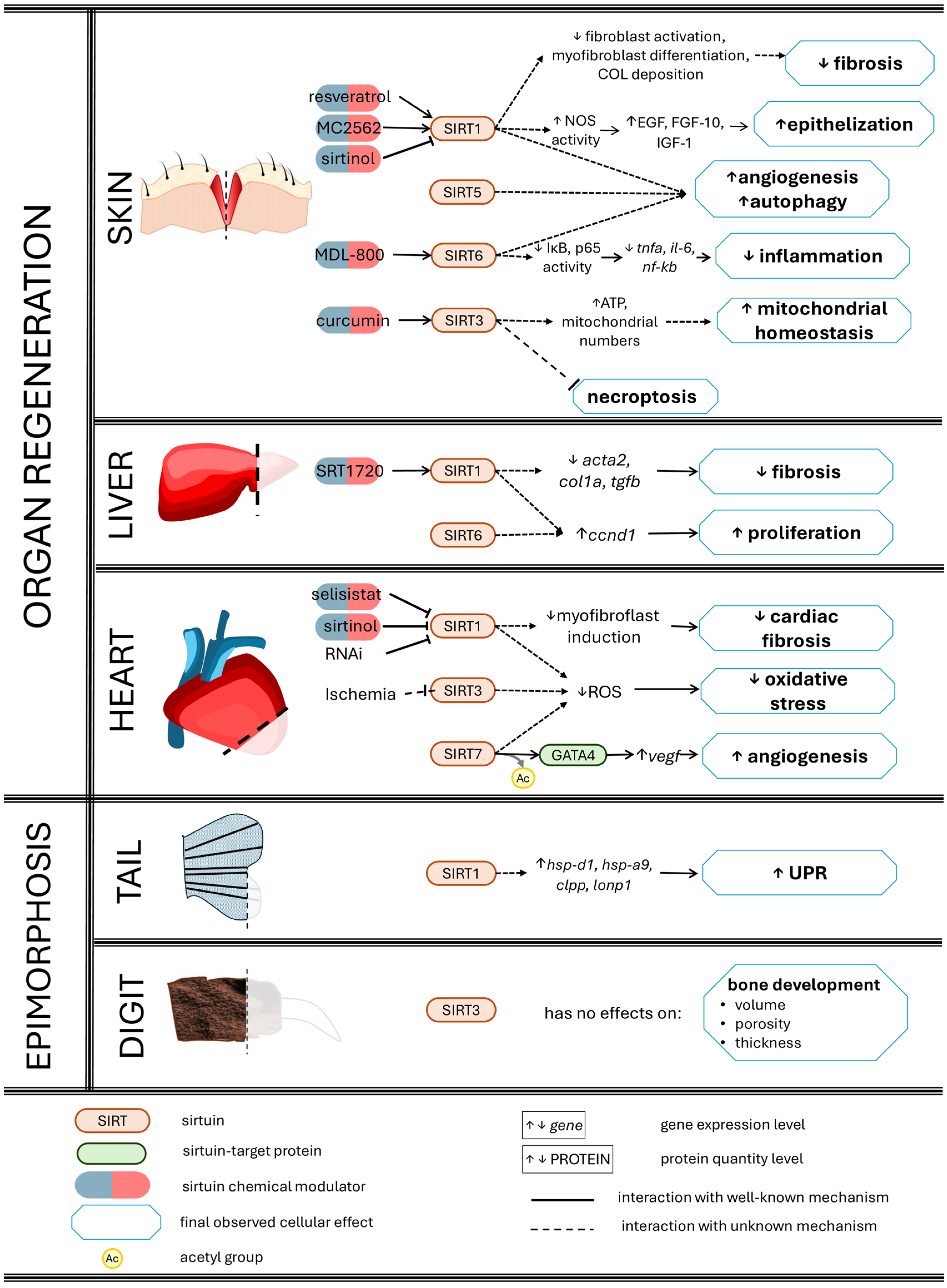
3.1. Skin Wound Healing

Skin wound closure consists of restoring the continuity of the epidermis and dermis after a cut or perforation of the skin [105]. Sirtuins promote skin wound closure in vivo. The activation of SIRT1 with resveratrol or MC2562 accelerates skin wound closure via nitric oxide production in mice. SIRT1 interacts with endothelial nitric oxide synthase, improves nitric oxide production, and increases the protein levels of growth factors such as EGF, FGF-10, and IGF-1 [106]. The activation of SIRT3 with curcumin accelerates wound closure in mice, and an injection of curcumin-treated BMSCs in punched skin produces faster closure compared to untreated BMSCs [107]. Curcumin changes the cellular physiology of these BMSCs: it reduces apoptosis and increases ATP production, mitochondrial quantity, and protein levels of antioxidant enzymes (such as SOD2 and PGC-1α) [107]. The activation of SIRT6 with MDL-800 enhances wound closure in mice: MDL-800 accelerates the thickening of granulation tissue and angiogenesis, while SIRT6 reduces the activation of the inflammatory transcription factors IκB and p65, and subsequently mitigates mRNA levels of the inflammatory cytokines Tnfa and Il6 [108]. Inflammation has negative effects on skin regeneration [109]; thus, the anti-inflammatory effect of SIRT6 is one of its molecular mechanisms that acts on skin regeneration. Conversely, the inhibition of SIRT1 with sirtinol delays wound closure in mice [106,110], highlighting the essential function of sirtuins in this process.
Sirtuins modulate diverse mechanisms in skin wound closure. For example, SIRT1, SIRT5, and SIRT6 induce autophagy in diabetic mice [111,112,113] and angiogenesis in mice in general [113,114]. Different in vivo models have been used to study skin regeneration, including epidermis-specific Sirt1-knockout mice using the Cre-Lox system [115], mice with implanted Sirt3-knockdown MSCs using siRNA [116], and Sirt6-knockdown mice using siRNA [113]. In these models, there was reduced angiogenesis, fibroblast activation, and decreased levels of inflammatory factors [113,115,116], such as the NF-κB protein [117]. Additionally, a Sirt1 knockdown delays the rate of wound closure [118] and increases fibrosis by elevating the mRNA and protein levels of COL1 and COL3 and the number of mouse myofibroblasts. This phenotype can be reversed by applying resveratrol [119], suggesting a potential therapeutic role for sirtuin activation during skin regeneration.
SIRT3 increased proliferation and cell migration in mouse wound closure [120]. Sirt3-/- transgenic mice showed increased oxidative stress and necroptosis and deregulated mitochondrial membrane potential [121], indicating that SIRT3 is necessary for mitochondrial well-being during wound healing in vivo. Furthermore, during wound healing, the inhibition of SIRT3 reduces cell proliferation and COL deposition [122].
Interestingly, the chemical activation of SIRT6 also increases COL1 and COL3 deposition and reduces the mRNA levels of inflammatory factors (such as Il6, Tnfa, and Nfkb) [108]. This suggests that sirtuins are necessary for the proper balance of COL deposition, allowing for ECM remodelling, and avoiding fibrosis in mice.
In conclusion, sirtuins are emerging as key regulators of skin wound healing and modulating multiple mechanisms, including the induction of autophagy and angiogenesis and the prevention of dermal fibrosis. Further research exploring the specific roles of sirtuins will help to elucidate the mechanisms of sirtuin signalling during skin regeneration, including studying how sirtuins modulate the expression of inflammatory factors or fibrosis factors, and how they do so in a skin regeneration model. Additionally, studies have predominantly focused on three out of the seven sirtuins, highlighting the need to investigate the remaining members to fully appreciate the significance of this protein family. Finally, sirtuin activators (resveratrol and curcumin) have potential as therapeutic targets to improve skin regeneration and wound healing after injuries.

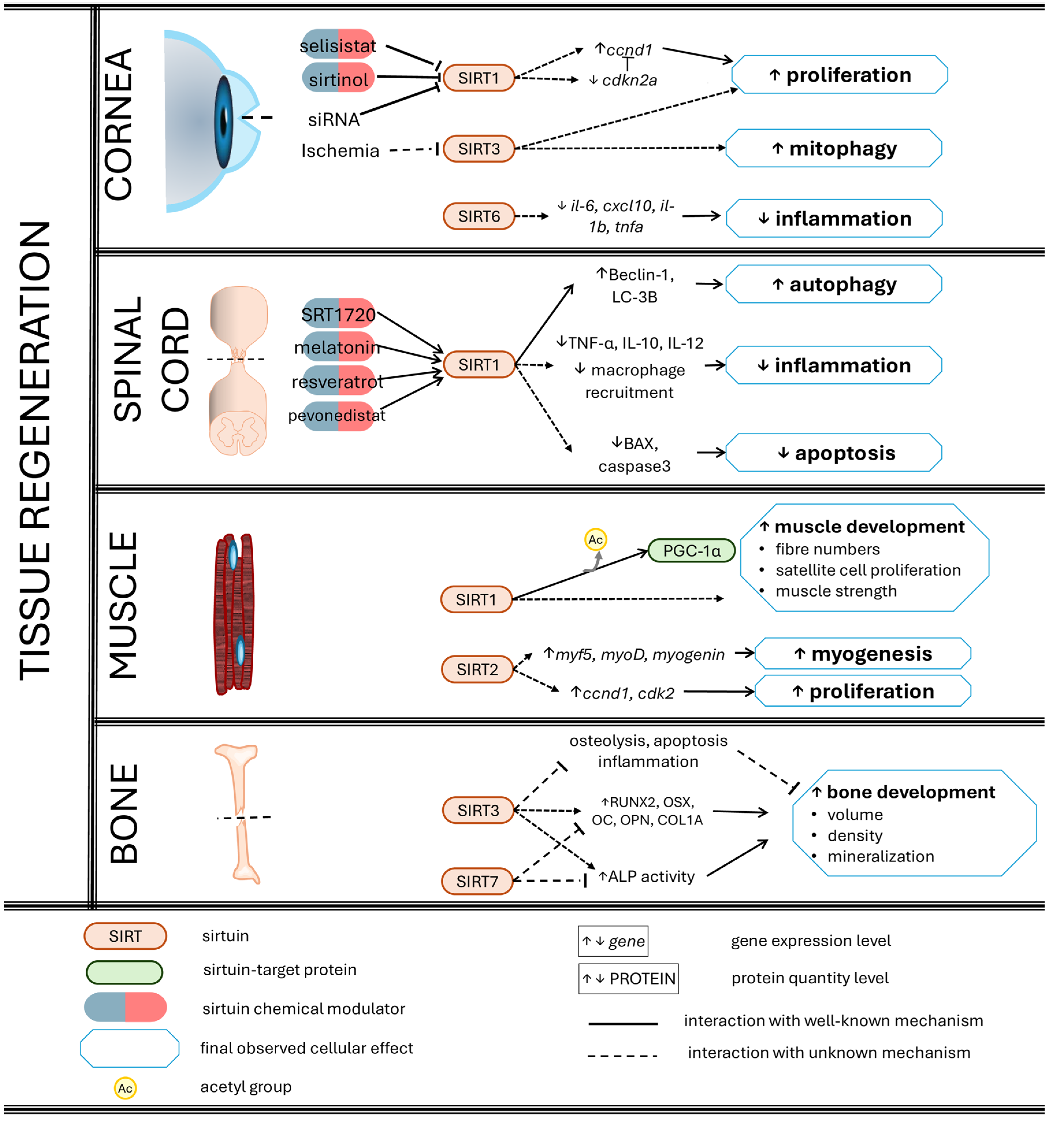
3.2. Corneal Re-Epithelialization

The cornea comprises a group of epithelial layers made up of a stratified epithelium on the outer surface, a stromal region of connective tissue, and a monostratified endothelium on the inner surface, which generates a translucent tissue that covers the eye [123]. Experimental injury to the cornea consists of making a surgical incision in the outer epithelium to interrupt its continuity. After injury, cells in the wound zone undergo apoptosis, and epithelial cells migrate and proliferate on the exposed stromal surface. Additionally, epithelial cells release inflammatory factors that induce the differentiation of keratinocytes to fibroblasts in the stroma, which then remodel the extracellular matrix to recover the original state. Later, they undergo apoptosis [124].
The overexpression of Sirt1 accelerates corneal regeneration [125] by increasing the mRNA level of Ccnd1 and by decreasing the mRNA level of Cdk2 (gene of p16; a tumour suppressor that inhibits CDKs) in juvenile male mice. This indicates that SIRT1 modulates the expression of genes related to cell proliferation in TKE2 corneal cells [126]. Additionally, the chemical activation of SIRT1 reduces oxidative stress in injured mouse corneas [127]. In contrast, corneal-specific knockout of Sirt1 reduces the rate of corneal re-epithelialization in mice, but it does not affect cell morphology [128]. SIRT3 and SIRT6 also have cytoprotective effects during corneal regeneration: mitochondrial SIRT3 regulates mitophagy [129] and SIRT6 reduces the mRNA levels of inflammatory genes such as Il6, Cxcl10, Il1b, and Tnfa [130].
In addition to in vivo experiments in mice, there are also results from in vitro experiments that provide more information about the relationship between sirtuins and corneal regeneration. In the 2040 pRSV-T human corneal epithelial cell line, activation of SIRT1 with SRT1720 increased viability and decreased apoptosis [131]. The aforementioned effects can be reversed through the chemical inhibition of SIRT1 [132]. Similarly, SIRT1 knockdown increased apoptosis, oxidative stress, and the expression of pro-inflammatory genes in the CRL-11135 human corneal epithelium cell line [133]. Other studies in the TKE2 murine corneal epithelial cell line have confirmed that SIRT1 and SIRT3 enhance re-epithelization by increasing epithelial cell proliferation and migration in vitro [126,129]. In summary, sirtuins are necessary for the regulation of proliferation and the cell cycle by increasing the expression of cyclins. Sirtuins also reduce inflammation by decreasing the expression of inflammatory cytokines in the corneal epithelium during wound healing.

3.3. Liver Regeneration

The liver is an organ composed mainly of hepatocytes. Partial hepatectomy, the surgical removal of a portion of the liver, induces the proliferation of hepatocytes. Other cells present in the liver also proliferate at the same time, such as cholangiocytes, hepatic stellate cells (HSCs), endothelial cells, and hepatic macrophages [134]. The proliferation of these cells occurs in a phenotypic fidelity or restricted-lineage manner, which means that cells of one phenotype only produce new cells of this phenotype. In other words, there is no differentiation into other phenotypes. Cell proliferation occurs until the liver recovers its original mass [134].
In a partial hepatectomy mouse model, knock-in of Sirt1 increased liver size during regeneration [135]. SIRT1 and SIRT6 promote liver regeneration by augmenting hepatocyte cell proliferation [136,137] by increasing Ccnd1 mRNA level and also by decreasing oxidative stress [138,139]. In mice with liver-specific knockdown of Sirt1 or Sirt6, the opposite effects were observed [140].
Knockout of Sirt1 generates fibrosis, and overexpression of Sirt1 reverses the fibrotic phenotype in mice [141]. Chemical activation of SIRT1 with SRT1720 also reduces fibrosis by decreasing the mRNA levels of Acta2, Col1, and Tgfb in rats [142]. Fibrosis is a pathology that is also observed in old mice [143]. In old mice, regenerative capacity decreases; however, the overexpression of Sirt1 improves regenerative capacity. Decreasing the gene expression of Sirt1 in young mice generates a phenotype similar to the liver of old mice [143], indicating that SIRT1 has anti-aging and pro-regenerative effects in old mice.
Sirtuins participate in liver regeneration, and their absence is associated with lipid accumulation, reduction in cell proliferation, or mitochondrial dysfunction [137,140,141]. All of these results are informative, but the complete mechanisms of sirtuin regulation are unknown; therefore, more studies are required to clarify how sirtuins participate in liver regeneration.

3.4. Ischaemia-Injured Hearts

The heart is an organ made up of an epicardium on the outer surface, a dense myocardium, and an endocardium on the inner surface. Ischaemia is a reduction in blood supply that leads to oxygen restriction and damage to the heart via cardiomyocyte necrosis [144]. Cells of the endocardium elongate to neighbour cardiomyocytes at the site of an injury. The endocardium cells then proliferate and migrate using filipodia. Simultaneously, epicardium cells induce angiogenesis at the injury site, undergo EMT, and cross the injury site. Finally, cardiomyocytes from the myocardium proliferate and restore the structure of the heart [145].
In rats with ischaemia-injured hearts, gene overexpression of Sirt1 decreases the infarct-injured area and oxidative stress while improving cardiac function [146]. In mice, chemical activation of SIRT1 by resveratrol [147,148] or melatonin also reduces the injured area and improves cardiac function, thus decreasing apoptosis. These effects can be reversed with the inhibitor selisistat [149], sirtinol, or RNA interference with anti-Sirt1 [150]. The inhibition of SIRT1 induces the differentiation of cardiac fibroblasts into myofibroblasts, inducing cardiac fibrosis; thus, SIRT1 improved heart regeneration and prevented fibrosis in a murine model [151].
Sirtuins have also been studied in minipigs, a rare model, but cardiologically more similar to humans. Knockdown of SIRT1 decreased cell proliferation and fibrosis and increased angiogenesis after an injury [152]. The decrease in fibrosis in this experiment may have been a result of a reduction in cell numbers and consequently a reduction in fibre deposition.
SIRT3 has also been evaluated during cardiac ischaemic injury in rats, showing that SIRT3 protein levels decrease after injury [153]. Sirt3 or Sirt7 knockout decrease cardiac reparative capacity, reducing the response against oxidative stress [154,155] and cardiac angiogenesis and increasing reactive oxygen species (ROS) levels in mice [156]. Additionally, SIRT7 deacetylates and activates GATA4 [157], a transcription factor that induces the gene expression of the angiogenic factor Vegf. This suggests that SIRT7 may modulate angiogenesis during cardiac regeneration.

3.5. Spinal Cord Regeneration and Motor Restoration

The spinal cord is a tubular structure that crosses through vertebrae and connects the brain to the peripheral neuronal system. It consists of meningeal cells, neurons, and glial cells [158]. In newts, when the continuity of the spinal cord is interrupted, both sides of the remnant spinal cord and axons retract in their own directions (either anterior or posterior) [159]. Leucocytes (particularly neutrophils and macrophages) then play an essential role in eliminating cellular debris and regulating inflammation [160]. Next, fibroblast-like meningeal cells from the outer layer migrate and cover the exposed cells (such as neurons and glial cells) [159]. In the injury zone, fibroblast-like cells deposit an extracellular matrix (ECM) to be used as a scaffold for future migrating cells. At the same time, glial cells close to the injury zone undergo EMT and begin migrating over to the ECM [161]. The anterior and posterior groups of migrating cells fuse, forming a structure called a ‘bridge’. Injured neurons regenerate their axons and elongate them by crossing the bridge and restoring the neural connections between both sides of the spinal cord [159].
The regeneration of the spinal cord has been studied in a model of spinal injury produced by laminectomy, the removal of a vertebral lamina. This process generates dysfunction in motor activity in animals [162]. SIRT1 levels in blood sera are lower in human patients with spinal cord injuries [163]. Spinal cord lesions also decrease Sirt1 mRNA levels in mice [164]. Indeed, in mice with more severe spinal injuries, serum SIRT1 levels are lower [165], indicating that there is an inverse correlation between SIRT1 level and injury severity and suggesting that SIRT1 alleviates or mitigates the negative effects of injuries.
After lumbar laminectomy in rats, various chemical activators of SIRT1 improve spinal cord regeneration and produce post-injury cytoprotective effects (Table 1). Sirtuins activators improve motor restoration and decrease inflammation in lumbar neurons [166], increase autophagy [167], decrease genotoxicity [168], and accelerate the restoration of motor ability [169].
The chemical activation of SIRT1 positively regulates cellular processes that participate in the regenerative capacity of the spinal cord (Table 1), while the genetic inhibition of SIRT1 mitigates regenerative capacity [164,170,173,174]. Therefore, transgenic Sirt1-/- mice have poor motor recovery after spinal injury evaluated using the Basso, Beattie, and Bresnahan (BBB) test [170] (this test can semiquantitatively assess motor activity based on limb movement, movement coordination, trunk position and stability, limb placement, and tail position during walking), which is caused by a reduction in the number of neurons [173], a reduction in their proliferation [172,174], and blockage of the restoration of the integrity and continuity of the blood–spinal cord barrier in mice [164]. The aforementioned results indicate that SIRT1 is necessary for appropriate spinal cord regeneration through the welfare of neurons, and the elimination of SIRT1 produces a reduction in regenerative capacity. However, although several animal models, including Xenopus frogs [175,176], zebrafish [160,161], and salamanders, have been used to study spinal cord regeneration [177,178], it is not known whether sirtuins are involved during the regeneration of this structure.

3.6. Bone Regeneration

When a long bone becomes fractured, a response mechanism is activated to restore its integrity. This response mechanism is divided into three stages: (1) haematoma formation and inflammation, (2) callus formation, and (3) bone remodelling [179,180]. During haematoma formation and inflammation, blood emerges from damaged vessels and accumulates in the fractured zone. Next, leucocytes arrive, eliminate the bone debris, degrade the bone ECM [181], and secrete inflammatory cytokines [182]. During soft callus formation, MSCs arrive at the fracture zone and differentiate into chondrocytes. These chondrocytes constitute the cartilage tissue that connects both sides of the fractured bone. Later, osteogenic cells arrive at blood vessels in the perichondrium and deposit mineralised ECM around the soft callus [182,183]. Eventually, the chondrocytes die, the cartilaginous callus is resorbed, and blood vessels penetrate the resorbing callus. Osteoblasts mineralise the inner side of the callus. The resulting hard callus is morphologically irregular and will be reshaped [184]. During bone remodelling, osteoclasts degrade the surface of the callus, and the original shape of the long bone is restored, while osteoblasts mediate the formation of new bone tissue [179,182].
Sirtuins have been less studied in bone regeneration. In distraction fracture models, overexpression of the SIRT1 gene results in a more regular distribution of trabeculae, more mature bone tissue, and higher mineral density in rabbit tibiae [185]. Similarly, chemical activation of SIRT3 accelerates fracture closure, while knockout of Sirt3 delays fracture closure and induces mitochondrial oxidative stress in mice [186]. SIRT3 increases bone volume and density, protein levels of RUNX2, OSX, and OC, and ALP activity. Additionally, SIRT3 reduces osteolysis, apoptosis, and inflammation in rat femurs. Moreover, SIRT3 overexpression raises the mineralization of MSCs in vitro [89]. All these results indicate that SIRT1 and SIRT3 improve bone regeneration.
On the other hand, SIRT7 negatively regulates bone regeneration. Knockdown of Sirt7 increased bone volume, bone density, and trabecular number and improved bone remodelling in a rat tibia fracture model [101]. In the same study, in vitro knockdown of SIRT7 increased mRNA and protein levels of RUNX2, OSX, OPN, and COL1A1, ALP activity, and mineral deposition in human BMSCs [101]. This suggests that SIRT7 downregulates osteogenic differentiation and impairs bone regeneration.

3.7. Muscle Repair

Muscle tissue is soft, contractile, and permits the locomotor movement of appendages and organs. It is made up of myocytes and satellite cells. The latter are stem cells that differentiate into myocytes during tissue homeostasis, injury, and regeneration [187]. SIRT1 activation using resveratrol and in a genetic gain-of-function model has been observed to positively regulate muscle regeneration in mice [188,189]. In fact, resveratrol increases the number of muscle progenitor cells and does not affect proliferation [189]; however, in other studies, SIRT1 modulated the proliferation of muscle precursors in rats [190]. Lee and Goldberg studied SIRT1 in a model of muscle atrophy caused by fasting or denervation and observed that overexpression of Sirt1 decreased atrophy and increased the number of muscle fibres [191]. In another study, aging in mice and muscle SIRT1 levels were related, with old mice showing lower levels of SIRT1. Additionally, knock-in of Sirt1 increased the proliferation of mouse satellite cells and muscle strength, and knockout had the opposite effect [190], indicating that SIRT1 can partially restore muscle deterioration caused by aging. Myers and colleagues also evaluated the acetylation level of PGC1-α (a target of SIRT1), but they did not find significant differences between satellite cell-specific knockout, skeletal muscle-specific knockout, young WT, and old WT groups; therefore, they did not find the target of SIRT1 that modulates muscle restoration in mice.
On the other hand, SIRT2 participates in muscle regeneration. Genetic inhibition of Sirt2 decreases regenerative capacity and increases muscle atrophy, and also decreases the mRNA levels of myogenic factors such as Myf5, MyoD, and myogenin. It reduces the mRNA levels of Ccnd1 and Cdk2, indicating that it decreases the proliferation of muscle cells [192].

3.8. Sirtuins as Potential Keys in Epimorphosis

Epimorphic regeneration is the process of limb or tail reconstruction directed by a blastema, which is a cluster of dedifferentiated cells (post-mitotic cells that lose the qualities of differentiated cells, such as morphology) found beneath the apical epithelial cap (AEC) (a stratified epithelium at the distal end of the regenerating structure) [193]. The cells comprising the blastema originate from various tissues, including epidermal, dermal, muscle, vascular, and connective and nervous endothelial tissues [194,195]. Although these cells present a dedifferentiation phenotype in the blastema, i.e., they have a mesenchymal morphology [193], they continue to have memory. Cellular memory in the blastema is evident when each cell re-differentiates into its same cell type or lineage of origin [196]. In addition, stem cells residing in the injured tissues can be recruited to form the blastema and contribute to regenerated tissue.
Sirtuins have been poorly studied in animal models that regenerate appendages, e.g., limbs and tails. In a recent study, the usage of sirtinol inhibited zebrafish tail regeneration: the length of the regenerated tail was shorter and the morphology was concave instead of convex. Some of the mechanisms in which SIRT1 is involved are increased mRNA levels of the mitochondrial chaperones heat shock protein (hsp) d1 and hsp a9 and the mitochondrial proteases caseinolytic protease proteolytic (clpp) and lon protease 1 (lonp1), suggesting that SIRT1 could regulate the unfolded protein response (UPR) during zebrafish tail regeneration [197]. SIRT3 has also been evaluated in epimorphic regeneration. Knockout of Sirt3 using the Cre-Lox system did not affect mouse digit regeneration: no differences were found in bone volume, porosity, or histology during regeneration. Furthermore, Sirt3-/- embryos developed limbs and digits normally [198]. Busse and colleagues proposed that an explanation of the differences absent in Sirt3-knockout mice may occur because sirtuins have redundant functions and because epimorphic regeneration is a complex process, interacting with other processes that are not affected by SIRT3’s absence. Further studies are needed to evaluate the involvement of sirtuins in limb regeneration, morphallaxis, and hypertrophic regeneration.
Sirtuins have been studied in zebrafish tail and mouse digit epimorphic regeneration, but not in limb regeneration. Among vertebrates, salamanders are the kings of regeneration: they can fully regenerate complex structures (limbs and tail) throughout their lifespan. In the 21st century, the axolotl (an aquatic salamander) has become the most studied model in regeneration research, and currently has one of the most complete molecular mechanism and pathway descriptions of epimorphic regeneration. The axolotl has sequenced transcriptomes and a sequenced genome, and there are transgenic line specimens, all of which make the axolotl an ideal model for investigating the role of sirtuins using ‘omics’ and molecular techniques.

4. Conclusions

Sirtuins, a family of deacetylase enzymes, play roles in the regulation of various cellular processes, including proliferation, migration, epithelial–mesenchymal transition, and differentiation. Sirtuins positively and negatively regulate the aforementioned cell processes; however, there is more evidence to support positive regulation. In this review, some molecular mechanisms are discussed. These mechanisms explain the roles of sirtuins during tissue regeneration, particularly of the cornea, spinal cord, bone, and muscle (Figure 2).
Sirtuins also participate in organ regeneration, such as skin, liver, and heart regeneration (Figure 3). In summary, sirtuins improve regeneration by increasing the wound healing rate, cell proliferation and migration, and autophagy, while also reducing oxidative stress, inflammation, apoptosis, and fibrosis, among other processes involved in regeneration.
By studying sirtuins, scientists may discover new pathways and molecular factors involved in critical cellular processes for regeneration, such as proliferation, differentiation, and migration. These discoveries could reveal new mechanisms by which cells respond to damage and initiate repair. Sirtuins have been shown to play roles in the regeneration of various organs and tissues, including the skin, liver, heart, and spinal cord, positioning them as promising targets for therapeutic interventions. Further exploration of sirtuin-mediated mechanisms may also lead to the development of therapeutic interventions for age-related degeneration and traumatic injuries.
In 2005, Porcu and Chiangi published a review presenting sirtuins as potential therapeutic targets, particularly in diseases such as neurodegeneration, diabetes, atherosclerosis, cardiovascular disorders, and age-related comorbidities [199]. Posteriorly, several articles had been published and analysed the evidence supporting the proposal of sirtuins as pharmacological targets for different diseases and disorders, i.e., Alzheimer’s disease [200], glycaemic disorders [201], mitochondrial myopathies, and cancer [202]. In this review, the referenced articles focused on basic regeneration biology or molecular biology, and none of them studied sirtuins in humans. As mammals, humans are poor regenerating animals, and future therapeutic interventions would improve human regeneration capacity. It is evident that there is a large gap between the current knowledge about sirtuins and clinical therapies of regenerative medicine. Comprehension of the molecular and cellular mechanisms of regeneration is necessary for the implementation of a real therapy. To that end, studying the participation of sirtuins (and other emergent players) in regeneration processes is relevant to achieve this clinical objective.
In regeneration research (and most biological research), SIRT1 is overrepresented compared to other sirtuins. This may be because SIRT1 was the first sirtuin discovered and the most biochemically investigated, but the study of the role of all seven sirtuins in regeneration is essential to comprehend and understand the participation of this protein family during regeneration.

Author Contributions

Conceptualisation, A.K.N., C.M.A.-G. and B.A.C.M.; writing—original draft preparation, A.K.N.; figure design, A.K.N.; review and editing, C.M.A.-G. and B.A.C.M. All authors have read and agreed to the published version of the manuscript.

Funding

This research received no external funding.

Acknowledgments

We would like to extend our sincere thanks to Emily Lucas for their invaluable assistance in enhancing the grammar and clarity of this work. Their support has been instrumental in helping us effectively communicate our ideas, especially as non-native English speakers.

Conflicts of Interest

The authors declare no conflicts of interest.

References

  1. Dai, H.; Sinclair, D.A.; Ellis, J.L.; Steegborn, C. Sirtuin Activators and Inhibitors: Promises, Achievements, and Challenges. Pharmacol. Ther. 2018, 188, 140–154. [Google Scholar] [CrossRef] [PubMed]
  2. Wu, S.; Liu, H. Sirtuins—Novel Regulators of Epigenetic Alterations in Airway Inflammation. Front. Genet. 2022, 13, 862577. [Google Scholar] [CrossRef] [PubMed]
  3. Zhao, D.; Kang, W.; Wang, Y.; Ge, J.; Huang, J.; Yang, J.; Yang, W.; Tang, X.; Xie, S. SIRT1 Promotes Osteogenic Differentiation in Human Dental Pulp Stem Cells through Counteracting the Activation of STAT3. Coatings 2021, 11, 1353. [Google Scholar] [CrossRef]
  4. Deng, Z.; Sun, M.; Wu, J.; Fang, H.; Cai, S.; An, S.; Huang, Q.; Chen, Z.; Wu, C.; Zhou, Z.; et al. SIRT1 Attenuates Sepsis-Induced Acute Kidney Injury via Beclin1 Deacetylation-Mediated Autophagy Activation. Cell Death Dis. 2021, 12, 217. [Google Scholar] [CrossRef] [PubMed]
  5. Pang, J.; Xiong, H.; Zhan, T.; Cheng, G.; Jia, H.; Ye, Y.; Su, Z.; Chen, H.; Lin, H.; Lai, L.; et al. Sirtuin 1 and Autophagy Attenuate Cisplatin-Induced Hair Cell Death in the Mouse Cochlea and Zebrafish Lateral Line. Front. Cell. Neurosci. 2019, 12, 515. [Google Scholar] [CrossRef] [PubMed]
  6. Ren, H.; Shao, Y.; Wu, C.; Ma, X.; Lv, C.; Wang, Q. Metformin Alleviates Oxidative Stress and Enhances Autophagy in Diabetic Kidney Disease via AMPK/SIRT1-FoxO1 Pathway. Mol. Cell. Endocrinol. 2020, 500, 110628. [Google Scholar] [CrossRef]
  7. Ben Salem, I.; Boussabbeh, M.; Da Silva, J.P.; Guilbert, A.; Bacha, H.; Abid-Essefi, S.; Lemaire, C. SIRT1 Protects Cardiac Cells against Apoptosis Induced by Zearalenone or Its Metabolites α- and β-Zearalenol through an Autophagy-Dependent Pathway. Toxicol. Appl. Pharmacol. 2017, 314, 82–90. [Google Scholar] [CrossRef]
  8. Luo, G.; Jian, Z.; Zhu, Y.; Zhu, Y.; Chen, B.; Ma, R.; Tang, F.; Xiao, Y. Sirt1 Promotes Autophagy and Inhibits Apoptosis to Protect Cardiomyocytes from Hypoxic Stress. Int. J. Mol. Med. 2019, 43, 2033–2043. [Google Scholar] [CrossRef] [PubMed]
  9. Gillum, M.P.; Kotas, M.E.; Erion, D.M.; Kursawe, R.; Chatterjee, P.; Nead, K.T.; Muise, E.S.; Hsiao, J.J.; Frederick, D.W.; Yonemitsu, S.; et al. SirT1 Regulates Adipose Tissue Inflammation. Diabetes 2011, 60, 3235–3245. [Google Scholar] [CrossRef]
  10. Wang, F.; Nguyen, M.; Qin, F.X.F.; Tong, Q. SIRT2 Deacetylates FOXO3a in Response to Oxidative Stress and Caloric Restriction. Aging Cell 2007, 6, 505–514. [Google Scholar] [CrossRef]
  11. Liu, S.; Gao, X.; Fan, Z.; Wang, Q. SIRT2 Affects Cell Proliferation and Apoptosis by Suppressing the Level of Autophagy in Renal Podocytes. Dis. Markers 2022, 2022, 4586198. [Google Scholar] [CrossRef] [PubMed]
  12. Inoue, T.; Nakayama, Y.; Li, Y.; Matsumori, H.; Takahashi, H.; Kojima, H.; Wanibuchi, H.; Katoh, M.; Oshimura, M. SIRT2 Knockdown Increases Basal Autophagy and Prevents Postslippage Death by Abnormally Prolonging the Mitotic Arrest That Is Induced by Microtubule Inhibitors. FEBS J. 2014, 281, 2623–2637. [Google Scholar] [CrossRef] [PubMed]
  13. Gal, J.; Bang, Y.; Choi, H.J. SIRT2 Interferes with Autophagy-Mediated Degradation of Protein Aggregates in Neuronal Cells under Proteasome Inhibition. Neurochem. Int. 2012, 61, 992–1000. [Google Scholar] [CrossRef] [PubMed]
  14. Zhao, L.; Cao, J.; Hu, K.; He, X.; Yun, D.; Tong, T.; Han, L. Sirtuins and Their Biological Relevance in Aging and Age-Related Diseases. Aging Dis. 2020, 11, 927–945. [Google Scholar] [CrossRef] [PubMed]
  15. Yu, L.; Li, Y.; Song, S.; Zhang, Y.; Wang, Y.; Wang, H.; Yang, Z.; Wang, Y. The Dual Role of Sirtuins in Cancer: Biological Functions and Implications. Front. Oncol. 2024, 14, 1384928. [Google Scholar] [CrossRef]
  16. Bhalla, S.; Gordon, L.I. Functional Characterization of NAD Dependent De-Acetylases SIRT1 and SIRT2 in B-Cell Chronic Lymphocytic Leukemia (CLL). Cancer Biol. Ther. 2016, 17, 300–309. [Google Scholar] [CrossRef] [PubMed]
  17. Lee, B.R.; Sanstrum, B.J.; Liu, Y.; Kwon, S.H. Distinct Role of Sirtuin 1 (SIRT1) and Sirtuin 2 (SIRT2) in Inhibiting Cargo-Loading and Release of Extracellular Vesicles. Sci. Rep. 2019, 9, 20049. [Google Scholar] [CrossRef]
  18. Kabiljo, J.; Murko, C.; Pusch, O.; Zupkovitz, G. Spatio-Temporal Expression Profile of Sirtuins during Aging of the Annual Fish Nothobranchius Furzeri. Gene Expr. Patterns 2019, 33, 11–19. [Google Scholar] [CrossRef]
  19. Pereira, T.C.B.; Rico, E.P.; Rosemberg, D.B.; Schirmer, H.; Dias, R.D.; Souto, A.A.; Bonan, C.D.; Bogo, M.R. Zebrafish as a Model Organism to Evaluate Drugs Potentially Able to Modulate Sirtuin Expression. Zebrafish 2011, 8, 9–16. [Google Scholar] [CrossRef]
  20. Sidorova-Darmos, E.; Wither, R.G.; Shulyakova, N.; Fisher, C.; Ratnam, M.; Aarts, M.; Lilge, L.; Monnier, P.P.; Eubanks, J.H. Differential Expression of Sirtuin Family Members in the Developing, Adult, and Aged Rat Brain. Front. Aging Neurosci. 2014, 6, 333. [Google Scholar] [CrossRef]
  21. Luo, H.; Zhou, M.; Ji, K.; Zhuang, J.; Dang, W.; Fu, S.; Sun, T.; Zhang, X. Expression of Sirtuins in the Retinal Neurons of Mice, Rats, and Humans. Front. Aging Neurosci. 2017, 9, 366. [Google Scholar] [CrossRef] [PubMed]
  22. Li, M.; Li, C.M.; Ye, Z.C.; Huang, J.; Li, Y.; Lai, W.; Peng, H.; Lou, T. qi Sirt3 Modulates Fatty Acid Oxidation and Attenuates Cisplatin-Induced AKI in Mice. J. Cell. Mol. Med. 2020, 24, 5109–5121. [Google Scholar] [CrossRef] [PubMed]
  23. Li, R.; Quan, Y.; Xia, W. SIRT3 Inhibits Prostate Cancer Metastasis through Regulation of FOXO3A by Suppressing Wnt/β-Catenin Pathway. Exp. Cell Res. 2018, 364, 143–151. [Google Scholar] [CrossRef] [PubMed]
  24. Ahmedy, O.A.; Abdelghany, T.M.; El-Shamarka, M.E.A.; Khattab, M.A.; El-Tanbouly, D.M. Apigenin Attenuates LPS-Induced Neurotoxicity and Cognitive Impairment in Mice via Promoting Mitochondrial Fusion/Mitophagy: Role of SIRT3/PINK1/Parkin Pathway. Psychopharmacology 2022, 239, 3903–3917. [Google Scholar] [CrossRef] [PubMed]
  25. D’onofrio, N.; Martino, E.; Mele, L.; Colloca, A.; Maione, M.; Cautela, D.; Castaldo, D.; Balestrieri, M.L. Colorectal Cancer Apoptosis Induced by Dietary δ-Valerobetaine Involves Pink1/Parkin Dependent-Mitophagy and Sirt3. Int. J. Mol. Sci. 2021, 22, 8117. [Google Scholar] [CrossRef]
  26. Laurent, G.; de Boer, V.C.J.; Finley, L.W.S.; Sweeney, M.; Lu, H.; Schug, T.T.; Cen, Y.; Jeong, S.M.; Li, X.; Sauve, A.A.; et al. SIRT4 Represses Peroxisome Proliferator-Activated Receptor α Activity To Suppress Hepatic Fat Oxidation. Mol. Cell. Biol. 2013, 33, 4552–4561. [Google Scholar] [CrossRef]
  27. Nasrin, N.; Wu, X.; Fortier, E.; Feng, Y.; Baré, O.C.; Chen, S.; Ren, X.; Wu, Z.; Streeper, R.S.; Bordone, L. SIRT4 Regulates Fatty Acid Oxidation and Mitochondrial Gene Expression in Liver and Muscle Cells. J. Biol. Chem. 2010, 285, 31995–32002. [Google Scholar] [CrossRef]
  28. Lang, A.; Piekorz, R.P. Novel Role of the SIRT4-OPA1 Axis in Mitochondrial Quality Control. Cell Stress 2018, 2, 1–3. [Google Scholar] [CrossRef]
  29. Lang, A.; Anand, R.; Altinoluk-Hambüchen, S.; Ezzahoini, H.; Stefanski, A.; Iram, A.; Bergmann, L.; Urbach, J.; Böhler, P.; Hänsel, J.; et al. SIRT4 Interacts with OPA1 and Regulates Mitochondrial Quality Control and Mitophagy. Aging 2017, 9, 2160–2186. [Google Scholar] [CrossRef]
  30. Jung, Y.H.; Chae, C.W.; Chang, H.S.; Choi, G.E.; Lee, H.J.; Han, H.J. Silencing SIRT5 Induces the Senescence of UCB-MSCs Exposed to TNF-α by Reduction of Fatty Acid β-Oxidation and Anti-Oxidation. Free Radic. Biol. Med. 2022, 192, 1–12. [Google Scholar] [CrossRef]
  31. Rardin, M.J.; He, W.; Nishida, Y.; Newman, J.C.; Carrico, C.; Danielson, S.R.; Guo, A.; Gut, P.; Sahu, A.K.; Li, B.; et al. SIRT5 Regulates the Mitochondrial Lysine Succinylome and Metabolic Networks. Cell Metab. 2013, 18, 920–933. [Google Scholar] [CrossRef] [PubMed]
  32. Zhang, Y.; Bharathi, S.S.; Rardin, M.J.; Lu, J.; Maringer, K.V.; Sims-Lucas, S.; Prochownik, E.V.; Gibson, B.W.; Goetzman, E.S. Lysine Desuccinylase SIRT5 Binds to Cardiolipin and Regulates the Electron Transport Chain. J. Biol. Chem. 2017, 292, 10239–10249. [Google Scholar] [CrossRef] [PubMed]
  33. Cardus, A.; Uryga, A.K.; Walters, G.; Erusalimsky, J.D. SIRT6 Protects Human Endothelial Cells from DNA Damage, Telomere Dysfunction, and Senescence. Cardiovasc. Res. 2013, 97, 571–579. [Google Scholar] [CrossRef] [PubMed]
  34. Cea, M.; Cagnetta, A.; Adamia, S.; Acharya, C.; Tai, Y.T.; Fulciniti, M.; Ohguchi, H.; Munshi, A.; Acharya, P.; Bhasin, M.K.; et al. Evidence for a Role of the Histone Deacetylase SIRT6 in DNA Damage Response of Multiple Myeloma Cells. Blood 2016, 127, 1138–1150. [Google Scholar] [CrossRef] [PubMed]
  35. Chen, Y.; Chen, J.; Sun, X.; Yu, J.; Qian, Z.; Wu, L.; Xu, X.; Wan, X.; Jiang, Y.; Zhang, J.; et al. The SIRT6 Activator MDL-800 Improves Genomic Stability and Pluripotency of Old Murine-Derived IPS Cells. Aging Cell 2020, 19, e13185. [Google Scholar] [CrossRef]
  36. Geng, A.; Tang, H.; Huang, J.; Qian, Z.; Qin, N.; Yao, Y.; Xu, Z.; Chen, H.; Lan, L.; Xie, H.; et al. The Deacetylase SIRT6 Promotes the Repair of UV-Induced DNA Damage by Targeting DDB2. Nucleic Acids Res. 2020, 48, 9181–9194. [Google Scholar] [CrossRef]
  37. Gao, Y.; Tan, J.; Jin, J.; Ma, H.; Chen, X.; Leger, B.; Xu, J.; Spagnol, S.T.; Dahl, K.N.; Levine, A.S.; et al. SIRT6 Facilitates Directional Telomere Movement upon Oxidative Damage. Sci. Rep. 2018, 8, 5407. [Google Scholar] [CrossRef]
  38. Nagai, K.; Matsushita, T.; Matsuzaki, T.; Takayama, K.; Matsumoto, T.; Kuroda, R.; Kurosaka, M. Depletion of SIRT6 Causes Cellular Senescence, DNA Damage, and Telomere Dysfunction in Human Chondrocytes. Osteoarthr. Cartil. 2015, 23, 1412–1420. [Google Scholar] [CrossRef]
  39. Tang, M.; Li, Z.; Zhang, C.; Lu, X.; Tu, B.; Cao, Z.; Li, Y.; Chen, Y.; Jiang, L.; Wang, H.; et al. SIRT7-Mediated ATM Deacetylation Is Essential for Its Deactivation and DNA Damage Repair. Sci. Adv. 2019, 5, eaav1118. [Google Scholar] [CrossRef]
  40. Su, Y.; Wu, C.; Chang, Y.; Li, L.; Chen, Y.; Jia, X.; Wang, X.; Lv, Y.; Yu, B.; Yuan, J. USP17L2-SIRT7 Axis Regulates DNA Damage Repair and Chemoresistance in Breast Cancer Cells. Breast Cancer Res. Treat. 2022, 196, 31–44. [Google Scholar] [CrossRef]
  41. Carlson, B.M. Principles of Regenerative Biology, 1st ed.; Academic Press: Cambridge, MA, USA, 2007; ISBN 9780123694393. [Google Scholar]
  42. Zeisberg, M.; Neilson, E.G. Biomarkers for Epithelial-Mesenchymal Transitions. J. Clin. Invest. 2009, 119, 1429–1437. [Google Scholar] [CrossRef] [PubMed]
  43. Loh, C.Y.; Chai, J.Y.; Tang, T.F.; Wong, W.F.; Sethi, G.; Shanmugam, M.K.; Chong, P.P.; Looi, C.Y. The E-Cadherin and n-Cadherin Switch in Epithelial-to-Mesenchymal Transition: Signaling, Therapeutic Implications, and Challenges. Cells 2019, 8, 1118. [Google Scholar] [CrossRef] [PubMed]
  44. Ishikawa, K.; He, S.; Terasaki, H.; Nazari, H.; Zhang, H.; Spee, C.; Kannan, R.; Hinton, D.R. Resveratrol Inhibits Epithelial-Mesenchymal Transition of Retinal Pigment Epithelium and Development of Proliferative Vitreoretinopathy. Sci. Rep. 2015, 5, 16386. [Google Scholar] [CrossRef]
  45. Watanabe, M.; Masuyama, N.; Fukuda, M.; Nishida, E. Regulation of Intracellular Dynamics of Smad4 by Its Leucine-Rich Nuclear Export Signal. EMBO Rep. 2000, 1, 176–182. [Google Scholar] [CrossRef] [PubMed]
  46. Simic, P.; Williams, E.O.; Bell, E.L.; Gong, J.J.; Bonkowski, M.; Guarente, L. SIRT1 Suppresses the Epithelial-to-Mesenchymal Transition in Cancer Metastasis and Organ Fibrosis. Cell Rep. 2013, 3, 1175–1186. [Google Scholar] [CrossRef] [PubMed]
  47. Gamart, J.; Barozzi, I.; Laurent, F.; Reinhardt, R.; Martins, L.R.; Oberholzer, T.; Visel, A.; Zeller, R.; Zuniga, A. SMAD4 Target Genes Are Part of a Transcriptional Network That Integrates the Response to BMP and SHH Signaling during Early Limb Bud Patterning. Development 2021, 148, dev200182. [Google Scholar] [CrossRef] [PubMed]
  48. Sarvagalla, S.; Kolapalli, S.P.; Vallabhapurapu, S. The Two Sides of YY1 in Cancer: A Friend and a Foe. Front. Oncol. 2019, 9, 1230. [Google Scholar] [CrossRef] [PubMed]
  49. Du, L.; Qian, X.; Li, Y.; Li, X.-Z.; He, L.-L.; Xu, L.; Liu, Y.-Q.; Li, C.-C.; Ma, P.; Shu, F.-L.; et al. Sirt1 Inhibits Renal Tubular Cell Epithelial–mesenchymal Transition through YY1 Deacetylation in Diabetic Nephropathy. Acta Pharmacol. Sin. 2021, 42, 242–251. [Google Scholar] [CrossRef]
  50. Chen, F.; Zhou, J.; Li, Y.; Zhao, Y.; Yuan, J.; Cao, Y.; Wang, L.; Zhang, Z.; Zhang, B.; Wang, C.C.; et al. YY 1 Regulates Skeletal Muscle Regeneration through Controlling Metabolic Reprogramming of Satellite Cells. EMBO J. 2019, 38, e99727. [Google Scholar] [CrossRef]
  51. Lin, Y.; Li, L.; Liu, J.; Zhao, X.; Ye, J.; Reinach, P.S.; Qu, J.; Yan, D. SIRT1 Deletion Impairs Retinal Endothelial Cell Migration through Downregulation of VEGF-A/VEGFR-2 and MMP14. Investig. Ophthalmol. Vis. Sci. 2018, 59, 5431–5440. [Google Scholar] [CrossRef]
  52. Kunimoto, R.; Jimbow, K.; Tanimura, A.; Sato, M.; Horimoto, K.; Hayashi, T.; Hisahara, S.; Sugino, T.; Hirobe, T.; Yamashita, T.; et al. SIRT1 Regulates Lamellipodium Extension and Migration of Melanoma Cells. J. Invest. Dermatol. 2014, 134, 1693–1700. [Google Scholar] [CrossRef] [PubMed]
  53. Li, Y.; Chen, X.; Cui, Y.; Wei, Q.; Chen, S.; Wang, X. Effects of SIRT1 Silencing on Viability, Invasion and Metastasis of Human Glioma Cell Lines. Oncol. Lett. 2019, 17, 3701–3708. [Google Scholar] [CrossRef] [PubMed]
  54. Sun, T.; Jiao, L.; Wang, Y.; Yu, Y.; Ming, L. SIRT1 Induces Epithelial-Mesenchymal Transition by Promoting Autophagic Degradation of E-Cadherin in Melanoma Cells Article. Cell Death Dis. 2018, 9, 136. [Google Scholar] [CrossRef] [PubMed]
  55. Han, L.L.; Jia, L.; Wu, F.; Huang, C. Sirtuin6 (SIRT6) Promotes the EMT of Hepatocellular Carcinoma by Stimulating Autophagic Degradation of e-Cadherin. Mol. Cancer Res. 2019, 17, 2267–2280. [Google Scholar] [CrossRef] [PubMed]
  56. Zeng, K.; Feng, Q.G.; Lin, B.T.; Ma, D.H.; Liu, C.M. Effects of MicroRNA-211 on Proliferation and Apoptosis of Lens Epithelial Cells by Targeting SIRT1 Gene in Diabetic Cataract Mice. Biosci. Rep. 2017, 37, BSR20170695. [Google Scholar] [CrossRef] [PubMed]
  57. Zhang, N.; Zhang, Y.; You, S.; Tian, Y.; Lu, S.; Cao, L.; Sun, Y. Septin4 Prevents PDGF-BB-Induced HAVSMC Phenotypic Transformation, Proliferation and Migration by Promoting SIRT1-STAT3 Deacetylation and Dephosphorylation. Int. J. Biol. Sci. 2020, 16, 708–718. [Google Scholar] [CrossRef] [PubMed]
  58. Lu, R.H.; Xiao, Z.Q.; Zhou, J.D.; Yin, C.Q.; Chen, Z.Z.; Tang, F.J.; Wang, S.H. MiR-199a-5p Represses the Stemness of Cutaneous Squamous Cell Carcinoma Stem Cells by Targeting Sirt1 and CD44ICD Cleavage Signaling. Cell Cycle 2020, 19, 1–14. [Google Scholar] [CrossRef]
  59. Deng, Y.W.; Shu, Y.G.; Sun, S.L. MiR-376a Inhibits Glioma Proliferation and Angiogenesis by Regulating YAP1/VEGF Signalling via Targeting of SIRT1. Transl. Oncol. 2022, 15, 101270. [Google Scholar] [CrossRef] [PubMed]
  60. Li, B.; Li, M.; Li, X.; Li, H.; Lai, Y.; Huang, S.; He, X.; Si, X.; Zheng, H.; Liao, W.; et al. Sirt1-Inducible Deacetylation of P21 Promotes Cardiomyocyte Proliferation. Aging 2019, 11, 12546–12567. [Google Scholar] [CrossRef]
  61. Wang, F.; Li, Z.; Zhou, J.; Wang, G.; Zhang, W.; Xu, J.; Liang, A. SIRT1 Regulates the Phosphorylation and Degradation of P27 by Deacetylating CDK2 to Promote T-Cell Acute Lymphoblastic Leukemia Progression. J. Exp. Clin. Cancer Res. 2021, 40, 259. [Google Scholar] [CrossRef]
  62. Mateo, F.; Vidal-laliena, M.; Canela, N.; Zecchin, A.; Martínez-balbás, M.; Agell, N.; Giacca, M.; Pujol, M.J.; Bachs, O. The Transcriptional Co-Activator PCAF Regulates Cdk2 Activity. Nucleic Acids Res. 2009, 37, 7072–7084. [Google Scholar] [CrossRef] [PubMed]
  63. Sheaff, R.J.; Groudine, M.; Gordon, M.; Roberts, J.M.; Clurman, B.E. Cyclin E-CDK2 Is a Regulator of P27(Kip1). Genes Dev. 1997, 11, 1464–1478. [Google Scholar] [CrossRef] [PubMed]
  64. Li, Y.; Cui, W.; Song, B.; Ye, X.; Li, Z.; Lu, C. Autophagy-Sirtuin1(SIRT1) Alleviated the Coronary Atherosclerosis (AS) in Mice through Regulating the Proliferation and Migration of Endothelial Progenitor Cells (EPCs) via Wnt/β-Catenin/GSK3β Signaling Pathway. J. Nutr. Health Aging 2022, 26, 297–306. [Google Scholar] [CrossRef] [PubMed]
  65. Li, Y.; Liu, X.; Wan, L.; Han, B.; Ma, S.; Pan, H.; Wei, J.; Cui, X. Metformin Suppresses Cardiac Fibroblast Proliferation under High-Glucose Conditions via Regulating the Mitochondrial Complex I Protein Grim-19 Involved in the Sirt1/Stat3 Signaling Pathway. Free Radic. Biol. Med. 2023, 206, 1–12. [Google Scholar] [CrossRef] [PubMed]
  66. Sherry, M.M.; Reeves, A.; Wu, J.K.; Cochran, B.H. STAT3 Is Required for Proliferation and Maintenance of Multipotency in Glioblastoma Stem Cells. Stem Cells 2009, 27, 2383–2392. [Google Scholar] [CrossRef] [PubMed]
  67. Zhu, H.; Xiao, F.; Wang, G.; Wei, X.; Jiang, L.; Chen, Y.; Zhu, L.; Wang, H.; Diao, Y.; Wang, H.; et al. STAT3 Regulates Self-Renewal of Adult Muscle Satellite Cells during Injury-Induced Muscle Regeneration. Cell Rep. 2016, 16, 2102–2115. [Google Scholar] [CrossRef]
  68. Liang, J.; Wang, D.; Renaud, G.; Wolfsberg, T.G.; Wilson, A.F.; Burgess, S.M. The Stat3/Socs3a Pathway Is a Key Regulator of Hair Cell Regeneration in Zebrafish Stat3/Socs3a Pathway: Regulator of Hair Cell Regeneration. J. Neurosci. 2012, 32, 10662–10673. [Google Scholar] [CrossRef]
  69. Fang, Y.; Gupta, V.; Karra, R.; Holdway, J.E.; Kikuchi, K.; Poss, K.D. Translational Profiling of Cardiomyocytes Identifies an Early Jak1/Stat3 Injury Response Required for Zebrafish Heart Regeneration. Proc. Natl. Acad. Sci. USA 2013, 110, 13416–13421. [Google Scholar] [CrossRef]
  70. Sánchez Alvarado, A.; Yamanaka, S. Rethinking Differentiation: Stem Cells, Regeneration, and Plasticity. Cell 2014, 157, 110–119. [Google Scholar] [CrossRef] [PubMed]
  71. Thompson, M.; Nejak-Bowen, K.; Monga, S.P.S. Crosstalk of the Wnt Signaling Pathway. In Targeting the Wnt Pathway in Cancer; Springer: New York, NY, USA, 2011; pp. 51–80. ISBN 9781441980229. [Google Scholar]
  72. Lu, Y.; Ma, Z.X.; Deng, R.; Jiang, H.T.; Chu, L.; Deng, Z.L. The SIRT1 Activator SRT2104 Promotes BMP9-Induced Osteogenic and Angiogenic Differentiation in Mesenchymal Stem Cells. Mech. Ageing Dev. 2022, 207, 111724. [Google Scholar] [CrossRef]
  73. Borojević, A.; Jauković, A.; Kukolj, T.; Mojsilović, S.; Obradović, H.; Trivanović, D.; Živanović, M.; Zečević, Ž.; Simić, M.; Gobeljić, B.; et al. Vitamin D3 Stimulates Proliferation Capacity, Expression of Pluripotency Markers, and Osteogenesis of Human Bone Marrow Mesenchymal Stromal/Stem Cells, Partly through SIRT1 Signaling. Biomolecules 2022, 12, 323. [Google Scholar] [CrossRef] [PubMed]
  74. Song, C.Y.; Guo, Y.; Chen, F.Y.; Liu, W.G. Resveratrol Promotes Osteogenic Differentiation of Bone Marrow-Derived Mesenchymal Stem Cells Through MiR-193a/SIRT7 Axis. Calcif. Tissue Int. 2022, 110, 117–130. [Google Scholar] [CrossRef] [PubMed]
  75. Xu, Y.; Wang, X.; Liu, W.; Lu, W. Thrombin-Activated Platelet-Rich Plasma Enhances Osteogenic Differentiation of Human Periodontal Ligament Stem Cells by Activating SIRT1-Mediated Autophagy. Eur. J. Med. Res. 2021, 26, 105. [Google Scholar] [CrossRef]
  76. Gao, L.; Gong, F.-Z.; Ma, L.-Y.; Yang, J.-H. Uncarboxylated Osteocalcin Promotes Osteogenesis and Inhibits Adipogenesis of Mouse Bone Marrow-derived Mesenchymal Stem Cells via the PKA-AMPK-SIRT1 Axis. Exp. Ther. Med. 2021, 22, 880. [Google Scholar] [CrossRef] [PubMed]
  77. Luo, B.; Yang, J.F.; Wang, Y.H.; Qu, G.B.; Hao, P.D.; Zeng, Z.J.; Yuan, J.; Yang, R.; Yuan, Y. MicroRNA-579-3P Promotes the Progression of Osteoporosis by Inhibiting Osteogenic Differentiation of Mesenchymal Stem Cells through Regulating Sirt1. Eur. Rev. Med. Pharmacol. Sci. 2019, 23, 6791–6799. [Google Scholar] [CrossRef]
  78. Lin, C.H.; Li, N.T.; Cheng, H.S.; Yen, M.L. Oxidative Stress Induces Imbalance of Adipogenic/Osteoblastic Lineage Commitment in Mesenchymal Stem Cells through Decreasing SIRT1 Functions. J. Cell. Mol. Med. 2018, 22, 786–796. [Google Scholar] [CrossRef]
  79. Zhu, C.; Ding, H.; Shi, L.; Zhang, S.; Tong, X.; Huang, M.; Liu, L.; Guan, X.; Zou, J.; Yuan, Y.; et al. Exercise Improved Bone Health in Aging Mice: A Role of SIRT1 in Regulating Autophagy and Osteogenic Differentiation of BMSCs. Front. Endocrinol. 2023, 14, 1156637. [Google Scholar] [CrossRef]
  80. Ouyang, X.; Ding, Y.; Yu, L.; Xin, F.; Yang, X. LncRNA TUG Regulates Osteogenic Differentiation of Bone Marrow Mesenchymal Stem Cells via MiRNA-204/SIRT 1. J. Musculoskelet. Neuronal Interact. 2022, 22, 401–410. [Google Scholar]
  81. Li, M.; Yan, J.; Chen, X.; Tam, W.; Zhou, L.; Liu, T.; Pan, G.; Lin, J.; Yang, H.; Pei, M.; et al. Spontaneous Up-Regulation of SIRT1 during Osteogenesis Contributes to Stem Cells’ Resistance to Oxidative Stress. J. Cell. Biochem. 2018, 119, 4928–4944. [Google Scholar] [CrossRef]
  82. Qu, B.; Gong, K.; Yang, H.S.; Li, Y.G.; Jiang, T.; Zeng, Z.M.; Cao, Z.R.; Pan, X.M. MiR-449 Overexpression Inhibits Osteogenic Differentiation of Bone Marrow Mesenchymal Stem Cells via Suppressing Sirt1/Fra-1 Pathway in High Glucose and Free Fatty Acids Microenvironment. Biochem. Biophys. Res. Commun. 2018, 496, 120–126. [Google Scholar] [CrossRef] [PubMed]
  83. Qu, B.; He, J.; Zeng, Z.; Yang, H.; Liu, Z.; Cao, Z.; Yu, H.; Zhao, W.; Pan, X. MiR-155 Inhibition Alleviates Suppression of Osteoblastic Differentiation by High Glucose and Free Fatty Acids in Human Bone Marrow Stromal Cells by Upregulating SIRT1. Pflugers Arch. Eur. J. Physiol. 2020, 472, 473–480. [Google Scholar] [CrossRef] [PubMed]
  84. Qu, H.; Li, T.; Jin, H.; Zhang, S.; He, B. Silent Mating Type Information Regulation 2 Homolog (SIRT1) Influences Osteogenic Proliferation and Differentiation of MC3T3-E1 Cells via Regulation of MiR-132-3p. Med. Sci. Monit. 2019, 25, 2289–2295. [Google Scholar] [CrossRef] [PubMed]
  85. Tseng, P.C.; Hou, S.M.; Chen, R.J.; Peng, H.W.; Hsieh, C.F.; Kuo, M.L.; Yen, M.L. Resveratrol Promotes Osteogenesis of Human Mesenchymal Stem Cells by Upregulating RUNX2 Gene Expression via the SIRT1/FOXO3A Axis. J. Bone Miner. Res. 2011, 26, 2552–2563. [Google Scholar] [CrossRef]
  86. Smith, C.A.; Humphreys, P.A.; Bates, N.; Naven, M.A.; Cain, S.A.; Dvir-Ginzberg, M.; Kimber, S.J. SIRT1 Activity Orchestrates ECM Expression during HESC-Chondrogenic Differentiation. FASEB J. 2022, 36, e22314. [Google Scholar] [CrossRef] [PubMed]
  87. Lu, Y.; Zhou, L.; Wang, L.; He, S.; Ren, H.; Zhou, N.; Hu, Z. The Role of SIRT1 in BMP2-Induced Chondrogenic Differentiation and Cartilage Maintenance under Oxidative Stress. Aging 2020, 12, 9000–9013. [Google Scholar] [CrossRef]
  88. Gao, J.; Feng, Z.; Wang, X.; Zeng, M.; Liu, J.; Han, S.; Xu, J.; Chen, L.; Cao, K.; Long, J.; et al. SIRT3/SOD2 Maintains Osteoblast Differentiation and Bone Formation by Regulating Mitochondrial Stress. Cell Death Differ. 2018, 25, 229–240. [Google Scholar] [CrossRef]
  89. Zheng, K.; Bai, J.; Li, N.; Li, M.; Sun, H.; Zhang, W.; Ge, G.; Liang, X.; Tao, H.; Xue, Y.; et al. Protective Effects of Sirtuin 3 on Titanium Particle-Induced Osteogenic Inhibition by Regulating the NLRP3 Inflammasome via the GSK-3β/β-Catenin Signalling Pathway. Bioact. Mater. 2021, 6, 3343–3357. [Google Scholar] [CrossRef]
  90. Zaganjor, E.; Yoon, H.; Spinelli, J.B.; Nunn, E.R.; Laurent, G.; Keskinidis, P.; Sivaloganathan, S.; Joshi, S.; Notarangelo, G.; Mulei, S.; et al. SIRT4 Is an Early Regulator of Branched-Chain Amino Acid Catabolism That Promotes Adipogenesis. Cell Rep. 2021, 36, 109345. [Google Scholar] [CrossRef] [PubMed]
  91. Hong, J.; Li, S.; Wang, X.; Mei, C.; Zan, L. Study of Expression Analysis of SIRT4 and the Coordinate Regulation of Bovine Adipocyte Differentiation by SIRT4 and Its Transcription Factors. Biosci. Rep. 2018, 38, BSR20181705. [Google Scholar] [CrossRef]
  92. Singh, C.K.; Chhabra, G.; Ndiaye, M.A.; Garcia-Peterson, L.M.; MacK, N.J.; Ahmad, N. The Role of Sirtuins in Antioxidant and Redox Signaling. Antioxid. Redox Signal. 2018, 28, 643–661. [Google Scholar] [CrossRef]
  93. Shuai, L.; Zhang, L.N.; Li, B.H.; Tang, C.L.; Wu, L.Y.; Li, J.; Li, J.Y. SIRT5 Regulates Brown Adipocyte Differentiation and Browning of Subcutaneous White Adipose Tissue. Diabetes 2019, 68, 1449–1461. [Google Scholar] [CrossRef] [PubMed]
  94. Molinari, F.; Feraco, A.; Mirabilii, S.; Saladini, S.; Sansone, L.; Vernucci, E.; Tomaselli, G.; Marzolla, V.; Rotili, D.; Russo, M.A.; et al. Sirt5 Inhibition Induces Brown Fat-like Phenotype in 3t3-L1 Preadipocytes. Cells 2021, 10, 1126. [Google Scholar] [CrossRef] [PubMed]
  95. Jia, B.; Chen, J.; Wang, Q.; Sun, X.; Han, J.; Guastaldi, F.; Xiang, S.; Ye, Q.; He, Y. SIRT6 Promotes Osteogenic Differentiation of Adipose-Derived Mesenchymal Stem Cells Through Antagonizing DNMT1. Front. Cell Dev. Biol. 2021, 9, 648627. [Google Scholar] [CrossRef]
  96. Zhao, J.; Liu, S.; Zhang, W.; Ni, L.; Hu, Z.; Sheng, Z.; Yin, B. MIR-128 Inhibits the Osteogenic Differentiation in Osteoporosis by down-Regulating SIRT6 Expression. Biosci. Rep. 2019, 39, BSR20191405. [Google Scholar] [CrossRef] [PubMed]
  97. Xiao, J.; Qin, S.; Li, W.; Yao, L.; Huang, P.; Liao, J.; Liu, J.; Li, S. Osteogenic Differentiation of Rat Bone Mesenchymal Stem Cells Modulated by MiR-186 via SIRT6. Life Sci. 2020, 253, 117660. [Google Scholar] [CrossRef] [PubMed]
  98. Xiao, F.; Zhou, Y.; Liu, Y.; Xie, M.; Guo, G. Inhibitory Effect of Sirtuin6 (SIRT6) on Osteogenic Differentiation of Bone Marrow Mesenchymal Stem Cells. Med. Sci. Monit. 2019, 25, 8412–8421. [Google Scholar] [CrossRef]
  99. Piao, J.; Tsuji, K.; Ochi, H.; Iwata, M.; Koga, D.; Okawa, A.; Morita, S.; Takeda, S.; Asou, Y. Sirt6 Regulates Postnatal Growth Plate Differentiation and Proliferation via Ihh Signaling. Sci. Rep. 2013, 3, 3022. [Google Scholar] [CrossRef] [PubMed]
  100. Yang, S.; Guo, S.; Tong, S.; Sun, X. Exosomal MiR-130a-3p Regulates Osteogenic Differentiation of Human Adipose-Derived Stem Cells through Mediating SIRT7/Wnt/β-Catenin Axis. Cell Prolif. 2020, 53, e12890. [Google Scholar] [CrossRef]
  101. Chen, E.E.M.; Zhang, W.; Ye, C.C.Y.; Gao, X.; Jiang, L.L.J.; Zhao, T.T.F.; Pan, Z.Z.J.; Xue, D.D.T. Knockdown of SIRT7 Enhances the Osteogenic Differentiation of Human Bone Marrow Mesenchymal Stem Cells Partly via Activation of the Wnt/β-Catenin Signaling Pathway. Cell Death Dis. 2017, 8, e3042. [Google Scholar] [CrossRef]
  102. Martinez-Pastor, B.; Mostoslavsky, R. Sirtuins, Metabolism, and Cancer. Front. Pharmacol. 2012, 3 FEB, 22. [Google Scholar] [CrossRef]
  103. German, N.J.; Haigis, M.C. Sirtuins and the Metabolic Hurdles in Cancer. Curr. Biol. 2015, 25, R569–R583. [Google Scholar] [CrossRef] [PubMed]
  104. Zhao, E.; Hou, J.; Ke, X.; Abbas, M.N.; Kausar, S.; Zhang, L.; Cui, H. The Roles of Sirtuin Family Proteins in Cancer Progression. Cancers 2019, 11, 1949. [Google Scholar] [CrossRef] [PubMed]
  105. Gantwerker, E.A.; Hom, D.B. Skin: Histology and Physiology of Wound Healing. Clin. Plast. Surg. 2011, 19, 441–453. [Google Scholar] [CrossRef] [PubMed]
  106. Spallotta, F.; Cencioni, C.; Straino, S.; Nanni, S.; Rosati, J.; Artuso, S.; Manni, I.; Colussi, C.; Piaggio, G.; Martelli, F.; et al. A Nitric Oxide-Dependent Cross-Talk between Class i and III Histone Deacetylases Accelerates Skin Repair. J. Biol. Chem. 2013, 288, 11004–11012. [Google Scholar] [CrossRef] [PubMed]
  107. Wang, X.; Shen, K.; Wang, J.; Liu, K.; Wu, G.; Li, Y.; Luo, L.; Zheng, Z.; Hu, D. Hypoxic Preconditioning Combined with Curcumin Promotes Cell Survival and Mitochondrial Quality of Bone Marrow Mesenchymal Stem Cells, and Accelerates Cutaneous Wound Healing via PGC-1α/SIRT3/HIF-1α Signaling. Free Radic. Biol. Med. 2020, 159, 164–176. [Google Scholar] [CrossRef]
  108. Jiang, X.; Yao, Z.; Wang, K.; Lou, L.; Xue, K.; Chen, J.; Zhang, G.; Zhang, Y.; Du, J.; Lin, C.; et al. MDL-800, the SIRT6 Activator, Suppresses Inflammation via the NF- κ B Pathway and Promotes Angiogenesis to Accelerate Cutaneous Wound Healing in Mice. Oxid. Med. Cell. Longev. 2022, 2022, 1619651. [Google Scholar] [CrossRef]
  109. Lei, M.; Lien, W.H.; Li, J. Editorial: Inflammation, Stem Cells and Wound Healing in Skin Aging. Front. Cell Dev. Biol. 2022, 10, 1046022. [Google Scholar] [CrossRef]
  110. Xiu, Y.; Su, Y.; Gao, L.; Yuan, H.; Xu, S.; Liu, Y.; Qiu, Y.; Liu, Z.; Li, Y. Corylin Accelerated Wound Healing through SIRT1 and PI3K/AKT Signaling: A Candidate Remedy for Chronic Non-Healing Wounds. Front. Pharmacol. 2023, 14, 1153810. [Google Scholar] [CrossRef]
  111. Shi, R.; Jin, Y.; Hu, W.; Lian, W.; Cao, C.; Han, S.; Zhao, S.; Yuan, H.; Yang, X.; Shi, J.; et al. Exosomes Derived from Mmu_circ_0000250-Modified Adipose-Derived Mesenchymal Stem Cells Promote Wound Healing in Diabetic Mice by Inducing MiR-128-3p/SIRT1-Mediated Autophagy. Am. J. Physiol. Cell Physiol. 2020, 318, C848–C856. [Google Scholar] [CrossRef]
  112. Shang, B.; Xu, T.; Hu, N.; Mao, Y.; Du, X. Circ-Klhl8 Overexpression Increased the Therapeutic Effect of EPCs in Diabetic Wound Healing via the MiR-212-3p/SIRT5 Axis. J. Diabetes Complicat. 2021, 35, 108020. [Google Scholar] [CrossRef]
  113. Thandavarayan, R.A.; Garikipati, V.N.S.; Joladarashi, D.; Suresh Babu, S.; Jeyabal, P.; Verma, S.K.; Mackie, A.R.; Khan, M.; Arumugam, S.; Watanabe, K.; et al. Sirtuin-6 Deficiency Exacerbates Diabetes-Induced Impairment of Wound Healing. Exp. Dermatol. 2015, 24, 773–778. [Google Scholar] [CrossRef] [PubMed]
  114. Wahedi, H.M.; Chae, J.K.; Subedi, L.; Kang, M.C.; Cho, H.; Kim, S.; Kim, S.Y. NED416, a Novel Synthetic Sirt1 Activator, Promotes Cutaneous Wound Healing via the MAPK/Rho Pathway. Int. J. Mol. Med. 2020, 46, 149–158. [Google Scholar] [CrossRef] [PubMed]
  115. Qiang, L.; Sample, A.; Liu, H.; Wu, X.; He, Y.Y. Epidermal SIRT1 Regulates Inflammation, Cell Migration, and Wound Healing. Sci. Rep. 2017, 7, 14110. [Google Scholar] [CrossRef] [PubMed]
  116. Jung, Y.H.; Lee, H.J.; Kim, J.S.; Lee, S.J.; Han, H.J. EphB2 Signaling-Mediated Sirt3 Expression Reduces MSC Senescence by Maintaining Mitochondrial ROS Homeostasis. Free Radic. Biol. Med. 2017, 110, 368–380. [Google Scholar] [CrossRef] [PubMed]
  117. Koo, J.H.; Jang, H.Y.; Lee, Y.; Moon, Y.J.; Bae, E.J.; Yun, S.K.; Park, B.H. Myeloid Cell-Specific Sirtuin 6 Deficiency Delays Wound Healing in Mice by Modulating Inflammation and Macrophage Phenotypes. Exp. Mol. Med. 2019, 51, 1–10. [Google Scholar] [CrossRef]
  118. Wang, Z.H.; Bao, X.G.; Hu, J.J.; Shen, S.B.; Xu, G.H.; Wu, Y.L. Nicotinamide Riboside Enhances Endothelial Precursor Cell Function to Promote Refractory Wound Healing Through Mediating the Sirt1/AMPK Pathway. Front. Pharmacol. 2021, 12, 671563. [Google Scholar] [CrossRef]
  119. Bai, X.Z.; Liu, J.Q.; Yang, L.L.; Fan, L.; He, T.; Su, L.L.; Shi, J.H.; Tang, C.W.; Zheng, Z.; Hu, D.H. Identification of Sirtuin 1 as a Promising Therapeutic Target for Hypertrophic Scars. Br. J. Pharmacol. 2016, 173, 1589–1601. [Google Scholar] [CrossRef]
  120. Zhang, Y.; Bai, X.; Shen, K.; Luo, L.; Zhao, M.; Xu, C.; Jia, Y.; Xiao, D.; Li, Y.; Gao, X.; et al. Exosomes Derived from Adipose Mesenchymal Stem Cells Promote Diabetic Chronic Wound Healing through SIRT3/SOD2. Cells 2022, 11, 2568. [Google Scholar] [CrossRef]
  121. Yang, S.; Xu, M.; Meng, G.; Lu, Y. SIRT3 Deficiency Delays Diabetic Skin Wound Healing via Oxidative Stress and Necroptosis Enhancement. J. Cell. Mol. Med. 2020, 24, 4415–4427. [Google Scholar] [CrossRef]
  122. Boniakowski, A.M.; denDekker, A.D.; Davis, F.M.; Joshi, A.; Kimball, A.S.; Schaller, M.; Allen, R.; Bermick, J.; Nycz, D.; Skinner, M.E.; et al. SIRT3 Regulates Macrophage-Mediated Inflammation in Diabetic Wound Repair. J. Invest. Dermatol. 2019, 139, 2528–2537.e2. [Google Scholar] [CrossRef]
  123. Chaurasia, S.; Lim, R.; Lakshminarayanan, R.; Mohan, R. Nanomedicine Approaches for Corneal Diseases. J. Funct. Biomater. 2015, 6, 277–298. [Google Scholar] [CrossRef] [PubMed]
  124. Park, M.; Richardson, A.; Pandzic, E.; Lobo, E.P.; Whan, R.; Watson, S.L.; Lyons, J.G.; Wakefield, D.; Di Girolamo, N. Visualizing the Contribution of Keratin-14+ Limbal Epithelial Precursors in Corneal Wound Healing. Stem Cell Reports 2019, 12, 14–28. [Google Scholar] [CrossRef] [PubMed]
  125. Wang, Y.; Zhao, X.; Shi, D.; Chen, P.; Yu, Y.; Yang, L.; Xie, L. Overexpression of SIRT1 Promotes High Glucose-Attenuated Corneal Epithelial Wound Healing via P53 Regulation of the IGFBP3/IGF-1R/AKT Pathway. Invest. Ophthalmol. Vis. Sci. 2013, 54, 3806–3814. [Google Scholar] [CrossRef] [PubMed]
  126. Gao, J.; Wang, Y.; Zhao, X.; Chen, P.; Xie, L. MicroRNA-204-5p–mediated Regulation of SIRT1 Contributes to the Delay of Epithelial Cell Cycle Traversal in Diabetic Corneas. Investig. Ophthalmol. Vis. Sci. 2015, 56, 1493–1504. [Google Scholar] [CrossRef] [PubMed]
  127. Dong, C.; Li, Z.; Wang, X.; Zou, D.; Duan, H.; Zhao, C.; Zhou, Q.; Shi, W. SRT1720 Attenuates UVA-Induced Corneal Endothelial Damage via Inhibition of Oxidative Stress and Cellular Apoptosis. Exp. Eye Res. 2023, 231, 109464. [Google Scholar] [CrossRef] [PubMed]
  128. Lin, Y.; Liu, Q.; Li, L.; Yang, R.; Ye, J.; Yang, S.; Luo, G.; Reinach, P.S.; Yan, D. Sirt1 Regulates Corneal Epithelial Migration by Deacetylating Cortactin. Investig. Ophthalmol. Vis. Sci. 2022, 63, 14. [Google Scholar] [CrossRef] [PubMed]
  129. Hu, J.; Kan, T.; Hu, X. Sirt3 Regulates Mitophagy Level to Promote Diabetic Corneal Epithelial Wound Healing. Exp. Eye Res. 2019, 181, 223–231. [Google Scholar] [CrossRef] [PubMed]
  130. Hu, X.; Zhu, S.; Liu, R.; Miller, J.D.; Merkley, K.; Tilton, R.G.; Liu, H. Sirt6 Deficiency Impairs Corneal Epithelial Wound Healing. Aging 2018, 10, 1932–1946. [Google Scholar] [CrossRef]
  131. Li, X.; Kang, B.; Eom, Y.; Zhong, J.; Lee, H.K.; Kim, H.M.; Song, J.S. SIRT1 Protects against Particulate Matter-Induced Oxidative Stress in Human Corneal and Conjunctival Epithelial Cells. Investig. Ophthalmol. Vis. Sci. 2022, 63, 19. [Google Scholar] [CrossRef]
  132. Jalil, H.A.; Al-Sudani, B.T.; Jasim, G.A. SIRT1720 Promotes Survival of Corneal Epithelial Cells via the P53 Pathway. J. Popul. Ther. Clin. Pharmacol. 2022, 29, e17–e33. [Google Scholar] [CrossRef]
  133. Dong, Y.; Ding, Y.Y.; Gao, W.P. Puerarin Alleviates Hyperosmotic Stress-Induced Oxidative Stress, Inflammation, Apoptosis and Barrier Damage of Human Corneal Epithelial Cells by Targeting SIRT1/NLRP3 Signaling. Toxicol. Vitr. 2024, 94, 105722. [Google Scholar] [CrossRef] [PubMed]
  134. Michalopoulos, G.K.; Bhushan, B. Liver Regeneration: Biological and Pathological Mechanisms and Implications. Nat. Rev. Gastroenterol. Hepatol. 2021, 18, 40–55. [Google Scholar] [CrossRef] [PubMed]
  135. Gong, J.; Cong, M.; Wu, H.; Wang, M.; Bai, H.; Wang, J.; Que, K.; Zheng, K.; Zhang, W.; Yang, X.; et al. P53/MiR-34a/SIRT1 Positive Feedback Loop Regulates the Termination of Liver Regeneration. Aging 2023, 15, 1859–1877. [Google Scholar] [CrossRef] [PubMed]
  136. Wan, H.F.; Li, J.X.; Liao, H.T.; Liao, M.H.; Luo, L.; Xu, L.; Yuan, K.F.; Zeng, Y. Nicotinamide Induces Liver Regeneration and Improves Liver Function by Activating SIRT1. Mol. Med. Rep. 2019, 19, 555–562. [Google Scholar] [CrossRef] [PubMed]
  137. Zhou, Y.; Fan, X.; Jiao, T.; Li, W.; Chen, P.; Jiang, Y.; Sun, J.; Chen, Y.; Chen, P.; Guan, L.; et al. SIRT6 as a Key Event Linking P53 and NRF2 Counteracts APAP-Induced Hepatotoxicity through Inhibiting Oxidative Stress and Promoting Hepatocyte Proliferation. Acta Pharm. Sin. B 2021, 11, 89–99. [Google Scholar] [CrossRef]
  138. Wang, Y.; Jiang, Y.; Fan, X.; Tan, H.; Zeng, H.; Wang, Y.; Chen, P.; Huang, M.; Bi, H. Hepato-Protective Effect of Resveratrol against Acetaminophen-Induced Liver Injury Is Associated with Inhibition of CYP-Mediated Bioactivation and Regulation of SIRT1-P53 Signaling Pathways. Toxicol. Lett. 2015, 236, 82–89. [Google Scholar] [CrossRef]
  139. Liu, Q.; Pu, S.; Chen, L.; Shen, J.; Cheng, S.; Kuang, J.; Li, H.; Wu, T.; Li, R.; Jiang, W.; et al. Liver-Specific Sirtuin6 Ablation Impairs Liver Regeneration after 2/3 Partial Hepatectomy. Wound Repair Regen. 2019, 27, 366–374. [Google Scholar] [CrossRef]
  140. Bellet, M.M.; Masri, S.; Astarita, G.; Sassone-Corsi, P.; Della Fazia, M.A.; Servillo, G. Histone Deacetylase SIRT1 Controls Proliferation, Circadian Rhythm, and Lipid Metabolism during Liver Regeneration in Mice. J. Biol. Chem. 2016, 291, 23318–23329. [Google Scholar] [CrossRef]
  141. Ramirez, T.; Li, Y.M.; Yin, S.; Xu, M.J.; Feng, D.; Zhou, Z.; Zang, M.; Mukhopadhyay, P.; Varga, Z.V.; Pacher, P.; et al. Aging Aggravates Alcoholic Liver Injury and Fibrosis in Mice by Downregulating Sirtuin 1 Expression. J. Hepatol. 2017, 66, 601–609. [Google Scholar] [CrossRef]
  142. Tian, X.F.; Ji, F.J.; Zang, H.L.; Cao, H. Activation of the MiR-34a/SIRT1/P53 Signaling Pathway Contributes to the Progress of Liver Fibrosis via Inducing Apoptosis in Hepatocytes but Not in HSCs. PLoS ONE 2016, 11, e0158657. [Google Scholar] [CrossRef]
  143. Jin, J.; Iakova, P.; Jiang, Y.; Medrano, E.E.; Timchenko, N.A. The Reduction of SIRT1 in Livers of Old Mice Leads to Impaired Body Homeostasis and to Inhibition of Liver Proliferation. Hepatology 2011, 54, 989–998. [Google Scholar] [CrossRef] [PubMed]
  144. Bertero, A.; Murry, C.E. Hallmarks of Cardiac Regeneration. Nat. Rev. Cardiol. 2018, 15, 579–580. [Google Scholar] [CrossRef] [PubMed]
  145. Fernandez, C.E.; Bakovic, M.; Karra, R. Endothelial Contributions to Zebrafish Heart Regeneration. J. Cardiovasc. Dev. Dis. 2018, 5, 56. [Google Scholar] [CrossRef] [PubMed]
  146. Ding, M.; Lei, J.; Han, H.; Li, W.; Qu, Y.; Fu, E.; Fu, F.; Wang, X. SIRT1 Protects against Myocardial Ischemia-Reperfusion Injury via Activating ENOS in Diabetic Rats. Cardiovasc. Diabetol. 2015, 14, 143. [Google Scholar] [CrossRef]
  147. Shalwala, M.; Zhu, S.G.; Das, A.; Salloum, F.N.; Xi, L.; Kukreja, R.C. Sirtuin 1 (SIRT1) Activation Mediates Sildenafil Induced Delayed Cardioprotection against Ischemia-Reperfusion Injury in Mice. PLoS ONE 2014, 9, e86977. [Google Scholar] [CrossRef] [PubMed]
  148. Wang, B.; Yang, Q.; Sun, Y.Y.; Xing, Y.F.; Wang, Y.B.; Lu, X.T.; Bai, W.W.; Liu, X.Q.; Zhao, Y.X. Resveratrol-Enhanced Autophagic Flux Ameliorates Myocardial Oxidative Stress Injury in Diabetic Mice. J. Cell. Mol. Med. 2014, 18, 1599–1611. [Google Scholar] [CrossRef]
  149. Yu, L.; Sun, Y.; Cheng, L.; Jin, Z.; Yang, Y.; Zhai, M.; Pei, H.; Wang, X.; Zhang, H.; Meng, Q.; et al. Melatonin Receptor-Mediated Protection against Myocardial Ischemia/Reperfusion Injury: Role of SIRT1. J. Pineal Res. 2014, 57, 228–238. [Google Scholar] [CrossRef]
  150. Yang, Y.; Duan, W.; Lin, Y.; Yi, W.; Liang, Z.; Yan, J.; Wang, N.; Deng, C.; Zhang, S.; Li, Y.; et al. SIRT1 Activation by Curcumin Pretreatment Attenuates Mitochondrial Oxidative Damage Induced by Myocardial Ischemia Reperfusion Injury. Free Radic. Biol. Med. 2013, 65, 667–679. [Google Scholar] [CrossRef]
  151. Luo, Y.; Lu, J.; Wang, Z.; Wang, L.; Wu, G.; Guo, Y.; Dong, Z. Small Ubiquitin-Related Modifier (SUMO)Ylation of SIRT1 Mediates (-)-Epicatechin Inhibited- Differentiation of Cardiac Fibroblasts into Myofibroblasts. Pharm. Biol. 2022, 60, 1762–1770. [Google Scholar] [CrossRef]
  152. Ozawa, H.; Miyagawa, S.; Fukushima, S.; Itoh, E.; Harada, A.; Saito, A.; Ueno, T.; Toda, K.; Kuratani, T.; Sawa, Y. Sirtuin1 Regulates the Stem Cell Therapeutic Effects on Regenerative Capability for Treating Severe Heart Failure in a Juvenile Animal Model. Ann. Thorac. Surg. 2016, 102, 803–812. [Google Scholar] [CrossRef]
  153. Klishadi, M.S.; Zarei, F.; Hejazian, S.H.; Moradi, A.; Hemati, M.; Safari, F. Losartan Protects the Heart against Ischemia Reperfusion Injury: Sirtuin3 Involvement. J. Pharm. Pharm. Sci. 2015, 18, 112–123. [Google Scholar] [CrossRef]
  154. Porter, G.A.; Urciuoli, W.R.; Brookes, P.S.; Nadtochiy, S.M. SIRT3 Deficiency Exacerbates Ischemia-Reperfusion Injury: Implication for Aged Hearts. Am. J. Physiol. Hear. Circ. Physiol. 2014, 306, H1602–H1609. [Google Scholar] [CrossRef] [PubMed]
  155. Araki, S.; Izumiya, Y.; Rokutanda, T.; Ianni, A.; Hanatani, S.; Kimura, Y.; Onoue, Y.; Senokuchi, T.; Yoshizawa, T.; Yasuda, O.; et al. Sirt7 Contributes to Myocardial Tissue Repair by Maintaining Transforming Growth Factor-β Signaling Pathway. Circulation 2015, 132, 1081–1093. [Google Scholar] [CrossRef] [PubMed]
  156. Zeng, H.; Li, L.; Chen, J.X. Loss of Sirt3 Limits Bone Marrow Cell-Mediated Angiogenesis and Cardiac Repair in Post-Myocardial Infarction. PLoS ONE 2014, 9, e107011. [Google Scholar] [CrossRef] [PubMed]
  157. Yamamura, S.; Izumiya, Y.; Araki, S.; Nakamura, T.; Kimura, Y.; Hanatani, S.; Yamada, T.; Ishida, T.; Yamamoto, M.; Onoue, Y.; et al. Cardiomyocyte Sirt (Sirtuin) 7 Ameliorates Stress-Induced Cardiac Hypertrophy by Interacting With and Deacetylating GATA4. Hypertension 2020, 75, 98–108. [Google Scholar] [CrossRef] [PubMed]
  158. Squire, L.R.; Berg, D.; Bloom, F.E.; Du Lac, S.; Ghosh, A.; Spitzer, N.C. Fundamental Neuroscience, 4th ed.; Academic Press: Cambridge, MA, USA, 2012; ISBN 9780123858719. [Google Scholar]
  159. Zukor, K.A.; Kent, D.T.; Odelberg, S.J. Meningeal Cells and Glia Establish a Permissive Environment for Axon Regeneration after Spinal Cord Injury in Newts. Neural Dev. 2011, 6, 1. [Google Scholar] [CrossRef] [PubMed]
  160. Anguita-Salinas, C.; Sánchez, M.; Morales, R.A.; Ceci, M.L.; Rojas-Benítez, D.; Allende, M.L. Cellular Dynamics during Spinal Cord Regeneration in Larval Zebrafish. Dev. Neurosci. 2019, 41, 112–122. [Google Scholar] [CrossRef]
  161. Tsata, V.; Wehner, D. Know How to Regrow—Axon Regeneration in the Zebrafish Spinal Cord. Cells 2021, 10, 1404. [Google Scholar] [CrossRef]
  162. Lochhead, R.; Sonntag, V.K.H. Laminectomy. In Encyclopedia of the Neurological Sciences; Academic Press: Cambridge, MA, USA, 2014; pp. 829–830. ISBN 9780123851574. [Google Scholar]
  163. Chen, J.; Qin, R. MicroRNA-138-5p Regulates the Development of Spinal Cord Injury by Targeting SIRT1. Mol. Med. Rep. 2020, 22, 328–336. [Google Scholar] [CrossRef]
  164. Jiang, T.; Qin, T.; Gao, P.; Tao, Z.; Wang, X.; Wu, M.; Gu, J.; Chu, B.; Zheng, Z.; Yi, J.; et al. SIRT1 Attenuates Blood-Spinal Cord Barrier Disruption after Spinal Cord Injury by Deacetylating P66Shc. Redox Biol. 2023, 60, 102615. [Google Scholar] [CrossRef]
  165. Zhong, G.; Yang, Y.; Huang, X.; Chen, J.; Feng, D.; Wei, K.; Chen, J.; Chen, H. The Serum SIRT1 Protein Is Associated with the Severity of Injury and Neurological Recovery in Mice with Traumatic Spinal Cord Injury. Neuroscience 2021, 469, 103–109. [Google Scholar] [CrossRef] [PubMed]
  166. Lu, P.; Han, D.; Zhu, K.; Jin, M.; Mei, X.; Lu, H. Effects of Sirtuin 1 on Microglia in Spinal Cord Injury: Involvement of Wnt/β-Catenin Signaling Pathway. Neuroreport 2019, 30, 867–874. [Google Scholar] [CrossRef] [PubMed]
  167. Gao, K.; Niu, J.; Dang, X. Neuroprotection of Melatonin on Spinal Cord Injury by Activating Autophagy and Inhibiting Apoptosis via SIRT1/AMPK Signaling Pathway. Biotechnol. Lett. 2020, 42, 2059–2069. [Google Scholar] [CrossRef]
  168. Yan, P.; Bai, L.; Lu, W.; Gao, Y.; Bi, Y.; Lv, G. Regulation of Autophagy by AMP-Activated Protein Kinase/Sirtuin 1 Pathway Reduces Spinal Cord Neurons Damage. Iran. J. Basic Med. Sci. 2017, 20, 1029–1036. [Google Scholar] [CrossRef] [PubMed]
  169. Feng, X.; Chen, X.; Zaeem, M.; Zhang, W.; Song, L.; Chen, L.; Mubwandarikwa, J.; Chen, X.; Xiao, J.; Xie, L.; et al. Sesamol Attenuates Neuroinflammation by Regulating the AMPK/SIRT1/NF- κ B Signaling Pathway after Spinal Cord Injury in Mice. Oxid. Med. Cell. Longev. 2022, 2022, 8010670. [Google Scholar] [CrossRef]
  170. Chen, H.; Ji, H.; Zhang, M.; Liu, Z.; Lao, L.; Deng, C.; Chen, J.; Zhong, G. An Agonist of the Protective Factor SIRT1 Improves Functional Recovery and Promotes Neuronal Survival by Attenuating Inflammation after Spinal Cord Injury. J. Neurosci. 2017, 37, 2916–2930. [Google Scholar] [CrossRef] [PubMed]
  171. Zhao, H.; Mei, X.; Yang, D.; Tu, G. Resveratrol Inhibits Inflammation after Spinal Cord Injury via SIRT-1/NF-ΚB Signaling Pathway. Neurosci. Lett. 2021, 762, 136151. [Google Scholar] [CrossRef]
  172. Yu, S.; Xie, L.; Liu, Z.; Li, C.; Liang, Y. MLN4924 Exerts a Neuroprotective Effect against Oxidative Stress via Sirt1 in Spinal Cord Ischemia-Reperfusion Injury. Oxid. Med. Cell. Longev. 2019, 2019, 7283639. [Google Scholar] [CrossRef]
  173. Zhong, G.; Yang, Y.; Feng, D.; Wei, K.; Chen, J.; Chen, J.; Deng, C. Melatonin Protects Injured Spinal Cord Neurons From Apoptosis by Inhibiting Mitochondrial Damage via the SIRT1/Drp1 Signaling Pathway. Neuroscience 2023, 534, 54–65. [Google Scholar] [CrossRef]
  174. Jablonska, B.; Gierdalski, M.; Chew, L.J.; Hawley, T.; Catron, M.; Lichauco, A.; Cabrera-Luque, J.; Yuen, T.; Rowitch, D.; Gallo, V. Sirt1 Regulates Glial Progenitor Proliferation and Regeneration in White Matter after Neonatal Brain Injury. Nat. Commun. 2016, 7, 13866. [Google Scholar] [CrossRef]
  175. Gargiolo, C.; Slack, J.M.W. Cell Lineage Tracing during Xenopus Tail Regeneration. Development 2004, 131, 2669–2679. [Google Scholar] [CrossRef] [PubMed]
  176. Sugiura, T.; Taniguchi, Y.; Tazaki, A.; Ueno, N.; Watanabe, K.; Mochii, M. Differential Gene Expression between the Embryonic Tail Bud and Regenerating Larval Tail in Xenopus Laevis. Dev. Growth Differ. 2004, 46, 97–105. [Google Scholar] [CrossRef] [PubMed]
  177. Davis, B.M.; Ayers, J.L.; Koran, L.; Carlson, J.; Anderson, M.C.; Simpson, S.B. Time Course of Salamander Spinal Cord Regeneration and Recovery of Swimming: HRP Retrograde Pathway Tracing and Kinematic Analysis. Exp. Neurol. 1990, 108, 198–213. [Google Scholar] [CrossRef] [PubMed]
  178. Davis, B.M.; Duffy, M.T.; Simpson, S.B. Bulbospinal and Intraspinal Connections in Normal and Regenerated Salamander Spinal Cord. Exp. Neurol. 1989, 103, 41–51. [Google Scholar] [CrossRef] [PubMed]
  179. Niu, Y.; Wang, Z.; Shi, Y.; Dong, L.; Wang, C. Modulating Macrophage Activities to Promote Endogenous Bone Regeneration: Biological Mechanisms and Engineering Approaches. Bioact. Mater. 2021, 6, 244–261. [Google Scholar] [CrossRef] [PubMed]
  180. Wang, W.; Yeung, K.W.K. Bone Grafts and Biomaterials Substitutes for Bone Defect Repair: A Review. Bioact. Mater. 2017, 2, 224–247. [Google Scholar] [CrossRef]
  181. Zhou, M.; Graves, D.T. Impact of the Host Response and Osteoblast Lineage Cells on Periodontal Disease. Front. Immunol. 2022, 13, 998244. [Google Scholar] [CrossRef]
  182. Pountos, I.; Giannoudis, P.V. Fracture Healing: Back to Basics and Latest Advances. In Fracture Reduction and Fixation Techniques: Upper Extremities; Springer: Cham, Switzerland, 2018; pp. 3–17. ISBN 9783319686288. [Google Scholar]
  183. Bahney, C.S.; Hu, D.P.; Miclau, T.; Marcucio, R.S. The Multifaceted Role of the Vasculature in Endochondral Fracture Repair. Front. Endocrinol. 2015, 6, 4. [Google Scholar] [CrossRef]
  184. Sheen, J.R.; Garla, V.V. Fracture Healing Overview. StatPearls: St. Petersburg, FL, USA, 2019. [Google Scholar]
  185. Song, D.; Xu, P.; Liu, S.; Wu, S. Dental Pulp Stem Cells Expressing Sirt1 Improve New Bone Formation during Distraction Osteogenesis. Am. J. Transl. Res. 2019, 11, 832–843. [Google Scholar]
  186. Huang, X.; Shu, H.; Ren, C.; Zhu, J. SIRT3 Improves Bone Regeneration and Rescues Diabetic Fracture Healing by Regulating Oxidative Stress. Biochem. Biophys. Res. Commun. 2022, 604, 109–115. [Google Scholar] [CrossRef]
  187. Dave, H.D.; Shook, M.; Varacallo, M. Anatomy, Skeletal Muscle. StatPearls: St. Petersburg, FL, USA, 2023. [Google Scholar]
  188. Chalkiadaki, A.; Igarashi, M.; Nasamu, A.S.; Knezevic, J.; Guarente, L. Muscle-Specific SIRT1 Gain-of-Function Increases Slow-Twitch Fibers and Ameliorates Pathophysiology in a Mouse Model of Duchenne Muscular Dystrophy. PLoS Genet. 2014, 10, e1004490. [Google Scholar] [CrossRef] [PubMed]
  189. Mañas-García, L.; Guitart, M.; Duran, X.; Barreiro, E. Satellite Cells and Markers of Muscle Regeneration during Unloading and Reloading: Effects of Treatment with Resveratrol and Curcumin. Nutrients 2020, 12, 1870. [Google Scholar] [CrossRef] [PubMed]
  190. Myers, M.J.; Shepherd, D.L.; Durr, A.J.; Stanton, D.S.; Mohamed, J.S.; Hollander, J.M.; Alway, S.E. The Role of SIRT1 in Skeletal Muscle Function and Repair of Older Mice. J. Cachexia. Sarcopenia Muscle 2019, 10, 929–949. [Google Scholar] [CrossRef] [PubMed]
  191. Lee, D.; Goldberg, A.L. SIRT1 Protein, by Blocking the Activities of Transcription Factors FoxO1 and FoxO3, Inhibits Muscle Atrophy and Promotes Muscle Growth. J. Biol. Chem. 2013, 288, 30515–30526. [Google Scholar] [CrossRef]
  192. Lee, E.J.; Lee, M.M.; Park, S.Y.; Jeong, K.S. Sirt2 Positively Regulates Muscle Regeneration after Notexin-Induced Muscle Injury. Exp. Mol. Pathol. 2022, 127, 104798. [Google Scholar] [CrossRef]
  193. Hay, E.D. The Fine Structure of Blastema Cells and Differentiating Cartilage Cells in Regenerating Limbs of Amblystoma Larvae. J. Biophys. Biochem. Cytol. 1958, 4, 583–591. [Google Scholar] [CrossRef]
  194. Gerber, T.; Murawala, P.; Knapp, D.; Masselink, W.; Schuez, M.; Hermann, S.; Gac-Santel, M.; Nowoshilow, S.; Kageyama, J.; Khattak, S.; et al. Single-Cell Analysis Uncovers Convergence of Cell Identities during Axolotl Limb Regeneration. Science 2018, 362, eaaq0681. [Google Scholar] [CrossRef]
  195. Bryant, S.V.; Endo, T.; Gardiner, D.M. Vertebrate Limb Regeneration and the Origin of Limb Stem Cells. Int. J. Dev. Biol. 2002, 46, 887–896. [Google Scholar]
  196. Kragl, M.; Knapp, D.; Nacu, E.; Khattak, S.; Maden, M.; Epperlein, H.H.; Tanaka, E.M. Cells Keep a Memory of Their Tissue Origin during Axolotl Limb Regeneration. Nature 2009, 460, 60–65. [Google Scholar] [CrossRef]
  197. Lin, Y.F.; Sam, J.; Evans, T. Sirt1 Promotes Tissue Regeneration in Zebrafish through Regulating the Mitochondrial Unfolded Protein Response. iScience 2021, 24, 103118. [Google Scholar] [CrossRef]
  198. Busse, E.; Simkin, J.; Marrero, L.; Stewart, K.; Brunauer, R.; Muneoka, K.; Guntur, A.; Lacey, M.; Sammarco, M. Sirtuin 3 Deficiency Does Not Impede Digit Regeneration in Mice. Sci. Rep. 2019, 9, 16491. [Google Scholar] [CrossRef] [PubMed]
  199. Porcu, M.; Chiarugi, A. The Emerging Therapeutic Potential of Sirtuin-Interacting Drugs: From Cell Death to Lifespan Extension. Trends Pharmacol. Sci. 2005, 26, 94–103. [Google Scholar] [CrossRef] [PubMed]
  200. Gan, L. Therapeutic Potential of Sirtuin-Activating Compounds in Alzheimer’s Disease. Drug News Perspect. 2007, 20, 233–239. [Google Scholar] [CrossRef] [PubMed]
  201. Balcerczyk, A.; Pirola, L. Therapeutic Potential of Activators and Inhibitors of Sirtuins. BioFactors 2010, 36, 383–393. [Google Scholar] [CrossRef]
  202. Lavu, S.; Boss, O.; Elliott, P.J.; Lambert, P.D. Sirtuins-Novel Therapeutic Targets to Treat Age-Associated Diseases. Nat. Rev. Drug Discov. 2008, 7, 841–853. [Google Scholar] [CrossRef]
Figure 1. Sirtuins regulate cell migration, proliferation, and differentiation. SIRT1 negatively (red grid) or positively (green grid) modulates cell migration. Deacetylation by SIRT1 activates or inhibits the functions of proteins, while SIRT1 inhibits SMAD and YY1 and activates beclin 1 through deacetylation. SIRT1 modulates cell proliferation through the JAK/STAT and WNT pathways, and the deacetylation of STAT3 permits its phosphorylation and (subsequent) activation. Similarly, the deacetylation of p21 permits its ubiquitination and (subsequent) degradation. SIRT1 deacetylates and activates FOXO3a, and FOXO3a binds to the FOXO response element (FRE) in the runx2 (an osteogenesis factor) promoter and enhances its expression. SIRT1 also indirectly participates in osteogenesis through the inhibition of osteogenesis inhibitors such as NF-κB and p65. In general, sirtuins promote osteogenesis and chondrogenesis and impair adipogenesis, except SIRT4, which increases adipogenesis. Abbreviations: ACSL1, acyl CoA synthetase long-chain family member 1; ACTA2, actin alpha 2; ALP, alkaline phosphatase; BSP, bone sialoprotein; C/EBP α, CCAAT/enhancer-binding protein α; CDK, cyclin-dependent kinase; COL, collagen; ECM, extracellular matrix; EMT, epithelial–mesenchymal transition; FABP4, fatty acid-binding protein 4; FAS, fatty acid synthase; FN, fibronectin; FRE, FOXO response element; FOXO3a, forkhead box O3a; IWP2, inhibitor of Wnt production 2; KLF5, Krüppel-like factor 5; MMP, matrix metalloproteinase; NF-κB, nuclear factor kappa-light-chain enhancer of activated B cells; OC, osteocalcin; OPN, osteopontin; OSX, osterix; PPARγ2, peroxisome proliferator-activated receptor gamma 2; PRDM16, positive regulatory domain zinc finger region protein 16; RUNX2, runt-related transcription factor 2; SMAD4, small mothers against decapentaplegic 4; STAT, signal transducer and activator of transcription; WNT, wingless-related integration; YY1, Yin Yang 1; α-SMA, alpha smooth muscle actin.
Ijtm 04 00048 g001
Figure 2. Sirtuins participate in tissue regeneration. Inhibition of sirtuins reduces proliferation, retarding wound closure in the corneal epithelium. Chemical activation of SIRT1 improves autophagy and reduces negative events (inflammation and apoptosis) in spinal cord regeneration. Additionally, in muscle regeneration, experimental modulation of PGC1-α has no effects on muscle regeneration. Abbreviations: ALP, alkaline phosphatase; CCND, cyclin D; CDK, cyclin-dependent kinase; COL, collagen; IL, interleukin; LC-3B, light chain 3 beta; MYF5, myogenic factor 5; MyoD1, myoblast determination protein 1; OC, osteocalcin; OPN, osteopontin; OSX, osterix; PGC1-α, peroxisome proliferator-activated receptor gamma coactivator 1 alpha; RUNX2, runt-related transcription factor 2; TNF-α, tumour necrosis factor alpha.
Ijtm 04 00048 g002
Figure 3. Sirtuins participate in organ regeneration. Sirtuins improve skin regeneration by increasing some cellular processes (angiogenesis, autophagy, etc.) and reducing other ones (fibrosis, inflammation, etc.). In heart regeneration, a direct target of a sirtuin is known: SIRT7 deacetylates and activates GATA4 (a transcription factor that induces the gene expression of Vegf). Additionally, SIRT3 has no effect on the bone parameters in mouse digit regeneration. Abbreviations: ATP, adenosine triphosphate; CCND, cyclin D; CLPP, caseinolytic protease proteolytic subunit; COL, collagen; EGF, epidermal growth factor; FGF, fibroblast growth factor; GATA4, guanine, adenine, thymine, adenine 4; HSP, heat shock protein; IGF, insulin-like growth factor; IκB, inhibitory kappa B; LONP1, Lon protease 1; NF-κB, nuclear factor kappa-light-chain enhancer of activated B cells; NOS, nitric oxide synthase; ROS, reactive oxygen species; TGF-β, transforming growth factor beta; TNF-α, tumour necrosis factor alpha; UPR, unfolded protein response; VEGF, vascular endothelial growth factor.
Ijtm 04 00048 g003
Table 1. Sirtuin chemical activators enhance spinal cord regeneration.
ActivatorsCellular EffectsMechanismsReferences
SRT1720It improves the motor restoration and decreases inflammation in lumbar neuronsIt reduces the levels of the proinflammatory cytokines TNF-α, IL-12, and IL-10 and it reduces the recruitment of inflammatory macrophages[166,170]
MelatoninIt increases autophagy and decreases the apoptosis of vertebral cellsIt increases the autophagic factors beclin 1 and LC-3B[167]
ResveratrolIt increased autophagy and reduces apoptosisIt decreases the levels of BAX and caspase-3[168,171]
PevonedistatIt reduces apoptosisIt decreases the levels of BAX and caspase-3[172]
SesamolIt accelerates the restoration of motor ability [169]
Disclaimer/Publisher’s Note: The statements, opinions and data contained in all publications are solely those of the individual author(s) and contributor(s) and not of MDPI and/or the editor(s). MDPI and/or the editor(s) disclaim responsibility for any injury to people or property resulting from any ideas, methods, instructions or products referred to in the content.

