Article

Regulation of Oxidative Stress-Related Signaling Pathways in Tetrahymena pyriformis Exposed to Micro- and Nanoplastics

by
Thavamani Rajapandi
*,
Adewale Adeleke
and
Mintesinot Jiru
Department of Natural Sciences, Coppin State University, Baltimore, MD 21216, USA
*
Author to whom correspondence should be addressed.
Microplastics 2025, 4(2), 33; https://doi.org/10.3390/microplastics4020033
Submission received: 30 March 2025 / Revised: 27 April 2025 / Accepted: 23 May 2025 / Published: 12 June 2025

Abstract

:
Micro and nanoplastics, pervasive environmental pollutants, pose significant threats to ecosystems and human health, necessitating urgent research and innovative solutions. Several research groups have investigated the uptake of synthetic microplastics (MPs) and nanoplastics (NPs) using various model organisms. We investigated the uptake and the growth inhibitory effect of polystyrene (PS) and polymethacrylate (PMA)-based MPs and NPs in Tetrahymena pyriformis. Carboxyl-modified PS-MPs showed a greater growth inhibitory effect than amine-modified PS-MPs and PMA-based MPs. We also studied the impact of these particles on the transcriptomics of T. pyriformis and observed that PS-MPs directly impact various signaling pathways related to oxidative stress. PMA-based MPs showed differential expressions of signaling pathways related to cancer and some related to oxidative stress. Using a fluorescent probe, we measured the reactive oxygen species (ROS) generated by carboxyl-modified PS-MPs and PMA-MPs and observed that PS-MPs generated greater ROS than PMA-MPs. This study suggests that it is important to understand the type and the nature of chemical modification of various MPs and the specific signaling pathways in particular oxidative-related pathways they target on diverse groups of organisms, as this will provide key information related to the effect of various modified MPs and NPs on human health.

1. Introduction

Micro (<5 mm) and Nano- (<1 or 0.1 µm) sized plastics are heterogeneous groups of particles, varying in size, shape, and chemical composition [1]. These microplastics (MP) and nanoplastics (NPs) result from the degradation of larger plastic items due to physical, chemical, or biological processes [2]. For example, plastic bags, bottles, and clothing can be torn, weathered, or degraded by sunlight, water, or microorganisms into smaller pieces [2]. Microbeads and microfibers produced for cosmetics, personal care products, and industrial abrasives are also sources of micro- and nano-sized plastics [3]. MPs and NPs can be found in water, soil, air, and food, and they can enter the organ systems of various organisms through ingestion, inhalation, or contact [4]. They are so tiny that they can pass through the intestines, lungs, and placentas and enter the bloodstream, traveling to different cells, organs, and even fetuses [4]. For example, 1 nm-sized plastics can diffuse through the membrane, up to 10 nm plastics by facilitated diffusion, and 200–500 nm by endocytosis [5]. Some potential threats of micro- and nanoplastics are inflammation, organ damage, and fibrosis in the targeted organs [4]. These particles can alter the gut microbiota and affect the gastrointestinal and immune systems. These plastic particles can also form biofilms and carry toxic chemicals or pathogens that can harm the health of the organisms [4]. The health effects of MPs and NPs are still not fully understood, and more research is needed to assess the risks and impacts of these particles [4].
Tetrahymena thermophila is a model organism used to study various biological pathways, notably telomere structure and function, because of its easy cultivation, short generation time, and genetic manipulability [6]. Tetrahymena is a single-celled ciliate belonging to the phylum Protozoa. These are 30–50 μm in size and divide every 2–3 h under optimal conditions [7]. They can indiscriminately ingest various particles, including latex beads, carbon nanotubes, bacteriophages, and bacteria [6]. Also, as major consumers of bacteria in soils, protists occupy an important position at the base of soil food webs and are potentially important vehicles for the delivery of microplastics into the soil and aquatic food chain [7]. Tetrahymena can ingest MPs/NPs by phagocytosis, and this process allows us to observe the uptake, accumulation, distribution, and discharge of MP/NPs in Tetrahymena cells using various techniques, such as microscopy, spectroscopy, and flow cytometry [7]. Tetrahymena can also serve as a bioindicator of MP/NP pollution in water bodies, as it can respond to MP/NP exposure by changing its behavior, morphology, physiology, and gene expression [6]. By studying how MPs/NPs affect Tetrahymena, researchers can gain insights into the possible effects of MP/NPs on other aquatic organisms, such as algae, invertebrates, and vertebrates such as fish [6].

Impact of Microplastics on Various Signaling Pathways

Preliminary studies have shown that MPs can interfere with signaling pathways such as phospho inositol signaling pathways [8], apoptosis by the activation of phospho inositol 3-kinase/Akt pathway [9], phosphatidylinositol kinase (PI3K), protein kinase B [10], phosphatidylinositol 3, 4, 5 triphosphate 2 and 3 (PI 3, 4, 5—P2 and PI 3, 4, 5—P3) in human lung epithelial cells and mouse liver cells and induce oxidative stress and apoptosis [11]. These studies suggest that MPs can disrupt the normal function of the phosphoinositol and phosphatidylinositol signaling pathways and potentially cause cellular damage and disease. Some studies have indicated that MPs can alter the expression and activity of peroxisomal enzymes such as peroxisomal catalases [12], ferroptosis in earthworms, and cancer cells mediated by ion metabolism and oxidative stress [13,14]. Additional signaling pathways such as mTOR can also be affected by MPs in different cell lines and tissues [15,16], and this may affect several cellular functions such as cell proliferation, apoptosis, autophagy, and responses to environmental stimuli regulated by mTOR [16]. Though several signaling pathways are modulated by MPs in various cell types, the direct interaction with the signaling molecules is still unclear and needs further investigation.
In this study, we used T. pyriformis as a model organism to understand the mechanism of uptake of polystyrene (PS), polymethacrylate (PMA) micro and nanobeads, their impact on growth, complete transcriptome T. pyriformis, and their role on selected physiological pathways of T. pyriformis. This study will allow us to compare the effect of different types of MPs on the transcriptome of model organisms.

2. Materials and Methods

2.1. Materials

Aqueous suspensions of polystyrene latex beads, including fluorescent red 0.5 µm carboxylate-functionalized beads (catalog # L3280), fluorescent blue 0.05 µm amine-functionalized beads (catalog # 0780), fluorescent blue 2 µm amine-functionalized beads (catalog # 0280), and a non-fluorescent 1 µM Polymethacrylate beads (catalog # 90875) used in this study were obtained from Sigma-Aldrich, St. Louis, MO USA (Table 1). 2′,7′-dichlorodihydrofluorescein diacetate (H2DCFDA) was purchased from Thermo Fisher Scientific, Waltham, MA 02451 USA. Tetrahymena pyriformis culture and the growth medium were purchased from Carolina Biologicals Supply Company, Burlington, NC 27215 USA. Pure Link RNA mini kit (Cat # 12183018A) was obtained from Thermo Fisher Scientific, Waltham, MA 02451 USA. Karnovsky’s fixative (2% paraformaldehyde, 2% glutaraldehyde in 0.1 M phosphate buffer, pH 7.2) was used to prepare all samples for fluorescence microscopy.

2.2. In Vitro Culture of Tetrahymena

Tetrahymena pyriformis (T. pyriformis) was cultured in vitro in protease peptone medium at 25 °C in 15 mL screwcap tubes or T25 culture flasks. The caps were loosely closed and kept away from the light source.

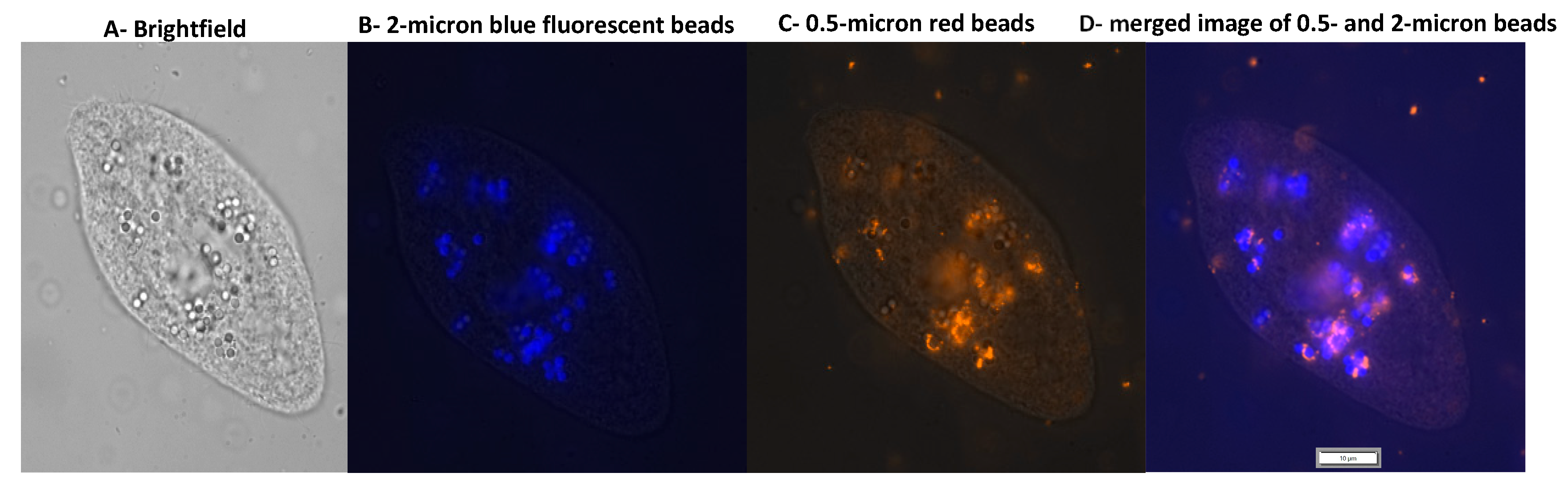
2.3. Uptake of MPs by T. pyriformis

The uptake and accumulation of MPs were measured by actively growing T. pyriformis culture, microscopic observation, and the observation of phagosomes containing MPs by compound light microscopy. For fluorescence microscopy, 50 µL of microbeads fed to T. pyriformis were collected by centrifugation at 1500 rpm for 2 min. Forty microliters of the supernatant were removed carefully, and the remaining 10 μL of the culture was mixed with 10 μL of Karnovsky’s fixative. About 10 μL of the sample was aliquoted on a glass slide, covered with glass coverslips, sealed at the edges, and viewed under fluorescence microscopy (Olympus IX83 microscope, Olympus Corporation of the Americas, PA 18034 USA).

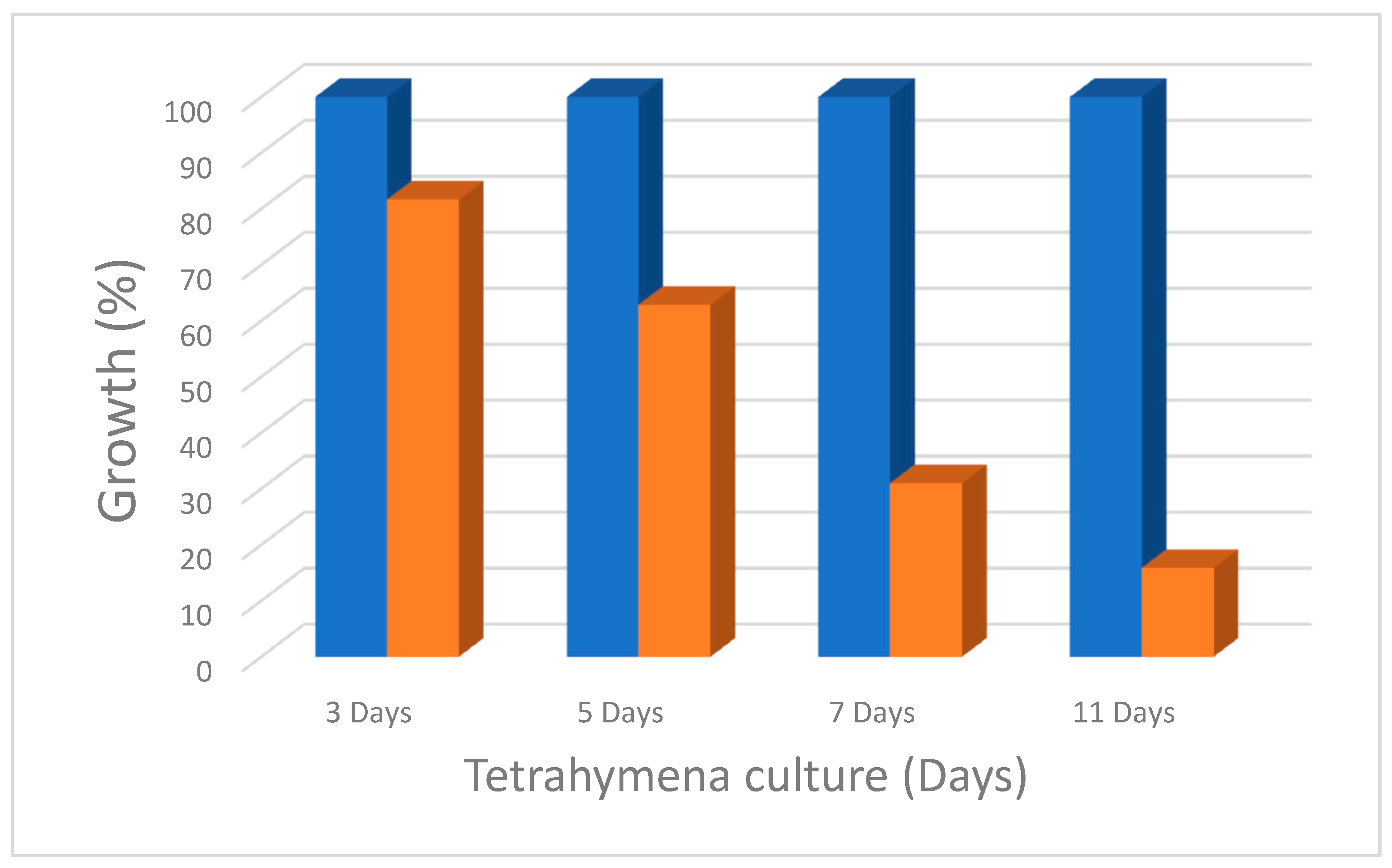
2.4. Measurement of Growth Inhibition

Growth inhibition of T. pyriformis by MP was measured using Lugol solution: T. pyriformis was stained/fixed with Lugol solution (Orange/brown solution), and the number of T. pyriformis was calculated by compound light microscopy. The actively growing T. pyriformis culture was diluted at a ratio of 1:20 in 5 mL protease peptone medium in 15 mL culture tubes, 10 μL of PBS was added into the culture, and this culture was used as a positive control for the growth inhibition assay. Three additional culture tubes were also prepared, and 10 μL of 0.05-, 0.5-, and 2-micron polystyrene beads diluted in PBS were added. All four cultures were maintained at 25 °C for 2 weeks. Every 24 h, five microliter aliquots of the cultures from all four tubes were mixed with 1 uL of Lugol solution, and the total number of T. pyriformis was obtained by counting the Lugol-stained T. pyriformis by microscopic observation of the organism.

2.5. Measurement of the Level of Reactive Oxygen Species

To examine whether polystyrene microbeads induce oxidative stress in T. pyriformis, intracellular ROS levels were measured using 2′7′-dichlorodihydrofluorescein diacetate (H2DCFDA; Life Technologies Corporation, Carlsbad, CA 92008 USA), which was oxidized to fluorescent dichlorofluorescein (DCF) by intracellular ROS. An actively growing T. pyriformis culture was fed with 0.05 µM plastic particles for 48 h. We observed that more than 90% of the cells contained plastic particles in their food vacuoles or cytoplasm. After exposure to different concentrations (0.1, 1, 10, and 20 μg/mL) of microbeads for 24 h, H2DCFDA was added to the culture and incubated for 2 and 16 h at 25 °C to allow the probe to enter the cells. After the incubation, equal volumes of the cultures from both MP-exposed and unexposed control samples were collected by centrifugation and processed for ROS measurement. T. pyriformis cells were homogenized in a buffer containing 0.32 M sucrose, 20 mM HEPES, 1 mM MgCl2, and 0.5 mM PMSF (pH 7.4) with a Teflon homogenizer. The homogenate was centrifuged at 10,000× g for 20 min at 4 °C. The supernatant was then collected for measurements. Measurements were obtained with an excitation wavelength of 485 nm and an emission wavelength of 520 nm with a fluorescence spectroscope.

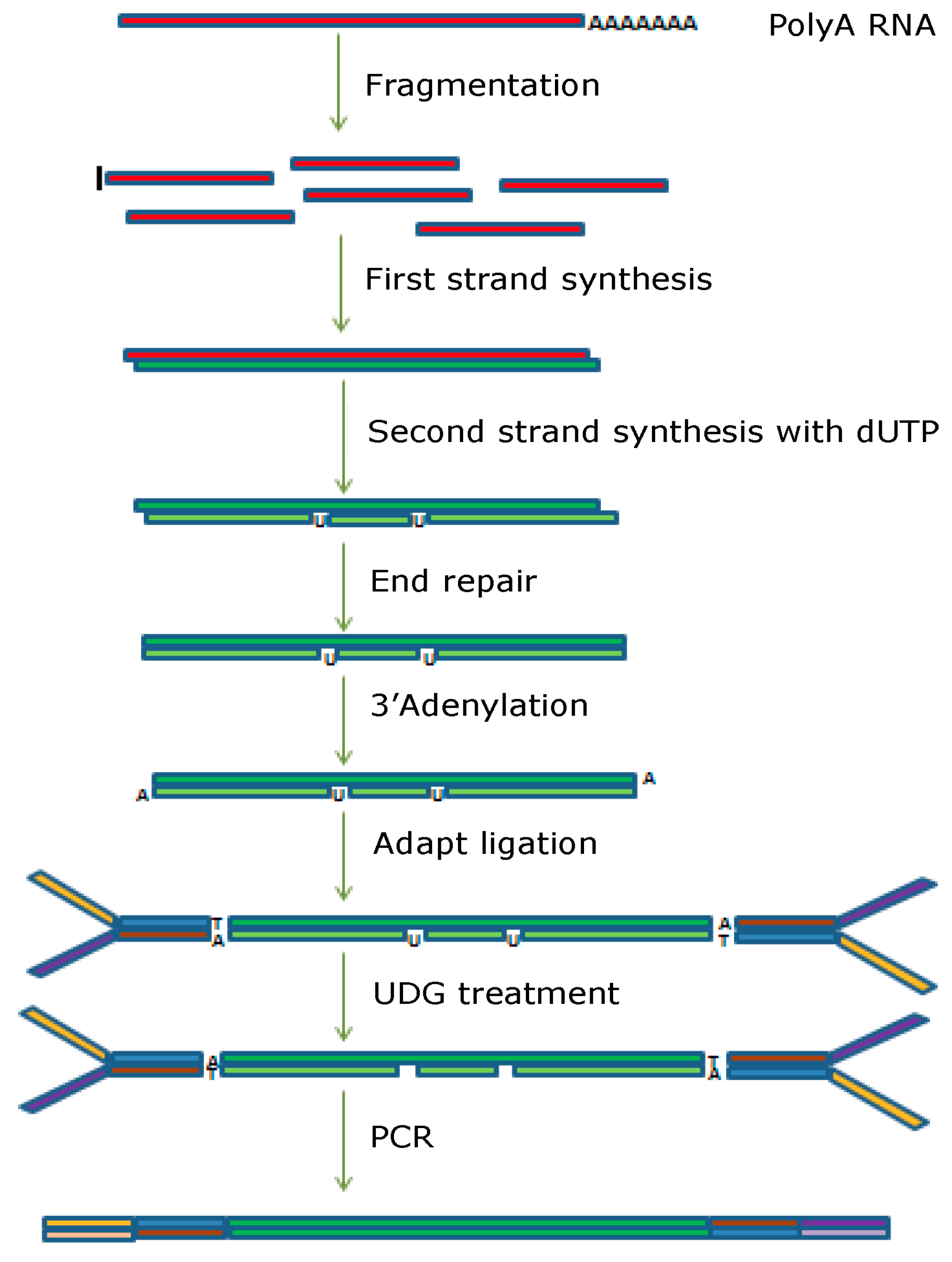
2.6. Differential Gene Expression Analysis by RNA Sequencing

RNA extraction—Total RNA was extracted using a Pure Link RNA mini kit as described by the manufacturer. The quality of the total RNA was determined by 1.2% agarose gel electrophoresis. The total RNA concentration was determined using Nanospec. The quality of RNA was further determined by analyzing the ratio of 28S and 18S RNA and the RNA integrity number (RIN). The RNA samples that showed a RIN value of 8.0 or above were selected and used for RNA-seq analysis. RNA integrity was checked with Agilent Technologies 2100 Bioanalyzer. The sample preparation for RNA-Seq analysis is provided in Figure 1.

Library Construction and Sequencing

Poly(A) RNA sequencing library was prepared following Illumina’s TruSeq-stranded-mRNA sample preparation protocol. RNA integrity was checked with Agilent Technologies 2100 Bioanalyzer. Poly(A) tail-containing mRNAs were purified using oligo-(dT) magnetic beads with two rounds of purification. After purification, poly(A) RNA was fragmented using a divalent cation buffer at elevated temperatures. The DNA library construction is shown in the following workflow. Quality control analysis and quantification of the sequencing library were performed using Agilent Technologies 2100 Bioanalyzer High Sensitivity DNA Chip. Paired-ended sequencing was performed on Illumina’s NovaSeq 6000 sequencing system.

2.7. Bioinformatics Analysis

2.7.1. De Novo Assembly, Unigene Annotation, and Functional Classification

Details for the bioinformatics analysis are provided in the Supplementary Materials. In short, firstly, Cutadapt and Perl scripts in-house developed at LC Sciences were used to remove the reads that contained adaptor contamination, low-quality bases and undetermined bases. Then, the sequence quality was verified using FastQC, including the Q20, Q30, and GC content of the clean data. All downstream analyses were based on clean, high-quality data. De novo assembly of the transcriptome was performed with Trinity 2.4.0. Trinity groups transcripts into clusters based on shared sequence content. Such a transcript cluster is very loosely referred to as a ‘gene’. The longest transcript in the cluster was chosen as the ‘gene’ sequence (aka Unigene). All assembled Unigenes were aligned against the non-redundant (Nr) protein database (the Supplementary Materials provide detailed information).

2.7.2. Differential Expression Analysis of Unigenes

Salmon is a system used for the quantification of transcript expression, which was used to perform expression level analysis for Unigenes. The differentially expressed Unigenes were selected with log2 (fold change) > 1 or log2 (fold change) < −1 and with statistical significance (p-value < 0.05) by R package edgeR (Please refer to Supplementary Materials S1 for the bioinformatics pipeline for de novo RNA sequencing).

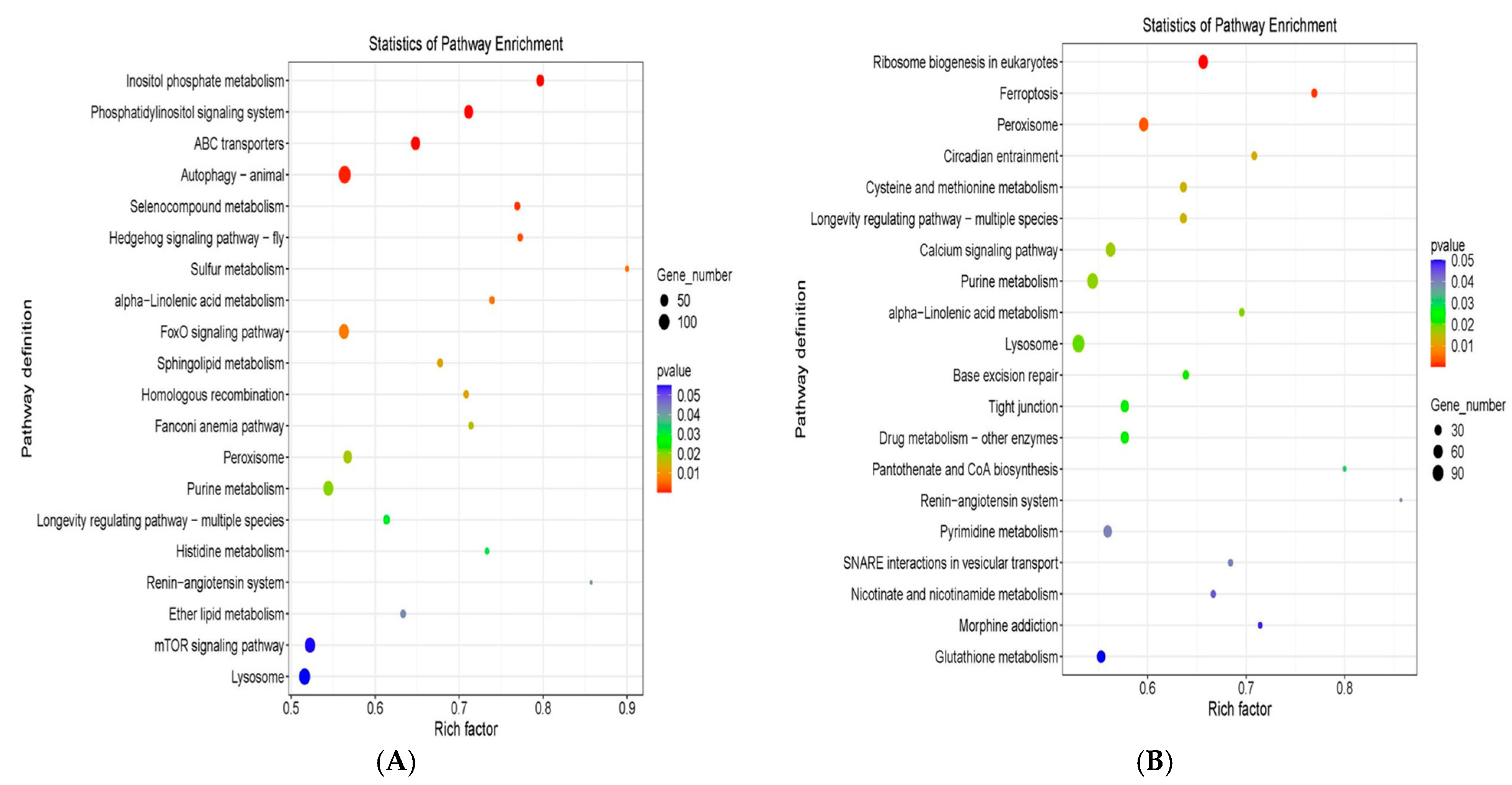
2.7.3. KEGG Enrichment Analysis of Differentially Expressed Genes

The Kyoto Encyclopedia of Genes and Genomes (KEGG) is a collection of databases dealing with genomes, biological pathways, diseases, drugs, and chemical substances. KEGG is utilized for bioinformatics research and education, including data analysis in genomics, metagenomics, metabolomics, and other omics studies, modeling and simulation in systems biology, and translational research in drug development. Significant KEGG pathways were calculated using the hypergeometric equation (please refer to the Supplementary Materials for additional details).

3. Results

3.1. Uptake of MPs and NPs by Tetrahymena pyriformis

Generally, the uptake of MPs and NPs is mediated by ingestion, absorption, inhalation, and food consumption, including water. The uptake in unicellular Eukaryotes such as Tetrahymena or other protozoans is either by phagocytosis or ingestion [17]. How the ingested particles are transported to various other tissues is not clearly defined or studied in multicellular organisms. This study investigated the uptake of MPs and NPs in a well-characterized model organism, T. pyriformis. In T. pyriformis, we measured the uptake of 0.05, 0.5, and 2-micron-sized fluorescent polystyrene beads using light and fluorescence microscopy. We observed 2.0- and 0.5-micron PS MP particles accumulated within phagosomes (Figure 2B,C); 0.05-micron PS MP beads were not visible under light microscopy even at 100× magnification but were observed throughout the cytoplasm in fluorescence microscopy images (Figure 2A). We believe that these smaller particles were taken up by endocytosis through the cell membrane (as [13,18] observed in human cells) and then dispersed into the cytosol and may be localized on other subcellular organelles. We observed that under long-term culture with 0.5- and 2-micron polystyrene beads, T. pyriformis phagocytosed both 0.5 and 2-micron size beads (Figure 3B,C). The ciliary action of T. pyriformis may propel beads in a non-selective manner into the cytostome and form discrete vacuoles containing mixed PS beads (Figure 3D). This observation is crucial for understanding how different particles interact with cellular structures and can provide insights into their potential impacts on cellular functions. By elucidating the mechanisms of endocytosis and subsequent intracellular trafficking, researchers can gain a deeper understanding of how particles are internalized and distributed within cells. This knowledge is essential for identifying the specific cellular pathways and organelles that are affected by particle uptake. Additionally, understanding these interactions can reveal how particles influence cellular processes such as signal transduction, gene expression, and metabolic activities. Such insights are vital for assessing the potential toxicological effects of various particles, including microplastics and nanoplastics, on cellular health and function. Ultimately, this information can inform the development of strategies to mitigate the adverse effects of particle exposure on human health and the environment.

3.2. Growth Inhibition of T. pyriformis by Polystyrene Micro and Nano Plastics

We also examined the physiological effect of PS-MP and PS-NP beads and found a profound inhibitory effect on the growth of T. pyriformis by all sizes of the tested beads. As shown in Figure 4, T. pyriformis, which ingested the 0.5-micron beads, showed a 90% reduction in growth rate. In particular, the T. pyriformis that ingested 0.05- and 2-micron beads showed 50–60% growth inhibition. The difference is likely due to the beads’ modification; the 0.5-micron size was carboxylated, while the others were amine-modified. The PMA-MPs showed 40–50% growth inhibition.

3.3. RNA-Seq (Transcriptome) Analysis of PS-MP and PMA-MP Exposed T. pyriformis

To understand the altered gene expression in PS-MP and PMA-MP-exposed T. pyriformis, we performed RNA-seq analysis to get the complete transcriptome as described in Section 2.1. All the experimental samples were repeated twice. The quality control and statistics details of the reads are provided in Table 2, and the bioinformatics analysis was carried out as described in Section 2.6.
The transcriptome of T. pyriformis exposed to PS-MPs and PMA-MPs reveals altered expressions of various signaling pathways.
As shown in Figure 5A, we observed an altered expression of inositol phosphate metabolism, Phosphatidylinositol signaling pathways, pathways related to autophagy, peroxisomes, mTOR signaling, and ABC transporters. Interestingly, the transcriptome profile of PMA-MP-exposed T. pyriformis differs completely except for a few pathways, such as the peroxisome, lysosome, and renin-angiotensin pathways (Figure 5B). Ferroptosis pathway genes are upregulated with a very high Rich factor and a p-value of less than 0.01. This is important because PMA-MP did not cause severe growth inhibition as we observed for PS-MP. The ferroptosis pathway is one of the significant regulators of cancer in higher-level organisms.

3.4. The Mechanism of Growth Inhibition

The transcriptome analysis revealed that several genes belonging to various signaling pathways that are related to oxidative damage response are modulated. We decided to measure oxidative damage in response to polystyrene and polymethacrylate. To this effect, T. pyriform was grown in nutrient broth, and polystyrene particles were added to the cultures and were grown for 48 h. The uptake of PS beads was confirmed by microscopy, and the presence of PS particles was observed in more than 90% of the cells. At this time, the oxidative damage caused by 0.5-micron PS MPs and PMA MPs was assessed by using the H2DCFDA compound. H2DCFDADA was added into the culture, and aliquots were taken 2 and 16 h after the treatment. Cells were collected by centrifugation, washed in PBS buffer, and lysed using Triton X-100. The cell debris was removed by centrifugation, and the cytoplasmic extract was used to measure the oxidative damage caused by PS MPs. As shown in Figure 6, the fluorescence of the DCFDA resulted from the reaction of H2DCFDA with the free radicals from the oxidative damage of PS MPs. Interestingly, the fluorescence intensity exhibited after 16 h of exposure is 1.5 times higher than T. pyriformis exposed for 2 h. We also tested the oxidative damage caused by PMA MPs to T. pyriformis and found that the PMA MPs did not cause any oxidative damage when compared with T. pyriformis not treated with MPs. This suggests that different types of micro- and nanoplastics may cause differential effects on the growth and metabolism of T. pyriformis. These results suggest that oxidative damage caused by various types of microplastics may not be the same. The growth inhibition observed may be because of oxidative stress or other pathways modulated by PSMPs, as observed by RNA-seq experiments.

4. Discussion

4.1. Chemical Modifications of PS-MP and PMA-MP Particles and Their Effect on the Growth Inhibition Response in T. pyriformis

In this study, we observed that T. pyriformis uptakes various-sized polystyrene micro and nanoparticles, and as shown in Figure 2C, the localization of the 0.05-micron size NPs indicated that they are distributed throughout the cytoplasm of T. pyriformis and 0.5 and 2-micron size particles are localized in vesicles [19]. This suggests that T. pyriformis may take up the particles both by endocytosis of the plasma membrane and the propulsion of the microparticles through the gullet and the eventual formation of the food vacuoles [20]. The uptake of various MPs indicated that there is no selective uptake of PS particles by T. pyriformis, but the uptake mechanism of various particles may be different [21]. Among the three PS particles used in this study, 0.05 and 2-micron PS are chemically modified at the amino-terminal end of the particles, and the 0.5-micron particles are modified at the carboxy-terminal end of the PS particles [19]. The growth inhibition studies reveal that carboxy-terminal modified 0.5-micron PS shows more than 85% growth inhibitory effect on T. pyriformis compared with amino-terminal modified 0.05 and 2.0-micron size beads. This finding also suggests that the growth inhibition effect is not directly correlated with the size of the beads, and it is more with the type of chemical modification of the MPs and NPs [22]. Microplastics can undergo intentional chemical modifications, such as the addition of functional groups like amino (NH2) or carboxyl (COOH) groups. These modifications can enhance the adsorption capacity of microplastics for specific pollutants. These modifications can significantly impact the behavior and ecological effects of microplastics on the environment. Understanding these processes is crucial for assessing the risks associated with microplastic pollution. Modified microplastics with amino or carboxyl groups can enhance the adsorption capacity of microplastics for heavy metals and organic compounds as well. Hence, we believe these modifications impact the biological functions of micro- and nanoplastics, and studies using microplastics with modifications are important to understanding their biological significance.

4.2. Variation in the Oxidative Damage Caused by Various Types of MPs and NPs

MPs and NPs have been shown to cause increased oxidative stress, inflammation, and altered metabolism, leading to cellular damage, which ultimately affects tissue and organismal homeostasis in numerous animal species and human cells [23,24]. A recent study on the effect of NPs on Tetrahymena thermophiles found that they led to the growth inhibition of the organism by affecting specific pathways, such as calcium signaling and phosphatidylinositol signaling [21]. In this study, we found that PS-MP particles altered various pathways, such as oxidative stress response, and many of these pathways may directly alter the oxidative response-related cellular process. Interestingly, PS-MS particle-induced oxidative response is much greater when compared with PMMS-induced oxidative response in T. pyriformis [19]. In general, oxidative response can affect various signaling pathways that regulate cell growth, survival, differentiation, and death, and further studies will be required to identify alterations of specific pathways or genes that are regulated by MPs and NPs uptake [20].

4.3. Alteration of PS-MP and PMS-MP-Induced Transcriptome Changes in T. pyriformis

Our study, along with several others, has indicated that exposure to microplastics (MPs) can result in significant changes in gene expression levels. These changes can manifest either up-regulation or down-regulation of specific genes. For instance, research has shown that exposure to MPs can lead to the up-regulation of genes involved in stress responses and detoxification pathways while simultaneously down-regulating genes associated with normal cellular functions and metabolic processes [25]. Genes related to immune response, stress, and metabolism may also show altered expression patterns [21]. This differential gene expression suggests that MPs can disrupt normal cellular activities and potentially lead to adverse health effects in organisms.
Oxidative stress can significantly impact various cellular processes in Tetrahymena, including inositol phosphate metabolism, autophagy, ABC transporters, and lipid peroxidation. Inositol phosphates play a crucial role in cellular signaling and stress responses. Under oxidative stress, the metabolism of inositol phosphates can be altered, affecting the regulation of ion channels, metabolic flux, and gene expression. This disruption can lead to impaired cellular homeostasis and survival, resulting in the growth inhibition we observed [26,27]. Autophagy is a cellular process that helps in the degradation and recycling of damaged organelles and proteins. Oxidative stress can induce autophagy as a protective mechanism to remove damaged components and maintain cellular health. However, excessive oxidative stress can overwhelm autophagic machinery, leading to cell damage and growth inhibition [28,29]. ATP-binding cassette (ABC) transporters are involved in the transport of various molecules across cellular membranes. Oxidative stress can affect the function of ABC transporters by altering their expression and activity. This can impact the efflux of toxic substances and the uptake of essential nutrients, further contributing to growth inhibition [30].
These and several other pathway processes are interconnected, and their disruption under oxidative stress can lead to significant cellular dysfunction in Tetrahymena. The observed growth inhibition and oxidative stress may be a cumulative effect of multiple pathways. Studies on individual pathway genes are required to identify the regulatory genes involved in oxidative stress and growth inhibition.

5. Conclusions

This study provides a comprehensive analysis of the uptake and growth inhibitory effects of microplastics (MPs) and nanoplastics (NPs) on the protozoan Tetrahymena pyriformis. Our results indicate that polystyrene (PS) MPs exert a significantly more pronounced inhibitory effect on the growth of T. pyriformis compared to polymethyl methacrylate (PMA) MPs. Detailed transcriptomic analysis revealed that exposure to PS-MPs and PMA-MPs led to differential expression of various signaling pathways within T. pyriformis. Specifically, PS-MPs were found to significantly impact oxidative stress-related pathways, while PMA-MPs influenced pathways associated with both carcinogenesis and oxidative stress.
The quantification of reactive oxygen species (ROS) using a fluorescent probe demonstrated that PS-MPs induced higher levels of ROS and oxidative stress than PMA-MPs. This suggests that the chemical composition and surface properties of MPs are critical determinants of their biological effects. The elevated oxidative stress observed with PS-MP exposure underscores the potential for these particles to cause significant cellular damage through oxidative mechanisms.
The findings underscore the necessity of understanding the specific chemical compositions and surface modifications of MPs to elucidate their distinct effects on various biological systems. Such knowledge is essential for accurately assessing the potential implications of MPs and NPs on human health. Our study highlights the need for further research into the mechanistic pathways underlying the biological impacts of different types of MPs and NPs. This will be critical for developing effective strategies to mitigate their adverse effects on environmental and human health.

Supplementary Materials

The following supporting information can be downloaded at: https://www.mdpi.com/article/10.3390/microplastics4020033/s1, Supplementary S1: Bioinformatics pipeline for de novo RNA sequencing.

Author Contributions

Conceptualization, T.R. and M.J.; methodology, T.R.; validation, T.R.; formal analysis, T.R.; investigation, T.R. and A.A.; resources, T.R. and M.J.; writing—original draft preparation, T.R.; writing—review and editing, T.R., M.J. and A.A.; project administration, T.R.; funding acquisition, M.J. and T.R.; All authors have read and agreed to the published version of the manuscript.

Funding

This research was funded by NSF (grant # 2040364—Establishing the Center for Emerging Contaminants (ICEC) at Coppin State University) and the Takeda STEM HBCU I3 Fellows Program grant.

Institutional Review Board Statement

Not applicable.

Informed Consent Statement

Not applicable.

Data Availability Statement

The original contributions presented in this study are included in the article/Supplementary Material. Further inquiries can be directed to the corresponding author.

Acknowledgments

William Ghann and Jude Namanga for their assistance in the fluorescence spectrophotometric analysis. We thank Sulakshana Bhatt and Kavita Hegde for reviewing the manuscript.

Conflicts of Interest

The authors declare that there are no conflicts of interest.

References

  1. Thompson, R.C.; Olsen, Y.; Mitchell, R.P.; Davis, A.; Rowland, S.J.; John, A.W.G.; McGonigle, D.; Russell, A.E. Lost at sea: Where is all the plastic? Science 2004, 304, 838. [Google Scholar] [CrossRef] [PubMed]
  2. Galloway, T.S. Micro- and nano-plastics and human health. In Marine Anthropogenic Litter; Bergmann, M., Gutow, L., Klages, M., Eds.; Springer: Berlin/Heidelberg, Germany, 2015; pp. 343–366. [Google Scholar] [CrossRef]
  3. Amobonye, A.; Bhagwat, P.; Raveendran, S.; Singh, S.; Pillai, S. Environmental impacts of microplastics and nanoplastics: A current overview. Front. Microbiol. 2021, 12, 768297. [Google Scholar] [CrossRef]
  4. Galloway, T.S. Micro- and nanoplastics in the environment: Research priorities for the near future. In Reviews of Environmental Contamination and Toxicology; Springer: Berlin/Heidelberg, Germany, 2021; Volume 257, pp. 163–218. [Google Scholar] [CrossRef]
  5. Hua, X.; Wang, D. Cellular uptake, transport, and organelle response after exposure to microplastics and nanoplastics: Current knowledge and perspectives for environmental and health risks. Rev. Environ. Contam. Toxicol. 2022, 260, 12. [Google Scholar] [CrossRef]
  6. Ruehle, M.D.; Orias, E.; Pearson, C.G. Tetrahymena as a unicellular model eukaryote: Genetic and genomic tools. Genetics 2016, 203, 649–665. [Google Scholar] [CrossRef] [PubMed]
  7. Xiong, J.; Lu, X.; Zhou, Z.; Chang, Y.; Yuan, D.; Tian, M.; Zhou, Z.; Wang, L.; Fu, C.; Orias, E.; et al. Transcriptome analysis of the model protozoan, Tetrahymena thermophila, using deep RNA sequencing. PLoS ONE 2012, 7, e30630. [Google Scholar] [CrossRef]
  8. Zhang, L.; Li, Y.; Li, W.; Liu, X. Immunotoxicity and intestinal effects of nano- and microplastics: A review of the literature. Part. Fibre Toxicol. 2020, 17, 57. [Google Scholar] [CrossRef]
  9. Li, Y.; Zhang, X.; Wang, L.; Chen, Z. Liver injury induced by exposure to polystyrene microplastics alone or in combination with cadmium in mice is mediated by oxidative stress and apoptosis. Biol. Trace Elem. Res. 2019, 191, 2170–2183. [Google Scholar] [CrossRef]
  10. Chu, E.; Mychasiuk, R.; Hibbs, M.L.; Semple, B.D. Dysregulated phosphoinositide 3-kinase signaling in microglia: Shaping chronic neuroinflammation. J. Neuroinflamm. 2021, 18, 276. [Google Scholar] [CrossRef]
  11. Katan, M.; Cockcroft, S. Phosphatidylinositol (4,5) bisphosphate: Diverse functions at the plasma membrane. Essays Biochem. 2020, 64, 513–531. [Google Scholar] [CrossRef]
  12. Kumar, R.; Islinger, M.; Worthy, H.; Carmichael, R.; Schrader, M. The peroxisome: An update on mysteries 3.0. Histochem. Cell Biol. 2024, 161, 99–132. [Google Scholar] [CrossRef]
  13. Chen, X.; Yu, C.; Kang, R.; Tang, D. Iron metabolism in ferroptosis. Front. Cell Dev. Biol. 2020, 8, 590226. [Google Scholar] [CrossRef] [PubMed]
  14. Piccolo, M.; Ferraro, M.G.; Iazzetti, F.; Santamaria, R.; Irace, C. Insight into iron, oxidative stress and ferroptosis: Therapy targets for approaching anticancer strategies. Cancers 2024, 16, 1220. [Google Scholar] [CrossRef] [PubMed]
  15. Brito WA, S.; Mutter, F.; Wende, K.; Cecchini, A.L.; Schmidt, A.; Bekeschus, S. Consequences of nano and microplastic exposure in rodent models: The known and unknown. Part. Fibre Toxicol. 2022, 19, 28. [Google Scholar] [CrossRef] [PubMed]
  16. Chen, P.Z.; Li, R.; Lai, K.P.; Zhang, X.X. Biological exposure to microplastics and nanoplastics and plastic additives: Impairment of glycolipid metabolism and adverse effects on metabolic diseases. Environ. Sci. Pollut. Res. 2024, 31, 60778–60791. [Google Scholar] [CrossRef]
  17. Mondellini, S.; Schwarzer, M.; Völkl, M.; Jasinski, J.; Jérôme, V.; Scheibel, T.; Laforsch, C.; Freitag, R. Size-dependent uptake and trophic transfer of polystyrene microplastics in unicellular freshwater eukaryotes. Sci. Total Environ. 2024, 929, 172470. [Google Scholar] [CrossRef]
  18. Kim, E.-H.; Choi, S.; Kim, D.; Park, H.J.; Bian, Y.; Choi, S.H.; Chung, H.Y.; Bae, O.-N. Amine-modified nanoplastics promote the procoagulant activation of isolated human red blood cells and thrombus formation in rats. Part. Fibre Toxicol. 2022, 19, 60. [Google Scholar] [CrossRef]
  19. Smith, A.; Johnson, B.; Williams, C. Localization of nanoplastics in T. pyriformis. J. Environ. Sci. 2022, 45, 123–135. [Google Scholar]
  20. Jones, D.; Lee, E. Uptake mechanisms of microplastics in protozoa. Environ. Toxicol. Chem. 2023, 42, 67–78. [Google Scholar]
  21. Brown, F.; Green, G.; Black, H. Chemical modifications and growth inhibition in T. pyriformis. Environ. Res. 2023, 56, 200–215. [Google Scholar]
  22. Reichelt, S.; Gorokhova, E. Micro- and nanoplastic exposure effects in microalgae: A meta-analysis of standard growth inhibition tests. Front. Environ. Sci. 2020, 8, 131. [Google Scholar] [CrossRef]
  23. Kiran, B.R.; Kopperi, H.; Mohan, S.V. Micro/nano-plastics occurrence, identification, risk analysis and mitigation: Challenges and perspectives. Rev. Environ. Sci. Bio/Technol. 2022, 21, 169–203. [Google Scholar] [CrossRef] [PubMed]
  24. Prattichizzo, F.; Ceriello, A.; Pellegrini, V.; La Grotta, R.; Graciotti, L.; Olivieri, F.; Paolisso, P.; D’Agostino, B.; Iovino, P.; Balestrieri, M.L. Micro-nanoplastics and cardiovascular diseases: Evidence and perspectives. Eur. Heart J. 2024, 45, 4099–4110. [Google Scholar] [CrossRef] [PubMed]
  25. Poma AM, G.; Morciano, P.; Aloisi, M. Beyond genetics: Can micro and nanoplastics induce epigenetic and gene-expression modifications? Front. Epigenetics Epigenomics 2023, 1, 1241583. [Google Scholar] [CrossRef]
  26. Berridge, M.J.; Irvine, R.F. Inositol phosphates and cell signalling. Nature 1989, 341, 197–205. [Google Scholar] [CrossRef] [PubMed]
  27. Divecha, N.; Irvine, R.F. Phospholipid signaling. Cell 1995, 80, 269–278. [Google Scholar] [CrossRef]
  28. Zhang, S.-W.; Feng, J.-N.; Cao, Y.; Meng, L.-P.; Wang, S.-L. Autophagy prevents autophagic cell death in Tetrahymena in response to oxidative stress. Zool. Res. 2015, 36, 167–176. [Google Scholar]
  29. Ye, Q.; Zhang, C.; Wang, Z.; Feng, Y.; Zhou, A.; Xie, S.; Xiang, Q.; Song, E.; Zou, J. Induction of oxidative stress, apoptosis, and DNA damage by koumine in Tetrahymena thermophila. PLoS ONE 2019, 14, e0212231. [Google Scholar] [CrossRef]
  30. Liu, Y.; Zhang, J.; Xiao, P.; Liu, X.; Ma, Y.; Zhang, J.; Zhang, B. Effects of pyraclostrobin on growth, oxidative stress, and gene expression related to stress and ATP-binding cassette transporters in Tetrahymena thermophila. J. Oceanol. Limnol. 2024, 42, 150–162. [Google Scholar] [CrossRef]
Figure 1. Workflow of the mRNA processing, cDNA preparation, repair, and sample preparation for RNA-seq analysis.
Microplastics 04 00033 g001
Figure 2. Uptake and localization of polystyrene beads by T. pyriformis. Uptake of 0.05-micron (A), 2-micron (B), 0.5-micron (C), and 1-micron Polymethacrylate. (A1,B1,C1)—Brightfield, (A2,B2,C2)—Fluorescence images collected using DAPI channel (A2,B2) or RED channel (C2) and merged with brightfield images, which show the 0.05-micron beads accumulated in the cytoplasm and 0.5 micron and 2-micron beads accumulated in food vacuoles. The bar is 20 µM.
Microplastics 04 00033 g002
Figure 3. Non-selective uptake of 0.5- and 2-micron polystyrene beads by T. pyriformis. Nonselective uptake of different-sized PS MPs was measured by fluorescent microscopy: (A) Brightfield, (B) Blue channel, (C) Red channel, and (D) merged image of (B,C). The scale bar is 10 µM.
Microplastics 04 00033 g003
Figure 4. Growth inhibition. Growth inhibition of T. pyriformis by 0.5-micron PS MPs was measured by culturing T. pyriformis with PS beads for 11 days, and the growth was monitored by microscopy. T. pyriformis was stained/fixed with Lugol solution, and the total number of T. pyriformis was calculated by microscopy. The orange bars represent the growth pattern of T. pyriformis in the presence of PS-MPs, and the blue bars represent the growth pattern without PS-MPs.
Microplastics 04 00033 g004
Figure 5. RNA-seq Enrichment Analysis of T. pyriformis exposed to 0.5-Micron PS MPs and PMA MPs. The scatter plot shows the top few KEGG enrichments. The Rich factor is the ratio of differentially expressed gene numbers annotated in this pathway term to all gene numbers annotated in this pathway term: the greater the Rich factor, the greater the degree of pathway enrichment. The x-axis represents the enrichment score, and the y-axis lists the pathways. Colors indicate the significance level (p-value), with darker shades representing higher significance. A p-value ranging from 0 to 1, and a lower value indicates greater pathway enrichment. (A) 50, 100 genes significance based on the p-value (red highly significant and blue least significant). (B) 30, 60, 90 genes significance based on the p-value (red highly significant and blue least significant).
Microplastics 04 00033 g005
Figure 6. Measurement of Reactive Oxygen Species (ROS): The total number of ROS as a measure of fluorescence density in the supernatant was determined using a Fluorescent Spectrophotometer (model). Measurements were obtained with an excitation wavelength of 485 nm and an emission wavelength of 520 nm with a fluorescence spectroscope (Horiba Nanolog iHR 320). The identity of the samples was presented as an insert within the figure. TP—Polystyrene and H2DCFDA are not added in the culture, and TP FDA and TP FDA16—only H2DCFDA incubated, but no polystyrene added, PS—Polystyrene exposed but no HEDCFDA, PS FDA and PS FDA 16—both polystyrene and H2DCFDA were added. Emission intensity means the non-fluorescent H2DCFDA, which is oxidized by ROS generated by PS MPs to green fluorescent DCFDA. The number 16000000 is 16,000,000, and 2000000 is 2,000,000.
Microplastics 04 00033 g006
Table 1. Composition and properties of polystyrene and polymethacrylate beads used in this study.
Cat #Type of MicroplasticsSurface Functional GroupAverage Particle Size Micron (Mean Diameter)Dye Content (%)Polymer Density (g/mL)Solid Content (%)
L0780PolystyreneAmine Modified0.045–0.055≥0.1 (blue Fluorescent)1.04–1.06≥2.5
L3280PolystyreneCarboxylate Modified0.4–0.60.2–0.6 (red fluorescent)1.04–1.052.5
L0280PolystyreneAmine Modified1.90–2.20≥0.1 (blue fluorescent)1.04–1.062.4–2.6
90,875PolymethacrylateNone0.92–1.0Non-fluorescent1.1910
Table 2. Quality Control and Statistics of Reads.
SampleRaw_ReadsRaw_BasesValid_Reads
PMMA142,055,9246.31G41,495,164
PMMA249,347,1447.40G48,728,388
PS146,459,3346.97G45,922,588
PS242,432,9426.36G41,856,014
Tp41,112,5446.17G40,552,504
Disclaimer/Publisher’s Note: The statements, opinions and data contained in all publications are solely those of the individual author(s) and contributor(s) and not of MDPI and/or the editor(s). MDPI and/or the editor(s) disclaim responsibility for any injury to people or property resulting from any ideas, methods, instructions or products referred to in the content.

