Article

Regulation of DNA Methylation Through EBP1 Interaction with NLRP2 and NLRP7

by
Nayeon Hannah Son
,
Matthew So
and
Christopher R. Lupfer
*,‡
Department of Biology, Missouri State University, Springfield, MO 65897, USA
*
Author to whom correspondence should be addressed.
Current address: Department of Immunology, St. Jude Children’s Research Hospital, Memphis, TN 38105, USA.
Current address: Department of Biology, Brigham Young University-Idaho, Rexburg, ID 83460, USA.
Submission received: 24 June 2025 / Revised: 11 September 2025 / Accepted: 30 September 2025 / Published: 17 October 2025

Abstract

Background/Objectives: Mutations in NACHT, LRR and PYD domain-containing protein 2 (NLRP2) and NLRP7 genes, members of the NOD-like receptor (NLR) family of innate immune sensors, result in recurrent miscarriages and reproductive wastage in women. These genes have been identified to be maternal effect genes in humans and mice regulating early embryo development. Previous research in vitro suggests that NLRP2 and NLRP7 regulate DNA methylation and/or immune signaling through inflammasome formation. However, the exact mechanisms underlying NLRP2 and NLRP7 function are not well defined. Methods: To determine the interacting proteins required for NLRP2/NLRP7-mediated regulation of DNA methylation, yeast 2-hybrid screens, coimmunoprecipitation, and FRET studies were performed and verified the ability of novel protein interactions to affect global DNA methylation by 5-methylcytosine-specific ELISA. Results: Various methodologies employed in this research demonstrate a novel protein interaction between human ErbB3-binding protein 1 (EBP1, also known as proliferation-associated protein 2G4 (PA2G4) and NLRP2 or NLRP7. In addition, NLRP2 and NLRP7 regulate EBP1 gene expression. Functionally, global DNA methylation levels appeared to decrease further when NLRP2 and NLRP7 were co-expressed with EBP1, although additional studies may need to confirm the significance of this effect. Conclusions: Since EBP1 is implicated in apoptosis, cell proliferation, DNA methylation, and differentiation, our discovery significantly advances our understanding of how mutations in NLRP2 or NLRP7 may contribute to reproductive wastage in women through EBP1.

Graphical Abstract

1. Introduction

Recurrent miscarriage (RM) is the spontaneous loss of three or more consecutive pregnancies before 20 weeks of gestation. RM affects 1–5% of all women of reproductive age [1,2]. Women with RM have a broad range of hormonal or genetic alteration, maternal infections, immune disorders, and other contributing factors [3]. However, the causes of RM are still not clearly understood, and many studies are contradictory, suggesting further investigation is needed to understand the pathological mechanisms involved in RM. The nucleotide-binding domain and leucine-rich repeat containing receptors (NLRs) are generally classified as innate immune receptors expressed by cells including macrophages, dendritic cells, and others. Many NLRs have a critical role in regulating inflammation, cell death, and immune responses [4,5]. Among them, NLR family pyrin domain containing 2 (NLRP2) and NLRP7 have been reported to be maternal effect genes, and mutations in both genes are associated with RM [6,7,8]. Germline mutations of NLRP7 are known to cause hydatidiform molar (HM) pregnancy and recurrent HM patients have a high rate of chromosomal abnormalities in early embryo development [9]. Also, mutations in NLRP2 are known to cause the human DNA methylation imprinting disorder Beckwith–Wiedemann syndrome (BWS), with dysregulated gene imprinting on chromosome 11p15.5 [10].
NLRP2 and NLRP7 are members of the pyrin domain containing NLR family group. Although proposed to have immune functions in vitro, most human studies associate polymorphisms in these NLRs with RMs [11,12]. Importantly, mutations in NLRP2 and NLRP7 result in aberrant DNA methylation patterns in developing embryos. Furthermore, NLRP2 and NLRP7 tend to have unique expression patterns in human oocytes compared to other NLRs during embryogenesis [13]. Therefore, understanding the protein interactions and cellular pathways regulated by NLRP2 and NLRP7 may provide important breakthroughs for epigenetics or embryogenesis research as well as elucidating any role for these proteins in the immune system. For these reasons, NLRP2 and NLRP7 were specifically selected for this study. Both genes are classified as maternal effect genes and have been strongly linked to reproductive failure and epigenetic dysregulation, particularly abnormal DNA methylation in oocytes and early embryos [6,7,8,9,10]. In contrast, other NLRP family members such as NLRP1 and NLRP3 are primarily involved in immune responses through inflammasome formation and have not been associated with early developmental regulation [4,5,14]. To validate the specificity of our observations, we also included NLRP12 as a negative control, as it is known to have an immune-specific function and lacks any known reproductive phenotype [11,12].
ErbB3-Binding Protein 1 (EBP1), also known as proliferation-associated 2G4 (PA2G4), is involved in the regulation of cellular growth and differentiation in a wide range of cancers, including neuroblastoma, cervical, brain, breast, and other tumors, and has also been shown to play a role in fetal development and DNA methylation [15,16]. Given its known roles in development and epigenetics regulation, EBP1 is a promising candidate for interaction with NLRP2 and NLRP7. This study aimed to uncover and analyze proteins interacting with NLRP2 and NLRP7 that may influence epigenetic pathways. The results of this study indicate that both NLR proteins associate with common regulatory factors such as EBP1, and that these interactions may impact DNA methylation processes critical to early developmental stages.

2. Materials and Methods

2.1. Plasmid Constructs

Plasmids for the yeast two-hybrid screen were generated by cloning NLRP2/7 genes into the pGBKT7 plasmid. The purified pGBKT7-NLRP2 and pGBKT7-NLRP7 plasmids were sequenced to verify proper gene insertion and the absence of any mutations. Expression of proteins containing the tags and correct molecular weights in yeast cells was verified by Western blot prior to performing experiments.
To study identified protein interactions in human cells, NLRP2/7 were cloned into a mammalian expression vector pCDNA3.1 3xMyc using Gibson cloning (Cat. E2621S/L/X, Cat. E5520S, New England Biolabs, Ipswich, MA, USA) following the manufacturer’s protocol. The purified pCDNA3.1 3xMyc-NLRP2 and pCDNA3.1 3xMyc-NLRP7 were sequenced to verify proper gene insertion and the absence of any mutations. The pCMV-HA-EBP1 plasmid was a gift from Stephen Goff (Addgene plasmid #67792; https://www.addgene.org/67792/ URL (accessed on 24 June 2025); RRID:Addgene_67792) [17]. Expression of proteins containing the tags and correct molecular weights in HEK293T cells were verified by Western blot prior to performing experiments.

2.2. Yeast Two-Hybrid Screen

To screen for novel protein interactions, Y2H Gold yeast were transformed with one of the bait plasmids (pGBKT7-hNLRP2, and pGBKT7-hNLRP7) using the Frozen-EZ Yeast Transformation II Kit (Cat. T2001, Zymo Research, Irvine, CA, USA). Yeast containing the bait plasmid was mated with yeast containing a human cDNA library (Cat. 630479, Clontech, Mountain View, CA, USA) following the Matchmaker GAL4-based two-hybrid assay protocol (Cat. 630466, 630489, 630498, 630499). Mated yeast was plated on triple dropout medium and over 100 colonies transferred to quadruple dropout medium to identify clones with strong interactions. Prey plasmids from yeast displaying strong interactions were purified via the EZ yeast plasmid kit (Cat. D2004, Zymogen, Irvine, CA, USA), and genes amplified by PCR using insert screening amplimers (Table 1) and DreamTaq polymerase (Cat. EP0701, ThermoFisher Scientific, Waltham, MA, USA) for 35 cycles at 95 °C for 10 s, 55 °C for 30 s, and 72 °C for 2 min after an initial denaturation step of 95 °C for 3 min. PCR products were sequenced by Sanger sequencing followed by analysis using BLASTN to identify the interacting protein genes.

2.3. Co-Immunoprecipitation

HEK293T cells were transfected with the indicated mammalian plasmids or GFP expression vector (as a control) using LipoFexin (Cat. TS310, LAMDA BIOTECH, Ballwin, MO, USA) with 2–3 µg of the Myc- and/or HA-tagged expression plasmid. After 24 h, media were removed and 0.5 mL of cold lysis buffer (1% NP-40 in PBS+ protease inhibitor (Cat. A32953, Pierce, Waltham, MA, USA) + phosphatase inhibitor (Cat. P2850-1ml, Sigma-Aldrich, St. Louis, MO, USA)) was added. Plates were incubated on a shaker for 30 min at 4 °C. After 30 min, cells and lysates were passed 10 times through a Dounce homogenizer on ice to fully disrupt cells. Then, homogenates were centrifuged at 5000× g for 10 min at 4 °C to remove cellular debris. After centrifugation, 300–500 µL of supernatant was transferred into new 1.5 mL micro-centrifuge tubes and 2 µg of appropriate antibody added (anti-Myc, Cat. 631206, Clontech, Mountain View, CA, USA; anti-HA, Cat. UF281924, Invitrogen, Carlsbad, CA, USA), and incubated on a shaker for one hour at 4 °C. 40 µL of protein A/G beads was then added to each tube, and samples incubated overnight on a shaker at 4 °C. Next day, samples were centrifuged at 2000× g for 1 min at 4 °C. Homogenates were removed, and protein A/G beads were washed 3–5 times with 1 mL of cold PBS with centrifugation at 2000× g for 1 min at 4 °C between each wash. Finally, 50 µL of 4X SDS-PAGE loading dye was added to protein A/G beads containing (co)-immunoprecipitates and samples boiled for 5 min at 95 °C.
To visualize the presence of proteins from the (co-)immunoprecipitation, 20 µL of each sample was separated by SDS-PAGE and immunoblotting performed after transfer to PVDF membranes with primary antibodies (1:1000; anti-Myc, Cat. 631206, Clontech, Mountain View, CA, USA; anti-HA, Cat. UF281924, Invitrogen, Carlsbad, CA, USA) and secondary antibodies (1:5000; anti-Mouse, Cat. 1706516, BIORAD, Hercules, CA, USA). The bands were detected with HRP-based chemiluminescence (Radiance Q, Cat. AC2101, Azure Biosystems, Dublin, CA, USA) on an Azure c300 digital imager.

2.4. Confocal Microscopy and Immunostaining

Clean glass coverslips were placed in each well prior to seeding HEK293T cells (1 × 10⁵ cells per well in 1 mL of media). Next day, cells were (co-)transfected with plasmids and as per the co-immunoprecipitation protocol above. After 24 h, cells were fixed using 4% paraformaldehyde (PFA) for 15 min at room temperature and washed with PBS 3 times. Cells were permeabilized with PBS containing 0.1% Triton X-100 for 1 h at 37 °C and washed with PBS 3 times. Afterward, cells were blocked with 1× ELISA/ELISPOT Diluent (Cat. 00-4202-56, Invitrogen, Carlsbad, CA, USA) for 30 min at room temperature and washed with PBS 3 times. Then, cells were incubated with each of the primary antibodies: anti-Myc Polyclonal (1:100) (Cat. PA1-981, Invitrogen, Carlsbad, CA, USA) and/or anti-HA Monoclonal (1:100) (Cat. 26183, Invitrogen, Carlsbad, CA, USA) overnight at 4 °C, followed by incubation with the appropriate secondary antibody for 1 h at 37 °C: Conjugated Goat anti-Mouse IgG (1:1000, FITC, Invitrogen, Carlsbad, CA, USA) or Conjugated Goat anti-Rabbit (1:1000, TxRed, Invitrogen, Carlsbad, CA, USA). All antibodies were diluted in 1× ELISA/ELISPOT Diluent. Stained cells were washed 4 times with PBS and mounted with DAPI Fluoromount-G® medium (Cat. 0100-20, SouthernBiotech, Birmingham, AL, USA) and examined with an Olympus FV1000 Confocal IX81 Microscope (Olympus, Center Valley, PA, USA) and Slidebook software v6 (intelligent Imaging Innovations, Denver, CO, USA).

2.5. Three-Channel Corrected FRET(c) Analysis

FRET imaging was performed on an Olympus FV1000 Confocal IX81 Microscope using Slidebook software v6. Transfected and stained cells were prepared following the confocal microscopy and immunostaining protocol above. The occurrence of FRET was determined by calculating donor/acceptor bleedthrough coefficients. Three FRET images (Donor: FITC, Acceptor: TxRed, Transfer: FRET G/R) were obtained from direct emission light passing through donor excitation and acceptor emission filters, or FRET filters. Donor/acceptor bleedthrough coefficients were calculated using the formula: Df/Dd (Donor emission bleedthrough) and Df/Da (Direct excitation of acceptor). Then, corrected FRET(c) was calculated using the following equation by subtracting the non-FRET portions from the raw FRET signal: FRETc = FRETraw − Df/Dd[FITC] − Df/Da[TxRED].

2.6. Global DNA Methylation Analysis

Global DNA methylation was performed by seeding 250,000 HEK293T cells per well in a 12-well plate with 1 mL of DMEM + 10% FBS + 100 U pen/strep. Next day, cells were (co-)transfected with the indicated expression vectors following the recommended protocol (Lipofexin; LAMDA BIOTECH). After an additional 24 h, genomic DNA (gDNA) was extracted using the GenCatch Blood and Tissue Mini-Prep Kit (Cat. 1460050, EPOCH Bio Labs, Austin, TX, USA) following the manufacturer’s protocol. Finally, 100 ng of purified gDNA from each sample was analyzed using the MethylFlash Methylated DNA Quantification kit (Cat. P-1030-48, EpiGentek, Farmingdale, NY, USA) or Imprint® Methylated DNA Quantification kit (Cat. MDQ1, Sigma-Aldrich). The relative amount of methylated DNA for each sample was calculated by normalizing to a control sample transfected with a GFP expression vector. Optical density (OD) at 450 nm was read with a microplate spectrophotometer (BioTek ELx808, Highland Park/Winooski, VT, USA).

2.7. RNA Isolation and q-PCR Analysis

Total RNA was isolated using RNAzol® (Molecular Research Center, Inc. Cincinnati, OH, USA) and cDNA was synthesized using the MultiScrib Reverse Transcriptase kit (Cat. 4311235, Thermo Fisher Scientific). Prepared cDNA was used for q-PCR in triplicate using the DyNAmo HS SYBR Green qPCR Kit (Cat. F-410L, Thermo Fisher Scientific) and analyzed on a QuantStudio 6 Pro for 40 cycles of 94 °C for 10 s and 60 °C for 60 s after an initial denaturation step of 95 °C for 15 min. Data were normalized to GAPDH. Primer sequences are listed in Table 1.

2.8. Statistical Analysis

Statistical analysis was performed using Prism6 (GraphPad) using a one-way ANOVA with Tukey’s post hoc analysis. A p < 0.05 was considered statistically significant.

3. Results

3.1. Identification of Potential Proteins That Interact with NLRP2 and NLRP7 Using Y2H Screening

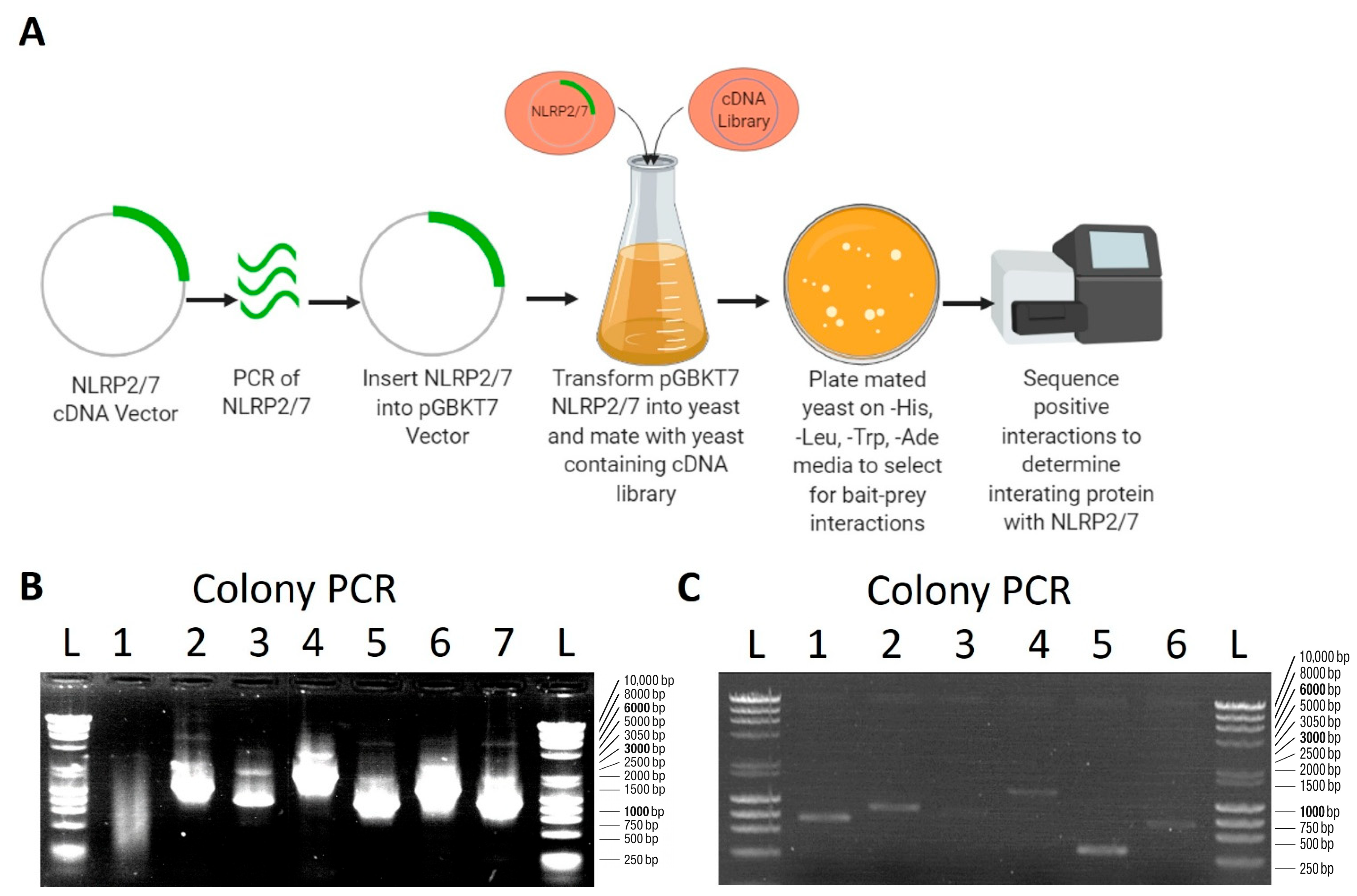
NLRP2 is a maternal effect gene regulating embryo development in mice and humans [10,18]. Mice lack NLRP7, but mutations in human NLRP7 cause recurrent molar pregnancies [19]. Although NLRP2 and NLRP7 are linked to reproductive wastage, the exact mechanisms involved are unclear, so potential NLRP2/7 interacting proteins were examined by screening a human Hela cell cDNA yeast 2-hybrid library with NLRP2 or NLRP7 as “bait”. The mated yeast were placed on triple dropout (TDO, -leu -trp -ade), and quadruple dropout (QDO, -leu -trp -ade -his) media (Figure 1A). Strong protein interactions characterized by growth on QDO medium were further characterized. Representative yeast colony growth on the QDO plate indicating interaction between NLRP2 and EBP1 is shown in Supplementary Figure S1. Prey plasmid DNA was extracted, amplified by PCR (Figure 1B,C), and sequenced to determine the gene of the novel interacting proteins. The screen revealed novel interactions with six proteins for NLRP2 and four proteins for NLRP7 (Table 2 and Table 3). Interestingly, there was one common protein-interaction with both NLRP2 and NLRP7: ErB3-Binding protein 1 (EBP1) also known as Proliferation-Associated 2G4 (PA2G4). EBP1 plays a role in proliferation and methylation through repressing DNA methyl transferase-1 (DNMT-1) mediated maintenance of DNA methylation. This common interaction with EBP1 could explain why mutations in either NLRP2 or NLRP7 result in DNA methylation defects and reproductive wastage. EBP1 was thus selected for further characterization.

3.2. Confirmation of NLRP2/7 and EBP1 Interactions in Human Cells

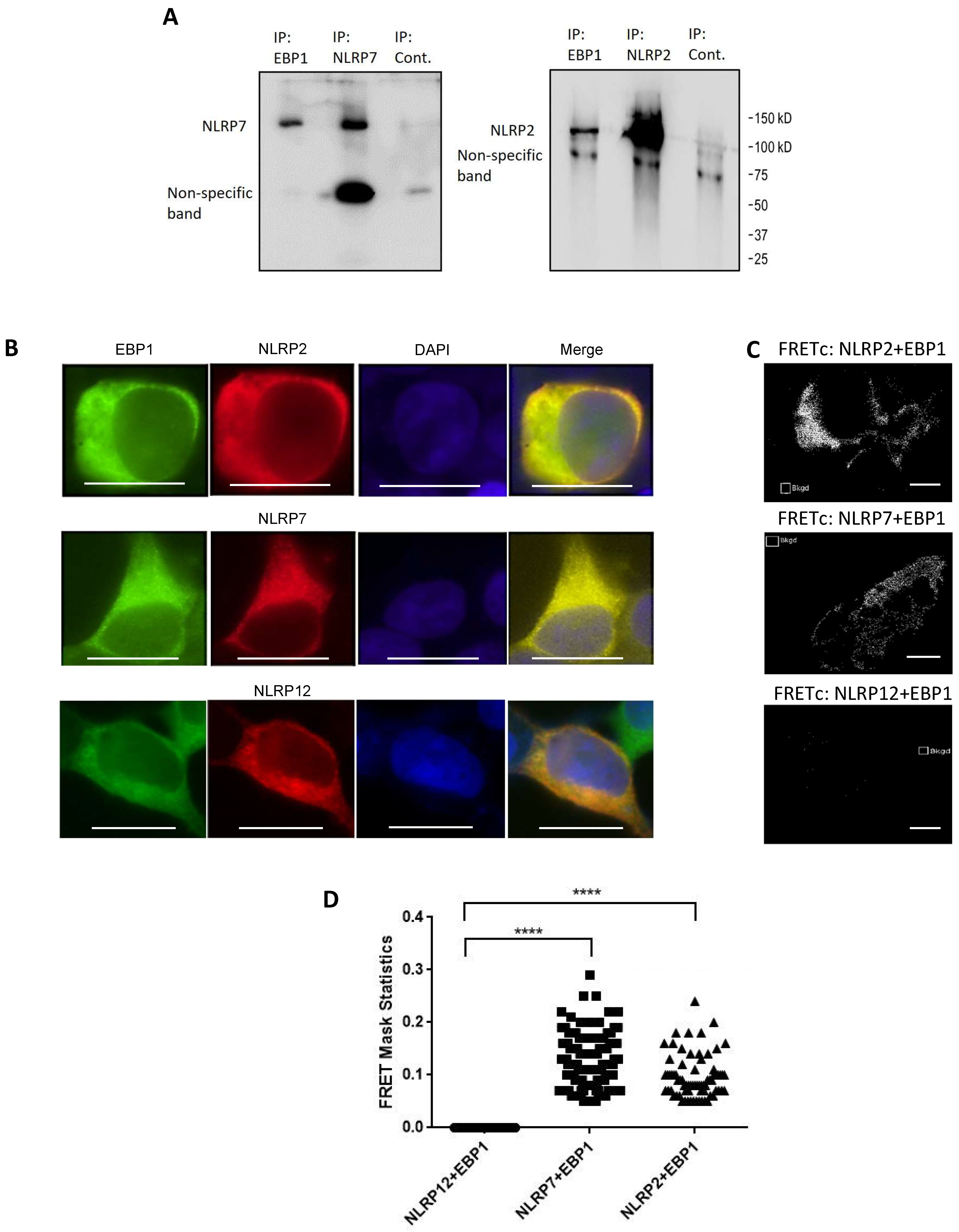
To confirm the interaction of interest in human cells, HEK293T cells were co-transfected with Myc-tagged NLRP2 and HA-tagged EBP1 or Myc-tagged NLRP7 and HA-tagged EBP1. Co-Immunoprecipitation was performed, and the presence of interacting proteins confirmed by immunoblotting. Importantly, the interactions between NLRP2/7 and EBP1 were both confirmed (Figure 2A).
Next, immunofluorescence staining was performed on HEK293T cells transfected with NLRP2/7/12 and EBP1 to visualize the co-localization and cellular location of the interaction. Significant cytoplasmic colocalization of NLRP2/7 with EBP1 was observed (Figure 2B). Further analysis by Förster’s resonance energy transfer (FRET) confirmed the interaction of NLRP2/7 with EBP1 in the cytoplasm (Figure 2C,D). Importantly, NLRP12, which is involved in immune signaling pathways and not fetal development [14], did not colocalize with EBP1 or result in FRET, which further validates the specificity of the interactions between NLRP2/7 and EBP1.

3.3. Combinatorial Effect of NLRP2/7 and EBP1 on DNA Methylation

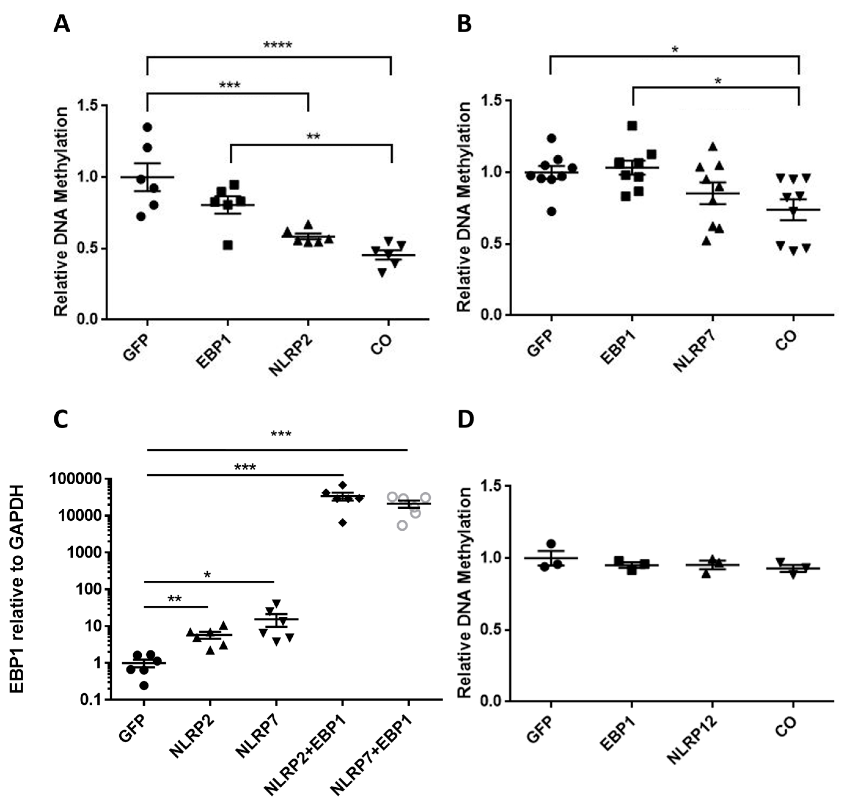
A previous study reported abnormal DNA methylation patterns on maternal alleles associated with NLRP7 mutations [20]. However, the exact mechanisms involved in NLRP2/7 mutations mediating methylation defects is not clearly understood. Studies have investigated altered DNA methylation in recurrent hydatidiform molar pregnancy as consequences of NLRP7 mutations, and hypomethylation is a common feature in these patients [21]. Therefore, the co-functional effects on DNA methylation levels of NLRP2-EBP1 and NLRP7-EBP1 transfected cells were examined. Although NLRP2 had an effect on DNA methylation alone, it had a more significant effect when co-transfected with EBP1 (Figure 3A) and similar results were observed for NLRP7 and EBP1 (Figure 3B). Interestingly, NLRP2 and NLRP7 overexpression enhanced the endogenous expression of EBP1, suggesting that NLRP2/7 not only interact with EBP1, but also regulate its expression (Figure 3C). In contrast, single or co-transfection of NLRP12 and EBP1 did not result in significant changes in DNA methylation (Figure 3D), again confirming the specificity of the interaction between NLRP2/7 and EBP1.

4. Discussion

Previous research demonstrates that mutations in NLRP2/7 are associated with the development of idiopathic recurrent miscarriages. To understand the mechanisms involved, screens for novel protein interactions with NLRP2/7 and other cellular proteins were performed. The results show that EBP1 is a specific binding partner of NLRP2/7. Protein interactions were further confirmed in human cells through co-immunoprecipitation and FRET. FRET analysis also provided a clear cytoplasmic co-localization of NLRP2/7 and EBP1 but not NLRP12 and EBP1, further demonstrating the specificity of these interactions. Unexpectedly, NLRP2/7 also regulate EBP1 gene expression. Importantly, basal expression or overexpression of EBP1 alone does not appear to be sufficient for regulating DNA methylation. Instead, NLRP2/7 may be required for EBP1 activation or proper localization to influence DNA methylation, although the exact mechanisms remain to be elucidated.
NLRP2 and NLRP7 play important roles during early embryogenesis [18,22]. NLRP2 is known as a maternal effect gene and the deletion of NLRP2 results in early embryonic arrest in mice [23] and an imprinting disorder called Beckwith–Wiedemann syndrome (BWS) is associated with NLRP2 mutations in humans [10]. Mutations in NLRP7 are linked to hydatidiform molar pregnancies [21]. Specifically, mutations in NLRP7 result in diploid androgenetic or triploid moles [24]. Recent studies have expanded the known functions of NLRP7, implicating it in regulating alternative splicing and DNA damage response during early embryogenesis in human stem cells and blastoids [25]. Likewise, NLRP2 has been increasingly recognized as a regulator of epigenetics processes, including DNA methylation and imprint maintenance [26].
EBP1 has been implicated in apoptosis, cell proliferation, and differentiation, all processes important in fetal development. EBP1 has two isoforms with differing functions, with the p48 isoform expressed predominantly during embryonic development [15,16]. More relevant to our research, EBP1 was shown to regulate embryonic development through DNA methylation [27], which is particularly important, as NLRP2/7 are associated with altered DNA methylation, but neither protein can do this directly [28]. EBP1 has also been shown to contribute to adult hippocampal neurogenesis through modulation of Bmp4 and Ascl1 signaling pathways, further supporting its role in developmentally regulated gene networks [29]. In conjunction with previous research showing interactions with NLRP7 and ZBTB16 or YY1 [21,30], our discovery that both NLRP2 and NLRP7 interact with EBP1 provides further understanding for the mechanisms governing methylation defects in patients with NLRP2/7 mutations.
A common feature of molar pregnancies is the loss of maternal methylation [31,32]. One study suggested the primary defect in recurrent HM pregnancy is due to a failure to maintain DNA methylation marks in oocytes [31]. In this study, co-expression of NLRP2/7 with EBP1 appeared to further reduce global DNA methylation levels in HEK293T cells, suggesting a possible co-regulatory relationship. However, as no statistical comparisons were made between single and co-transfection conditions, it is not clear how NLRP2/7 and EBP1 interactions affect DNA methylation and whether they are synergistic or co-functional. In conclusion, our identification of a novel protein interaction between NLRP2/7 and EBP1 provides a potential mechanism by which NLRP2/7 mutations could impact epigenetic regulation during early embryo development. Future studies are planned in H9 human embryonic stem cells to study how NLRP2/7 and EBP1 function in more relevant cells in epigenetic control of cell differentiation.

Supplementary Materials

The following supporting information can be downloaded at https://www.mdpi.com/article/10.3390/dna5040049/s1, Figure S1: Representative yeast two-hybrid assay showing positive protein–protein interactions between NLRP2 and EBP1. Yeast cells co-transformed with bait and prey constructs were spotted in duplicate on QDO (quadruple dropout) selective medium lacking adenine, histidine, leucine, and tryptophan. Growth indicates a positive interaction. Each column corresponds to a different co-transformation condition, labeled from 1 to 5. Colony 1, located in the top-right corner, represents the interaction between NLRP2 and EBP1.

Author Contributions

N.H.S. and M.S. acquired, analyzed and interpreted the data, and they assisted in writing the manuscript. C.R.L. developed the concept and designed experiments, assisted with analysis and interpretation of data, and assisted in writing the manuscript. All authors have read and agreed to the published version of the manuscript.

Funding

This research was funded by Missouri State University F07434 to CRL and Tri-Beta Research Scholarship Fund to NHS.

Data Availability Statement

The raw data supporting the conclusions of this article will be made available by the authors on request.

Acknowledgments

The pCMV-HA-EBP1 plasmid was a gift from Stephen Goff (Addgene plasmid #67792; https://www.addgene.org/67792/; RRID: Addgene_67792).

Conflicts of Interest

The authors declare no conflicts of interest.

Abbreviations

The following abbreviations are used in this manuscript:
PCRPolymerase Chain Reaction
SDS-PAGESodium dodecyl sulphate-polyacrylamide gel electrophoresis
FRETFörster’s Resonance Energy Transfer
ELISAEnzyme Linked Immunosorbent Assay
DMEMDulbecco’s Modified Eagle Medium
FBSFetal Bovine Serum

References

  1. Huang, J.Y.; Su, M.; Lin, S.H.; Kuo, P.L. A genetic association study of NLRP2 and NLRP7 genes in idiopathic recurrent miscarriage. Hum. Reprod. 2013, 28, 1127–1134. [Google Scholar] [CrossRef]
  2. Baek, K.H.; Lee, E.J.; Kim, Y.S. Recurrent pregnancy loss: The key potential mechanisms. Trends Mol. Med. 2007, 13, 310–317. [Google Scholar] [CrossRef] [PubMed]
  3. Aruna, M.; Nagaraja, T.; Andal, S.; Tarakeswari, S.; Sirisha, P.V.; Reddy, A.G.; Thangaraj, K.; Singh, L.; Reddy, B.M. Role of progesterone receptor polymorphisms in the recurrent spontaneous abortions: Indian case. PLoS ONE 2010, 5, e8712. [Google Scholar] [CrossRef] [PubMed]
  4. Davis, B.K.; Wen, H.; Ting, J.P. The inflammasome NLRs in immunity, inflammation, and associated diseases. Annu. Rev. Immunol. 2011, 29, 707–735. [Google Scholar] [CrossRef] [PubMed]
  5. Yeretssian, G. Effector functions of NLRs in the intestine: Innate sensing, cell death, and disease. Immunol. Res. 2012, 54, 25–36. [Google Scholar] [CrossRef]
  6. Huang, T.C.; Chang, K.C.; Chang, J.Y.; Tsai, Y.S.; Yang, Y.J.; Chang, W.C.; Mo, C.F.; Yu, P.H.; Chiang, C.T.; Lin, S.P.; et al. Variants in Maternal Effect Genes and Relaxed Imprinting Control in a Special Placental Mesenchymal Dysplasia Case with Mild Trophoblast Hyperplasia. Biomedicines 2021, 9, 544. [Google Scholar] [CrossRef]
  7. Begemann, M.; Rezwan, F.I.; Beygo, J.; Docherty, L.E.; Kolarova, J.; Schroeder, C.; Buiting, K.; Chokkalingam, K.; Degenhardt, F.; Wakeling, E.L.; et al. Maternal variants in NLRP and other maternal effect proteins are associated with multilocus imprinting disturbance in offspring. J. Med. Genet. 2018, 55, 497–504. [Google Scholar] [CrossRef] [PubMed]
  8. Soellner, L.; Begemann, M.; Degenhardt, F.; Geipel, A.; Eggermann, T.; Mangold, E. Maternal heterozygous NLRP7 variant results in recurrent reproductive failure and imprinting disturbances in the offspring. Eur. J. Hum. Genet. 2017, 25, 924–929. [Google Scholar] [CrossRef]
  9. Rubio, C.; Pehlivan, T.; Rodrigo, L.; Simón, C.; Remohí, J.; Pellicer, A. Embryo aneuploidy screening for unexplained recurrent miscarriage: A minireview. Am. J. Reprod. Immunol. 2005, 53, 159–165. [Google Scholar] [CrossRef]
  10. Meyer, E.; Lim, D.; Pasha, S.; Tee, L.J.; Rahman, F.; Yates, J.R.; Woods, C.G.; Reik, W.; Maher, E.R. Germline mutation in NLRP2 (NALP2) in a familial imprinting disorder (Beckwith-Wiedemann Syndrome). PLoS Genet. 2009, 5, e1000423. [Google Scholar] [CrossRef]
  11. Van Gorp, H.; Kuchmiy, A.; Van Hauwermeiren, F.; Lamkanfi, M. NOD-like receptors interfacing the immune and reproductive systems. FEBS J. 2014, 281, 4568–4582. [Google Scholar] [CrossRef]
  12. Zhong, Y.; Kinio, A.; Saleh, M. Functions of NOD-Like Receptors in Human Diseases. Front. Immunol. 2013, 4, 333. [Google Scholar] [CrossRef]
  13. Zhang, P.; Dixon, M.; Zucchelli, M.; Hambiliki, F.; Levkov, L.; Hovatta, O.; Kere, J. Expression analysis of the NLRP gene family suggests a role in human preimplantation development. PLoS ONE 2008, 3, e2755. [Google Scholar] [CrossRef] [PubMed]
  14. Bracey, N.A.; Gershkovich, B.; Chun, J.; Vilaysane, A.; Meijndert, H.C.; Wright, J.R., Jr.; Fedak, P.W.; Beck, P.L.; Muruve, D.A.; Duff, H.J. Mitochondrial NLRP3 protein induces reactive oxygen species to promote Smad protein signaling and fibrosis independent from the inflammasome. J. Biol. Chem. 2014, 289, 19571–19584. [Google Scholar] [CrossRef]
  15. Stevenson, B.W.; Gorman, M.A.; Koach, J.; Cheung, B.B.; Marshall, G.M.; Parker, M.W.; Holien, J.K. A structural view of PA2G4 isoforms with opposing functions in cancer. J. Biol. Chem. 2020, 295, 16100–16112. [Google Scholar] [CrossRef]
  16. Hwang, I.; Ko, H.R.; Ahn, J.Y. The roles of multifunctional protein ErbB3 binding protein 1 (EBP1) isoforms from development to disease. Exp. Mol. Med. 2020, 52, 1039–1047. [Google Scholar] [CrossRef] [PubMed]
  17. Wang, G.Z.; Wolf, D.; Goff, S.P. EBP1, a novel host factor involved in primer binding site-dependent restriction of moloney murine leukemia virus in embryonic cells. J. Virol. 2014, 88, 1825–1829. [Google Scholar] [CrossRef] [PubMed]
  18. Peng, H.; Chang, B.; Lu, C.; Su, J.; Wu, Y.; Lv, P.; Wang, Y.; Liu, J.; Zhang, B.; Quan, F.; et al. Nlrp2, a maternal effect gene required for early embryonic development in the mouse. PLoS ONE 2012, 7, e30344. [Google Scholar] [CrossRef]
  19. Fallahi, J.; Fallahi, J.; Razban, V.; Momtahan, M.; Akbarzadeh-Jahromi, M.; Namavar-Jahromi, B.; Anvar, Z.; Fardaei, M. A Novel Mutation in NLRP7 Related to Recurrent Hydatidiform Mole and Reproductive Failure. Int. J. Fertil. Steril. 2019, 13, 135–138. [Google Scholar]
  20. Djuric, U.; El-Maarri, O.; Lamb, B.; Kuick, R.; Seoud, M.; Coullin, P.; Oldenburg, J.; Hanash, S.; Slim, R. Familial molar tissues due to mutations in the inflammatory gene, NALP7, have normal postzygotic DNA methylation. Hum. Genet. 2006, 120, 390–395. [Google Scholar] [CrossRef]
  21. Singer, H.; Biswas, A.; Nuesgen, N.; Oldenburg, J.; El-Maarri, O. NLRP7, Involved in Hydatidiform Molar Pregnancy (HYDM1), Interacts with the Transcriptional Repressor ZBTB16. PLoS ONE 2015, 10, e0130416. [Google Scholar] [CrossRef]
  22. Li, G.; Tian, X.; Lv, D.; Zhang, L.; Zhang, Z.; Wang, J.; Yang, M.; Tao, J.; Ma, T.; Wu, H.; et al. NLRP7 is expressed in the ovine ovary and associated with in vitro pre-implantation embryo development. Reproduction 2019, 158, 415–427. [Google Scholar] [CrossRef]
  23. Peng, H.; Liu, H.; Liu, F.; Gao, Y.; Chen, J.; Huo, J.; Han, J.; Xiao, T.; Zhang, W. NLRP2 and FAF1 deficiency blocks early embryogenesis in the mouse. Reproduction 2017, 154, 245–251. [Google Scholar] [CrossRef]
  24. Andreasen, L.; Christiansen, O.B.; Niemann, I.; Bolund, L.; Sunde, L. NLRP7 or KHDC3L genes and the etiology of molar pregnancies and recurrent miscarriage. Mol. Hum. Reprod. 2013, 19, 773–781. [Google Scholar] [CrossRef]
  25. Chen, Z.; Jiang, L.; Su, M.; Zeng, Q.; Luo, P.; Chu, L. NLRP7 maintains genomic stability during early human embryogenesis via mediating alternative splicing. Commun. Biol. 2025, 8, 125. [Google Scholar] [CrossRef]
  26. Zhang, R.; Liu, J.; Zhao, F.; Sun, Y.; Chen, Z. NLRP2 in health and disease: Emerging roles in epigenetic regulation. Immunology 2024, 171, 245–256. [Google Scholar] [CrossRef] [PubMed]
  27. Ko, H.R.; Hwang, I.; Jin, E.J.; Yun, T.; Ryu, D.; Kang, J.S.; Park, K.W.; Shin, J.H.; Cho, S.W.; Lee, K.H.; et al. Roles of ErbB3-binding protein 1 (EBP1) in embryonic development and gene-silencing control. Proc. Natl. Acad. Sci. USA 2019, 116, 24852–24860. [Google Scholar] [CrossRef] [PubMed]
  28. Mahadevan, S.; Sathappan, V.; Utama, B.; Lorenzo, I.; Kaskar, K.; Van den Veyver, I.B. Maternally expressed NLRP2 links the subcortical maternal complex (SCMC) to fertility, embryogenesis and epigenetic reprogramming. Sci. Rep. 2017, 7, 44667. [Google Scholar]
  29. Kim, Y.; Ko, H.R.; Hwang, I.; Ahn, J.Y. ErbB3 binding protein 1 contributes to adult hippocampal neurogenesis by modulating Bmp4 and Ascl1 signaling. BMB Rep. 2024, 57, 182–187. [Google Scholar] [CrossRef] [PubMed]
  30. Mahadevan, S.; Wen, S.; Wan, Y.W.; Peng, H.H.; Otta, S.; Liu, Z.; Iacovino, M.; Mahen, E.M.; Kyba, M.; Sadikovic, B.; et al. NLRP7 affects trophoblast lineage differentiation, binds to overexpressed YY1 and alters CpG methylation. Hum. Mol. Genet. 2014, 23, 706–716. [Google Scholar] [CrossRef] [PubMed]
  31. Kou, Y.C.; Shao, L.; Peng, H.H.; Rosetta, R.; del Gaudio, D.; Wagner, A.F.; Al-Hussaini, T.K.; Van den Veyver, I.B. A recurrent intragenic genomic duplication, other novel mutations in NLRP7 and imprinting defects in recurrent biparental hydatidiform moles. Mol. Hum. Reprod. 2008, 14, 33–40. [Google Scholar] [CrossRef] [PubMed]
  32. El-Maarri, O.; Seoud, M.; Coullin, P.; Herbiniaux, U.; Oldenburg, J.; Rouleau, G.; Slim, R. Maternal alleles acquiring paternal methylation patterns in biparental complete hydatidiform moles. Hum. Mol. Genet. 2003, 12, 1405–1413. [Google Scholar] [CrossRef] [PubMed]
Figure 1. Identification of novel protein interactions. (A) Overview of yeast-2 hybrid screening. (B,C) PCR of extracted prey pGAD-T7 plasmids from mated and screened yeast was performed using vector-specific primers (Yeast Amplimer For/Rev; see Table 1), and products were examined by gel electrophoresis prior to Sanger sequencing. (L = DNA Ladder).
Dna 05 00049 g001
Figure 2. Confirmation of protein interactions in human cells. (A) Co-Immunoprecipitation of transiently transfected NLRP2/7 (Myc-tagged) and EBP1 (HA-tagged) in HEK293T cells. Anti-Myc and anti-HA specific antibodies were used for co-immunoprecipitation and Western blot was subsequently performed with Myc-epitope tag specific antibodies. “IP cont.” refers to immunoprecipitation with anti-HA antibody in cells transfected with GFP (without HA-tagged EBP1), included as a control for non-specific binding. (B) Co-localization of transiently transfected EBP1 and NLRP2/7/12 in HEK293T cells. Confocal images were acquired with a 63× objective. Scale bar = 10 µm. (C) Three-channel corrected FRET(c) (Donor normalization). FRET appears as white pixels. (D) FRET intensities were determined by creating a background region for background corrected intensity calculations, any pixels with intensity values above 0.05 were analyzed by Slidebook 6 and Prism 6 using one-way ANOVA and Tukey test. **** p < 0.0001.
Dna 05 00049 g002
Figure 3. Examination of DNA methylation changes in HEK293 T cells overexpressing NLRP2/7 and EBP1. (A,B,D) Global 5-methylcytosine (5-mC) was examined by ELISA assay. ‘CO’ refers to co-transfection of NLRP2 or NLRP7 with EBP1. GFP transfection served as the control. (C) RT-PCR analysis of endogenous Ebp1 gene expression following single and co-transfection with NLRP2 or NLRP7. The expression levels were normalized to GAPDH. Data indicate that co-expression enhances EBP1 mRNA levels compared to single transfections. Data are from 2–3 independent experiments with n = 3 per experiment. One-way ANOVA and Tukey test. * p < 0.05, ** p < 0.01, *** p < 0.001 **** p < 0.0001.
Dna 05 00049 g003
Table 1. Primer sequences.
PrimerSequence
Yeast Amplimer ForwardCTA TTC GAT GAT GAA GAT ACC CCA CCA AAC C
Yeast Amplimer ReverseGTG AAC TTG CGG GGT TTT TCA GTA TCT ACG AT
hEBP1 ForwardAGA GCA TTT GAA GAT GAG
hEBP1 ReverseTCAGTCCCCAGCTTCATT
hGAPDH ForwardACCACCCTGTTGCTGTAGCCAA
hGAPDH ReverseGTCTCCTCTGACTTCAACAGCG
Table 2. Results from Yeast 2-Hybrid Screen for NRLP2.
Protein NameAbbreviationDescription
ErbB3-binding protein 1EBP1 (PA2G4)RNA-binding protein involved in growth regulation
Mannan binding lectin serine peptidase 1MASP1Synthesis of proteins involved in the lectin complement pathway
Kinesin family member 20AKIF20AKinesin-like protein
Protein tyrosine phosphatase receptor type MPTPRMInvolved in cell growth, differentiation, and oncogenic transformation
Ring finger protein 170RNF170RING domain-containing protein involved in ubiquitination.
Charged multivesicular body protein 3CHMP3Protein sorting through the multivesicular body
Table 3. Results from Yeast 2-Hybrid Screen for NRLP7.
Protein NameAbbreviationDescription
ErbB3-binding protein 1EBP1 (PA2G4)RNA-binding protein involved in growth regulation.
SNAP Associated ProteinSNAPINSNARE complex involved in protein and membrane trafficking
ArfGAP with SH3 domain, ankyrin repeat and PH domain 1ASAP1Membrane trafficking and cytoskeleton remodeling
Cannabinoid receptor interacting protein 1CNRIP1Cannabinoid receptor 1 interacting protein
Disclaimer/Publisher’s Note: The statements, opinions and data contained in all publications are solely those of the individual author(s) and contributor(s) and not of MDPI and/or the editor(s). MDPI and/or the editor(s) disclaim responsibility for any injury to people or property resulting from any ideas, methods, instructions or products referred to in the content.