Review

Genomic, Epigenomic, and Immuno-Genomic Regulations of Vitamin D Supplementation in Multiple Sclerosis: A Literature Review and In Silico Meta-Analysis

by
Preetam Modak
1,2,
Pritha Bhattacharjee
3 and
Krishnendu Ghosh
1,4,*
1
Department of Zoology, Ramakrishna Mission Vidyamandira, Belur Math, Howrah 711202, West Bengal, India
2
Institute of Bioinformatics and Applied Biotechnology (IBAB), Bengaluru 560100, Karnataka, India
3
Environmental Epigenomics Lab, Department of Environmental Science, University of Calcutta, Kolkata 700073, West Bengal, India
4
Department of Pathology, Carver College of Medicine, University of Iowa, Iowa City, IA 52242, USA
*
Author to whom correspondence should be addressed.
DNA 2025, 5(4), 48; https://doi.org/10.3390/dna5040048 (registering DOI)
Submission received: 1 June 2025 / Revised: 22 July 2025 / Accepted: 19 September 2025 / Published: 10 October 2025

Abstract

Multiple sclerosis (MS) is a chronic autoimmune neurodegenerative disorder characterized by progressive demyelination and axonal degeneration within the central nervous system, driven by complex genomic and epigenomic dysregulation. Its pathogenesis involves aberrant DNA methylation patterns at CpG islands of numbers of genes like OLIG1 and OLIG2 disrupting protein expression at myelin with compromised oligodendrocyte differentiation. Furthermore, histone modifications, particularly H3K4me3 and H3K27ac, alter the promoter regions of genes responsible for myelination, affecting myelin synthesis. MS exhibits chromosomal instability and copy number variations in immune-regulatory gene loci, contributing to the elevated expression of genes for pro-inflammatory cytokines (TNF-α, IL-6) and reductions in anti-inflammatory molecules (IL-10, TGF-β1). Vitamin D deficiency correlates with compromised immune regulation through hypermethylation and reduced chromatin accessibility of vitamin D receptor (VDR) dysfunction and is reported to be associated with dopaminergic neuronal loss. Vitamin D supplementation demonstrates therapeutic potential through binding with VDR, which facilitates nuclear translocation and subsequent transcriptional activation of target genes via vitamin D response elements (VDREs), resulting in suppression of NF-κB signalling, enhancement of regulatory T-cell (Treg) responses due to upregulation of specific genes like FOXP3, downregulation of pro-inflammatory pathways, and potential restoration of the chromatin accessibility of oligodendrocyte-specific gene promoters, which normalizes oligodendrocyte activity. Identification of differentially methylated regions (DMRs) and differentially expressed genes (DEGs) that are in proximity to VDR-mediated gene regulation supports vitamin D supplementation as a promising, economically viable, and sustainable therapeutic strategy for MS. This systematic review integrates clinical evidence and eventual bioinformatical meta-analyses that reference transcriptome and methylome profiling and identify prospective molecular targets that represent potential genetic and epigenetic biomarkers for personalized therapeutic intervention.

1. Introduction

Multiple sclerosis (MS) is a chronic neurodegenerative disorder which is mostly characterized by demyelination and axonal loss in the CNS. Multiple sclerosis leads to degeneration of the myelin sheath and a decrease in the white and grey matter of the brain. T1 back holes have been observed by gadolinium-enhanced MRI. Immunoglobin IgG bands have also been observed in CSF extracted by lumbar puncture [1]. Subtypes of MS (Figure 1) include clinically isolated syndrome (CIS), which is initially attributed to 80% of MS cases that can be converted to RRMS (relapsing remitting multiple sclerosis) around 20 years. 80% of affected individuals tend to develop secondary progressive MS (SPMS) [2]. The credible cause of MS-associated demyelination is usually considered to have an inflammatory pathological cue with migration of auto-immunologically active lymphocytes across the blood–brain barrier (BBB), which triggers a series of inflammatory reactions with subsets of clonally expressed T- and B-cells, microglial activation, damages by oxidative induction, mitochondrial damage, and energy letdown, which collectively cater to the development of the characteristic plaque of MS [3]. Associative injury and atrophy in both white and grey matter is further associated with MS and is thought to be mostly associated with neurodegenerative pathogenesis with, however, less inflammation in secondary progressive multiple sclerosis (SPMS). Additionally, there happens to be an active and an inactive form of multiple sclerosis. In the active form, occurrence of a clinical relapse, new T2, or gadolinium-enhancing lesions on MRI over at least one year are observed. There is anterograde and retrograde neurodegeneration with more oxidation-oriented damage and failure in energy maintenance [4]. MS has a complex genetic architecture that includes both uncommon monogenic mimics and common variants with small effect sizes, according to recent genome-wide and multi-omics studies. Numerous non-HLA genes also play a crucial factor in susceptibility and the inception and progression of disease, but the major histocompatibility complex (MHC) contains the most consistently linked loci. Additionally, more recent research demonstrates that gene expression and epigenetic regulation vary among MS subtypes, as well as between sexes and ancestries, which suggests that MS is a genetically diverse illness [5,6].

2. Epidemiology

Multiple sclerosis (MS) affects approximately 2.8–2.9 million people globally as of 2020–2023, with its global prevalence estimated at 23.9 cases per 100,000 individuals. The disease demonstrates marked geographical variation, with North America and Western Europe exhibiting the highest prevalence rates, while Nordic countries show prevalence rates exceeding 150 per 100,000 individuals [7]. MS occurs in India with a lower incidence compared to European and American populations. Genetic analysis has revealed that Indian patients with MS share similar gene profiles with Western populations, which explains why the clinical presentation of MS in India closely resembles that observed in Western countries. Within India, geographical variations exist, with Northern India showing higher prevalence rates compared to Southern India. Demographic analysis reveals distinct patterns in the disease onset: males typically develop MS at a mean age of 31 years, while females present earlier at 29 years. This results in a female/male incidence ratio of 1.32:1, which is consistent with global patterns showing female predominance in MS occurrence [7,8].

3. Factors Leading to Multiple Sclerosis

There are several factors that lead to the occurrence of MS. These epigenetic mechanisms collectively demonstrate the complex regulatory networks underlying MS development.

3.1. Epigenetic Factors

Multiple sclerosis patients exhibit distinct DNA methylation patterns, with studies revealing 33% reduced methylation in the white matter of the brain. DNA methylation serves as a critical epigenetic mechanism that thwarts the binding of transcription factor, establishing strong repression of transcriptional process. The methylation machinery involves specific enzymes with distinct functions: DNMT1 maintains existing methylation patterns during DNA replication, ensuring proper transmission of methylation patterns from parent to progeny cells, while DNMT3a and DNMT3b establish de novo methylation patterns on previously unmethylated DNA regions [9]. Concurrently, DNA demethylation occurs through ten-eleven translocation methyl cytosine dioxygenases (TETs). A significant finding involves hypomethylation at the promoter region of peptidyl arginine deiminase, type II (PADI2), that interferes with the citrullination of myelin basic protein (MBP). This process inhibits MBP production, consequently altering the myelin ultrastructure and compromising its function [10]. Additionally, CD4+ T helper cells demonstrate decreased DNA methylation during differentiation into Th1 and Th2 subsets. This hypomethylation promotes enhanced differentiation into Th17 cells, which produce IL-17A, a major inflammatory mediator in MS pathogenesis. The resulting increased IL-17A production amplifies central nervous system inflammation, thereby promoting multiple sclerosis progression (Table 1).
Histone acetylation represents another crucial epigenetic mechanism in MS development. Histone acetyltransferases (HATs), including KAT2A, EP300, KAT6–8, and KAT2B, catalyse acetylation of lysine residues in histone tails, enhancing transcriptional activity [17]. Histone deacetylases (HDACs) counteract this process and are classified into four groups: Classes I, II, and IV operate through zinc-dependent mechanisms, while Class III HDACs require NAD+ cofactors and thus ultimately lead to gene repression. During brain development, oligodendrocyte differentiation from progenitor cells involves global histone deacetylation mediated by HDAC1 and HDAC2 activity. Patients with MS show altered histone deacetylation in oligodendrocytes, which results in decreased transcriptional activity and subsequent myelin sheath degeneration. Since oligodendrocytes are specialized glial cells responsible for myelin production, this dysfunction directly contributes to MS pathology. HDACs also influence T-cell activation and promote Th1 differentiation, potentially initiating pathogenesis and exacerbating disease severity. Research demonstrates that HDAC inhibition reduces IFNγ production (responsible for effector T-cell activity) while promoting differentiation into regulatory T cells (Tregs), thereby suppressing immune responses. Consequently, HDAC inhibitors have emerged as a potential therapeutic application due to the restoration of immune homeostasis, that leads to decreased Th1 and Th17 cell populations and increased Th2 and Treg cell counts [16]. IL-33 expression regulates genes through HDAC activity, thereby influencing myelin production and MS progression.
MicroRNAs (miRNAs) constitute small non-coding RNAs that regulate post-transcriptional gene expression and contribute significantly to MS pathogenesis through miRNA-mediated gene silencing. These molecules modulate cellular development, differentiation, proliferation, and apoptosis through interactions with target mRNAs [24]. Several miRNAs show overexpression in patients with MS, including miR-326, miR-155, miR-146a, and hsa-miR-142-3p. Upregulation of miR-18b and miR-599 correlates with MS relapses, while downregulation of miR-96 is associated with disease remission. Interestingly, miR-96 demonstrates resistance to development of experimental autoimmune encephalomyelitis (EAE) [11], which suggests a potential protective role in MS pathogenesis (Table 1).

3.2. Genetic Factors

Numerous important genes have been shown to be dysregulated in MS brain tissue by extensive transcriptomic and protein-wide association studies (Table 1). Significant upregulation of SHMT1 and FAM120B in white matter lesions indicates changed lipid and folate metabolism, respectively, which may impact immune signalling and myelination. On the other hand, grey matter lesions exhibit downregulation of genes linked to cerebrovascular disease, such as ICA1L. Other genes that exhibit region-specific or stage-specific changes include MTHFR, TRAF3, WARS, and GALC, which may indicate their involvement in axonal degeneration and oxidative stress responses [6]. Furthermore, epigenome-wide studies have shown that NAV2, KCNQ4, and CADM1 are hypermethylated in MS associated brains, indicating potential neurodevelopmental dysregulation. On the other hand, the protein methylation gene METTL21C is hypomethylated, which indicates an upregulation associated with inflammation [21].
Females are more prone to MS than males, and sex-specific gene expression patterns may help to explain this difference. According to transcriptomic profiling, female patients with MS have markedly elevated levels of the X chromosome-based TLR7 and TLR8, which are involved in innate immune responses. These results provide evidence for a possible mechanistic explanation of the sex bias in MS prevalence, indicating that increased immune reactivity through X-linked genes may play a pivotal role in the development and severity of the disease in women [22].
Next-generation sequencing has revealed that a few monogenic disorders, especially those with atypical progression, can mimic multiple sclerosis. For instance, a patient who was initially diagnosed with multiple sclerosis had a mutation in the NOTCH3 gene, which is linked to CADASIL (cerebral autosomal dominant arteriopathy with subcortical infarcts and leukoencephalopathy). This discovery highlights how crucial genomic screening is for differential diagnosis, especially in cases with a young onset or those that are resistant to treatment. Future improvements in MS classification and therapeutic response prediction could be aided by thorough genomic profiling [23].

3.3. Immunological Factors

Contemporary evidence from experimental animal models and comprehensive immunological investigations in patients with multiple sclerosis (MS) reveals a biphasic disease progression characterized by distinct immunological phases. During the early relapsing-remitting stages, peripheral immune responses targeting central nervous system (CNS) antigens constitute the primary pathogenic mechanism. However, as the disease transitions to progressive phases, intrathecal immune reactions within the CNS compartment become the dominant pathological driver.
B lymphocytes orchestrate meningeal inflammation through multifaceted effector functions that encompass production of immunoglobulins, secretion of cytokines, presentation of antigen, and ectopic lymphoid organogenesis [14]. Dysregulated cytokine homeostasis, characterized by excessive production of pro-inflammatory intermediaries, including lymphotoxin, tumour necrosis factor-α (TNF-α), and interleukin-6 (IL-6), coupled with regulatory cytokine synthesis (IL-10, IL-35), results into aberrant T-cell activation and subsequent MS pathogenesis. B cells in CNS directly mediate neural tissue injury through the establishment of ectopic lymphoid follicles within the subarachnoid space of the leptomeninges, particularly in proximity to inflamed vasculature [13]. These tertiary lymphoid structures comprise proliferating B lymphocytes, plasma cells, helper T lymphocytes, and specialized follicular dendritic cell networks. Follicular dendritic cells synthesize CXCL13, a critical chemokine that facilitates B-cell recruitment, maturation, and antigenic selection. Notably, these follicle-like structures demonstrate selective prevalence in about 40–70% of SPMS cases, while remaining absent in RRMS and PPMS phenotypes. Clonal B-cell expansion within the cerebrospinal fluid-containing thecal sac suggests these structures maintain sustained humoral immune responses and autoimmune mechanisms independent of peripheral inflammatory processes. Epidemiological and molecular investigations have established a striking association between Epstein–Barr virus (EBV) infection and MS susceptibility. The EBV lytic proteins BZLF1 and BERF1 demonstrate specific localization to plasma cells within active cortical lesions [25]. This viral reactivation triggers CD8+ cytotoxic T lymphocyte responses that identify antigens presented by oligodendrocytes and/or neurons, contributing to demyelination and axonal damage. The synergistic interaction between EBV reactivation and CD8+ antiviral responses drives widespread inflammation across white matter, grey matter, and meningeal compartments [26].
CD8+ cytotoxic T lymphocytes execute neural tissue destruction through multiple molecular pathways. These cells tend to secrete pro-inflammatory cytokines (TNF-α, IFNγ) alongside cytotoxic effector molecules, including perforin and granzymes A and B [27]. The cytotoxic mechanism involves perforin-mediated membrane pore formation, facilitating granzyme entry and subsequent target cell apoptosis induction. Neuronal vulnerability involves multiple death receptor pathways. TNF-α triggers apoptosis through p55 receptor engagement on neurons, while interferon-γ enhances glutamate neurotoxicity and calcium influx via IFN-γ/AMPA GluR1 complex modulation [15]. Additionally, Fas antigen–Fas ligand interactions between cytotoxic T lymphocytes and neurons activate intracellular caspase cascades, culminating in axonal and neuronal damage. Fas ligand, a transmembrane protein (type-II) of the TNF family, induces receptor-mediated apoptosis upon binding [12].
Active demyelination and neurodegeneration result from microglial activation and macrophage accumulation in injured tissues, facilitating antigen presentation and T-cell activation. This process initiates both retrograde and anterograde neurodegeneration. Recent experimental autoimmune encephalomyelitis (EAE) investigations demonstrate that astrocyte depletion during chronic disease phases ameliorates severity through elimination of deleterious effects mediated by preferential β-1,4-galactosyltransferase 5 and 6 expression [28]. These enzymes synthesize lactosylceramide (LacCer), a signalling molecule that is significantly elevated in the CNS during the progressive phases of EAE. LacCer promotes astrocyte activation, inducing granulocyte-macrophage colony-stimulating factor and CCL2 gene expression, and therefore activates microglia [29]. The pathological cascade is further amplified by neurotoxic products from activated innate immune cells, particularly reactive oxygen species (ROS), reactive nitrogen species (RNS), and nitric oxide (NO) generated by macrophages, microglia, and astrocytes. These mediators are also major contributors to tissue damage in MS.

3.4. Substantial Nigra in MS

Multiple sclerosis is known to affect the basal ganglia and other grey matter regions. Involvement with grey matter correlates with numerous clinical appearances of the disease such as fatigue, physical disability, and cognitive impairment. Vitamin D receptors and 1-alpha hydroxylase, an enzyme which converts vitamin D to its active form, were shown to be vastly expressed in the substantial nigra [30]. It was initially thought that inadequate levels of circulating vitamin D may cause dysfunction or cell death within the substantia nigra. Patients with MS were more likely to show hyperechogenicity in the substantial nigra, lentiform nucleus, caudate nucleus, and thalamus [31]. Current usage of transcranial neuro-sonology imaging systems on the basal ganglia in 75 patients with MS (with 55 healthy controls) revealed that, compared to the controls, the patients with MS were more likely to show hyperechogenicity in the substantial nigra, lentiform nucleus, caudate nucleus, and thalamus. The size of the echogenic area in some of the grey matter structures corelated with hypointensity on T2 weighted magnetic resonance images, suggesting that iron deposition was responsible for hyperechogenicity [32]. Apart from dopaminergic neuron death, research indicates that vitamin D-metabolizing enzymes like CYP27B1 and CYP2R1 may have genetic regulation in their regional expression. Reduced expression of these genes in brains with MS may lead to insufficient neuroprotection, and polymorphisms in these genes are linked to changed levels of activated vitamin D [18]. Furthermore, the incapacity to mount suitable anti-inflammatory responses has also been thought to have a link with vitamin D receptor (VDR) gene polymorphisms and bacteria-induced VDR dysfunction, which exacerbates neuroinflammation in MS [23,33].
The dopaminergic pars compacta (SNpc) and the GABAergic pars reticulata (SNpr) constitute the substantia nigra (SN), which regulates the circuitry of the basal ganglia and is essential for motor control. The SNpr suppresses unwanted movements by blocking thalamic and brainstem targets, whereas the SNpc promotes movement initiation by adjusting the direct and indirect pathways through dopaminergic projections to the striatum [34]. Although SN involvement is less common in MS than cortical or spinal lesions, imaging and postmortem studies have confirmed demyelinating plaques and neuronal loss in the SN, which are specifically linked to iron accumulation (estimated to be four to five times higher than in healthy controls) and the dysregulation of metal-binding proteins like ferredoxin reductase and calreticulin, which contribute to oxidative stress and neurodegeneration [35,36]. Clinically, motor impairments in MS that are directly caused by SN damage are uncommon; instead, parkinsonian symptoms like bradykinesia or rigidity usually only appear when demyelinating lesions impact key nigrostriatal pathway regions, which may temporarily improve with levodopa [37]. The fact that substantia nigra is highly expressed for both vitamin D receptors (VDRs) and 1-α-hydroxylase, is the enzyme that transforms inactive vitamin D into its active form, indicates that vitamin D metabolism is linked to SN vulnerability in MS [38]. Low levels of vitamin D in the blood have been proposed as a cause of dopaminergic neuron death or impairment of SN function [38]. Furthermore, genetic variations in the enzymes that metabolize vitamin D, like CYP27B1 and CYP2R1, may be a factor in the brain’s inadequate activation of vitamin D, which would exacerbate oxidative stress and neuroinflammation in multiple sclerosis (MS). Local neuroprotection is further diminished in MS brains due to decreased expression of these genes [39]. Furthermore, ineffective anti-inflammatory responses have been linked to VDR gene polymorphisms and bacteria-induced VDR dysfunction, which can exacerbate neurodegenerative cascades in multiple sclerosis susceptibility in in different study populations [40,41].

3.5. Vitamin D Receptor (VDR) Dysfunction in MS

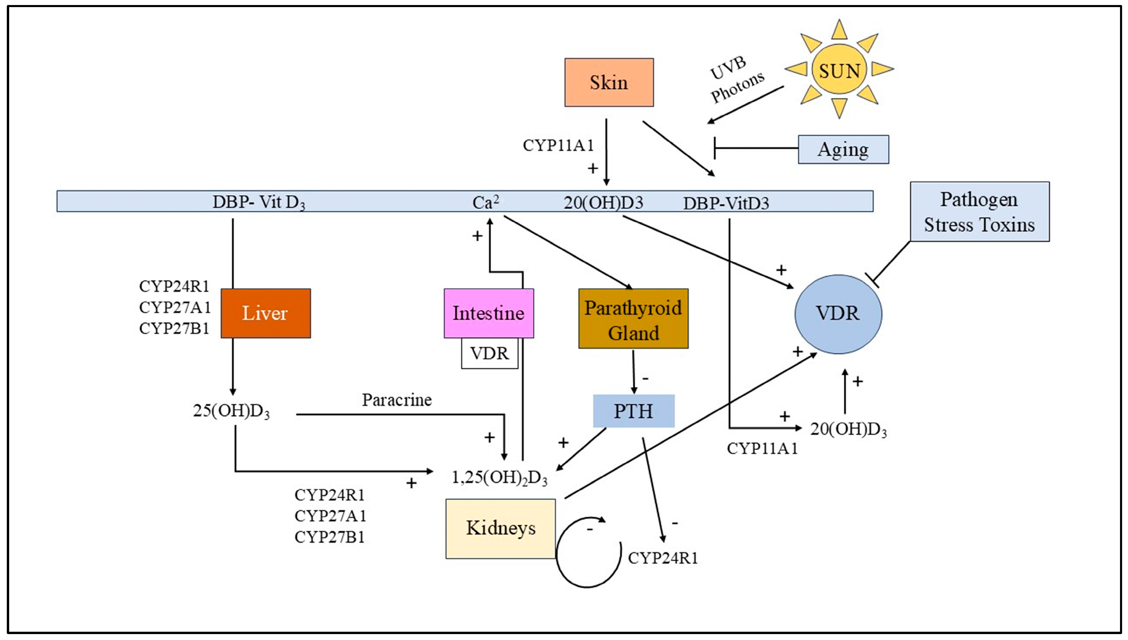
Vitamin D synthesis initiates through cutaneous photochemical conversion of 7-dehydrocholesterol following exposure to ultraviolet-B (UVB) radiation from the sun (Figure 2). The synthesized cholecalciferol undergoes systemic circulation via transport by vitamin D-binding protein (DBP/Gc-globulin) to hepatic tissue, where initial hydroxylation occurs through the cytochrome P450 enzymes CYP2R1, CYP27A1, or CYP27B1, generating 25-hydroxyvitamin D3 [25(OH)D3] [42]. Subsequent renal 1α-hydroxylase (CYP27B1) activity converts 25(OH)D3 to the bioactive hormone 1,25-dihydroxyvitamin D3 [1,25(OH)2D3 or calcitriol] through precise hydroxylation at the C1α position [43]. The active metabolite 1,25(OH)2D3 functions through nuclear vitamin D receptor (VDR) binding, forming heterodimeric complexes with retinoid X receptors (RXRs) that regulate transcriptional activity of vitamin D response elements (VDREs). Classical genomic actions include enhanced intestinal calcium absorption through upregulation of calcium-binding proteins (calbindin-D9k and calbindin-D28k) and calcium transport proteins (TRPV6, NCX1), and thereby maintaining calcium homeostasis and triggering parathyroid hormone (PTH) feedback regulation.
The potentiality of causative infectious agents is considered to be one of the major contributing factors for autoimmune diseases. During chronic mycobacterial infections, for example, the specific disease markers for autoimmune conditions can show high elevation [44]. Recent advancements in genome- and culturing-based diagnosis and detection techniques have greatly revolutionized the scope of the number of pathogens implicated in chronic diseases. In many autoimmune diseases, intracellular inclusions of bacterial species have been observed, and these persist within phagocytes. Many studies have reported that a large number of intracellular bacteria are capable of producing a molecular substance called sulfonolipid capnine (chemically 2-amino-3-hydroxy-15-methylhexadecane-1-sulfonate, a sulfonolipid that is present in the outer membrane of bacteria in the phylum bacteroidetes and a sulfur analog of sphingosine-1-phosphate), which blocks the VDR, although the actual mechanism is poorly understood [45,46,47]. Activation of the successive immune system owing to the killing of bacteria often leads to transient increases in symptoms, which sometime become severe and life-threatening. Vitamin D metabolic pathways that show how bacteria-induced VDR dysfunction occurs could explain the varied levels of 25-D and 1,25-D observed in many autoimmune diseases [48]. Although tightly regulated by the kidneys, 1,25-D production can be elevated in inflammatory diseases owing to an unregulated extra-renal condition governed by conditional activation of macrophages and dendritic cells [49]. When 1,25-D levels become too high, this active vitamin D form binds to the PXR receptor and disrupts the body’s natural cleanup system. Physiologically, the enzyme CYP24A1 breaks down excess 1,25-D to maintain homeostasis. However, when 1,25-D occupies PXR receptors, CYP24A1 eventually slows down the usual cleanup process, which leads to elevated 1,25-D levels in autoimmune diseases. This altered condition also prevents proper vitamin D processing in the liver, blocking conversion of 1,25-D to 25-D [20]. Recent in silico studies reveal that vitamin D supplementation may backfire by interfering with VDR function and reorients the innate immune response. Interestingly, research also shows that autoimmune diseases can enter remission with restoration of innate immune functions by proper activation of VDR receptors [50]. The VDR activator olmesartan elicits significant improvements in most autoimmune patients through its dual mode of actions: directly activating VDR receptors while providing additional therapeutic effects on other cellular receptors. Its high affinity for VDR makes it selectively effective [51]. This treatment approach also requires a fundamentally different antibiotic strategy that incorporates carefully timed, lower doses that inhibit bacterial reproduction rather than attempting to kill bacteria outright [19]. This shift is necessary because most conventional antibiotic regimes fail against chronic inflammatory diseases. The causative bacterial species tend to develop resistance and, thus, ironically, many conventional antibiotics that target mostly the cell walls drive bacteria to survive by altering minute biochemical features of their cell walls, causing them to continue to thrive and jeopardizing the treatment process.

4. Bioinformatics-Based Meta-Analysis of Molecular Dysregulation in Multiple Sclerosis

We combined DNA methylation studies, gene expression profiling and protein–protein interaction (PPI) networks in a multi-layered bioinformatics analysis to better understand the molecular basis of multiple sclerosis (MS) and the possible mechanisms through which vitamin D supplementation may have therapeutic effects. Transcriptomic and epigenomic datasets from the UK cohort of patients with MS that were made publicly available through the Gene Expression Omnibus (GEO) database were used in this analysis. Python 3.10, in a Jupyter Notebook v6, was used for data processing and visualization, utilizing libraries like pandas, matplotlib, seaborn, numpy, scipy, and plotly.

4.1. Protein–Protein Interactions in MS

Using STRING (https://string-db.org/), a PPI network was built, and the Network Analyzer plugin in Cytoscape v3.9.1 was used for analysis. Important proteins found by transcriptomic, immunologic, and epigenetic research were included in the network. High-degree (hub) nodes like CASP3 (degree = 18) and CD86 (degree = 16) have been identified as essential regulators of apoptosis and antigen presentation, respectively. The significance of B-cell maturation and T-cell co-stimulation in MS pathophysiology was highlighted by a closely related cluster comprising CD40, CD80, and CD86. The association between immune dysfunction, cell death, and chromatin dynamics was further supported by the presence of proteins implicated in apoptosis (FAS, FASLG, CASPA8, TP53) and epigenetic regulation (HDAC1/2, DNMT1/3A) in the network. Oxidative stress response proteins like NFE2L2, GPX, and SOD were among the peripheral but functionally significant nodes, which indicated possible modifiable targets of vitamin D signalling. Through epigenetic crosstalk, other important regulators like BACH2, JDP2, MAPK3, IKZF3, and RGS1 have also been linked to the control of T-cell differentiation and immune homeostasis.

4.2. Box-Plot Analysis of the Retrieved Data in MS

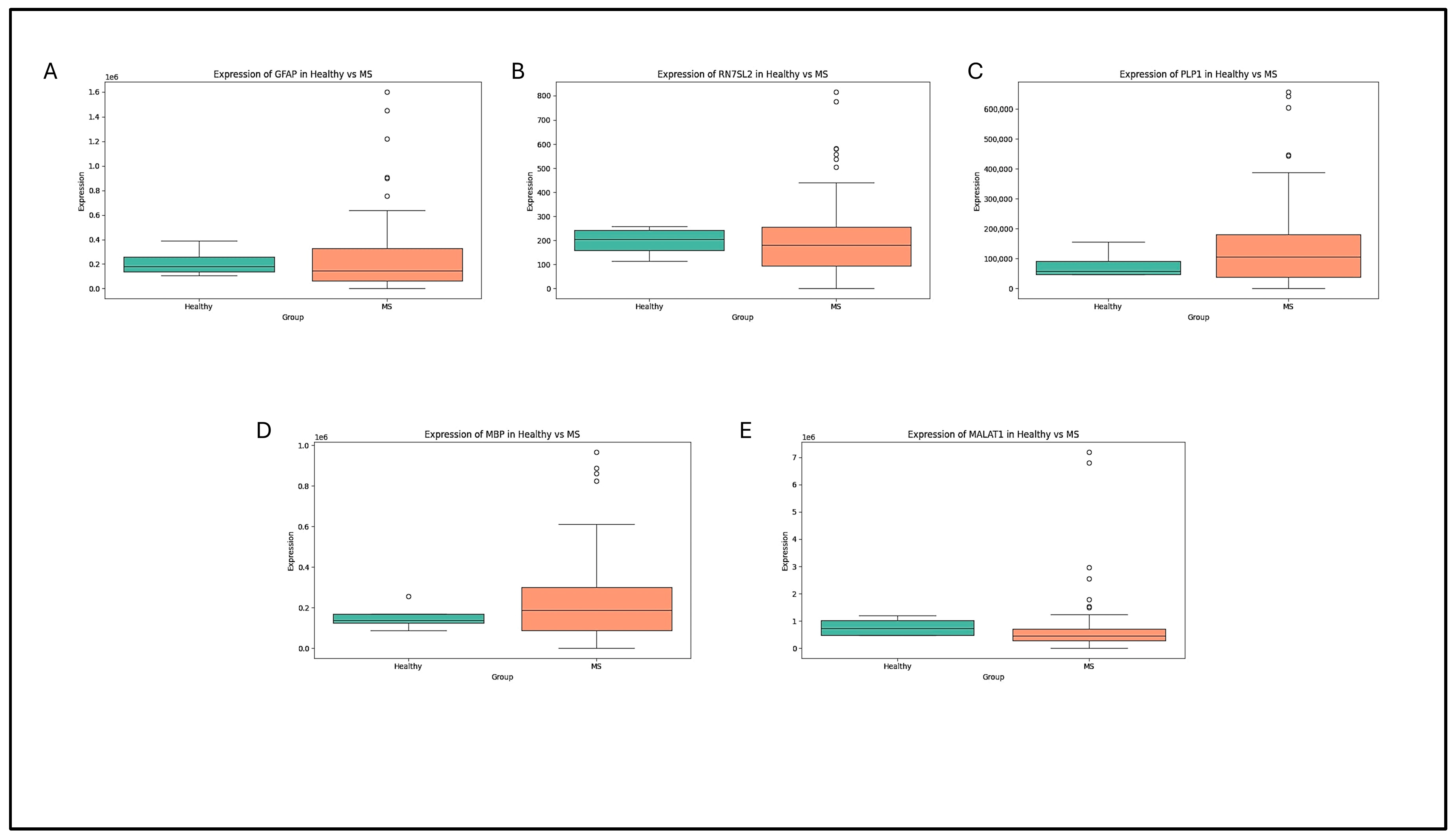
The expression variabilities in brain tissues from control (non-MS) individuals (as available in public genome domain) were examined using box-plot visualization (Figure 3). Five genes were found to be highly variable: RN7SL2, MALAT1, GFAP, MBP, and PLP1. The function of glial cells and the integrity of the myelin sheath, which are essential components of MS pathology, are directly linked to GFAP, MBP, and PLP1. Significant variability was shown by MALAT1, a long non-coding RNA involved in transcriptional regulation and chromatin remodelling, which may indicate a connection to neuroinflammatory signalling. Extreme outliers in RN7SL2, a protein involved in RNA transport and cellular stress responses, suggested regulatory or environmental heterogeneity. These results lend credence to the idea that vitamin D supplementation may restore dysregulated expression patterns of important neuroimmune genes when considered in the context of vitamin D’s epigenetic regulatory function via VDR.

4.3. Volcano-Plot Analysis of the Retrieved Data in MS

A genome-wide view of the differences in methylation between the MS and control groups was provided by a volcano plot analysis conducted by us (Figure 4). TRAIL (TNF-related apoptosis-inducing ligand) serves as a critical immunoregulatory protein that maintains immune homeostasis through selective induction of apoptosis in target cells. This type II transmembrane protein, primarily expressed by immune cells including T cells, natural killer cells, and dendritic cells, binds to the death receptors DR4 and DR5 to trigger apoptotic cascades in susceptible cells while sparing most normal tissues due to their expression of decoy receptors [52]. TRAIL’s immunoregulatory functions include eliminating activated T cells to prevent excessive immune responses, clearing infected or transformed cells, and maintaining peripheral immune tolerance [52,53,54]. TRAIL signalling has been reported earlier to be related to a number of CNS pathologies and neurodegenerative diseases like Alzheimer’s disease [55] and experimental autoimmune encephalomyelitis (EAE) exhibiting autoimmune inflammation mediated by CD4+ T-cell activation [56]. In MS, TRAIL’s role appears paradoxical, although polymorphism in the TRAIL gene has been reported in be associated with MS [57]. While TRAIL can potentially limit autoimmune inflammation by inducing apoptosis in autoreactive T cells and activated microglia within the central nervous system, dysregulated TRAIL signalling may contribute to MS pathogenesis. Studies have shown altered TRAIL expression in patients with MS, with some research suggesting that impaired TRAIL-mediated apoptosis of autoreactive immune cells may perpetuate neuroinflammation. Conversely, excessive TRAIL activity might contribute to oligodendrocyte death and demyelination, which is why TRAIL might serve as a potent biomarker for MS [58,59]. The promoter region of TNFRSF10C (tumour necrosis factor receptor superfamily member 10C) was found to contain a significantly hypomethylated CpG site (cg14521995), with a mean methylation difference of −0.2402 and a p-value of 5.82 × 10⁻⁶. The decoy receptor TRAIL-R3, encoded by TNFRSF10C, binds TRAIL but does not have a functional death domain, which stops immune cells from undergoing apoptosis. This site’s hypomethylation indicates elevated TNFRSF10C expression, which may prolong autoreactive T-cell survival and disrupt immune homeostasis in multiple sclerosis. This observation is consistent with more general epigenetic changes in multiple sclerosis (MS), including hypermethylation of FOXP3, which affects Treg function, and hypomethylation of PADI2, which causes demyelination and citrullination of myelin basic protein (MBP). Overexpression of TNFRSF10C may worsen these effects by inhibiting oxidative stress brought on by TRAIL, indirectly maintaining PADI2 activity and decreasing Treg-mediated regulation.
Furthermore, considering the observed hypomethylation of IL17A and STAT3, which promote Th17 cell survival and proliferation, TNFRSF10C-mediated blockade of TRAIL signalling may stabilize pro-inflammatory Th17 pathways. According to these results, TNFRSF10C (anti-apoptotic) and VDR (anti-inflammatory, pro-apoptotic) have an antagonistic relationship, forming a regulatory axis that may affect the pathophysiology of MS and the effectiveness of vitamin D. Crucially, microRNAs like miR-155, miR-146a, and miR-326 that are dysregulated in MS are also impacted by TRAIL signalling. Elevated TNFRSF10C expression may contribute to the maintenance of miR-155 by reducing the suppressive effects of TRAIL and thus strengthen inflammatory cascades. As a result, cg14521995 shows promise as an epigenetic marker and a possible therapeutic target for modulation in conjunction with vitamin D intervention.
From protein interaction mapping to transcriptomic visualization and methylation profiling, these multi-layered analyses collectively reveal a complex yet cohesive molecular framework in multiple sclerosis. They highlight how vitamin D may regulate inflammation, apoptosis, and remyelination through its genomic and epigenomic effects. Targeting vitamin D pathways in conjunction with innovative epigenetic interventions in multiple sclerosis holds therapeutic promise, as evidenced by the convergence of findings across methylation, expression, and network domains.

5. Effect of Vitamin D in Multiple Sclerosis

There is a trend of growing evidence that a complex interaction of genes and the environment facilitate the risk of development and progression of multiple sclerosis. Multiple studies have shown the risk of developing MS with low serum levels (an inverse correlation) of 25(OH)D [60]. Higher serum vitamin D is therefore considered as a crucial shielding factor against MS. Additionally, a low vitamin D level has also been shown to be associated with an increased relapse rate and disabilities [61]. Since both of our literature study as well as our predictive in silico analyses furnished key connections between vitamin D and various pathways of MS, vitamin D supplementation in patients with MS can be of holistic benefits in the following ways.

5.1. Effects of Vitamin D on Immune System

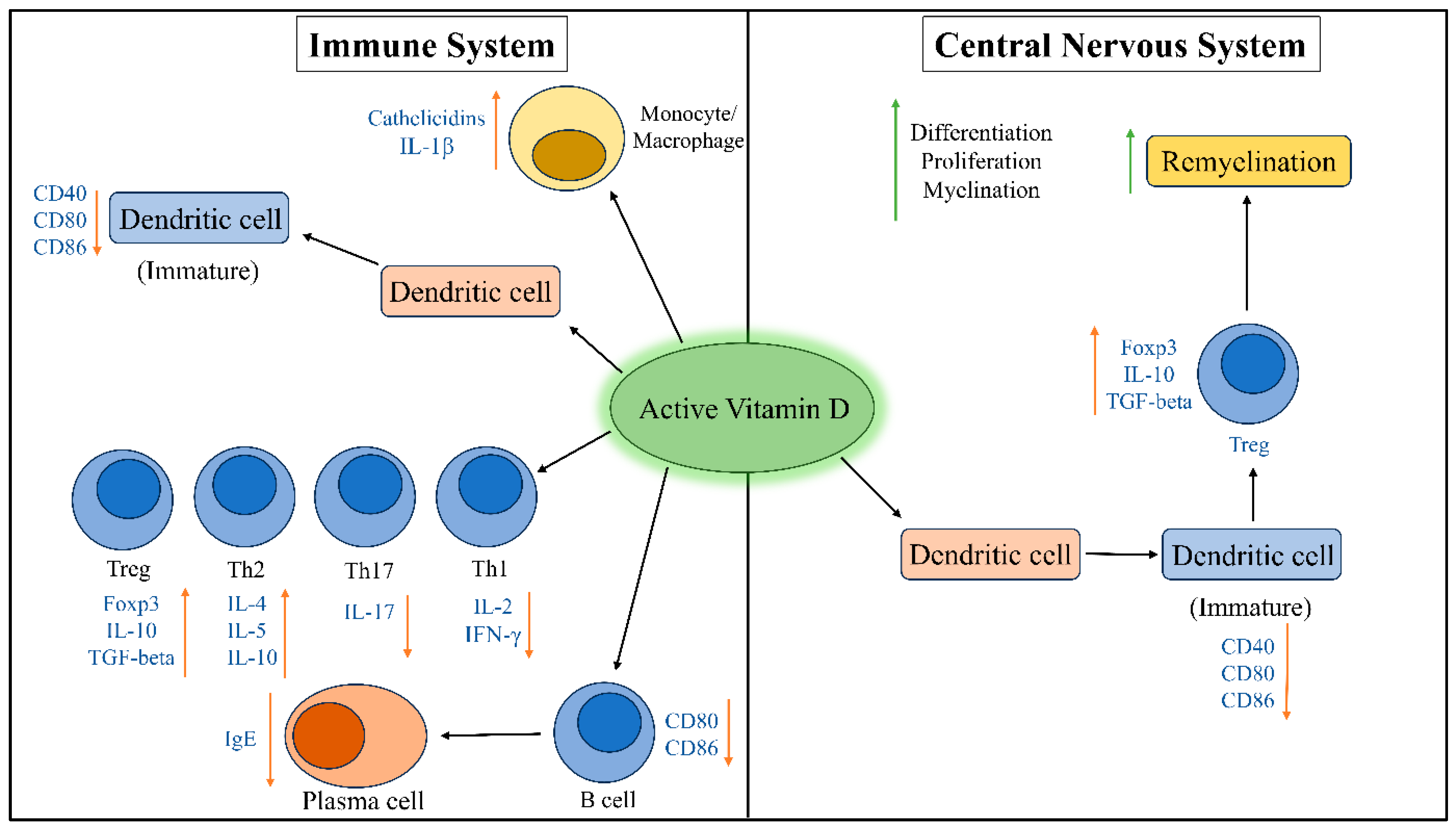
Vitamin D is known for its regulatory action in calcium homeostasis, since it stimulates intestinal calcium absorption. Vitamin D is also crucial for functional development of CNS and the brain, with an array of additional roles in cell proliferation, apoptosis, regulation of blood pressure, insulin secretion, differentiation of immune cells, and modulation of immune responses. Many of its immune modulatory functions are exerted via interaction with the VDR (Figure 5). Owing to have multiple important roles in lymphocyte activation and proliferation and T-helper cell differentiation and its regulatory effects on the immune response, vitamin D is a key player in the pathogenesis of MS [62]. Reportedly, vitamin D also suppresses type 1 T-helper cells (Th1) and mediates production of inflammatory cytokines. Vitamin D supplementation has shown to significantly decrease the levels of IL-10 and IL-17 significantly in multiple studies. High doses of vitamin D have also been reported to reduce the percentage of IL-17-producing CD4+ T-cells and increase central memory CD4+ T-cells and naive CD4+ T-cells [63]. 1,25(OH)2D3-supplemented culture of monocytes showed VDR-dependent loss of MHCII and a reduction in co-stimulatory molecules (CD40, CD80, CD86, etc.) [64]. Furthermore, 1,25(OH)2D3-reinforced monocytes also showed a decline in the release of Th1 and Th17 cell-inducing cytokines (IL12 and IL-23) and upregulated production of IL-10 and CCL22 by regulatory T cells and Th2 [65]. The exposure of B cells to 1,25(OH)2D3 has been reported to lower proliferation, plasma cell differentiation (decreasing IgM and IgG secretion), production of memory B cells, and induced B-cell apoptosis [66]. It is noteworthy that, although a moderate level of vitamin D exerts a direct immunoregulatory effect, an excessively high dose could implicate potential clinical diseases activity by increasing the levels of T-cell-excitatory calcium. Infection with EBV during or shortly after childhood in an experimental animal model has long been considered a significant environmental risk factor for the inception and development of MS and quicker accumulation of neurological disabilities [67]. Vitamin D supplementation (higher serum level of vitamin D) has been experimentally shown to reduce EBV-mediated MS onset with significant reduction in anti-EBNA-1 IgG levels [67].

5.2. Effects of Vitamin D on Substantial Nigra

Substantial nigra produces dopamine, which plays a major role in movement. In MS, it has been seen that, due to reduced levels of circulating vitamin D (reduced activation of vitamin D due to reduced 1 alpha hydroxylase enzyme), increased death of the cells in the substantial nigra leads to impairment in movement. Therefore, Vitamin D supplementation can inhibit the cell death in substantial nigra and thereby regulate the levels of activated vitamin D and increased levels of dopamine to normal, decreasing the pathogenesis of MS [68]. Substantial nigra, in MS, has been thoroughly reviewed to be highly involved in mediating glial cell actions. Microglia for example, has been reported to be involved in a number of MS-associated issues like MS-associated fatigue (MSAF) [69], which has been positively modulated by vitamin D signalling through VDR found in microglia [70]. Metal ions, like iron, have been reported to be associated with MS. Substantial nigra tends to develop iron dys-homeostasis, resulting in deregulated ferredoxin reductase, which significantly implies higher iron deposition in the MS condition than in normal individuals [71]. Zughaier et. al., in 2014, reported that vitamin D plays a crucial role in the iron metabolism in monocytes through hepcidin–ferroportin axes [72], which might also indicate an effect of vitamin D of relieving the MS condition at substantial nigra.

5.3. Effects of Vitamin D in Epigenetic Regulation of MS

Calcitriol also reduces cell proliferation and enhances cell differentiation. These lead to a higher rate of differentiation of progenitor cells to oligodendrocytes and thus enhance remyelination. In an array of HDAC inhibitors (inhibiting aberrant T-cell activation), a major treatment strategy for MS, vitamin D supplementation seems to orient differentiation, providing an overall protection against MS [73]. Calcitriol binding to VDR recruits coactivator complexes containing histone-modifying enzymes, including histone acetyltransferases and demethylases, which establish permissive chromatin states at anti-inflammatory gene loci while simultaneously promoting repressive histone modifications at pro-inflammatory genes [74]. This is particularly evident in the modulation of Foxp3 expression in regulatory T cells (Tregs), where vitamin D enhances the acetylation of histones H3 and H4 at the Foxp3 promoter region, facilitating increased Treg differentiation and immunosuppressive function [75]. Conversely, vitamin D suppresses the expression of pro-inflammatory cytokines such as IL-17 and IFN-γ through the recruitment of histone deacetylases and DNA methyltransferases to their respective promoter regions [76]. Vitamin D supplementation has been shown to reverse hypermethylation of CpG islands in genes encoding anti-inflammatory mediators, including IL-10 and TGF-β, while promoting hypermethylation of pro-inflammatory gene promoters. The vitamin D–VDR complex interacts with DNA methyltransferase 1 (DNMT1) and ten-eleven translocation (TET) enzymes, which catalyse DNA demethylation, thereby fine-tuning the methylation landscape of immune cells [77]. Non-coding RNAs, including microRNAs and long non-coding RNAs, constitute an additional layer of epigenetic regulation influenced by vitamin D in MS. Vitamin D modulates the expression of several microRNAs like miR-342 and miR-377, which target mRNAs that encode inflammatory mediators and transcription factors involved in altered T-cell responses [78]. Furthermore, vitamin D influences the expression of long non-coding RNAs that act as molecular scaffolds for chromatin-modifying complexes, thereby amplifying its epigenetic effects on MS-relevant gene networks [79].

5.4. Effects of Vitamin D on Oxidative Stress

Vitamin D can also regulate oxidative stress, a potent factor influencing risks of multiple sclerosis. People with chronic non-communicable diseases (like cardiovascular disease, T2DM, arthritis, etc.) have been observed to express a high chronic level of NF-κB [80]. NF-κB tends to act increasingly towards oxidative stress and cellular inflammatory responses. Calcitriol suppresses NF-κB and thereby reduces chronic inflammation. Following calcitriol–VDR interaction, the nuclear factor erythroid 2–related factor 2 (Nrf2) translocate from the cytoplasm to the nucleus. Nrf2 activates several antioxidants. Nrf2 is currently emerging as a regulator of cellular resistance to oxidative conditions [81]. Nrf2 controls the basal and induced expression of an array of antioxidant response element-dependent genes to regulate the physiological and pathophysiological outcomes of oxidant exposure, with special reference to MS [82]. Calcitriol functions not only as a hormone that regulate calcium homeostasis but also as a potent modulator of immune responses and cellular antioxidant mechanisms. In MS conditions, vitamin D deficiency correlates with increased disease activity, higher relapse rates, and accelerated disability progression, which suggests that suboptimal vitamin D status may compromise the body’s ability to counteract oxidative stress, especially in RRMS [82,83]. Reports suggest that calcitriol upregulates the expression of antioxidant enzymes, including glutathione peroxidase, superoxide dismutase, and catalase, which collectively neutralize harmful free radicals that would otherwise damage myelin sheaths and neuronal membranes. Additionally, vitamin D enhances the synthesis of glutathione, the brain’s primary endogenous antioxidant, thereby strengthening cellular defence mechanisms against oxidative damage [84]. Patients with MS with higher serum 25-hydroxyvitamin D levels and exhibit reduced concentrations of oxidative stress biomarkers such as malondialdehyde, protein carbonyls, and 8-hydroxy-2′-deoxyguanosine, while simultaneously showing increased total antioxidant capacity in both serum and cerebrospinal fluid [85,86].

6. Conclusions

Vitamin D deficiency triggers a chain of deleterious transcriptional and epigenetic dysregulation that extends beyond classical calcium homeostatic dysfunctions. Epidemiological and clinical investigations consistently indicate a potential significance of vitamin D insufficiency in patients with MS that correlates with altered expression of immunoregulatory genes and aberrant DNA methylation patterns at a number of CpG sites within inflammatory gene loci. This further correlates with upregulated pro-inflammatory gene networks, enhanced Th1/Th17 cell differentiation, and accelerated demyelination characterized by oligodendrocyte inefficacies that are coherent with the activities of CNS macrophage/microglia. Vitamin D supplementation emerges as a cornerstone epigenetic therapeutic intervention in MS management, demonstrating immunosuppressive efficacy through VDR-mediated transcriptional repression of proinflammatory molecules along with chromatin remodelling via histone deacetylase and microRNA mediated post-transcriptional regulation. Vitamin D supplementation represents an advantageous yet cost-effective pharmacogenomic therapeutic strategy with reliable clinical efficacy. Based on the orientation of vitamin D pathway-associated genes like CYP2R1 and CYP27B1, personalized treatment approaches like natural UV-mediated cutaneous vitamin D biosynthesis, dietary intake through fatty fish consumption (rich in cholecalciferol), and oral supplementations are now coming forward. Genomic evidence from genome-wide association studies (GWAS), VDR signalling cascade, and clinical trial data indicate vitamin D supplementation as a paradigm shift towards MS management as it modulates Tregs and suppresses inflammatory cascades. Our future research rationale includes optimization of various dosing regimens based on genetic orientations in vitamin D metabolic pathways, identification of prominent epigenetic biomarkers for personalized supplementation, investigation of synergistic effects with conventional therapies, and development of combinatorial approaches targeting multiple genetic and epigenetic pathways simultaneously.

Author Contributions

Writing—original draft preparation, P.M.; writing—review, editing and analysis, K.G.; Guidance, K.G.; Senior guidance/author, P.B. All authors have read and agreed to the published version of the manuscript.

Funding

This research received no external funding.

Data Availability Statement

Data used for bioinformatic analysis are already available in public domain. However, if needed, they can be submitted by first and/or corresponding author upon official request.

Acknowledgments

We sincerely thank Swami Mahaprajnananda, Principal, Ramakrishna Mission Vidyamandira, Belur Math, Howrah, West Bengal, for his academic fervor and warm support.

Conflicts of Interest

The authors declare no conflicts of interest.

References

  1. Doshi, A.; Chataway, J. Multiple Sclerosis, a Treatable Disease. Clin. Med. 2017, 17, 530–536. [Google Scholar] [CrossRef] [PubMed]
  2. Klineova, S.; Lublin, F.D. Clinical Course of Multiple Sclerosis. Cold Spring Harb. Perspect. Med. 2018, 8, a028928. [Google Scholar] [CrossRef]
  3. Wekerle, H.; Lassmann, H. The Immunology of Inflammatory Demyelinating Disease. In McAlpine’s Multiple Sclerosis; Elsevier: Amsterdam, The Netherlands, 2006; pp. 491–555. ISBN 9780443072710. [Google Scholar]
  4. Popescu, B.F.G.; Pirko, I.; Lucchinetti, C.F. Pathology of Multiple Sclerosis: Where Do We Stand? Continuum 2013, 19, 901–921. [Google Scholar] [CrossRef] [PubMed]
  5. Srinivasan, S.; Di Dario, M.; Russo, A.; Menon, R.; Brini, E.; Romeo, M.; Sangalli, F.; Costa, G.D.; Rodegher, M.; Radaelli, M.; et al. Dysregulation of MS Risk Genes and Pathways at Distinct Stages of Disease. Neurol. Neuroimmunol. Neuroinflamm. 2017, 4, e337. [Google Scholar] [CrossRef]
  6. Jia, T.; Ma, Y.; Qin, F.; Han, F.; Zhang, C. Brain Proteome-wide Association Study Linking-genes in Multiple Sclerosis Pathogenesis. Ann. Clin. Transl. Neurol. 2023, 10, 58–69. [Google Scholar] [CrossRef] [PubMed]
  7. Shi, M.; Liu, Y.; Gong, Q.; Xu, X. Multiple Sclerosis: An Overview of Epidemiology, Risk Factors, and Serological Biomarkers. Acta Neurol. Scand. 2024, 2024, 7372789. [Google Scholar] [CrossRef]
  8. Bhatia, R.; Bali, P.; Chowdhary, R. Epidemiology and Genetic Aspects of Multiple Sclerosis in India. Ann. Indian. Acad. Neurol. 2015, 18, 6. [Google Scholar] [CrossRef]
  9. Castro, K.; Casaccia, P. Epigenetic Modifications in Brain and Immune Cells of Multiple Sclerosis Patients. Mult. Scler. 2018, 24, 69–74. [Google Scholar] [CrossRef]
  10. Moscarello, M.A.; Mastronardi, F.G.; Wood, D.D. The Role of Citrullinated Proteins Suggests a Novel Mechanism in the Pathogenesis of Multiple Sclerosis. Neurochem. Res. 2007, 32, 251–256. [Google Scholar] [CrossRef]
  11. Ma, X.; Zhou, J.; Zhong, Y.; Jiang, L.; Mu, P.; Li, Y.; Singh, N.; Nagarkatti, M.; Nagarkatti, P. Expression, Regulation and Function of MicroRNAs in Multiple Sclerosis. Int. J. Med. Sci. 2014, 11, 810–818. [Google Scholar] [CrossRef]
  12. Suda, T.; Takahashi, T.; Golstein, P.; Nagata, S. Molecular Cloning and Expression of the Fas Ligand, a Novel Member of the Tumor Necrosis Factor Family. Cell 1993, 75, 1169–1178. [Google Scholar] [CrossRef]
  13. Haugen, M.; Frederiksen, J.L.; Degn, M. B Cell Follicle-like Structures in Multiple Sclerosis—With Focus on the Role of B Cell Activating Factor. J. Neuroimmunol. 2014, 273, 1–7. [Google Scholar] [CrossRef]
  14. Michel, L.; Touil, H.; Pikor, N.B.; Gommerman, J.L.; Prat, A.; Bar-Or, A. B Cells in the Multiple Sclerosis Central Nervous System: Trafficking and Contribution to CNS-Compartmentalized Inflammation. Front. Immunol. 2015, 6, 636. [Google Scholar] [CrossRef]
  15. Fang, C.; Weng, T.; Hu, S.; Yuan, Z.; Xiong, H.; Huang, B.; Cai, Y.; Li, L.; Fu, X. IFN-γ-Induced ER Stress Impairs Autophagy and Triggers Apoptosis in Lung Cancer Cells. OncoImmunology 2021, 10, 1962591. [Google Scholar] [CrossRef]
  16. Akimova, T.; Beier, U.H.; Liu, Y.; Wang, L.; Hancock, W.W. Histone/Protein Deacetylases and T-Cell Immune Responses. Blood 2012, 119, 2443–2451. [Google Scholar] [CrossRef] [PubMed]
  17. He, H.; Hu, Z.; Xiao, H.; Zhou, F.; Yang, B. The Tale of Histone Modifications and Its Role in Multiple Sclerosis. Hum. Genom. 2018, 12, 31. [Google Scholar] [CrossRef] [PubMed]
  18. Martinez-Hernandez, A.; Perez-Guerrero, E.E.; Macias-Islas, M.A.; Nava-Valdivia, C.A.; Villagomez-Vega, A.; Contreras-Haro, B.; Garcia-Ortega, Y.E.; Esparza-Guerrero, Y.; Gallardo-Moya, S.G.; Gamez-Nava, J.I.; et al. Polymorphisms CYP2R1 Rs10766197 and CYP27B1 Rs10877012 in Multiple Sclerosis: A Case-Control Study. J. Immunol. Res. 2021, 2021, 1–11. [Google Scholar] [CrossRef]
  19. Waterhouse, J.C.; Perez, T.H.; Albert, P.J. Reversing Bacteria-induced Vitamin D Receptor Dysfunction Is Key to Autoimmune Disease. Ann. N. Y. Acad. Sci. 2009, 1173, 757–765. [Google Scholar] [CrossRef] [PubMed]
  20. Christakos, S.; Dhawan, P.; Verstuyf, A.; Verlinden, L.; Carmeliet, G. Vitamin D: Metabolism, Molecular Mechanism of Action, and Pleiotropic Effects. Physiol. Rev. 2016, 96, 365–408. [Google Scholar] [CrossRef]
  21. Baldrighi, G.N.; Cavagnola, R.; Sacco, D.; Costantino, L.; Bernardinelli, L.; Gentilini, D. Exploring the Complexities of Epigenetics in Multiple Sclerosis: A Study Involving Meta-Analysis of DNA Methylation Profiles, Epigenetic Drift, and Rare Epivariations. Multiple Sclerosis. J. Exp. Transl. Clin. 2024, 10, 20552173241296726. [Google Scholar] [CrossRef]
  22. Gribbin, J.; Wolujewicz, P.; Soda, K.J. X Chromosome Analysis in Multiple Sclerosis for Identification of Genes Implicated in Female-Biased Disease Presentation (S42.007). Neurology 2024, 102, 3700. [Google Scholar] [CrossRef]
  23. Mandler, J.M.; Härtl, J.; Cordts, I.; Sturm, M.; Hedderich, D.M.; Bafligil, C.; Baki, E.; Becker, B.; Machetanz, G.; Haack, T.B.; et al. Uncovering Genetic Mimics in Multiple Sclerosis: A Single-Center Clinical Exome Sequencing Study. Mult. Scler. J. Exp. Transl. Clin. 2024, 10, 20552173241263491. [Google Scholar] [CrossRef] [PubMed]
  24. Hwang, H.-W.; Mendell, J.T. MicroRNAs in Cell Proliferation, Cell Death, and Tumorigenesis. Br. J. Cancer 2006, 94, 776–780. [Google Scholar] [CrossRef]
  25. Bar-Or, A.; Pender, M.P.; Khanna, R.; Steinman, L.; Hartung, H.-P.; Maniar, T.; Croze, E.; Aftab, B.T.; Giovannoni, G.; Joshi, M.A. Epstein–Barr Virus in Multiple Sclerosis: Theory and Emerging Immunotherapies. Trends Mol. Med. 2020, 26, 296–310. [Google Scholar] [CrossRef]
  26. Kerr, J.R. Epstein-Barr Virus (EBV) Reactivation and Therapeutic Inhibitors. J. Clin. Pathol. 2019, 72, 651–658. [Google Scholar] [CrossRef]
  27. Raskov, H.; Orhan, A.; Christensen, J.P.; Gögenur, I. Cytotoxic CD8+ T Cells in Cancer and Cancer Immunotherapy. Br. J. Cancer 2021, 124, 359–367. [Google Scholar] [CrossRef]
  28. Mahad, D.H.; Trapp, B.D.; Lassmann, H. Pathological Mechanisms in Progressive Multiple Sclerosis. Lancet Neurol. 2015, 14, 183–193. [Google Scholar] [CrossRef]
  29. Mayo, L.; Trauger, S.A.; Blain, M.; Nadeau, M.; Patel, B.; Alvarez, J.I.; Mascanfroni, I.D.; Yeste, A.; Kivisäkk, P.; Kallas, K.; et al. Regulation of Astrocyte Activation by Glycolipids Drives Chronic CNS Inflammation. Nat. Med. 2014, 20, 1147–1156. [Google Scholar] [CrossRef]
  30. Cui, X.; Pelekanos, M.; Liu, P.-Y.; Burne, T.H.J.; McGrath, J.J.; Eyles, D.W. The vitamin D receptor in dopamine neurons; its presence in human substantia nigra and its ontogenesis in rat midbrain. Neuroscience 2013, 236, 77–87. [Google Scholar] [CrossRef]
  31. Krogias, C.; Gold, R.; Chan, A.; Triantafyllou, N.; Voumvourakis, K.; Tsivgoulis, G. Brain Hyperechogenicities Are Not Associated with Venous Insufficiency in Multiple Sclerosis: A Pilot Neurosonology Study. J. Neuroimaging 2016, 26, 150–155. [Google Scholar] [CrossRef] [PubMed]
  32. Haller, S.; Vernooij, M.W.; Kuijer, J.P.A.; Larsson, E.-M.; Jäger, H.R.; Barkhof, F. Cerebral Microbleeds: Imaging and Clinical Significance. Radiology 2018, 287, 11–28. [Google Scholar] [CrossRef]
  33. Imani, D.; Razi, B.; Motallebnezhad, M.; Rezaei, R. Association between Vitamin D Receptor (VDR) Polymorphisms and the Risk of Multiple Sclerosis (MS): An Updated Meta-Analysis. BMC Neurol 2019, 19, 339. [Google Scholar] [CrossRef] [PubMed]
  34. Sonne, J.; Reddy, V.; Beato, M.R. Neuroanatomy, Substantia Nigra. In StatPearls; StatPearls Publishing: Treasure Island, FL, USA, 2024. [Google Scholar]
  35. Carmona, A.; Carboni, E.; Gomes, L.C.; Roudeau, S.; Maass, F.; Lenz, C.; Ortega, R.; Lingor, P. Metal Dyshomeostasis in the Substantia Nigra of Patients with Parkinson’s Disease or Multiple Sclerosis. J. Neurochem. 2024, 168, 128–141. [Google Scholar] [CrossRef]
  36. Vercellino, M.; Masera, S.; Lorenzatti, M.; Condello, C.; Merola, A.; Mattioda, A.; Tribolo, A.; Capello, E.; Mancardi, G.L.; Mutani, R.; et al. Demyelination, Inflammation, and Neurodegeneration in Multiple Sclerosis Deep Gray Matter. J. Neuropathol. Exp. Neurol. 2009, 68, 489–502. [Google Scholar] [CrossRef]
  37. Ghosh, R.; Roy, D.; Dubey, S.; Das, S.; Benito-León, J. Movement Disorders in Multiple Sclerosis: An Update. Tremor Other Hyperkinetic Mov. 2022, 12, 14. [Google Scholar] [CrossRef]
  38. Franczyk, A.; Stolarz-Skrzypek, K.; Wesołowska, A.; Czarnecka, D. Vitamin D and vitamin D receptor activators in treatment of hypertension and cardiovascular disease. Cardiovasc. Hematol. Disord.-Drug Targets 2014, 14, 34–44. [Google Scholar] [CrossRef]
  39. Wang, W.; Li, Y.; Meng, X. Vitamin D and Neurodegenerative Diseases. Heliyon 2023, 9, e12877. [Google Scholar] [CrossRef] [PubMed]
  40. Hosni, H.A.; Fouad, A.M.; Ibrahim, N.W.; Sharaf, S.A.E.-A. Investigating the Role of VDR Gene Variants in Multiple Sclerosis Susceptibility: A Case–Control Study in Egypt. Egypt J. Neurol. Psychiatry Neurosurg. 2024, 60, 51. [Google Scholar] [CrossRef]
  41. Sioka, C.; Papakonstantinou, S.; Markoula, S.; Gkartziou, F.; Georgiou, A.; Georgiou, I.; Pelidou, S.-H.; Kyritsis, A.P.; Fotopoulos, A. Vitamin D Receptor Gene Polymorphisms in Multiple Sclerosis Patients in Northwest Greece. J Negat Results BioMed 2011, 10, 3. [Google Scholar] [CrossRef]
  42. Zhu, J.G.; Ochalek, J.T.; Kaufmann, M.; Jones, G.; DeLuca, H.F. CYP2R1 Is a Major, but Not Exclusive, Contributor to 25-Hydroxyvitamin D Production in Vivo. Proc. Natl. Acad. Sci. USA 2013, 110, 15650–15655. [Google Scholar] [CrossRef]
  43. Deluca, H.F. Recent Advances in Our Understanding of the Vitamin D Endocrine System. J. Steroid Biochem. 1979, 11, 35–52. [Google Scholar] [CrossRef]
  44. Sharma, S.K.; Mohan, A.; Sharma, A. Challenges in the diagnosis & treatment of miliary tuberculosis. Indian J. Med. Res. 2012, 135, 703–730. [Google Scholar]
  45. Śliwka, P.; Ochocka, M.; Skaradzińska, A. Applications of Bacteriophages against Intracellular Bacteria. Crit. Rev. Microbiol. 2022, 48, 222–239. [Google Scholar] [CrossRef]
  46. Liu, Y.; Wei, Y.; Teh, T.M.; Liu, D.; Zhou, Y.; Zhao, S.; Ang, E.L.; Zhao, H.; Zhang, Y. Identification and Characterization of the Biosynthetic Pathway of the Sulfonolipid Capnine. Biochemistry 2022, 61, 2861–2869. [Google Scholar] [CrossRef]
  47. Abbanat, D.R.; Leadbetter, E.R.; Godchaux, W.; Escher, A. Sulphonolipids Are Molecular Determinants of Gliding Motility. Nature 1986, 324, 367–369. [Google Scholar] [CrossRef]
  48. Daryabor, G.; Gholijani, N.; Kahmini, F.R. A Review of the Critical Role of Vitamin D Axis on the Immune System. Exp. Mol. Pathol. 2023, 132–133, 104866. [Google Scholar] [CrossRef] [PubMed]
  49. Ao, T.; Kikuta, J.; Ishii, M. The Effects of Vitamin D on Immune System and Inflammatory Diseases. Biomolecules 2021, 11, 1624. [Google Scholar] [CrossRef] [PubMed]
  50. Grant, W.; Lahore, H.; McDonnell, S.; Baggerly, C.; French, C.; Aliano, J.; Bhattoa, H. Evidence That Vitamin D Supplementation Could Reduce Risk of Influenza and COVID-19 Infections and Deaths. Nutrients 2020, 12, 988. [Google Scholar] [CrossRef] [PubMed]
  51. Stumpe, K.O. Olmesartan Compared with Other Angiotensin II Receptor Antagonists: Head-to-Head Trials. Clin. Ther. 2004, 26, A33–A37. [Google Scholar] [CrossRef]
  52. Sag, D.; Ayyildiz, Z.O.; Gunalp, S.; Wingender, G. The Role of TRAIL/DRs in the Modulation of Immune Cells and Responses. Cancers 2019, 11, 1469. [Google Scholar] [CrossRef]
  53. Falschlehner, C.; Schaefer, U.; Walczak, H. Following TRAIL’s path in the immune system. Immunology 2009, 127, 145–154. [Google Scholar] [CrossRef] [PubMed]
  54. Rossin, A.; Miloro, G.; Hueber, A.O. TRAIL and FasL Functions in Cancer and Autoimmune Diseases: Towards an Increasing Complexity. Cancers 2019, 11, 639. [Google Scholar] [CrossRef]
  55. Tisato, V.; Gonelli, A.; Voltan, R.; Secchiero, P.; Zauli, G. Clinical Perspectives of TRAIL: Insights into Central Nervous System Disorders. Cell. Mol. Life Sci. 2016, 73, 2017–2027. [Google Scholar] [CrossRef]
  56. Chyuan, I.-T.; Tsai, H.-F.; Wu, C.-S.; Sung, C.-C.; Hsu, P.-N. TRAIL-Mediated Suppression of T Cell Receptor Signaling Inhibits T Cell Activation and Inflammation in Experimental Autoimmune Encephalomyelitis. Front. Immunol. 2018, 9, 15. [Google Scholar] [CrossRef]
  57. Kikuchi, S.; Miyagishi, R.; Fukazawa, T.; Yabe, I.; Miyazaki, Y.; Sasaki, H. TNF-Related Apoptosis Inducing Ligand (TRAIL) Gene Polymorphism in Japanese Patients with Multiple Sclerosis. J. Neuroimmunol. 2005, 167, 170–174. [Google Scholar] [CrossRef] [PubMed]
  58. López-Gómez, C.; Fernández, Ó.; García-León, J.A.; Pinto-Medel, M.J.; Oliver-Martos, B.; Ortega-Pinazo, J.; Suardíaz, M.; García-Trujillo, L.; Guijarro-Castro, C.; Benito-León, J.; et al. TRAIL/TRAIL Receptor System and Susceptibility to Multiple Sclerosis. PLoS ONE 2011, 6, e21766. [Google Scholar] [CrossRef] [PubMed]
  59. Wandinger, K.-P.; Lünemann, J.D.; Wengert, O.; Bellmann-Strobl, J.; Aktas, O.; Weber, A.; Grundström, E.; Ehrlich, S.; Wernecke, K.-D.; Volk, H.-D.; et al. TNF-Related Apoptosis Inducing Ligand (TRAIL) as a Potential Response Marker for Interferon-Beta Treatment in Multiple Sclerosis. Lancet 2003, 361, 2036–2043. [Google Scholar] [CrossRef]
  60. Wang, Y.; Lin, X.P.; Zhang, L.; Jin, Y. Expression of 25-hydroxyvitamin D in serum of patients with periodontitis and rheumatoid arthritis and its correlation with these diseases. Chin. J. Dent. Res. 2022, 57, 849–854. [Google Scholar] [CrossRef]
  61. Hanaei, S.; Sahraian, M.; Mohammadifar, M.; Ramagopalan, S.; Ghajarzadeh, M. Effect of Vitamin D Supplements on Relapse Rate and Expanded Disability Status Scale (EDSS) in Multiple Sclerosis (MS): A Systematic Review and Meta-Analysis. Int. J. Prev. Med. 2021, 12, 42. [Google Scholar] [CrossRef] [PubMed]
  62. Simpson, S., Jr.; Der Mei, I.V.; Taylor, B. The Role of Vitamin D in Multiple Sclerosis: Biology and Biochemistry, Epidemiology and Potential Roles in Treatment. Med. Chem. 2018, 14, 129–143. [Google Scholar] [CrossRef]
  63. Feige, J.; Moser, T.; Bieler, L.; Schwenker, K.; Hauer, L.; Sellner, J. Vitamin D Supplementation in Multiple Sclerosis: A Critical Analysis of Potentials and Threats. Nutrients 2020, 12, 783. [Google Scholar] [CrossRef]
  64. Cohen, M.S.; Mesler, D.E.; Snipes, R.G.; Gray, T.K. 1,25-Dihydroxyvitamin D3 activates secretion of hydrogen peroxide by human monocytes. J. Immunol. 1986, 136, 1049–1053. [Google Scholar] [CrossRef] [PubMed]
  65. Alonso, M.N.; Wong, M.T.; Zhang, A.L.; Winer, D.; Suhoski, M.M.; Tolentino, L.L.; Gaitan, J.; Davidson, M.G.; Kung, T.H.; Galel, D.M.; et al. TH1, TH2, and TH17 Cells Instruct Monocytes to Differentiate into Specialized Dendritic Cell Subsets. Blood 2011, 118, 3311–3320. [Google Scholar] [CrossRef]
  66. Chen, S.; Sims, G.P.; Chen, X.X.; Gu, Y.Y.; Chen, S.; Lipsky, P.E. Modulatory Effects of 1,25-Dihydroxyvitamin D3 on Human B Cell Differentiation. J. Immunol. 2007, 179, 1634–1647. [Google Scholar] [CrossRef]
  67. Csuka, D.; Simon, D.; Hóbor, R.; Uray, K.; Prohászka, Z.; Bánlaki, Z.; Jani, P.K.; Szilágyi, Á.; Hudecz, F.; Rajczy, K.; et al. Serum Concentration of Immunoglobulin G-Type Antibodies against the Whole Epstein–Barr Nuclear Antigen 1 and Its Aa35–58 or Aa398–404 Fragments in the Sera of Patients with Systemic Lupus Erythematosus and Multiple Sclerosis. Clin. Exp. Immunol. 2013, 171, 255–262. [Google Scholar] [CrossRef]
  68. Pludowski, P.; Holick, M.F.; Grant, W.B.; Konstantynowicz, J.; Mascarenhas, M.R.; Haq, A.; Povoroznyuk, V.; Balatska, N.; Barbosa, A.P.; Karonova, T.; et al. Vitamin D Supplementation Guidelines. J. Steroid Biochem. Mol. Biol. 2018, 175, 125–135. [Google Scholar] [CrossRef] [PubMed]
  69. Singhal, T.; Cicero, S.; Pan, H.; Carter, K.; Dubey, S.; Chu, R.; Glanz, B.; Hurwitz, S.; Tauhid, S.; Park, M.-A.; et al. Regional Microglial Activation in the Substantia Nigra Is Linked with Fatigue in MS. Neurol. Neuroimmunol. Neuroinflamm. 2020, 7, e854. [Google Scholar] [CrossRef]
  70. Eyles, D.W.; Smith, S.; Kinobe, R.; Hewison, M.; McGrath, J.J. Distribution of the vitamin D receptor and 1 alpha-hydroxylase in human brain. J. Chem. Neuroanat. 2005, 29, 21–30. [Google Scholar] [CrossRef]
  71. Hamdy, E.; Galeel, A.A.; Ramadan, I.; Gaber, D.; Mustafa, H.; Mekky, J. Iron Deposition in Multiple Sclerosis: Overall Load or Distribution Alteration? Eur. Radiol. Exp. 2022, 6, 49. [Google Scholar] [CrossRef] [PubMed]
  72. Zughaier, S.M.; Alvarez, J.A.; Sloan, J.H.; Konrad, R.J.; Tangpricha, V. The Role of Vitamin D in Regulating the Iron-Hepcidin-Ferroportin Axis in Monocytes. J. Clin. Transl. Endocrinol. 2014, 1, e19–e25. [Google Scholar] [CrossRef]
  73. Ferrara, G.; Benzi, A.; Sturla, L.; Marubbi, D.; Frumento, D.; Spinelli, S.; Abbotto, E.; Ivaldi, F.; Von Holtey, M.; Murone, M.; et al. Sirt6 Inhibition Delays the Onset of Experimental Autoimmune Encephalomyelitis by Reducing Dendritic Cell Migration. J. Neuroinflammation 2020, 17, 228. [Google Scholar] [CrossRef] [PubMed]
  74. Sangha, A.; Quon, M.; Pfeffer, G.; Orton, S.-M. The Role of Vitamin D in Neuroprotection in Multiple Sclerosis: An Update. Nutrients 2023, 15, 2978. [Google Scholar] [CrossRef]
  75. Boltjes, R.; Knippenberg, S.; Gerlach, O.; Hupperts, R.; Damoiseaux, J. Vitamin D Supplementation in Multiple Sclerosis: An Expert Opinion Based on the Review of Current Evidence. Expert Rev. Neurother. 2021, 21, 715–725. [Google Scholar] [CrossRef]
  76. Ayuso, T.; Aznar, P.; Soriano, L.; Olaskoaga, A.; Roldán, M.; Otano, M.; Ajuria, I.; Soriano, G.; Lacruz, F.; Mendioroz, M. Vitamin D Receptor Gene Is Epigenetically Altered and Transcriptionally Up-Regulated in Multiple Sclerosis. PLoS ONE 2017, 12, e0174726. [Google Scholar] [CrossRef]
  77. Fetahu, I.S.; Habaus, J.; Kállay, E. Vitamin D and the Epigenome. Front. Physiol. 2014, 5. [Google Scholar] [CrossRef]
  78. Chen, D.-J.; Li, L.-J.; Yang, X.-K.; Yu, T.; Leng, R.-X.; Pan, H.-F.; Ye, D.-Q. Altered microRNAs Expression in T Cells of Patients with SLE Involved in the Lack of Vitamin D. Oncotarget 2017, 8, 62099–62110. [Google Scholar] [CrossRef]
  79. Huynh, J.L.; Casaccia, P. Epigenetic Mechanisms in Multiple Sclerosis: Implications for Pathogenesis and Treatment. Lancet Neurol. 2013, 12, 195–206. [Google Scholar] [CrossRef]
  80. Wang, H.; Chen, W.; Li, D.; Yin, X.; Zhang, X.; Olsen, N.; Zheng, S.G. Vitamin D and Chronic Diseases. Aging Dis. 2017, 8, 346. [Google Scholar] [CrossRef]
  81. Ngo, V.; Duennwald, M.L. Nrf2 and Oxidative Stress: A General Overview of Mechanisms and Implications in Human Disease. Antioxidants 2022, 11, 2345. [Google Scholar] [CrossRef] [PubMed]
  82. Lis, M.; Niedziela, N.; Adamczyk-Zostawa, J.; Zalejska-Fiolka, J.; Szczygieł, J.; Sowa, A.; Świętek, A.; Adamczyk-Sowa, M. Comparative Effects of Vitamin D Supplementation on Oxidative Stress in Relapsing–Remitting Multiple Sclerosis. Curr. Issues Mol. Biol. 2024, 46, 14119–14131. [Google Scholar] [CrossRef] [PubMed]
  83. De Oliveira, L.R.C.; Mimura, L.A.N.; Fraga-Silva, T.F.D.C.; Ishikawa, L.L.W.; Fernandes, A.A.H.; Zorzella-Pezavento, S.F.G.; Sartori, A. Calcitriol Prevents Neuroinflammation and Reduces Blood-Brain Barrier Disruption and Local Macrophage/Microglia Activation. Front. Pharmacol. 2020, 11, 161. [Google Scholar] [CrossRef] [PubMed]
  84. Bhargava, P.; Fitzgerald, K.; Steele, S.; Cassard, S.; Waubant, E.; Calabresi, P.; Mowry, E. Vitamin D Supplementation Reduces Markers of Oxidative Stress Measured by Untargeted Metabolomics in Healthy Controls but Not in Multiple Sclerosis Patients (P3.006). Neurology 2016, 86, P3.006. [Google Scholar] [CrossRef]
  85. Jiménez-Jiménez, F.J.; Alonso-Navarro, H.; Salgado-Cámara, P.; García-Martín, E.; Agúndez, J.A.G. Oxidative Stress Markers in Multiple Sclerosis. Int. J. Mol. Sci. 2024, 25, 6289. [Google Scholar] [CrossRef] [PubMed]
  86. Piñar-Morales, R.; Durán, R.; Bautista-García, A.; García-Mansilla, M.J.; Aliaga-Gaspar, P.; Vives-Montero, F.; Barrero-Hernández, F.J. The Impact of Oxidative Stress on Symptoms Associated with Multiple Sclerosis. Sci. Rep. 2025, 15, 22983. [Google Scholar] [CrossRef] [PubMed]
Figure 1. Clinical types, subtypes, and progression of MS.
Dna 05 00048 g001
Figure 2. Synthesis and transport of vitamin D. Vitamin D3 biosynthesis begins with UVB-triggered conversion of 7-dehydrocholesterol to vitamin D3 in skin (positively regulated by CYP11A1 but negatively impacted by aging), followed by DBP-mediated circulation to the liver, where CYP24R1, CYP27A1, and CYP27B1 enzymes perform initial hydroxylation to form 25-hydroxyvitamin D3 [25(OH)D3], then subsequent kidney hydroxylation by the same enzyme set to produce the active hormone 1,25-dihydroxyvitamin D3 [1,25(OH)2D3]. This active form binds to vitamin D receptors (VDR) in intestines to enhance calcium absorption, positively regulating bone mineralization while negatively regulating parathyroid hormone (PTH) secretion, with PTH serving dual functions of increasing intestinal calcium absorption and stimulating kidney production of more 1,25(OH)2D3 during low-calcium states, creating negative feedback through CYP24R1 regulation to prevent excessive vitamin D metabolite breakdown. Additionally, toxins, stress, and infections positively affect VDR activation for immune regulation, while active VDR increases CYP11A1 expression to convert DBP–vitamin D3 to immunomodulatory 20(OH)D3, which further activates VDR+ in a positive feedback loop that enhances immune preparedness. These processes collectively demonstrate highly regulated endocrine system controlling skin synthesis, hepatic and renal activation, PTH-mediated hormonal regulation, intestinal calcium homeostasis, and VDR-mediated immune signalling.
Dna 05 00048 g002
Figure 3. A box-plot analysis of the top five most variable genes in control (non-MS) brain tissue showed significant transcriptional diversity across genes like GFAP (A), RN7SL2 (B), PLP1 (C), MBP (D), and MALAT1 (E). Control groups have been shown in green while MS groups are in orange. These genes may be important molecular players during the progression of multiple sclerosis (MS). Importantly, GFAP, MBP, and PLP1 are directly linked to the integrity of the myelin sheath and the function of glial cells—two essential components impacted in MS pathology. MALAT1 is a long non-coding RNA that is known to be involved in chromatin remodelling and the regulation of gene expression. Its high variability raises the possibility of epigenetic modulation related to immunological or neurodegenerative processes. The expression of RN7SL2, which is linked to RNA transport and stress responses, also displayed extreme outliers, which suggests that transcriptional or environmental control mechanisms may have an impact on this heterogeneity. These results suggest a potential connection between vitamin D status and the dynamics of these important genes’ expression when combined with vitamin D3′s known function in regulating gene expression via VDR-mediated epigenetic pathways.
Dna 05 00048 g003
Figure 4. A volcano plot analysis of DNA methylation profiling identified statistically significant hypomethylation at CpG site cg14521995 (mean difference = −0.2402, p = 5.82 × 10⁻⁶) in the promoter region of TNFRSF10C (chr7:157,263,638, hg19) in patients with MS versus controls. TNFRSF10C encodes TRAIL-R3, a decoy receptor lacking functional death domains that prevents TRAIL-mediated apoptosis in activated immune cells and thereby promotes autoreactive T-cell survival and disrupts immune homeostasis. The observed hypomethylation indicates TNFRSF10C overexpression, which mechanistically intersects with established MS epigenetic dysregulation including PADI2 hypomethylation (enhanced MBP citrullination), FOXP3 hypermethylation (reduced Treg function), and dysregulated microRNA expression (miR-155, miR-146a, miR-326). TNFRSF10C hypomethylation represents a novel epigenetic biomarker that functionally opposes vitamin D receptor signalling. This makes it a central node in MS pathogenesis through coordinated regulation of apoptosis resistance, inflammatory signalling, and immune dysregulation. This finding establishes TNFRSF10C as a potential therapeutic target, depending on its role in vitamin D and epigenetic intervention strategies in multiple sclerosis management.
Dna 05 00048 g004
Figure 5. Interaction between immune system and CNS in MS condition where active vitamin D is a key modulator lowering the risk of disease progression. Vitamin D is essential for calcium absorption and plays crucial roles in brain function, immune system regulation, and various cellular processes throughout the body. In MS, vitamin D helps regulate immune responses by suppressing inflammatory T-helper cells and reducing the production of harmful inflammatory molecules while promoting beneficial regulatory immune cells. The vitamin also affects B cells by inhibiting their proliferation and antibody production and can reduce levels of antibodies against the Epstein–Barr virus (EBV), which is considered a risk factor for MS development. However, while moderate vitamin D levels provide immune benefits, excessively high doses may worsen MS symptoms by increasing calcium levels that overstimulate immune cells. Research shows that vitamin D supplementation in patients with MS can significantly reduce EBV antibody levels and help modulate the immune system’s inflammatory responses.
Dna 05 00048 g005
Table 1. Certain genes, their expressions, and their functions in MS.
Gene/miRNAExpression in MSFunction/RoleReference
PADI2Upregulated (hypomethylation)Citrullinates MBP → demyelination[10]
MBPDownregulatedMajor myelin protein; degraded during demyelination[10]
FOXP3Downregulated (hypermethylation)Treg suppression → immune imbalance[9]
miR-155UpregulatedPro-inflammatory; TRAIL-regulated[11]
miR-326UpregulatedPromotes Th17 cell development[11]
miR-146aUpregulatedRegulates innate immune responses[11]
miR-142-3pUpregulatedImmune regulation[11]
miR-18b/599UpregulatedRelapse markers[11]
miR-96DownregulatedRemission marker[11]
FAS/FASLUpregulatedFas–FasL mediated apoptosis in oligodendrocytes[12]
CXCL13UpregulatedB-cell recruitment into CNS[13]
IL-6UpregulatedPro-inflammatory cytokine[14]
IL-10/IL-35DownregulatedAnti-inflammatory cytokines[14]
IFNG (IFN-γ)UpregulatedEnhances glutamate toxicity in neurons[15]
HDAC1/2UpregulatedHistone deacetylation → transcriptional repression[16]
DNMT1/3ADysregulatedDNA methylation; gene silencing or activation[9]
KAT2A/B, CREBBP, EP300UpregulatedHistone acetyltransferases → promote gene expression[17]
TET enzymesDysregulatedDNA demethylation; affects T-cell lineage commitment[9]
CYP27B1DownregulatedVitamin D activation enzyme[18]
VDRDysfunctionalVitamin D receptor; immune modulation[19]
NFE2L2 (Nrf2)DownregulatedRedox master regulator[20]
SHMT1/FAM120BUpregulatedFolate metabolism, lipid metabolism[6]
ICA1L/TRIM47DownregulatedVascular remodelling, protein degradation[6]
NAV2/KCNQ4/KAZN/CADM1HypermethylatedNeurodevelopment, cell adhesion regulation[21]
METTL21CHypomethylatedProtein methylation[21]
JDP2/MAF/MAPK3/RGS1/BACH2/IKZF3/FOXP1/ZNF438/IL7RUp/Down regulatedStage-specific MS gene expression[22]
TLR7/TLR8Female-specific upregulationInnate immune response (X chromosome)[22]
NOTCH3Mutated (monogenic mimic)CADASIL—MS-like clinical presentation[23]
Disclaimer/Publisher’s Note: The statements, opinions and data contained in all publications are solely those of the individual author(s) and contributor(s) and not of MDPI and/or the editor(s). MDPI and/or the editor(s) disclaim responsibility for any injury to people or property resulting from any ideas, methods, instructions or products referred to in the content.