Article

BUB1 Inhibition Induces Ferroptosis in Triple-Negative Breast Cancer Cell Lines

by
Sushmitha Sriramulu
1,
Shivani Thoidingjam
1,
Stephen L. Brown
1,2,3,
Farzan Siddiqui
1,2,3,
Benjamin Movsas
1,2,3 and
Shyam Nyati
1,2,3,*
1
Department of Radiation Oncology, Henry Ford Cancer Institute, Henry Ford Health, 1 Ford Place, Detroit, MI 48202, USA
2
Henry Ford Health + Michigan State University Health Sciences, Detroit, MI 48202, USA
3
Department of Radiology, Michigan State University, East Lansing, MI 48824, USA
*
Author to whom correspondence should be addressed.
Submission received: 20 December 2024 / Revised: 27 January 2025 / Accepted: 7 February 2025 / Published: 12 March 2025

Abstract

:
Background: Triple-negative breast cancer (TNBC) is a highly aggressive subtype with limited effective treatments available, including targeted therapies, often leading to poor prognosis. Mitotic checkpoint kinase BUB1 is frequently overexpressed in TNBC and correlates with poor survival outcomes suggesting its potential as a therapeutic target. This study explores the cytotoxicity of TNBC cells to BUB1 inhibition, alone or in combination with radiation and demonstrates that ferroptosis, an iron-dependent form of programmed cell death, has a role. Methods: TNBC cell lines (SUM159, MDA-MB-231, and BT-549) were treated with a BUB1 inhibitor BAY1816032 (BUB1i) alone or in combination with the ferroptosis activator RSL3 with or without 4 Gy irradiation. Cell viability assays were conducted to assess treatment effects, qPCR analyses measured expression of key ferroptosis markers including ACSL4, GPX4, PTGS2, SLC7A11, NCOA4, IREB2, NFS1, and TFRC expression, and TBARS assay measured the lipid peroxidation levels. Ferroptosis specificity was confirmed through co-treatment with the ferroptosis inhibitor Ferrostatin-1 (F-1). Results: In all TNBC cell lines studied, BUB1 inhibition significantly induced ferroptosis, marked by increased expression of ACSL4 and PTGS2, decreased expression of GPX4 and SLC7A11, and increased lipid peroxidation levels. The combination of BUB1i with RSL3 further amplified these ferroptotic markers, suggesting at least an additive effect, which was not present with the combination of BUB1i and radiation. Co-treatment with Ferrostatin-1 reversed the expression of ferroptosis markers, suggesting that BUB1i-mediated cell death may involve ferroptotic signaling in TNBC cell lines. Conclusions: This study demonstrates that BUB1 inhibition may independently induce ferroptosis in TNBC cell lines, which is enhanced when combined with a ferroptosis activator. Further research is warranted to delineate the molecular mechanism of BUB1-mediated ferroptosis in TNBC.

1. Introduction

Triple-negative breast cancer (TNBC) is a subtype of breast cancer characterized by the absence of estrogen, progesterone, and HER2 receptors, rendering it unresponsive to conventional hormone therapies and HER2-targeted treatments [1]. Additionally, chemotherapy options for TNBC, although they are the standard of care, are not always effective [2,3,4,5]. While the FDA has approved several newer therapies for TNBC, cytotoxic chemotherapies such as carboplatin, docetaxel, and doxorubicin remain the mainstay of treatment [6,7,8]. Novel therapeutic approaches, including immunotherapies like atezolizumab and pembrolizumab and antibody–drug conjugates such as sacituzumab govitecan and trastuzumab deruxtecan (for low/ultra-low HER2 expression), provide targeted treatments for different TNBC subtypes [8,9,10]. Furthermore, PARP inhibitors like olaparib and talazoparib have proven effective in BRCA-mutated TNBC cases [8,11,12]. Despite these advancements, TNBC continues to pose a significant clinical challenge due to its aggressive nature, high recurrence rates, and poor prognosis [13]. Consequently, the development of better therapeutic strategies that specifically target TNBC cells is needed.
One potential new therapeutic strategy against TNBC is to exploit ferroptosis [14,15,16,17]. Ferroptosis is a unique form of cell death, distinct from apoptosis, necrosis, and autophagy. It is specifically characterized by the accumulation of lipid peroxides within cellular membranes, driven by intracellular iron and the generation of reactive oxygen species (ROS) leading to cell death [18,19]. Key markers of ferroptosis include the accumulation of lipid peroxides, particularly malondialdehyde (MDA), which is a product of oxidative degradation of polyunsaturated fatty acids (PUFAs) in cell membranes [20]. Several genes and pathways are involved in the regulation of ferroptosis. For instance, ACSL4 (Acyl-CoA synthetase long-chain family member 4) is a key enzyme involved in the incorporation of PUFAs into phospholipids, and its upregulation has been associated with enhanced lipid peroxidation during ferroptosis [21]. Additionally, GPX4 (glutathione peroxidase 4), an antioxidant enzyme, plays a crucial role in mitigating lipid peroxidation. Inhibition or depletion of GPX4 sensitizes cells to ferroptosis [21]. NCOA4 (Nuclear Receptor Coactivator 4), IREB2 (Iron Regulatory Element Binding Protein 2), and TFRC (transferrin receptor) are also implicated in ferroptosis through their roles in iron homeostasis. NCOA4 regulates intracellular iron levels, which can influence ferroptosis sensitivity [22], while IREB2 regulates the expression of ferritin and transferrin receptor genes in response to iron overload [23]. Increased expression of the transferrin receptor enhances cellular iron uptake, a key step in ferroptosis induction [24]. Furthermore, NFS1 (Nucleoside Diphosphate Kinase 1), involved in iron–sulfur cluster biosynthesis, is necessary for maintaining cellular redox balance, and its downregulation has been shown to promote ferroptosis [25]. Given the intricate relationship between iron homeostasis and lipid peroxidation in ferroptosis, understanding the molecular markers and regulatory pathways of this cell death process is critical for developing targeted therapies.
Ferroptosis exploits the unique vulnerabilities of certain cancer types including TNBC [26,27,28]. Several agents, including erastin and sulfasalazine, have been shown to induce ferroptosis in TNBC cells by targeting key metabolic pathways related to oxidative stress and iron metabolism [17,29]. A combination of siramesine and lapatinib was shown to trigger ferroptosis in MDA-MB-231 and SKBR3 cells [30], while DMOCPTL, a derivative of parthenolide, was identified as a potent inducer of ferroptosis and apoptosis, offering a novel approach for targeting TNBC [31]. DMOCPTL induces ferroptosis by promoting the ubiquitination and degradation of GPX4, a critical enzyme that protects cells from lipid peroxidation [31]. These findings underscore the therapeutic promise of ferroptosis induction in TNBC.
A recent study has identified a role for mitotic checkpoint serine/threonine kinase BUB1 in regulating ferroptosis in cancer cells [32]. BUB1 plays a critical role in ensuring proper chromosomal segregation during mitosis [33]. We identified that pharmacological inhibition or CRISPR ablation of BUB1 reduced tumor growth and improved the efficacy of radiotherapy, chemotherapy, and chemoradiotherapy in pre-clinical models of TNBC [34,35] and lung cancer [36]. While BUB1 has been traditionally studied in the context of its role in mitotic regulation, new evidence linking BUB1 to ferroptosis in pancreatic cancer cells prompted us to hypothesize that BUB1 regulates ferroptosis in TNBC cells. In the present study, we sought to test whether BUB1 inhibition induces ferroptosis in TNBC cell lines and if these effects are altered when combined with irradiation. Previous research has demonstrated that ferroptosis can be triggered by a variety of mechanisms, including iron overload and oxidative stress, both of which are exacerbated by the disruption of cellular division [37]. Given that TNBC cells exhibit high rates of proliferation and are often dependent on intact mitotic checkpoints to maintain genomic stability [38], we hypothesized that BUB1 inhibition would create conditions vulnerable to ferroptosis.
Recently, we demonstrated that BUB1 is overexpressed in TNBC, and that this overexpression correlates with poorer patient outcomes [35]. Additionally, we confirmed that both pharmacological inhibition and genomic ablation of BUB1 were cytotoxic [35], supporting a new therapeutic strategy. Here, we employed a pharmacological BUB1 inhibitor BAY1816032 in multiple TNBC cell lines and assessed cell viability in the presence of the ferroptosis activator RSL3 and the inhibitor Ferrostatin-1. We also investigated the combined effects of BUB1 inhibition and irradiation to determine whether there is any potential therapeutic synergy between these treatments. Additionally, we evaluated how BUB1 inhibition influences the expression of key ferroptosis-related genes. Our data provide a foundation for developing approaches that combine BUB1 inhibition with ferroptosis induction to improve treatments for TNBC.

2. Materials and Methods

2.1. Chemicals

BUB1 kinase inhibitor BAY1816032 (Catalog No. HY-103020), ferroptosis inducer RSL3 (Catalog No. HY-100218A), and ferroptosis inhibitor Ferrostatin-1 (Catalog No. HY-100579) were obtained from MedChemExpress (Monmouth Junction, NJ, USA).

2.2. Cell Lines and Culture

The SUM159 cell line was originally established by Stephen P. Ethier and was sourced from Sofia Merajver at the University of Michigan. SUM159 cells were cultured in HAM’s F-12 media (Catalog No. 31765305, Thermo Fisher Scientific; Waltham, MA, USA) supplemented with 5% FBS, 10 mM HEPES, 1 μg/mL Hydrocortisone, 6 μg/mL Insulin, and 1% Penicillin-Streptomycin. The MDA-MB-231 cell line, obtained from the American Type Culture Collection (ATCC), was grown in DMEM media (Catalog No. 30-2002, ATCC) supplemented with 10% FBS and 1% Penicillin-Streptomycin. BT-549 cells were maintained in RPMI-1640 media (Catalog No. 30-2001, ATCC) supplemented with 10% FBS, 0.023 U/mL Insulin, and 1% Penicillin-Streptomycin. All cell lines were maintained at 37 °C in a 5% CO2 incubator and passaged when they reached 70% confluence, with passage numbers for SUM159, MDA-MB-231, and BT-549 ranging from P20 to P25. Mycoplasma contamination was regularly tested using the MycoAlert PLUS kit (Lonza, Walkersville, MD, USA, Cat. No. LT07-705).

2.3. Drug Treatment and Radiation

The stock solutions of BAY1816032 (20 mM), RSL3 (10 mM), and Ferrostatin-1 (10 mM) were made in DMSO and stored at −80 °C. For each experiment, a new vial was thawed, and any unused stock solution was discarded. Working concentrations were prepared in media containing serum and supplements. Irradiation was conducted 1 h after drug treatment using a CIX-3 orthovoltage unit (Xstrahl Inc., Suwanee, GA, USA), operating at 320 kV and 10 mA with a 1 mm Cu filter.

2.4. Cell Viability

SUM159, MDA-MB-231, and BT-549 cells were seeded at a density of 2000 cells per well in 96-well plates. After 24 h, the cells were treated with BAY1816032 (1 μM), RSL3 (62.5 nM), Ferrostatin-1 (10 µM), or a combination of BAY1816032 with RSL3 and Ferrostatin-1 for 72 h. Cytotoxicity was evaluated using the alamarBlue cell viability kit (Thermo Fisher Scientific, Cat. No. DAL1100) following the manufacturer’s instructions. Absorbance was measured at 570 nm using a Synergy H1 Hybrid Reader (BioTek Instruments, Winooski, VT, USA), and the results were compared to those of vehicle/mock-treated cells. All the experiments were performed in triplicates and were repeated at least three times. The data were analyzed and plotted on GraphPad Prism (V9).

2.5. Quantitative PCR

Cells (1.5 × 10⁵) were seeded in 6-well plates 24 h before treatment with BUB1i (1 μM), RSL3 (0.25 µM), or Ferrostatin-1 (10 µM) along with irradiation (4 Gy). After 24 h, the cells were harvested and stored at −80 °C. Total RNA was extracted using TRIzol (Catalog No. 15596026, Thermo Fisher Scientific), and RNA concentration was determined on Nanodrop 2000c (Thermo Fisher Scientific). RNA was reverse-transcribed into cDNA with the Super Script III Reverse Transcriptase kit (Catalog No. 18080044, Thermo Fisher Scientific), dNTPs (Catalog No. R0191, Thermo Fisher Scientific), and Random Primers (Catalog No. 48190011, Thermo Fisher Scientific). Quantitative PCR (qPCR) was conducted using Takyon Low ROX SYBR 2X MasterMix (Catalog No. UF-LSMT-B0701, Eurogentec, Seraing, Belgium) and KiCqStart pre-designed SYBR green gene-specific primers in a QuantStudio 6 Flex Real-Time PCR System (Applied Biosystems, Foster City, CA, USA). The expression levels of each gene were normalized to GAPDH for each experiment. Primer sequences are provided in Table 1 and Supplementary Table S1. All qRT-PCR reactions were performed in triplicates, and all experiments were repeated at least three times.

2.6. CRISPR-CAS9 RNP Transfection

BUB1 knockout cell lines were generated by transfecting TNBC cell lines (SUM159 and MDA-MB-231) with CRISPR/Cas9 ribonucleoproteins (RNPs) containing two sgRNAs targeting exons 2 and 3 for enhanced knockout efficiency. Cells were plated in a 24-well plate and transfected using Lipofectamine RNAiMAX. Post-transfection, single cells were seeded in a 96-well plate, grown into colonies, and expanded. Genomic DNA from clones was extracted, PCR-amplified, and sequence-verified using Sanger sequencing to confirm BUB1 knockout. The absence of BUB1 protein was validated via Western blot, and knockout efficiency was assessed using Synthego ICE software v3. Additional details, including sgRNA and primer sequences, are provided in our previous studies [35].

2.7. Lipid Peroxidation Assay (TBARS)

Lipid peroxidation in SUM159 cells was measured using the TBARS assay kit (Catalog No. 700870, Cayman Chemicals, Ann Arbor, MI, USA) with a colorimetric detection method, following the manufacturer’s protocol. Cells were treated with DMSO (mock), BUB1 inhibitor (BAY1816032, 1 µM), ferroptosis activator RSL3 (0.25 µM), and ferroptosis inhibitor (F-1, 10 µM) for 24 h. After treatment, cell lysates were prepared, and 100 µL of the lysate was mixed with the TBA reagent. The reaction mixtures were incubated at 95 °C for 1 h, cooled on ice, and centrifuged to remove precipitates. The absorbance of the supernatants was measured at 530–540 nm using a microplate reader. Malondialdehyde (MDA) levels, indicating lipid peroxidation, were quantified using the standard curve provided with the kit.

3. Results

3.1. BUB1 Inhibition Increases Cell Death Induced by Ferroptosis Activator RSL3

The effect of BUB1 inhibitor BAY1816032 with ferroptosis activator RSL3 on cell viability was evaluated in SUM159, MDA-MB-231 and BT-549 cells (Figure 1). A dose–response experiment was conducted in SUM159 cells to determine the effective concentrations of BUB1i, RSL3, and Ferrostatin-1 (Figure S1). Single-agent IC70–80 (BAY1816032 1 μM, RSL3 62.5 nM and Ferrostatin-1 10 μM) was selected for drug-combination experiments.
BUB1 inhibitor (BUB1i) alone reduced cell viability in TNBC cell lines SUM159, MDA-MB-231, and BT-549 by 20–30% which significantly increased when BUB1i was combined with the ferroptosis activator RSL3 (Figure 1A–C). There was a slight increase in cell death when cells were irradiated (4 Gy; Figure 1D–F). BUB1i and RSL3 combination treatment had the most significant effect on SUM159 cells (Figure 1A,D).

3.2. Ferroptosis Inhibitor Ferrostatin-1 Reverses BUB1 Inhibition-Induced Cell Death

Cells were treated with BUB1i in combination with Ferrostatin-1 (F-1), a well-established ferroptosis inhibitor (Figure 2). Co-treatment with F-1 mitigated the cell death caused by BUB1 inhibition, with cell viability reducing the BUB1 inhibition-induced cytotoxicity toward levels comparable to untreated controls (Figure 2A–C). F-1 mediated reversal in cell viability was also observed in irradiated cells (Figure 2D–F). Interestingly, F-1 did not completely abrogate the effect of BUB1i in all cell lines and whether radiation was included or not. Nonetheless, the observation consistent across SUM159, MDA-MB-231, and BT-549 cell lines that a reduction in cytotoxicity occurred when ferroptosis was reduced further supports that BUB1i-induced cell death was at least partly due to ferroptosis.

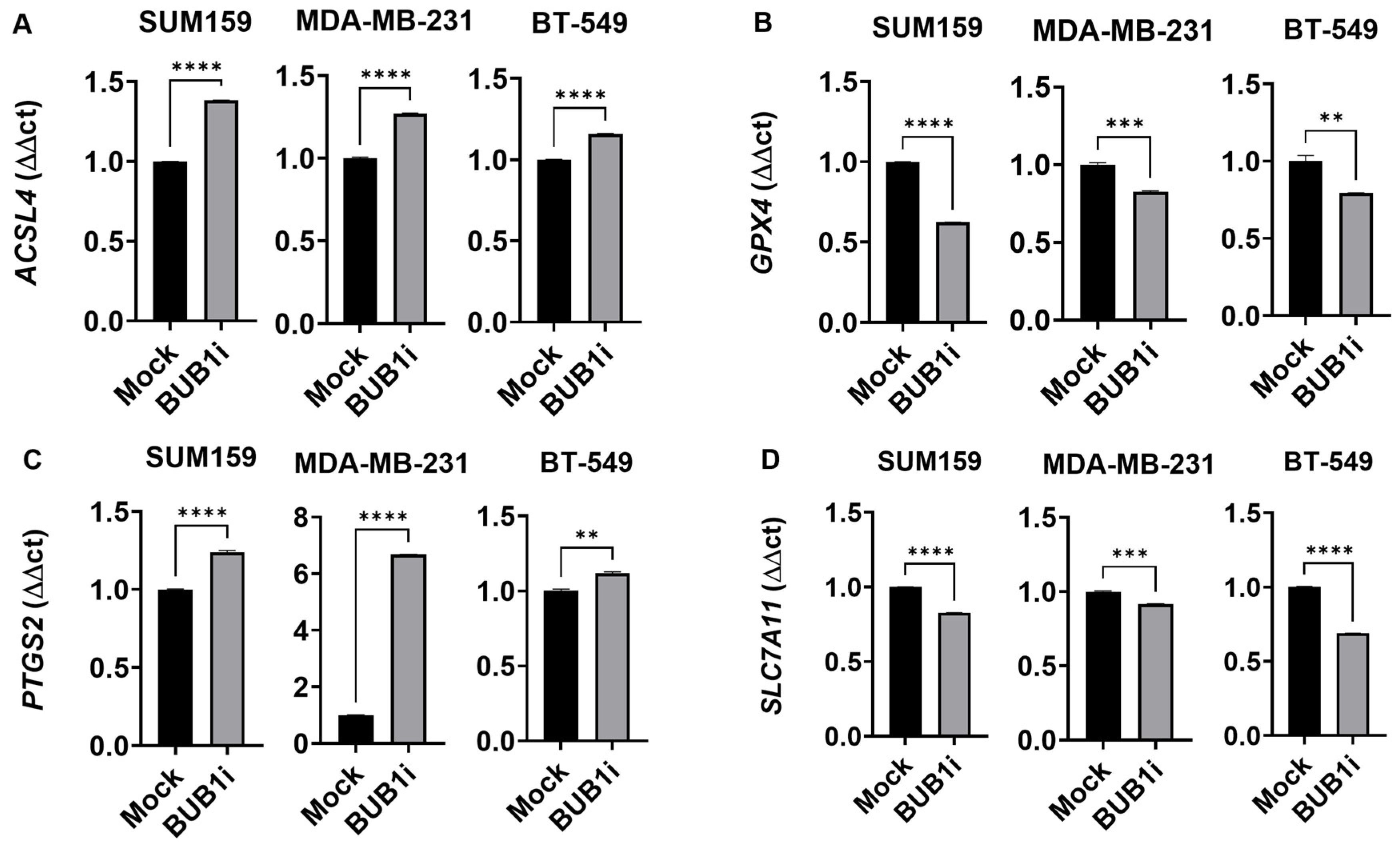
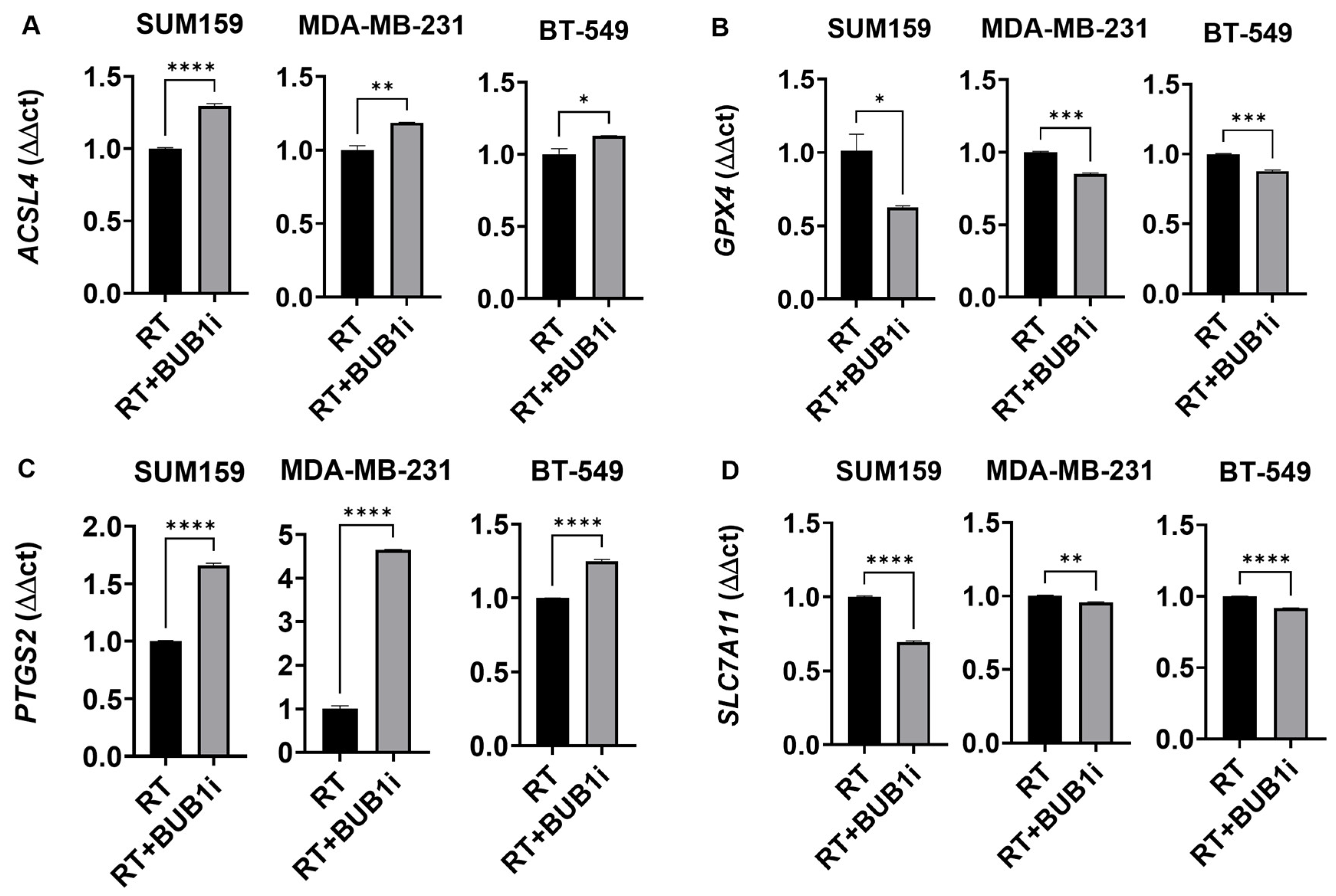
3.3. BUB1 Inhibition Alters Expression of Key Ferroptosis Genes

qRT-PCR assays were performed to measure the effect of BUB1 inhibition on ferroptosis markers in TNBC cell lines. Cells were treated with either a mock control or 1 μM BUB1 inhibitor (BUB1i) for 24 h, and total RNA was isolated to assess the expression of ACSL4, PTGS2 (Prostaglandin-Endoperoxide Synthase 2), GPX4, and SLC7A11 (Solute Carrier Family 7 Member 11) in SUM159, MDA-MB-231, and BT-549 cells. We observed that BUB1 inhibition significantly upregulated the expression of key ferroptosis markers ACSL4 and PTGS2 (Figure 3A,C) and downregulated GPX4 and SLC7A11 (Figure 3B,D) compared to the mock treatment. We also performed qPCR for NCOA4, IREB2, NFS1, and TFRC in SUM159 cells treated with DMSO (mock) and BUB1 inhibitor (Figure S2). Our analysis revealed a modest upregulation of NCOA4, IREB2, and TFRC (Figure S2A,B,D). NFS1 was significantly downregulated following treatment (Figure S2C). To further confirm the effect of BUB1 inhibition on ferroptosis, we performed a lipid peroxidation assay using the TBARS (thiobarbituric acid reactive substances) method. The TBARS assay was performed to measure MDA levels, a marker of lipid peroxidation, in SUM159 cells treated with DMSO (mock), ferroptosis inducer RSL3, ferroptosis inhibitor (F-1), or BUB1 inhibitor (BUB1i) (Figure S3). A standard curve was generated to quantify MDA levels in the samples (Figure S3A). The mock-treated control and the F-1-treated (negative control) samples showed MDA levels of 0.3 µM, indicating baseline lipid peroxidation. As expected, RSL3 (positive control) induced a significant increase in MDA levels to 1.2 µM, confirming lipid peroxidation and ferroptotic activity (Figure S3B). BUB1 inhibitor-treated samples exhibited MDA levels of 0.6 µM, which was higher than the mock control but lower than RSL3 (Figure S3B). This increase suggests that BUB1 inhibition induces lipid peroxidation, potentially contributing to ferroptosis in SUM159 cells.
TNBC cells were treated with sham or 4 Gy radiation for 24 h and monitored for the expression of ACSL4, PTGS2, GPX4 and SLC7A11 genes (Figure S4) in an attempt to confirm published reports that radiation causes ferroptotic cell death [39]. As expected, ACSL4 and PTGS2 expression increased in irradiation cells compared to sham-irradiated cells while GPX4 and SLC7A11 expression decreased. We next evaluated if BUB1 played any role in radiation-mediated ferroptosis in TNBC cells. The cell lines were treated with 1 μM BUB1 inhibitor (BUB1i) and 4 Gy irradiation for 24 h, and the expressions of the same four genes were evaluated. BUB1i combined with irradiation significantly upregulated ACSL4 expression in all three cell lines compared to radiation-only-treated cells (Figure 4A), suggesting enhanced ferroptotic activity. BUB1i significantly reduced the expression of GPX4 compared to radiation-only-treated cells (Figure 4B). The downregulation of GPX4 in irradiated cells treated with BUB1i underscores the role of this treatment in overcoming cellular resistance to ferroptosis. Furthermore, BUB1i caused a marked increase in PTGS2 and decreases in SLC7A11 expression compared to radiation only (Figure 4C,D), confirming the activation of ferroptotic signaling. This response was notably higher in irradiated cells than in non-irradiated cells, indicating at least an additive effect of BUB1 inhibition and radiation in amplifying ferroptosis-related oxidative stress (Figure S5). The collective upregulation of ACSL4 and PTGS2, coupled with the downregulation of GPX4 and SLC7A11, strongly indicates a pro-ferroptotic shift in TNBC cells treated with BUB1i and irradiation (Figure S5). These changes suggest that the combination treatment disrupts cellular defenses against lipid peroxidation, promoting ferroptosis as a potential mechanism for radiosensitization in TNBC.
We next assessed if BUB1i affected the expression of ferroptosis genes in the long term; TNBC cell lines were treated for 72 h with BUB1i, and 4 Gy radiation and gene expression were quantitated. ACSL4 expression remained elevated in irradiated BUB1i-treated cells across all cell lines (Figure S6A), indicating prolonged ferroptotic response. GPX4 expression continued to be suppressed at 72 h (Figure S6B). PTGS2 expression also stayed elevated (Figure S6C), while SLC7A11 remained downregulated (Figure S6D). This extended suppression of ferroptosis defenses further supports a role for BUB1 in potentially mediating radiation-induced ferroptotic cell death. A role for BUB1 in radiation-mediated ferroptotic cell death was further confirmed by using BUB1-depleted cell lines [35]. BUB1 CRISPR knockout (KO) TNBC cells were sham- or 4 Gy-irradiated, and gene expression was evaluated after 72 h. In SUM159 and MDA-MB-231 BUB1 KO cells, ACSL4, GPX4, PTGS2, and SLC7A11 expressions were comparable to those of BUB1i-treated cells. A pro-ferroptotic effect of radiation and BUB1 KO was evident in the expression of ACSL4 and PTGS2 (Figure S7A,C), with no pro-ferroptotic effect evident in the other markers in BUB1 KO cells.

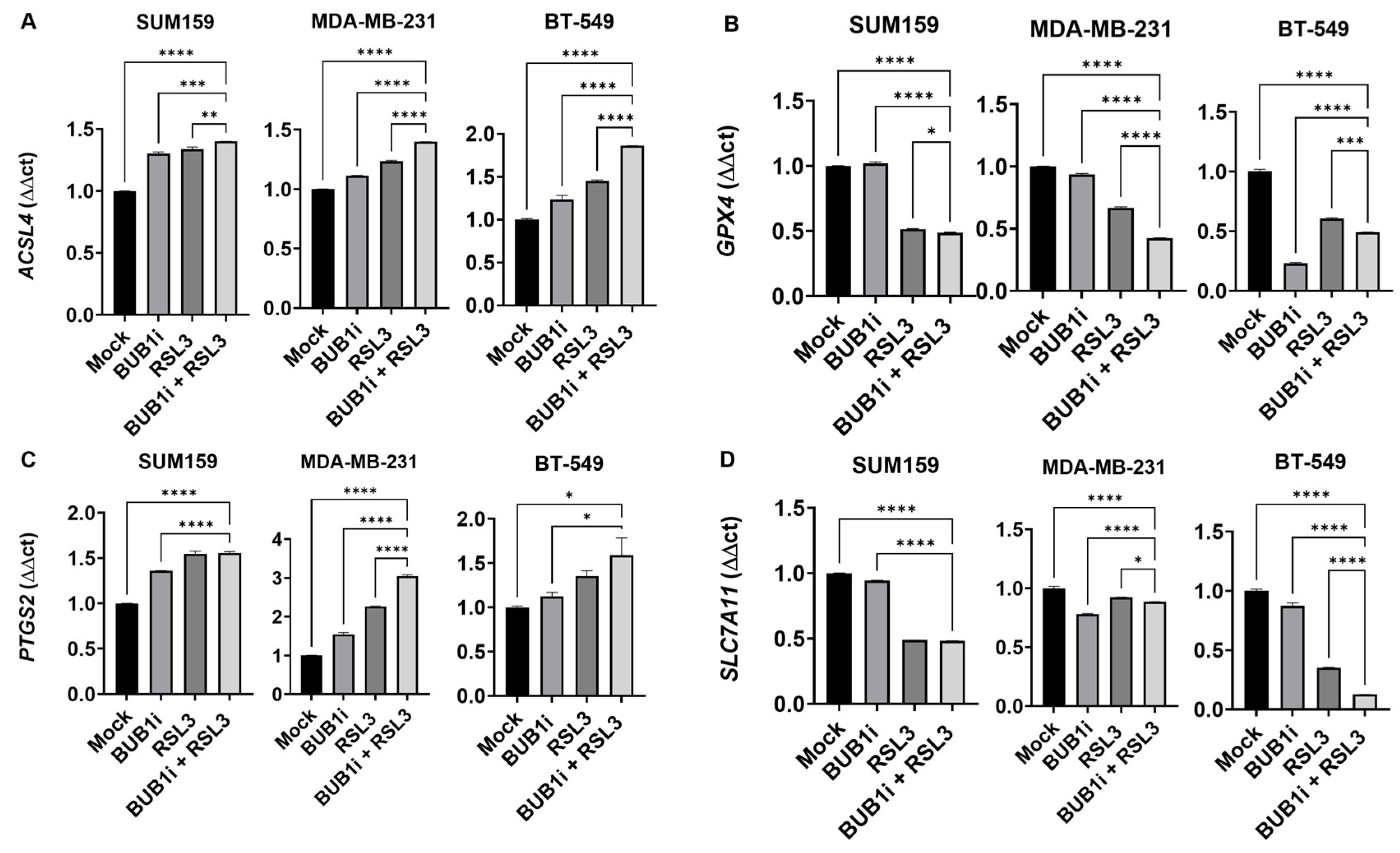
3.4. BUB1i and RSL3 Regulate Ferroptosis Markers in TNBC Cell Lines

The expression of ferroptosis markers ACSL4, GPX4, PTGS2 and SLC7A11 was assessed in SUM159, MDA-MB-231, and BT-549 cell lines that were treated with BUB1i and ferroptosis activator RSL3 alone or in combination for 24 h. The combined treatment of BUB1i and RSL3 significantly upregulated ACSL4 expression across all cell lines compared to individual treatments or mock controls (Figure 5A). Expression of GPX4 was notably downregulated in cells treated with BUB1i and RSL3 (Figure 5B), while PTGS2 expression was significantly elevated and SLC7A11 expression was significantly reduced in BUB1i and RSL3-treated cells (Figure 5D).

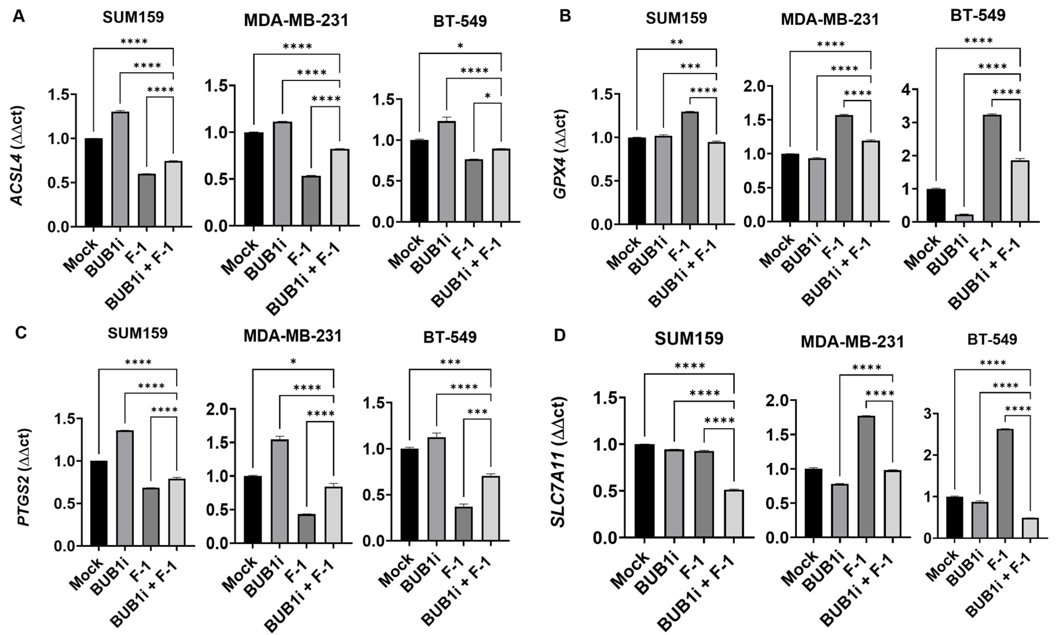
3.5. Ferrostatin-1 Reverses BUB1i-Induced Ferroptosis in TNBC Cell Lines

TNBC cell lines were treated with BUB1 inhibitor and Ferrostatin-1, and ferroptosis marker expression was evaluated to further demonstrate the specificity of ferroptosis induced by BUB1 inhibition. Co-treatment with F-1 effectively reduced the expression of ACSL4 and PTGS2 in BUB1i-treated cells (Figure 6A,C), indicating that F-1 suppresses the ferroptotic response initiated by BUB1 inhibition. In the presence of F-1, GPX4 expression increased minimally (Figure 6B) while SLC7A11 expression remained stable (Figure 6D) compared to the BUB1i treatment without F-1.

4. Discussion

Our recent studies demonstrated that BUB1 was significantly overexpressed in several breast cancers including TNBC compared to normal tissues, and its overexpression was associated with poorer patient survival [35]. Additionally, BUB1 was expressed higher in TNBC cell lines (SUM159, MDA-MB-231, and BT-549) compared to non-cancerous cells. Together, these observations provided a compelling rationale for investigating BUB1 inhibition as a radiosensitization strategy in TNBC [35]. Building on our findings and recent studies that radiation may induce ferroptosis [40], we wanted to test the hypothesis that BUB1, either alone or in combination with radiation, could induce ferroptosis and potentially serve as a novel therapeutic approach.
Ferroptosis, an iron-dependent form of cell death driven by lipid peroxidation, has emerged as a promising therapeutic target, particularly in apoptosis-resistant cancers such as TNBC [41]. This study reveals that BUB1 inhibition significantly promotes ferroptosis in TNBC cells, especially when combined with the ferroptosis activator RSL3, but is likely independent of irradiation (Figure 1). The ability of F-1 to reverse BUB1i-induced ferroptosis suggests exploiting BUB1 as a therapeutic target to induce ferroptosis in TNBC (Figure 2). These findings underscore the therapeutic potential of BUB1 as a target in TNBC, where treatment options are limited due to a lack of hormone receptors and poor responsiveness to conventional therapies. BUB1 inhibition significantly alters the expression of ferroptotic genes, indicating its potential to induce a ferroptotic response in TNBC cell lines. This is demonstrated by the upregulation of ACSL4 and PTGS2 (Figure 3A,C) alongside the downregulation of GPX4 and SLC7A11 (Figure 3B,D). These changes suggest that BUB1 inhibition alone is sufficient to trigger ferroptosis. The upregulation of NCOA4, IREB2, and TFRC, alongside the downregulation of NFS1 (Figure S2), supports the notion that ferroptosis is induced by BUB1i alone, further corroborating our findings from the TBARS assay (Figure S3). These results demonstrate that ferroptosis in SUM159 cells, induced by BUB1 inhibition, is associated with changes in the expression of genes involved in iron regulation, lipid peroxidation, and oxidative stress response. The hypothesis that BUB1 inhibition can lead to lipid peroxidation as part of the ferroptotic process is consistent with our findings in Supplementary Figure S3 that the partial elevation of MDA levels in BUB1i compared to RSL3 indicates that BUB1 inhibition alone may trigger ferroptosis to a moderate extent, but not as potently as a direct ferroptosis activator like RSL3. The lack of increased MDA levels in the F-1-treated samples further validates the specificity of the assay, as F-1 effectively blocks ferroptotic lipid peroxidation. Overall, these data support the role of BUB1 inhibition in inducing ferroptosis. Future studies may further investigate the molecular mechanisms linking BUB1 inhibition to ferroptosis induction in TNBC.
Furthermore, the upregulation of ACSL4 and PTGS2, combined with the suppression of GPX4 and SLC7A11, highlights an at-least-additive effect of BUB1 inhibition and RSL3 in driving ferroptosis in TNBC cells. ACSL4 is crucial for incorporating polyunsaturated fatty acids into membrane phospholipids, enhancing lipid peroxidation, which is a hallmark of ferroptosis [42]. This aligns with studies by Guo et al., who reported that ACSL4 upregulation is essential for sensitizing cancer cells to ferroptosis, particularly in contexts where traditional apoptotic pathways are inactive [43]. In TNBC, a subtype that often resists apoptosis, targeting ferroptosis through BUB1 inhibition could be a viable approach for inducing cell death. GPX4 is essential for detoxifying lipid peroxides, and its inhibition is a common feature in ferroptosis [44]. Downregulating GPX4 through BUB1 inhibition reduces the cellular defense against ferroptotic cell death, a finding consistent with studies showing that GPX4 is a critical regulator of ferroptosis and that its suppression enhances cancer cell susceptibility to lipid peroxidation-induced cell death [44]. The observed changes in ferroptotic gene expression highlight BUB1’s potential as a novel therapeutic target to induce cell death in TNBC through ferroptotic mechanisms. These findings align with growing evidence that targeting oxidative stress and lipid peroxidation pathways could provide innovative treatment strategies for aggressive cancers such as TNBC [45]. To confirm the specificity of ferroptosis in BUB1i-treated TNBC cells, we used F-1. Our results show that F-1 successfully reversed cell viability loss caused by BUB1 inhibition (Figure 2), indicating that the cell death observed was ferroptosis-specific. This finding complements the work of Skouta et al., who demonstrated that Ferrostatin-1 inhibits lipid peroxidation, thereby preventing ferroptotic cell death in cells undergoing oxidative stress-induced lipid damage [46]. In the context of our study, the reversal of ferroptosis markers by F-1 provides a confirmation that the cell death induced by BUB1 inhibition is mediated through ferroptotic mechanisms, validating BUB1’s role in ferroptosis regulation in TNBC. This suggests that F-1 protects against ferroptosis by sustaining antioxidant defenses and that BUB1 inhibition may induce ferroptotic cell death. Moreover, BUB1 KO experiments demonstrated increased ferroptotic marker expression and increased sensitivity to irradiation (Figure S7), indicating that BUB1 itself is crucial for maintaining ferroptotic activity under radiotherapy. This is consistent with Wang et al., who reported that BUB1 is integral to various cancer-related cell death pathways including ferroptosis and it influences oxidative stress and lipid metabolism [32].
Our findings indicate that targeting BUB1, especially in combination with ferroptosis activators like RSL3, may be a promising therapeutic approach for TNBC. This approach leverages ferroptosis to overcome the resistance typically seen with apoptosis-based therapies. Given that ferroptosis bypasses apoptotic mechanisms, which are often inactive in TNBC, therapies that induce ferroptosis could be particularly effective in this subtype. Recent studies suggest that targeting ferroptosis can improve therapeutic outcomes in various cancers, including pancreatic and colorectal cancers [47], by complementing traditional treatments and reducing tumor resistance. Our study adds to this growing body of research, suggesting that BUB1-targeted therapy could similarly benefit TNBC patients, particularly those who may not respond well to existing therapies. Artemisinin derivatives, such as artesunate, disrupt iron homeostasis, increasing free iron levels and lipid peroxidation [48], and enhance ferroptosis by modulating the p38 and ERK signaling pathways, as observed in glioblastoma cells [49]. Similarly, sorafenib, a multikinase inhibitor, promotes ferroptosis by inhibiting the cystine/glutamate antiporter (System Xc−), leading to glutathione depletion and lipid peroxidation [50]. Clinical trials, including the ARTIC M33/2 study on oral artesunate in metastatic breast cancer [51] and the Phase II trial (N0336) evaluating sorafenib [52], underscore the potential of ferroptosis-targeting agents, despite some challenges in clinical efficacy. These findings emphasize the importance of regulatory targets like GPX4 and System Xc− in ferroptosis, highlighting the promise of novel therapeutic strategies to overcome cancer resistance mechanisms. Our study of BUB1 inhibition aligns with these approaches, supporting this pathway to leverage ferroptosis in aggressive cancers like TNBC [53,54].

5. Conclusions

In conclusion, this study highlights the potential of BUB1 inhibition as a therapeutic strategy to induce ferroptosis in TNBC. Further in vivo studies and clinical trials will be essential to validate the efficacy of BUB1-targeted treatments, possibly combined with ferroptosis activators. These insights contribute to the ongoing development of innovative therapies aimed at exploiting ferroptosis for cancer treatment and suggest that BUB1 inhibition should be further investigated as a potential strategy to induce ferroptosis and enhance therapeutic efficacy in aggressive cancers like TNBC.

Supplementary Materials

The following supporting information can be downloaded at: www.mdpi.com/article/10.3390/dna5010016/s1, Table S1: List of primer sequences used in qRT-PCR for the genes NCOA4, IREB2, NFS1, and TFRC. Figure S1: Dose-response analysis of BUB1i, RSL3, and Ferrostatin-1 in SUM159 and MDA-MB-231 cell lines. p-values were defined as *** p ≤ 0.001, **** p ≤ 0.0001, Figure S2: BUB1 inhibition alters ferroptosis marker expression in TNBC cell lines. TNBC cell lines (SUM159, MDA-MB-231, BT-549) were treated with BUB1 inhibitor (1 µM) or mock control for 24 h, and ferroptosis markers were assessed via qPCR. (A) NCOA4 was significantly upregulated; (B) IREB2 was significantly upregulated; (C) NFS1 was significantly downregulated; and (D) TFRC was significantly upregulated, suggesting that BUB1 inhibition may trigger a ferroptotic response in TNBC cell lines. p-values were defined as ** p ≤ 0.01, *** p ≤ 0.001, **** p ≤ 0.0001, Figure S3: TBARS assay measuring MDA levels as a marker of lipid peroxidation in SUM159 cells treated with DMSO (mock), RSL3 (ferroptosis inducer), F-1 (ferroptosis inhibitor), or BUB1 inhibitor (BUB1i). (A) Standard curve for MDA quantification. (B) MDA levels: mock and F-1 treatments showed baseline levels (0.3 µM), RSL3 induced a significant increase (1.2 µM), and BUB1i treatment resulted in moderate lipid peroxidation (0.6 µM), Figure S4: Validation of ferroptosis marker expression in TNBC cell lines following irradiation (4 Gy). p-values were defined as * p ≤ 0.05, ** p ≤ 0.01, **** p ≤ 0.0001, Figure S5: Effects of BUB1 inhibition and irradiation on ferroptosis-related gene expression in TNBC cell lines. p-values were defined as ** p ≤ 0.01, *** p ≤ 0.001, **** p ≤ 0.0001, Figure S6: Long-term effects of BUB1 inhibition and irradiation on ferroptosis-related gene expression in TNBC cell lines. p-values were defined as ** p ≤ 0.01, **** p ≤ 0.0001, Figure S7: Regulation of ACSL4 and PTGS2 expression in BUB1 knockout cells following irradiation. p-values were defined as ** p ≤ 0.01, *** p ≤ 0.001, **** p ≤ 0.0001.

Author Contributions

S.S. performed all the experiments with help from S.T., S.S. also drafted the manuscript with input from all other authors. S.S., S.T., S.L.B., F.S., B.M. and S.N. analyzed the data. S.N. conceptualized and directed the study. All authors have read and agreed to the published version of the manuscript.

Funding

This work was supported by NCI R21 (1R21CA252010-01A1), the HFHS Research Administration Start Up, the HFHS Proposal Development Award, the HFHS-Rad Onc Start Up, and the Game on Cancer Award to Shyam Nyati. We also thank HFCI for providing a Translational Oncology Postdoctoral Fellowship to Sushmitha Sriramulu.

Institutional Review Board Statement

Not applicable.

Informed Consent Statement

Not applicable.

Data Availability Statement

The data can be obtained from the corresponding author upon request.

Conflicts of Interest

S.S., S.T., S.L.B., S.N.: no COI; F.S.: Varian Medical Systems Inc.—Honorarium and travel reimbursement for lectures and talks, Varian Noona—Member of Medical Advisory Board—Honorarium (no direct conflict); B.M.: Research support from Varian, ViewRay, and Philips (no direct conflict).

References

  1. Yin, L.; Duan, J.-J.; Bian, X.-W.; Yu, S.-C. Triple-negative breast cancer molecular subtyping and treatment progress. Breast Cancer Res. 2020, 22, 61. [Google Scholar] [CrossRef] [PubMed]
  2. Li, Y.; Zhang, H.; Merkher, Y.; Chen, L.; Liu, N.; Leonov, S.; Chen, Y. Recent advances in therapeutic strategies for triple-negative breast cancer. J. Hematol. Oncol. 2022, 15, 121. [Google Scholar] [CrossRef] [PubMed]
  3. Obidiro, O.; Battogtokh, G.; Akala, E.O. Triple Negative Breast Cancer Treatment Options and Limitations: Future Outlook. Pharmaceutics 2023, 15, 1796. [Google Scholar] [CrossRef]
  4. Zhu, S.; Wu, Y.; Song, B.; Yi, M.; Yan, Y.; Mei, Q.; Wu, K. Recent advances in targeted strategies for triple-negative breast cancer. J. Hematol. Oncol. 2023, 16, 100. [Google Scholar] [CrossRef]
  5. Zagami, P.; Carey, L.A. Triple negative breast cancer: Pitfalls and progress. NPJ Breast Cancer 2022, 8, 95. [Google Scholar] [CrossRef]
  6. Bou Zerdan, M.; Ghorayeb, T.; Saliba, F.; Allam, S.; Bou Zerdan, M.; Yaghi, M.; Bilani, N.; Jaafar, R.; Nahleh, Z. Triple Negative Breast Cancer: Updates on Classification and Treatment in 2021. Cancers 2022, 14, 1253. [Google Scholar] [CrossRef]
  7. Twelves, C.; Jove, M.; Gombos, A.; Awada, A. Cytotoxic chemotherapy: Still the mainstay of clinical practice for all subtypes metastatic breast cancer. Crit. Rev. Oncol./Hematol. 2016, 100, 74–87. [Google Scholar] [CrossRef]
  8. Sriramulu, S.; Thoidingjam, S.; Speers, C.; Nyati, S. Present and Future of Immunotherapy for Triple-Negative Breast Cancer. Cancers 2024, 16, 3250. [Google Scholar] [CrossRef]
  9. Alaluf, E.; Shalamov, M.M.; Sonnenblick, A. Update on current and new potential immunotherapies in breast cancer, from bench to bedside. Front. Immunol. 2024, 15, 1287824. [Google Scholar] [CrossRef]
  10. Rossi, V.; Turati, A.; Rosato, A.; Carpanese, D. Sacituzumab govitecan in triple-negative breast cancer: From bench to bedside, and back. Front. Immunol. 2024, 15, 1447280. [Google Scholar] [CrossRef]
  11. Cortesi, L.; Rugo, H.S.; Jackisch, C. An Overview of PARP Inhibitors for the Treatment of Breast Cancer. Target Oncol. 2021, 16, 255–282. [Google Scholar] [CrossRef] [PubMed]
  12. Eikesdal, H.P.; Yndestad, S.; Elzawahry, A.; Llop-Guevara, A.; Gilje, B.; Blix, E.S.; Espelid, H.; Lundgren, S.; Geisler, J.; Vagstad, G.; et al. Olaparib monotherapy as primary treatment in unselected triple negative breast cancer. Ann. Oncol. 2021, 32, 240–249. [Google Scholar] [CrossRef] [PubMed]
  13. Xiong, N.; Wu, H.; Yu, Z. Advancements and challenges in triple-negative breast cancer: A comprehensive review of therapeutic and diagnostic strategies. Front. Oncol. 2024, 14, 1405491. [Google Scholar] [CrossRef] [PubMed]
  14. Liu, Y.; Hu, Y.; Jiang, Y.; Bu, J.; Gu, X. Targeting ferroptosis, the achilles’ heel of breast cancer: A review. Front. Pharmacol. 2022, 13, 1036140. [Google Scholar] [CrossRef]
  15. Qi, X.; Wan, Z.; Jiang, B.; Ouyang, Y.; Feng, W.; Zhu, H.; Tan, Y.; He, R.; Xie, L.; Li, Y. Inducing ferroptosis has the potential to overcome therapy resistance in breast cancer. Front. Immunol. 2022, 13, 1038225. [Google Scholar] [CrossRef]
  16. Xu, C.; Chen, Y.; Yu, Q.; Song, J.; Jin, Y.; Gao, X. Compounds targeting ferroptosis in breast cancer: Progress and their therapeutic potential. Front. Pharmacol. 2023, 14, 1243286. [Google Scholar] [CrossRef]
  17. Ge, A.; He, Q.; Zhao, D.; Li, Y.; Chen, J.; Deng, Y.; Xiang, W.; Fan, H.; Wu, S.; Li, Y.; et al. Mechanism of ferroptosis in breast cancer and research progress of natural compounds regulating ferroptosis. J. Cell Mol. Med. 2024, 28, e18044. [Google Scholar] [CrossRef]
  18. Li, J.; Cao, F.; Yin, H.-l.; Huang, Z.-j.; Lin, Z.-t.; Mao, N.; Sun, B.; Wang, G. Ferroptosis: Past, present and future. Cell Death Dis. 2020, 11, 88. [Google Scholar] [CrossRef]
  19. Yan, H.-f.; Zou, T.; Tuo, Q.-z.; Xu, S.; Li, H.; Belaidi, A.A.; Lei, P. Ferroptosis: Mechanisms and links with diseases. Signal Transduct. Target. Ther. 2021, 6, 49. [Google Scholar] [CrossRef]
  20. Tang, D.; Chen, X.; Kang, R.; Kroemer, G. Ferroptosis: Molecular mechanisms and health implications. Cell Res. 2021, 31, 107–125. [Google Scholar] [CrossRef]
  21. Tomitsuka, Y.; Imaeda, H.; Ito, H.; Asou, I.; Ohbayashi, M.; Ishikawa, F.; Kuwata, H.; Hara, S. Gene deletion of long-chain acyl-CoA synthetase 4 attenuates xenobiotic chemical-induced lung injury via the suppression of lipid peroxidation. Redox Biol. 2023, 66, 102850. [Google Scholar] [CrossRef] [PubMed]
  22. Santana-Codina, N.; Del Rey, M.Q.; Kapner, K.S.; Zhang, H.; Gikandi, A.; Malcolm, C.; Poupault, C.; Kuljanin, M.; John, K.M.; Biancur, D.E. NCOA4-mediated ferritinophagy is a pancreatic cancer dependency via maintenance of iron bioavailability for iron–sulfur cluster proteins. Cancer Discov. 2022, 12, 2180–2197. [Google Scholar] [CrossRef]
  23. Yao, F.; Cui, X.; Zhang, Y.; Bei, Z.; Wang, H.; Zhao, D.; Wang, H.; Yang, Y. Iron regulatory protein 1 promotes ferroptosis by sustaining cellular iron homeostasis in melanoma. Oncol. Lett. 2021, 22, 657. [Google Scholar] [CrossRef] [PubMed]
  24. Feng, H.; Schorpp, K.; Jin, J.; Yozwiak, C.E.; Hoffstrom, B.G.; Decker, A.M.; Rajbhandari, P.; Stokes, M.E.; Bender, H.G.; Csuka, J.M.; et al. Transferrin Receptor Is a Specific Ferroptosis Marker. Cell Rep. 2020, 30, 3411–3423.e3417. [Google Scholar] [CrossRef]
  25. Lee, J.; Roh, J.-L. Targeting Iron-Sulfur Clusters in Cancer: Opportunities and Challenges for Ferroptosis-Based Therapy. Cancers 2023, 15, 2694. [Google Scholar] [CrossRef]
  26. Lei, G.; Zhuang, L.; Gan, B. The roles of ferroptosis in cancer: Tumor suppression, tumor microenvironment, and therapeutic interventions. Cancer Cell 2024, 42, 513–534. [Google Scholar] [CrossRef]
  27. Diao, J.; Jia, Y.; Dai, E.; Liu, J.; Kang, R.; Tang, D.; Han, L.; Zhong, Y.; Meng, L. Ferroptotic therapy in cancer: Benefits, side effects, and risks. Mol. Cancer 2024, 23, 89. [Google Scholar] [CrossRef]
  28. Lei, G.; Zhuang, L.; Gan, B. Targeting ferroptosis as a vulnerability in cancer. Nat. Rev. Cancer 2022, 22, 381–396. [Google Scholar] [CrossRef]
  29. Han, C.; Liu, Y.; Dai, R.; Ismail, N.; Su, W.; Li, B. Ferroptosis and Its Potential Role in Human Diseases. Front. Pharmacol. 2020, 11, 239. [Google Scholar] [CrossRef]
  30. Ma, S.; Henson, E.S.; Chen, Y.; Gibson, S.B. Ferroptosis is induced following siramesine and lapatinib treatment of breast cancer cells. Cell Death Dis. 2016, 7, e2307. [Google Scholar] [CrossRef]
  31. Ding, Y.; Chen, X.; Liu, C.; Ge, W.; Wang, Q.; Hao, X.; Wang, M.; Chen, Y.; Zhang, Q. Identification of a small molecule as inducer of ferroptosis and apoptosis through ubiquitination of GPX4 in triple negative breast cancer cells. J. Hematol. Oncol. 2021, 14, 19. [Google Scholar] [CrossRef] [PubMed]
  32. Wang, W.; Zhou, X.; Kong, L.; Pan, Z.; Chen, G. BUB1 Promotes Gemcitabine Resistance in Pancreatic Cancer Cells by Inhibiting Ferroptosis. Cancers 2024, 16, 1540. [Google Scholar] [CrossRef] [PubMed]
  33. Klebig, C.; Korinth, D.; Meraldi, P. Bub1 regulates chromosome segregation in a kinetochore-independent manner. J. Cell Biol. 2009, 185, 841–858. [Google Scholar] [CrossRef] [PubMed]
  34. Sriramulu, S.; Thoidingjam, S.; Siddiqui, F.; Brown, S.L.; Movsas, B.; Walker, E.; Nyati, S. BUB1 Inhibition Sensitizes TNBC Cell Lines to Chemotherapy and Radiotherapy. Biomolecules 2024, 14, 625. [Google Scholar] [CrossRef]
  35. Sriramulu, S.; Thoidingjam, S.; Chen, W.-M.; Hassan, O.; Siddiqui, F.; Brown, S.L.; Movsas, B.; Green, M.D.; Davis, A.J.; Speers, C.; et al. BUB1 regulates non-homologous end joining pathway to mediate radioresistance in triple-negative breast cancer. J. Exp. Clin. Cancer Res. 2024, 43, 163. [Google Scholar] [CrossRef]
  36. Thoidingjam, S.; Sriramulu, S.; Hassan, O.; Brown, S.L.; Siddiqui, F.; Movsas, B.; Gadgeel, S.; Nyati, S. BUB1 Inhibition Overcomes Radio- and Chemoradiation Resistance in Lung Cancer. Cancers 2024, 16, 3291. [Google Scholar] [CrossRef]
  37. Xu, L.; Liu, Y.e.; Chen, X.; Zhong, H.; Wang, Y. Ferroptosis in life: To be or not to be. Biomed. Pharmacother. 2023, 159, 114241. [Google Scholar] [CrossRef]
  38. Thu, K.L.; Soria-Bretones, I.; Mak, T.W.; Cescon, D.W. Targeting the cell cycle in breast cancer: Towards the next phase. Cell Cycle 2018, 17, 1871–1885. [Google Scholar] [CrossRef]
  39. Berry, C.E.; Kendig, C.B.; An, N.; Fazilat, A.Z.; Churukian, A.A.; Griffin, M.; Pan, P.M.; Longaker, M.T.; Dixon, S.J.; Wan, D.C. Role of ferroptosis in radiation-induced soft tissue injury. Cell Death Discov. 2024, 10, 313. [Google Scholar] [CrossRef]
  40. Lei, G.; Zhang, Y.; Koppula, P.; Liu, X.; Zhang, J.; Lin, S.H.; Ajani, J.A.; Xiao, Q.; Liao, Z.; Wang, H.; et al. The role of ferroptosis in ionizing radiation-induced cell death and tumor suppression. Cell Res. 2020, 30, 146–162. [Google Scholar] [CrossRef]
  41. Li, J.; He, D.; Li, S.; Xiao, J.; Zhu, Z. Ferroptosis: The emerging player in remodeling triple-negative breast cancer. Front. Immunol 2023, 14, 1284057. [Google Scholar] [CrossRef] [PubMed]
  42. Jia, B.; Li, J.; Song, Y.; Luo, C. ACSL4-Mediated Ferroptosis and Its Potential Role in Central Nervous System Diseases and Injuries. Int. J. Mol. Sci. 2023, 24, 21. [Google Scholar] [CrossRef] [PubMed]
  43. Guo, N. Identification of ACSL4 as a biomarker and contributor of ferroptosis in clear cell renal cell carcinoma. Transl. Cancer Res. 2022, 11, 2688–2699. [Google Scholar] [CrossRef] [PubMed]
  44. Xie, Y.; Kang, R.; Klionsky, D.J.; Tang, D. GPX4 in cell death, autophagy, and disease. Autophagy 2023, 19, 2621–2638. [Google Scholar] [CrossRef]
  45. Nandi, I.; Ji, L.; Smith, H.W.; Avizonis, D.; Papavasiliou, V.; Lavoie, C.; Pacis, A.; Attalla, S.; Sanguin-Gendreau, V.; Muller, W.J. Targeting fatty acid oxidation enhances response to HER2-targeted therapy. Nat. Commun. 2024, 15, 6587. [Google Scholar] [CrossRef]
  46. Skouta, R.; Dixon, S.J.; Wang, J.; Dunn, D.E.; Orman, M.; Shimada, K.; Rosenberg, P.A.; Lo, D.C.; Weinberg, J.M.; Linkermann, A.; et al. Ferrostatins Inhibit Oxidative Lipid Damage and Cell Death in Diverse Disease Models. J. Am. Chem. Soc. 2014, 136, 4551–4556. [Google Scholar] [CrossRef]
  47. Yan, H.; Talty, R.; Aladelokun, O.; Bosenberg, M.; Johnson, C.H. Ferroptosis in colorectal cancer: A future target? Br. J. Cancer 2023, 128, 1439–1451. [Google Scholar] [CrossRef]
  48. Chen, G.-Q.; Benthani, F.A.; Wu, J.; Liang, D.; Bian, Z.-X.; Jiang, X. Artemisinin compounds sensitize cancer cells to ferroptosis by regulating iron homeostasis. Cell Death Differ. 2020, 27, 242–254. [Google Scholar] [CrossRef]
  49. Song, Q.; Peng, S.; Che, F.; Zhu, X. Artesunate induces ferroptosis via modulation of p38 and ERK signaling pathway in glioblastoma cells. J. Pharmacol. Sci. 2022, 148, 300–306. [Google Scholar] [CrossRef]
  50. Li, Q.; Chen, K.; Zhang, T.; Jiang, D.; Chen, L.; Jiang, J.; Zhang, C.; Li, S. Understanding sorafenib-induced ferroptosis and resistance mechanisms: Implications for cancer therapy. Eur. J. Pharmacol. 2023, 955, 175913. [Google Scholar] [CrossRef]
  51. von Hagens, C.; Walter-Sack, I.; Goeckenjan, M.; Osburg, J.; Storch-Hagenlocher, B.; Sertel, S.; Elsässer, M.; Remppis, B.A.; Edler, L.; Munzinger, J.; et al. Prospective open uncontrolled phase I study to define a well-tolerated dose of oral artesunate as add-on therapy in patients with metastatic breast cancer (ARTIC M33/2). Breast Cancer Res. Treat. 2017, 164, 359–369. [Google Scholar] [CrossRef] [PubMed]
  52. Moreno-Aspitia, A.; Morton, R.F.; Hillman, D.W.; Lingle, W.L.; Rowland, K.M., Jr.; Wiesenfeld, M.; Flynn, P.J.; Fitch, T.R.; Perez, E.A. Phase II trial of sorafenib in patients with metastatic breast cancer previously exposed to anthracyclines or taxanes: North Central Cancer Treatment Group and Mayo Clinic Trial N0336. J. Clin. Oncol. 2009, 27, 11–15. [Google Scholar] [CrossRef] [PubMed]
  53. Du, Y.; Guo, Z. Recent progress in ferroptosis: Inducers and inhibitors. Cell Death Discov. 2022, 8, 501. [Google Scholar] [CrossRef] [PubMed]
  54. Sun, S.; Shen, J.; Jiang, J.; Wang, F.; Min, J. Targeting ferroptosis opens new avenues for the development of novel therapeutics. Signal Transduct. Target. Ther. 2023, 8, 372. [Google Scholar] [CrossRef]
Figure 1. Effects of BUB1 inhibition and ferroptosis activation on TNBC cell viability. (AC) Viability assays in TNBC cell lines (SUM159, MDA-MB-231, BT-549) show BUB1 inhibitor (BUB1i, 1 µM) alone reduces viability, while co-treatment with ferroptosis activator RSL3 (62.5 nM) further decreases survival. (DF) Adding irradiation (4 Gy) to BUB1i and RSL3 slightly increases cell death. (DF) represent the irradiated samples. p-values were defined as * p ≤ 0.05, ** p ≤ 0.01, *** p ≤ 0.001, **** p ≤ 0.0001.
Dna 05 00016 g001
Figure 2. Ferrostatin-1 confirms ferroptosis-specific cell death induced by BUB1 inhibition in TNBC cell lines. TNBC cell lines (SUM159, MDA-MB-231, BT-549) were treated with BUB1 inhibitor (BUB1i, 1 µM) alone or with Ferrostatin-1 (F-1, 10 µM). (AC) In non-irradiated cells, F-1 reversed BUB1i-induced cell death, restoring viability to control levels. (DF) F-1 mediated reversal in cell viability even in irradiated cells (4 Gy). (DF) represent the irradiated samples. p-values were defined as * p ≤ 0.05, ** p ≤ 0.01, *** p ≤ 0.001.
Dna 05 00016 g002
Figure 3. BUB1 inhibition alters ferroptosis marker expression in TNBC cell lines. TNBC cell lines (SUM159, MDA-MB-231, BT-549) were treated with BUB1 inhibitor (1 µM) or mock control for 24 h, and ferroptosis markers were assessed via qPCR. (A) ACSL4 was significantly upregulated; (B) GPX4 was significantly downregulated; (C) PTGS2 was significantly upregulated; and (D) SLC7A11 was significantly downregulated, suggesting that BUB1 inhibition may trigger a ferroptotic response in TNBC cell lines. p-values were defined as ** p ≤ 0.01, *** p ≤ 0.001, **** p ≤ 0.0001.
Dna 05 00016 g003
Figure 4. Upregulation of ferroptosis markers in TNBC cell lines following BUB1 inhibition and irradiation. TNBC cell lines (SUM159, MDA-MB-231, BT-549) were treated with BUB1 inhibitor (1 µM) and irradiation (4 Gy) for 24 h, and ferroptosis marker expression was analyzed by qPCR. (A) ACSL4 levels increased, indicating enhanced lipid peroxidation. (B) GPX4 was significantly downregulated, reducing antioxidant defenses. (C) PTGS2 expression was elevated, reflecting increased oxidative stress. (D) SLC7A11 was downregulated, impairing cystine import and sensitizing cells to ferroptosis. (AD) represent the irradiated samples. p-values were defined as * p ≤ 0.05, ** p ≤ 0.01, *** p ≤ 0.001, **** p ≤ 0.0001.
Dna 05 00016 g004
Figure 5. Regulation of ferroptosis markers by BUB1i and RSL3 in TNBC cell lines. Ferroptosis markers ACSL4, GPX4, PTGS2, and SLC7A11 were analyzed in SUM159, MDA-MB-231, and BT-549 cells treated for 24 h with BUB1i (1 µM), RSL3 (0.25 µM), or their combination. (A) Combined treatment significantly increased ACSL4 expression and (C) elevated PTGS2 levels, indicating enhanced lipid peroxidation and oxidative stress. (B) GPX4 was markedly downregulated, reducing antioxidant defenses, while (D) SLC7A11 was significantly decreased, impairing cystine import and glutathione synthesis. p-values were defined as * p ≤ 0.05, ** p ≤ 0.01, *** p ≤ 0.001, **** p ≤ 0.0001.
Dna 05 00016 g005
Figure 6. Ferrostatin-1 reverses BUB1i-induced ferroptosis in TNBC cell lines. TNBC cell lines were treated with BUB1 inhibitor (1 µM) alone or combined with Ferrostatin-1 (F-1, 10 µM) for 24 h. (A) F-1 co-treatment significantly reduced ACSL4 and (C) PTGS2 expression. (B) GPX4 showed a slight increase with F-1, while (D) SLC7A11 levels remained unchanged compared to BUB1i-only treatment. p-values were defined as * p ≤ 0.05, ** p ≤ 0.01, *** p ≤ 0.001, **** p ≤ 0.0001.
Dna 05 00016 g006
Table 1. List of primer sequences used in qRT-PCR.
ACSL4
Forward: 5′-ATAAAGCAGAGTACCCTGAAG
Reverse: 5′-CAAGTTTTCTGGGTTAGATCC
GPX4
Forward: 5′-GAAGTAAACTACACTCAGCTC
Reverse: 5′-CTCTTTGATCTCTTCGTTACTC
PTGS2
Forward: 5′-AAGCAGGCTAATACTGATAGG
Reverse: 5′-TGTTGAAAAGTAGTTCTGGG
SLC7A11
Forward: 5′-GGTTATTCTATGTTGCGTCTC
Reverse: 5′-AATAACAGCTGGTAGAGGAG
Disclaimer/Publisher’s Note: The statements, opinions and data contained in all publications are solely those of the individual author(s) and contributor(s) and not of MDPI and/or the editor(s). MDPI and/or the editor(s) disclaim responsibility for any injury to people or property resulting from any ideas, methods, instructions or products referred to in the content.

