Article

Low-Intensity Pulsed Ultrasound in the Treatment of Nonunions and Fresh Fractures: A Case Series

1
Studio Medico Associato Cecca-Romanò, 20121 Milan, Italy
2
External Fixation Unit, Humanitas Clinical and Research Center IRCCS, Via Manzoni 56, Rozzano, 20089 Milan, Italy
3
Osteoarticular Inflammatory Diseases Unit (MIOA), Department of Infectious Diseases and Septic Orthopaedics (MIOS), Santa Corona Hospital, 17027 Pietra Ligure (SV), Italy
4
Department of Medical and Surgical Specialties and Dentistry, University of Campania “Luigi Vanvitelli”, 80100 Naples, Italy
5
Unit of Orthopaedics and Traumatology, “Villa Malta” Hospital, ASL Salerno, 84087 Sarno, Italy
6
Rehabilitation Department, INI Città Bianca, 03029 Veroli, Italy
7
Orthopaedics and Traumatology Complex Operative Unit, DEA ASL Roma 5, 00019 Tivoli, Italy
8
Orthopaedics and Traumatology Department, AULSS 2 Marca Trevigiana Presidi Ospedali (P.O.) of Conegliano and Vittorio Veneto, 31015 Conegliano, Italy
9
Orthopaedics and Traumatology Department, A.O. “G.Rummo”, 82100 Benevento, Italy
10
IGEA Clinical Biophysics, 41012 Carpi, Italy
11
Department of Osteoarticular Infections, G. Pini Orthopaedic Institute, 20122 Milan, Italy
*
Author to whom correspondence should be addressed.
Trauma Care 2022, 2(2), 174-184; https://doi.org/10.3390/traumacare2020014
Submission received: 28 January 2022 / Revised: 1 April 2022 / Accepted: 3 April 2022 / Published: 5 April 2022

Abstract

:
It is estimated that approximately 5% to 10% of fractures will evolve into nonunions. Nonunions have a significant impact on patient quality of life and on socioeconomic costs. Low-intensity pulsed ultrasound (LIPUS) is a non-invasive therapy widely used within the orthopedic community to accelerate the healing of fresh fractures, to minimize delayed healing, and to promote healing of nonunions. In this case series, 46 nonunions and 19 fresh fractures were treated with LIPUS for at least three months or until fracture healing. Bone healing was assessed both at a radiological and a functional level. Of the nonunions healed, 89% had a mean healing time of 89 ± 53 days. In the group of fresh fractures, the healing percentage was 95% with a mean healing time of 46 ± 28 days. LIPUS treatment is proven to be safe and well tolerated; there were no adverse events related to the use of the device, even in the presence of internal fixations and infections. LIPUS therapy should be considered a low-risk option both as an adjunct to surgery or as a standalone therapy in the management of nonunion and fresh fractures.

1. Introduction

The incidence of fracture nonunion is estimated to be between 5% and 10%. The risk of nonunion is related to several factors: fracture severity, localization, comorbidities, and other medication use [1]. Nonunions have a significant impact on patients’ quality of life, leading to additional suffering and prolonged functional impairment. Nonunions often require further complex surgical procedures to heal. The surgical management of nonunions has a large socioeconomic impact on both patients and the healthcare system [2]. The success rate of the surgical treatment of nonunions is between 70% and 90%, depending on fracture location and surgical technique [3,4]. Nonunions still represent a challenge in clinical practice and are associated with an increased risk of further complications.
Avoiding additional surgery through conservative management of nonunion, as well as accelerating the healing of fresh fractures or reducing the risk of delayed unions, can have a significant impact on patients as well as on the healthcare system. The possibility of stimulating bone healing through the application of physical energies has been widely demonstrated over the last 50 years [5].
Low intensity pulsed ultrasound (LIPUS) is a non-invasive therapy widely used within the orthopedic community to accelerate the healing of fresh fractures, minimize delayed healing, and to stimulate the healing of nonunions. Specific ultrasound signal characteristics and treatment regimens have been reported to significantly enhance the fracture healing process [6,7]. The Food and Drug Administration (FDA) approved the use of LIPUS to accelerate the healing of fresh fractures and for the treatment of nonunions in 1994 and 2000, respectively [8]. The FDA-approved LIPUS signal consists of a 200 µs burst of 1.5 MHz sine waves repeating at 1 kHz and delivering 30 mW/cm2 spatial average and temporal average (SATA) applied at the fracture site for 20 min per day. LIPUS is widely recommended as a standalone or adjuvant treatment for nonunions, delayed unions, and fresh fractures. In the last 20 years, data in the literature support the efficacy of LIPUS in all types of fractures. The use of physical stimuli to enhance reparative osteogenesis has been widely investigated and its clinical use is based on sound scientific evidence. The orthopedic community undoubtedly played a central role in the development and understanding of the importance of physical stimuli to control biological activities [5,9].
In the current scenario of varying clinical indications, we designed a prospective observational case series to evaluate, in different Italian regional settings, (i) the most frequently LIPUS-treated sites, (ii) the average stimulation time for fresh fractures and nonunions, (iii) the average healing time for fresh fractures and nonunions treated with LIPUS, (iv) the healing rate of nonunions and fresh fractures treated with LIPUS, and (v) patient compliance.

2. Materials and Methods

LIPUS treatment was applied as part of the orthopedic treatment protocol, and written informed consent was given by all patients. Data from patients treated since 2005 were retrospectively reviewed using a shared case report form.
Patients with fresh fractures or nonunions (a fracture that persisted at least 6 months after trauma and 3 months after the last surgery) were treated. According to the indication for the use of biophysical stimulation [9], fractures not adequately stabilized, aligned, or with bone gap bigger than half the bone diameter were not treated.

2.1. LIPUS Treatment Protocol

To apply LIPUS, the FAST® device (IGEA SpA, Carpi, Italy) was used. This medical device delivers an ultrasound signal composed of a burst width of 200 µs, containing 1.5 MHz sine waves, with a repetition rate of 1 kHz and delivers 30 mW/cm2 of spatial averaged and temporal averaged (SATA) intensity. The therapeutic signal generated by the device is delivered through a transducer positioned on the skin overlying the fracture with ultrasonic coupling gel. Treatment time was 20 min per day with a warning signal that sounded if there was not proper coupling to the skin. The operating unit contained an integrated timer that monitored treatment times and automatically turned the unit off after the 20 min of daily treatment. A visual and audible signal alerted the patient that the treatment was completed.
Patients were instructed on the use of the device and performed the treatment at home. Treatment was continued for at least three months or until fracture healing, whichever came first.

2.2. Data Collection and Clinical Assessment

For all patients, data collected during baseline and follow-up visits were analyzed: personal data, fracture characteristics (date of fracture, location, type, trauma, infection, stability, alignment, and bone gap), treatments (cast, brace, type of fixation), and risk factors (smoking, diabetes, corticosteroids use, osteoporosis, serious osteomalacia, osteogenesis imperfecta).
Bone healing was assessed both at a radiological and functional level. Radiological healing was defined by the presence of bridging callus on radiographs in 3 out of 4 cortices. Clinical healing was defined by the absence of pain at the fracture site. The need for further surgery or the persistence of nonunion were considered treatment failures.

2.3. Statistical Methods
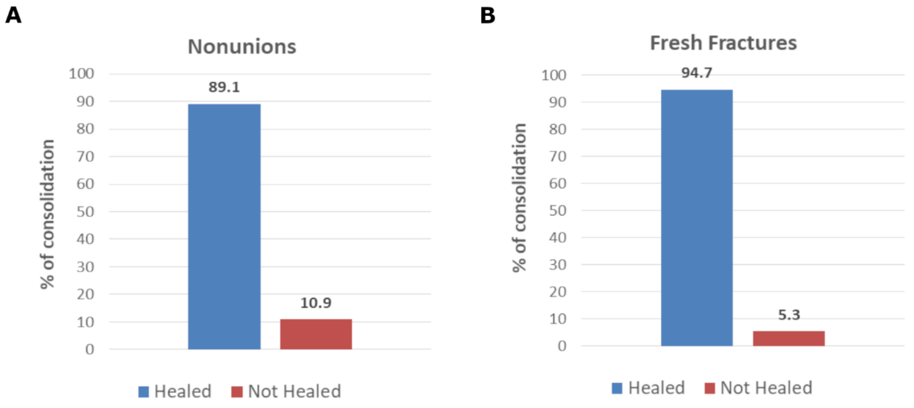
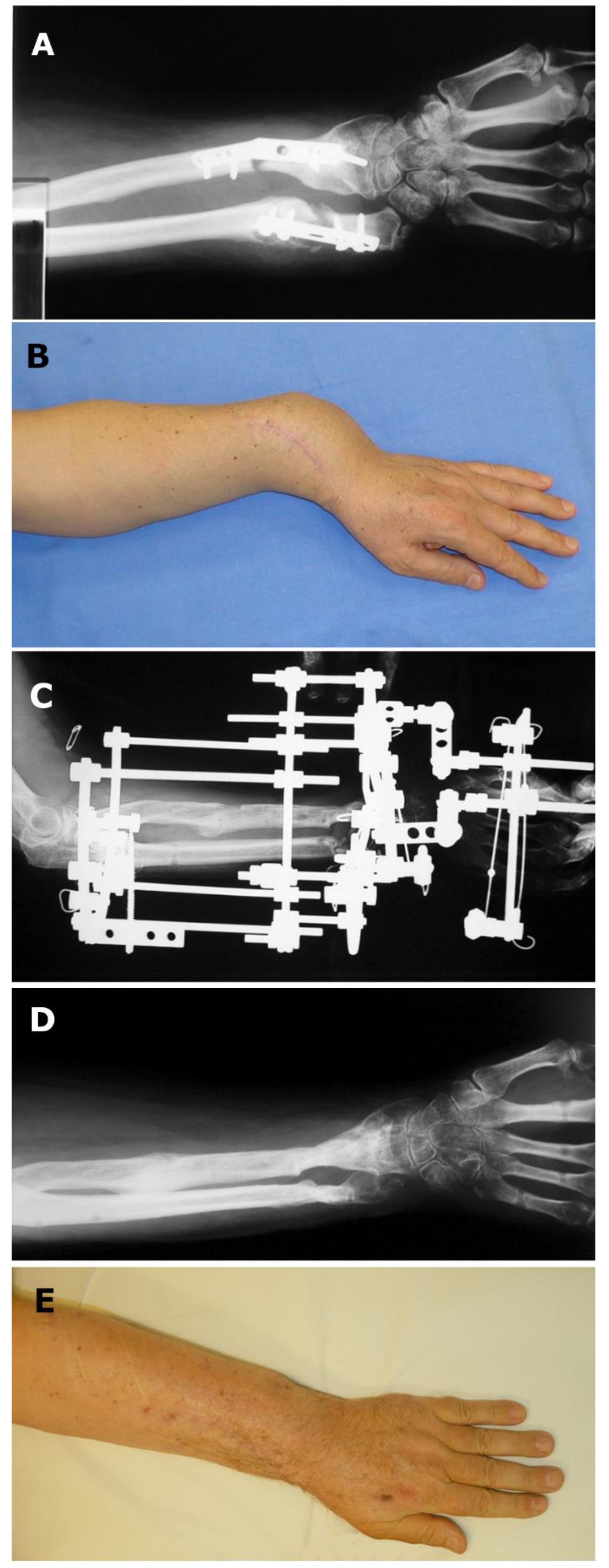
Continuous variables were described by mean value and standard deviation, and categorical variables by absolute number and percentage. Analysis of the occurrence of bone healing in the groups and relative comparison was performed by contingency table and the chi-square test, together with the relative risk calculation. A p-value of 0.05 was considered statistically significant. The statistical analysis was performed with GraphPad Instat software (version 3.06, GraphPad Software 2365 Northside Dr. Suite 560 San Diego, CA, USA).

3. Results

Seventy-three patients were treated, and seventy-one met the criteria for treatment; for six patients, follow-up visits were not possible. Sixty-five patients (46 M, 19 F) were analyzed. Demographics and clinical characteristics at baseline are reported in Table 1 and Table 2.
Forty-six nonunions and nineteen fresh fractures were treated with LIPUS for at least three months or until fracture healing, whichever came first.
No adverse events related to the use of the device were recorded, even in the presence of metal implants (plate, screws, or nails) and infections.
Forty-six patients (70.8%) suffered from nonunions (both infected and uninfected), twenty-eight in upper limbs and thirty-seven in lower limbs (Table 1). The mean age in the nonunion group was 44 ± 18 years. The average time between fracture and LIPUS therapy was 14 months. The average time between the last surgery and LIPUS therapy was 83 ± 165 days. The average treatment time was 100 ± 72 days. Of the patients, 60% showed good compliance to the treatment (at least 75% of the treatment sessions completed), whilst 70% of the patients performed at least 50% of the treatment sessions.
Eighty-nine percent of the nonunions healed within 89 ± 53 days (Figure 1, panel A).
Figure 2 shows a typical case that has reached bone healing.
Of the recovered patients, 34% were smokers, whilst in the group of non-healed patients, the percentage of smokers was 80% (p-value = 0.0237). Chi-square analysis revealed a significant difference in healing rates between smokers and non-smokers (p < 0.05) with a relative risk of nonunion equal to 3.29 in the smoking group compared with the non-smoking group. Among nonunions, 15 were infected, whilst 31 were uninfected: the healing rate of infected nonunions was 86.7% whilst the healing rate for uninfected nonunions was 90.3% (p-value = 0.71). No statistically significant difference was found. Likewise, there was no statistically significant difference in the healing rates between nonunions deriving from open or closed fractures (86.7% vs. 90.3% respectively, p-value = 0.71).
The five patients who had not healed had, on average, two prior surgeries, four out of the five patients were smokers, bone loss was present in three patients suffering from nonunions, two patients had infected nonunions, and two patients required further surgery.
Within the group of nonunions, a subset of 16 patients treated with surgery before LIPUS stimulation can be identified. In this group of patients, the mean age was 40 ± 13 years. Sixty-nine percent (69%) of these patients had previous unsuccessful surgeries (mean two, min one, max three). Table 3 shows patients’ characteristics at baseline. The average treatment time was 104 ± 79 days. Healing rate was 81% with a mean healing time of 79 ± 50 days. There was no statistically significant difference in the healing rate achieved in nonunions treated either conservatively or surgically before LIPUS stimulation (p-value = 0.21).
In the group of fresh fractures, the mean age was 49 ± 21 years. Table 2 shows patient characteristics at baseline. Treatment compliance (calculated as a percentage of patients completing at least 75% of the treatment sessions) was 70%, whilst 80% of the patients performed at least 50% of the treatment sessions. The percentage of healing was 95% with a mean healing time of 46 ± 28 days (Figure 2 panel B).
Among nonunion patients, 21 were less than 40 years old. Table 4 shows that the healing rate was no different between patients younger or older than 40. In the fresh fractures group, one patient did not heal after LIPUS: a female aged 77 suffering from a comminuted femoral fracture.

4. Discussion

Five to ten percent of fractures may evolve in nonunions. Factors like soft tissue damage and open comminuted fractures could lead to an increased risk of nonunions or delayed unions. It was reported that nonunions occurred in 4.7% of distal femoral fractures and 2.8% of shaft femoral fractures [10]. To enhance the healing process, therapeutic strategies, including mechanical environment control, use of scaffolds, growth factors, and cell therapies (the diamond concept) showed convincing results [11]. Recently, pharmacological systemic treatment with teriparatide was suggested to favor bone healing in the case of nonunions [12].
In addition to orthobiologics and pharmacological treatments, therapies based on physical stimuli are increasingly gaining importance in the orthopedic and traumatology practice.
The first report describing the possibility of promoting osteogenesis with physical stimuli such as ultrasound dates back to 1953, when Corradi and Cozzolino showed the accelerated bone healing of fresh fractures in the rabbit radius compared to control [13]. In the same year, the authors also reported similar results in a limited clinical series [14]. Duarte, in 1983, using a low-intensity pulsed ultrasound (LIPUS) system with the following characteristics: 1.5 MHz, SATA intensity of 30 mW/cm2, described a 28% increase in the ossification of rabbit fibular osteotomies in the limbs treated with LIPUS for 15 min/day [6]. In agreement with these results, Pilla reported a shortening of the bone healing process in rabbit fibular osteotomies upon exposure to LIPUS for 20 min/day [7].
The ability of LIPUS to stimulate fresh fracture healing in clinical settings was finally proven in a randomized, double-blind, placebo-controlled trial on closed or grade-I open tibia fractures. Heckman et al. studied LIPUS for simple closed or Gustilo I open tibial shaft fractures immobilized by a cast and showed statistically significant improvements in time of clinical healing to overall (clinical and radiographic) healing (with a 38% decrease in time), in the proportion of fractures healed within 120 days (LIPUS, 88%; control, 44%) and proportion of fractures healed within 150 days (LIPUS, 94%; control, 62%) [15]. In agreement with these results, Leung et al., in a randomized placebo-controlled clinical trial on open and/or severely comminuted tibial shaft fractures immobilized by intramedullary nailing or external fixation, showed that LIPUS stimulation accelerates full weight-bearing and bone callus formation by 40% compared to the control group (p < 0.05) [16]. These results were confirmed by Kristiansen et al. in a randomized, double-blind, placebo-controlled study on distal radial fractures treated by closed reduction and immobilization by casting; the authors reported that the time to radiographic fracture healing, as assessed by orthopedic surgeons, was 61 days in the LIPUS group and 98 days in the control group [17].
Schofer et al. performed a level-I study, based on bone mineral density analysis and gap area measured by computed tomography scans, on 101 tibial fractures. The authors demonstrated significantly greater progress toward bone healing after LIPUS treatment compared with no LIPUS treatment in subjects with established delayed unions of the tibia [18]. Finally, Zura et al. reported, in the largest cohort of chronic nonunion patients treated with LIPUS (767 patients), a healing rate of 86.2% in fractures that failed to heal for at least one year before treatment [19].
Our results further strengthened the positive findings on LIPUS treatment for both infected and uninfected nonunions and fresh fractures. First of all, LIPUS treatment proved to be safe, as no side effects were reported by the patients. It was also found to be easy to perform and well-tolerated. Compliance with the therapy was excellent, with 70% of patients completing at least 75% of the treatment sessions. Moreover, the present study showed that LIPUS enhances bone healing through evaluation by both clinical examination and radiological assessment in both nonunions and fresh fractures. Our data show an 89% healing rate with a mean healing time equal to 89 days for nonunions and a 95% healing rate with a mean healing time of 46 days for fresh fractures. These results are in line with previous findings from our group, where an 85.1% healing rate was achieved in a cohort of 49 patients suffering from infected nonunions treated with LIPUS therapy [20].
In the subgroup of patients affected by nonunions treated with surgery before LIPUS therapy, the healing rate, equal to 81%, was no different to the healing rate achieved in nonunions conservatively managed (p-value = 0.21). Moreover, no statistically significant difference in healing rates was found between infected and uninfected nonunions, as well as between nonunions deriving from open or closed fractures. These results are in agreement with previous findings on biophysical treatment for both infected and uninfected nonunions. Traina, in 1991, reported no difference in the healing rates between infected and uninfected nonunions upon pulsed electromagnetic field (PEMF) exposure [21]. Altogether, our data suggest that LIPUS stimulation might mitigate the presence of fracture characteristics associated with an increased risk of healing failure.
The healing rates reported in our study are in line with previous reports showing an overall success rate greater than 80% for LIPUS therapy in the treatment of nonunions [22], which is comparable with the success rate achieved with surgical management of uninfected nonunions. Wanatabe et al. already suggested that LIPUS can be applied as adjunctive therapy in combination with surgical intervention for an established nonunion [23]. Other authors questioned whether surgical management of an established nonunion represents a better option than LIPUS therapy, considering that both have a similar success rate and LIPUS therapy is much is safer due to its non-invasive nature [24,25].
Majeed et al. evaluated the effect of LIPUS following post-traumatic and post-surgical nonunions in the foot and ankle on patients who refused revision surgery. Seventy-eight percent (78%) of the patients showed clinically and/or radiologically improvement without the need for further intervention. LIPUS treatment proved to be a safe, valuable, and economically viable clinical option as an alternative to revision surgery for established nonunion in the foot and ankle [26].
The National Institute for Health and Care Excellence (NICE, UK) evaluated the effect of LIPUS treatment on nonunions and concluded that LIPUS stimulation is associated with an estimated cost saving of £2400 per patient (https://www.nice.org.uk/guidance/mtg12 (accessed on 27 January 2022)). Thus, LIPUS therapy should be considered a valuable and economic alternative to revision surgery based on its similar success rate. Busse et al., in a survey among Canadian orthopedic trauma surgeons, reported a substantial proportion of orthopedic surgeons (45%) currently making use of bone stimulators as part of their management strategy for complicated tibia fractures. Eighty percent (80%) of respondents evaluated a reduction in healing time of 6 weeks or more, attributed to a bone stimulator, to be clinically important [27].
As far as fresh fractures are concerned, LIPUS has been proved in the literature to be able to shorten the time to union [28,29] as well as to decrease the chance of delayed unions [15,17]. Our results on bone healing rates and times for fresh fractures are in line with previous findings [15,17]: 95% of fresh fractures healed with an average healing time of 46 days, regardless of the presence of risk factors, such as smoking or diabetes.

5. Conclusions

The main limitation of this case series is the lack of untreated controls and the absence of blinding; both patients and physicians were aware of the type of treatment. The multicenter clinical experience here reported indicates that LIPUS therapy should be considered a low-risk option both as an adjunct to surgery or as a standalone therapy in the management of nonunions and fresh fractures.

Author Contributions

C.L.R. and E.M. designed the study, and analyzed and interpreted the data; A.K., C.S., G.T., A.T., E.P.V., M.C., M.I. and G.F. collected the data; S.S. wrote the paper. All authors have read and agreed to the published version of the manuscript.

Funding

This research received no external funding.

Institutional Review Board Statement

At the time of patient enrollment, LIPUS treatment was an approved procedure. Written informed consent was obtained from all patients for review of treatment results and publication.

Informed Consent Statement

Informed consent was obtained from all subjects involved in the study. Written informed consent has been obtained from the patient(s) to publish this paper.

Data Availability Statement

The data presented in this study is available upon request from the corresponding author.

Conflicts of Interest

SS is an employee of IGEA. IGEA provided medical writing support. All the other authors declare no conflict of interest.

References

  1. Zura, R.; Xiong, Z.; Einhorn, T.; Watson, J.T.; Ostrum, R.F.; Prayson, M.J.; Della Rocca, G.J.; Mehta, S.; McKinley, T.; Wang, Z.; et al. Epidemiology of Fracture Nonunion in 18 Human Bones. JAMA Surg. 2016, 151, e162775. [Google Scholar] [CrossRef] [PubMed] [Green Version]
  2. Ekegren, C.; Edwards, E.; de Steiger, R.; Gabbe, B. Incidence, Costs and Predictors of Non-Union, Delayed Union and Mal-Union Following Long Bone Fracture. Int. J. Environ. Res. Public. Health 2018, 15, 2845. [Google Scholar] [CrossRef] [PubMed] [Green Version]
  3. Wu, C.-C.; Shih, C.-H. Distal Tibial Nonunion Treated by Intramedullary Reaming with External Immobilization. J. Orthop. Trauma 1996, 10, 45–49. [Google Scholar] [CrossRef] [PubMed]
  4. Marsh, D.R.; Shah, S.; Elliott, J.; Kurdy, N. The Ilizarov method in nonunion, malunion and infection of fractures. J. Bone Joint Surg. Br. 1997, 79-B, 273–279. [Google Scholar] [CrossRef]
  5. Massari, L.; Benazzo, F.; Falez, F.; Perugia, D.; Pietrogrande, L.; Setti, S.; Osti, R.; Vaienti, E.; Ruosi, C.; Cadossi, R. Biophysical Stimulation of Bone and Cartilage: State of the Art and Future Perspectives. Int. Orthop. 2019, 43, 539–551. [Google Scholar] [CrossRef] [PubMed] [Green Version]
  6. Duarte, L.R. The Stimulation of Bone Growth by Ultrasound. Arch. Orthop. Trauma. Surg. Arch. Orthop. Unf.-Chir. 1983, 101, 153–159. [Google Scholar] [CrossRef]
  7. Pilla, A.A.; Mont, M.A.; Nasser, P.R.; Khan, S.A.; Figueiredo, M.; Kaufman, J.J.; Siffert, R.S. Non-Invasive Low-Intensity Pulsed Ultrasound Accelerates Bone Healing in the Rabbit. J. Orthop. Trauma 1990, 4, 246–253. [Google Scholar] [CrossRef]
  8. Rubin, C.; Bolander, M.; Ryaby, J.P.; Hadjiargyrou, M. The Use of Low-Intensity Ultrasound to Accelerate the Healing of Fractures. J. Bone Jt. Surg.-Am. Vol. 2001, 83, 259–270. [Google Scholar] [CrossRef]
  9. Massari, L.; Cadossi, R. Physical Regulation of Skeletal Repair. In American Academy of Orthopaedic Surgeons: Rosemont, Ill, 1st ed.; American Academy of Orthopaedic Surgeons, Aaron, R.K., Bolander, M.E., Eds.; 2005; ISBN 978-0-89203-363-8. [Google Scholar]
  10. Koso, R.E.; Terhoeve, C.; Steen, R.G.; Zura, R. Healing, Nonunion, and Re-Operation after Internal Fixation of Diaphyseal and Distal Femoral Fractures: A Systematic Review and Meta-Analysis. Int. Orthop. 2018, 42, 2675–2683. [Google Scholar] [CrossRef]
  11. Marongiu, G.; Contini, A.; Cozzi Lepri, A.; Donadu, M.; Verona, M.; Capone, A. The Treatment of Acute Diaphyseal Long-Bones Fractures with Orthobiologics and Pharmacological Interventions for Bone Healing Enhancement: A Systematic Review of Clinical Evidence. Bioengineering 2020, 7, 22. [Google Scholar] [CrossRef] [Green Version]
  12. Ismailidis, P.; Suhm, N.; Clauss, M.; Mündermann, A.; Cadosch, D. Scope and Limits of Teriparatide Use in Delayed and Nonunions: A Case Series. Clin. Pract. 2021, 11, 47–57. [Google Scholar] [CrossRef] [PubMed]
  13. Corradi, C.; Cozzolino, A. Effect of ultrasonics on the development of osseous callus in fractures. Arch. Ortop. 1953, 66, 77–98. [Google Scholar] [PubMed]
  14. Corradi, C.; Mattai Del Moro, V. Two years experience of ultrasonic therapy in orthopedics. Arch. Ortop. 1953, 66, 52–76. [Google Scholar]
  15. Heckman, J.D.; Ryaby, J.P.; McCabe, J.; Frey, J.J.; Kilcoyne, R.F. Acceleration of Tibial Fracture-Healing by Non-Invasive, Low-Intensity Pulsed Ultrasound. J. Bone Joint Surg. Am. 1994, 76, 26–34. [Google Scholar] [CrossRef]
  16. Leung, K.-S.; Lee, W.-S.; Tsui, H.-F.; Liu, P.P.-L.; Cheung, W.-H. Complex Tibial Fracture Outcomes Following Treatment with Low-Intensity Pulsed Ultrasound. Ultrasound Med. Biol. 2004, 30, 389–395. [Google Scholar] [CrossRef]
  17. Kristiansen, T.K.; Ryaby, J.P.; McCabe, J.; Frey, J.J.; Roe, L.R. Accelerated Healing of Distal Radial Fractures with the Use of Specific, Low-Intensity Ultrasound. A Multicenter, Prospective, Randomized, Double-Blind, Placebo-Controlled Study. J. Bone Joint Surg. Am. 1997, 79, 961–973. [Google Scholar] [CrossRef] [PubMed] [Green Version]
  18. Schofer, M.D.; Block, J.E.; Aigner, J.; Schmelz, A. Improved Healing Response in Delayed Unions of the Tibia with Low-Intensity Pulsed Ultrasound: Results of a Randomized Sham-Controlled Trial. BMC Musculoskelet. Disord. 2010, 11, 1–6. [Google Scholar] [CrossRef] [PubMed] [Green Version]
  19. Zura, R.; Della Rocca, G.J.; Mehta, S.; Harrison, A.; Brodie, C.; Jones, J.; Steen, R.G. Treatment of Chronic (>1 Year) Fracture Nonunion: Heal Rate in a Cohort of 767 Patients Treated with Low-Intensity Pulsed Ultrasound (LIPUS). Injury 2015, 46, 2036–2041. [Google Scholar] [CrossRef] [Green Version]
  20. Romano, C.L.; Romano, D.; Logoluso, N. Low-Intensity Pulsed Ultrasound for the Treatment of Bone Delayed Union or Nonunion: A Review. Ultrasound Med. Biol. 2009, 35, 529–536. [Google Scholar] [CrossRef]
  21. Traina, G.C.; Fontanesi, G.; Costa, P.; Mammi, G.I.; Pisano, F.; Giancecchi, F.; Adravanti, P. Effect of Electromagnetic Stimulation on Patients Suffering from Non-Union. A Retrospective Study with a Control Group. J. Bioelectr. 1991, 10, 101–117. [Google Scholar] [CrossRef]
  22. Leighton, R.; Watson, J.T.; Giannoudis, P.; Papakostidis, C.; Harrison, A.; Steen, R.G. Healing of Fracture Nonunions Treated with Low-Intensity Pulsed Ultrasound (LIPUS): A Systematic Review and Meta-Analysis. Injury 2017, 48, 1339–1347. [Google Scholar] [CrossRef] [PubMed] [Green Version]
  23. Watanabe, Y.; Arai, Y.; Takenaka, N.; Kobayashi, M.; Matsushita, T. Three Key Factors Affecting Treatment Results of Low-Intensity Pulsed Ultrasound for Delayed Unions and Nonunions: Instability, Gap Size, and Atrophic Nonunion. J. Orthop. Sci. 2013, 18, 803–810. [Google Scholar] [CrossRef] [PubMed]
  24. Bhan, K.; Patel, R.; Hasan, K.; Pimplé, M.; Sharma, S.; Nandwana, V.; Basta, M. Fracture Nonunions and Delayed Unions Treated With Low-Intensity Pulsed Ultrasound Therapy: A Clinical Series. Cureus 2021, 13, e17067. [Google Scholar] [CrossRef] [PubMed]
  25. Nolte, P.; Anderson, R.; Strauss, E.; Wang, Z.; Hu, L.; Xu, Z.; Steen, R.G. Heal Rate of Metatarsal Fractures: A Propensity-Matching Study of Patients Treated with Low-Intensity Pulsed Ultrasound (LIPUS) vs. Surgical and Other Treatments. Injury 2016, 47, 2584–2590. [Google Scholar] [CrossRef] [Green Version]
  26. Majeed, H.; Karim, T.; Davenport, J.; Karski, M.; Smith, R.; Clough, T.M. Clinical and Patient-Reported Outcomes Following Low Intensity Pulsed Ultrasound (LIPUS, Exogen) for Established Post-Traumatic and Post-Surgical Nonunion in the Foot and Ankle. Foot Ankle Surg. 2020, 26, 405–411. [Google Scholar] [CrossRef] [PubMed]
  27. Busse, J.W.; Morton, E.; Lacchetti, C.; Guyatt, G.H.; Bhandari, M. Current Management of Tibial Shaft Fractures: A Survey of 450 Canadian Orthopedic Trauma Surgeons. Acta Orthop. 2008, 79, 689–694. [Google Scholar] [CrossRef] [PubMed] [Green Version]
  28. Hannemann, P.F.W.; Mommers, E.H.H.; Schots, J.P.M.; Brink, P.R.G.; Poeze, M. The Effects of Low-Intensity Pulsed Ultrasound and Pulsed Electromagnetic Fields Bone Growth Stimulation in Acute Fractures: A Systematic Review and Meta-Analysis of Randomized Controlled Trials. Arch. Orthop. Trauma Surg. 2014, 134, 1093–1106. [Google Scholar] [CrossRef]
  29. Lou, S.; Lv, H.; Li, Z.; Zhang, L.; Tang, P. The Effects of Low-Intensity Pulsed Ultrasound on Fresh Fracture: A Meta-Analysis. Medicine 2017, 96, e8181. [Google Scholar] [CrossRef]
Figure 1. Percentage of healing in nonunions (A) and fresh fractures (B).
Traumacare 02 00014 g001
Figure 2. Nonunion. Male, 57 years old. Post-traumatic nonunion of the forearm, treated with plates (A,B). Ilizarov treatment for 6.5 months. Lengthening of the radius and compression at the level of the nonunion. Started LIPUS treatment (and continued for 60 days) (C). X-ray 3 months after fixator removal (D). Clinical healing with the restoration of forearm length, normal distal radius and ulna ratios, and wrist function (E).
Traumacare 02 00014 g002
Table 1. Nonunions: demographics and clinical characteristics at baseline.
ParameterN%
Sex
Male3576.1
Female1123.9
Fracture type
Closed3167.4
Open1532.6
Location of fracture
Femur1328.3
Tibia/Fibula1226.1
Humerus817.4
Radius/Ulna817.4
Phalanges24.3
Metatarsus12.2
Patella12.2
Scaphoid12.2
Treatment
Cast36.5
Brace510.9
Fixator1941.3
Nail919.6
Plate510.9
Wire48.7
ND12.2
Infection
Yes1532.6
No3167.4
Risk factors
Smoke1839.1
Diabetes24.3
Osteoporosis12.2
None2554.3
Table 2. Fresh fractures: demographics and clinical characteristics at baseline.
ParameterN%
Sex
Male1157.9
Female842.1
Fracture type
Closed1684.2
Open315.8
Location of fracture
Femur631.6
Tibia/Fibula315.8
Humerus210.5
Radius/Ulna526.3
Clavicle210.5
Metatarsus15.3
Treatment
Cast736.8
Brace526.3
External Fixator315.8
Nail315.8
Wire421.1
ND15.3
Infection
Yes210.5
No1789.5
Risk factors
Smoke421.1
Diabetes210.5
Osteoporosis15.3
Corticosteroids15.3
Serious Osteomalacia15.3
None1052.6
Table 3. Nonunions treated with surgery and LIPUS: demographics and clinical characteristics at baseline.
ParameterN%
Sex
Male1593.7
Female16.3
Fracture type
Closed1062.5
Open637.5
Location of fracture
Femur531.3
Tibia/Fibula531.3
Humerus425.0
Radius/Ulna212.5
Treatment
Fixator743.7
Nail425.0
Plate318.7
Wire16.3
Removal of infected tissue16.3
Infection
Yes850.0
No850.0
Risk factors
Smoke1168.7
Diabetes16.3
None425.0
Table 4. Healing rate among patients younger or older than 40 years.
AgeN of PatientsHealedHealing Rate
≤40 years211781%
>41 years212095%
p = 0.343.
Publisher’s Note: MDPI stays neutral with regard to jurisdictional claims in published maps and institutional affiliations.

