Article

Medial Sural Perforator “Nerve through Flap”: Anatomical Study and Clinical Application

by
Pierfrancesco Pugliese
1,2,
Francesco De Francesco
1,*,
Andrea Campodonico
1,
Pier Paolo Pangrazi
1,
Andrea Antonini
3 and
Michele Riccio
1
1
Department of Reconstructive and Hand Surgery, AOU “Ospedali Riuniti”, Via Conca, 71, 60126 Ancona, Italy
2
Section of Plastic and Reconstructive Surgery, Department of Surgical, Oncological and Oral Sciences, University of Palermo, 90127 Palermo, Italy
3
Department of Infectious Disease and Septic Orthopedics, S. Corona Hospital, ASL2 Savonese, 17027 Pietra Ligure, Italy
*
Author to whom correspondence should be addressed.
Trauma Care 2021, 1(1), 15-22; https://doi.org/10.3390/traumacare1010002
Submission received: 5 February 2021 / Revised: 8 March 2021 / Accepted: 12 March 2021 / Published: 26 March 2021

Abstract

:
Background: Nerve recovery after a complex trauma is affected by many factors and a poorly vascularized bed is often the cause of failure and perineural scar. Many techniques have been devised to avoid this problem and the possibility to transfer a nerve with a surrounding viable sliding tissue could help in this purpose; Methods: We performed an anatomic study on 8 injected specimens to investigate the possibility to raise a medial sural artery perforator (MSAP) flap including the sural nerve within its vascularized sheath; Results: In anatomic specimens, a visible direct nerve vascularization was present in 57% of legs (8 out of 14). In 43% a vascular network was visible in the fascia layer. There were no vascular anomalies. In one patient the MSAP flap was raised including the sural nerve with its proximal tibial and peroneal components within the deep sheath. The tibial and peroneal component of the sural nerve were anastomized independently with the common digital nerve of 4th and 5th fingers and with the collateral nerve for the ulnar aspect of the 5th. After 9 months, the patient showed an improving nerve function both clinically and electromyographically without any problem due to nerve adherence; Conclusions: Given the still debated advantage of a vascularized nerve graft versus a non-vascularized one, this flap could be useful in those cases of composite wounds with nerve lesions acting as a “nerve through flap”, in order to reduce nerve adherence with a viable surrounding gliding tissue.

1. Introduction

Upper limb composite wounds are increasingly frequent due to motor vehicle accidents or other high energy traumas. Vessels and nerves are often involved because of their superficial location. Hand skin is thick and specialized on the palmar surface and very thin dorsally. Soft tissue reconstruction can be performed in many ways, but can be very thick (more often than not unless thinned) as with an anterolateral thigh flap, or with other thinner flaps as radial forearm, superficial circumflex iliac perforator or medial sural artery perforator (MSAP) flap [1,2,3,4,5].
As a single nerve innervates a specific region of the hand and has a specific function, an injury to the nerve results in a peculiar deficit with loss of sensation and functional disabilities, differently from an isolated interruption of radial or ulnar artery. The complex vascular network in most cases guarantees the possibility to maintain a viable hand despite radial or ulnar interruption. Among nerve lesions, ulnar nerve injury is most frequently involved in hospital admission, when compared with median and radial nerve injuries. The functional outcome after repair through primary neurorrhaphy or grafting is relatively poor when compared with those after radial or median nerve repair [6]. Peripheral nerve regeneration is affected by different factors, including the time span occurring between injury and repair [7], but in polytrauma patients nerve reconstruction might be delayed in time up to several weeks to allow the resolution of concomitant more severe injuries [8,9,10]. Currently, the gold standard technique used to bridge large peripheral nerve defects is the autologous nerve graft, and the clinical advantage of a vascularized graft is still controversially [11,12,13,14] in terms of recovery of function. Perineural adherences can become a major problem after any surgery involving the peripheral nerve system, independently from vascularization of the nerve transfer and this is more evident in mangled limbs. Despite the numerous anti-adherence devices and techniques employing autologous tissues that have been developed in the last years to prevent the physiologic onset of perineural fibrosis after a surgical trauma, few reports are available in literature regarding the efficacy of these types of barriers in the peripheral neural system [15,16].
We present an anatomical study performed to investigate the possibility to harvest the MSAP including both components of the sural nerve within vascularized fascia as a gliding nerve tissue to inset in the recipient site.

2. Materials and Methods

An anatomic study of the MSAP flap was undertaken using 14 legs from 7 fresh cadavers, four males and three females, performed by same author in two different cadaver lab sessions. The popliteal artery was injected with liquid latex 24 h earlier after lateral sural vessel ligation. A skin paddle of 8 × 10 cm was designed slightly more lateral than in the originally described technique [17,18,19] with the posterior border of the flap on the cleft between the medial and lateral heads of the gastrocnemius muscles. The dissection started with knee in flexed position and the leg slightly adducted. The sural nerve was first isolated through traditional segmental incision for the length needed. Thanks to a gentle traction, it was possible to ensure that the preoperative flap design was in the correct position. Then, with the leg positioned in a “frog” position, flap harvesting was performed on a subfascial plane, according with the described technique, involving one good caliber perforator coming from the lateral row of the medial sural artery. Once the medial part of the flap was dissected, it was possible to see the nerve in the most proximal part, the tibial and peroneal components of the sural nerve were raised within the fascia flap. The mean dimension of the flap was not analyzed nor systematically increased in size during dissection to include the nerve. The flap was just positioned slightly more laterally at the border of the midline of the leg.
The small saphenous vein, normally located within the flap, was included in all cases. All flaps were then de-epithelized with scalpel n°10 and backlight studied.

3. Results

In 57% of the specimens, a direct connection between the pedicle branches from lateral row and the nerve was visible through the fascia. In the other 43%, a branched vascular network coming from the perforator of the lateral row was visible within the fascia layer, but without any visible direct connection with the nerve. In all cases it was possible to harvest the flap with both tibial and peroneal components of the sural nerve and the small saphenous vein included in the fascia. Tibial and peroneal components appeared to join together, drawing a sort of “Y” within the flap. In no cases we documented any anatomical variations or pedicle absence nor difference in caliber or vascular pattern distribution. (Figure 1A,B).

Case Report

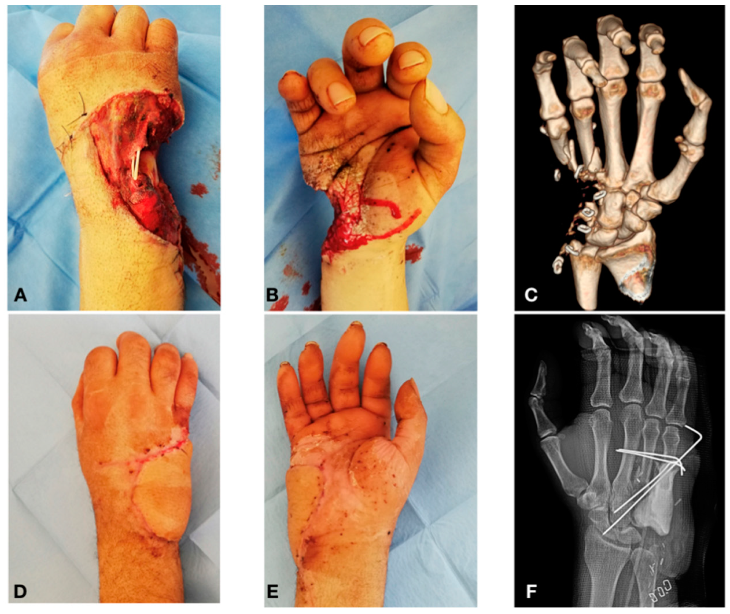
The investigation was performed in compliance with the Declaration of Helsinki and the Guidelines for Good Clinical Practice. The patient provided appropriate informed consent. A 50-year-old man, smoker, with right dominant hand suffered a firecracker trauma on New Year’s Eve. He presented with a complex wound on the right wrist on the ulnar side, with destruction of Guyon’s canal (Figure 2A–C). That explosion had caused an ulnar pedicle interruption proximal to the wrist crease, a fracture of 4th and 5th metacarpal bones, and an avulsion of hamate, triquetrum and pisiform. Flexor Carpi Ulnaris (FCU) was interrupted with a skin wound of 10 × 12 cm. The carpal tunnel and the radio-lunate joint were exposed but spared by trauma. First the right wrist was stabilized. Then negative pressure therapy (NPT) was applied and maintained for 10 days, during which the patient underwent microbiological wound exams based on multiple tissue cultures and antibiotics therapy. Radiological and vascular investigation in order to assess the real clinical deficit were performed and physiotherapy was administered to evaluate the wrist’s range of motion (ROM). Wrist movement was possible, but constrained by pain, with a loss of sensitivity on the 4th and 5th fingers, and intrinsic muscle impairment. We planned soft tissue reconstruction with a MSAP flap and sural nerve. Bone reconstruction was delayed in order to leave the patient free to move the wrist in the early postoperative period and to avoid post-traumatic stiffness.
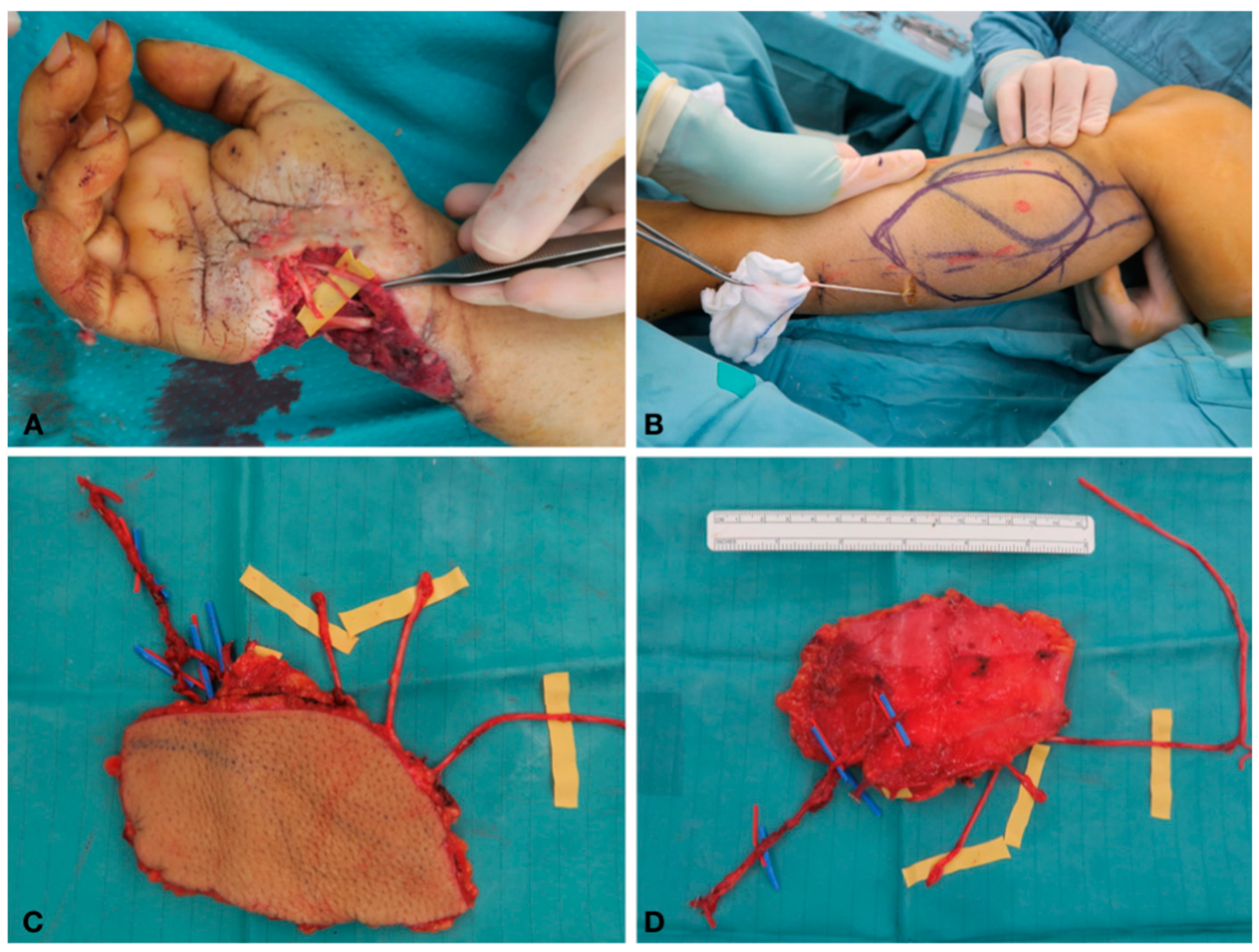
Perforators were preoperatively marked with a handheld Doppler probe. Multiple incisions on the postero-lateral leg surface were performed according to the traditional technique to raise sural nerve graft. Then with the patient in frog position, the skin paddle was incised slightly more laterally than in the traditional MSAP flap to include both tibialis and peroneal components of the sural nerve, from the popliteal fossa to the common trunk (Figure 3B–D). Hand fractures were treated with K-wires and bone spacers (Figure 2E and Figure 4B). Ulnar nerve stump was identified proximally to the wrist crease while distally were available just common sensitive trunk for the 4th and 5th finger and the branch for the ulnar side of the 5th (Figure 3A) with a gap of 3 cm.
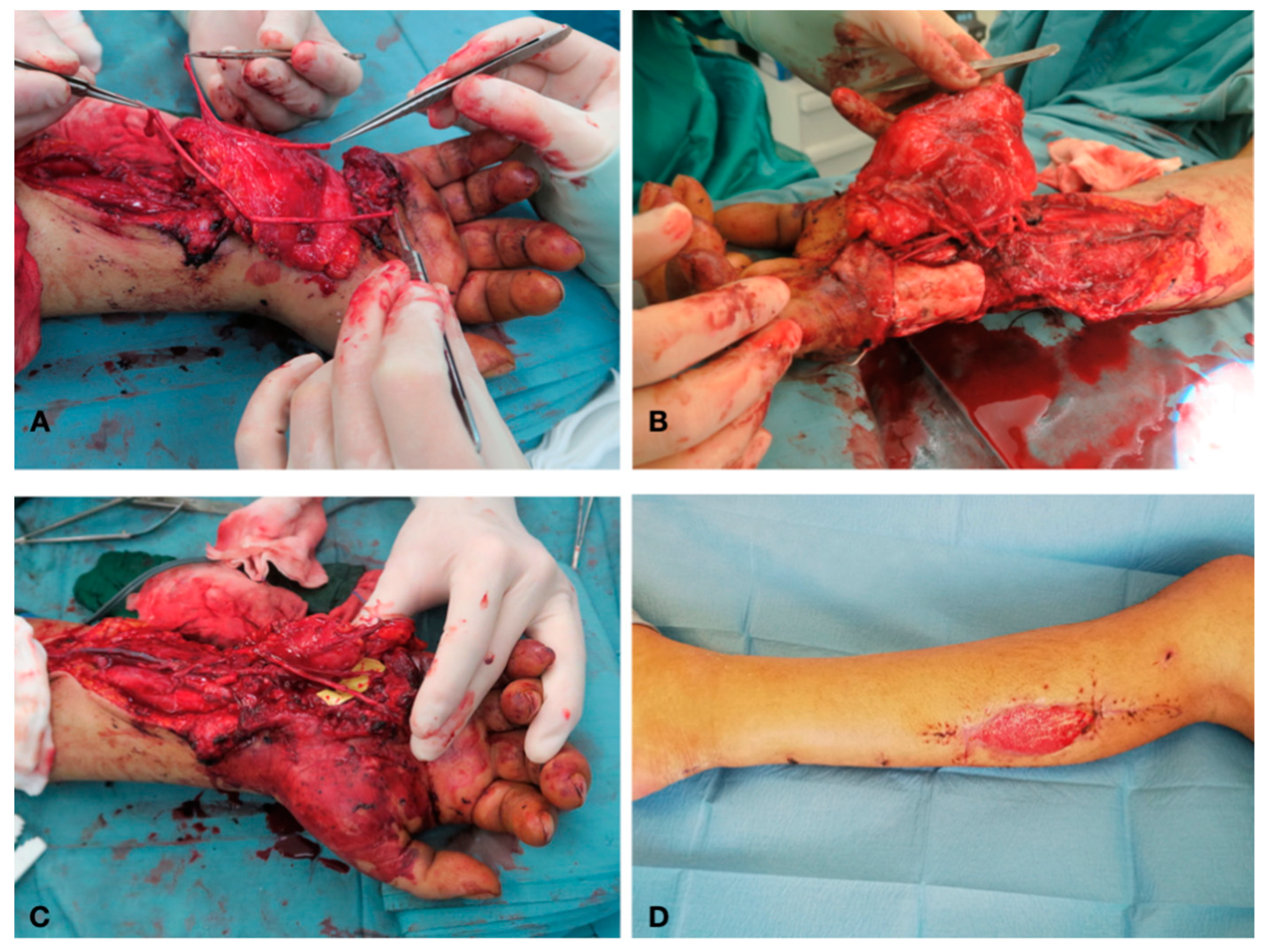
The motor branch was destroyed by explosion and the distal stump was not visible without a further incision in the palm, not indicated because of the poor functional prognosis in intrinsic muscle reinnervation. The patient was informed of the aim to restore an “elementary” hand function, without intrinsic function. The flap was raised on a subfascial plane, based on two perforators and anastomosed in an end-to-end fashion on the ulnar pedicle at wrist with a second vein anastomosed to the basilic vein. The tibialis component of the sural nerve, bigger than the other, was used for the 4th–5th common sensory digital nerves, the thinner peroneal component for the ulnar branch of the 5th digit (Figure 4A–C).
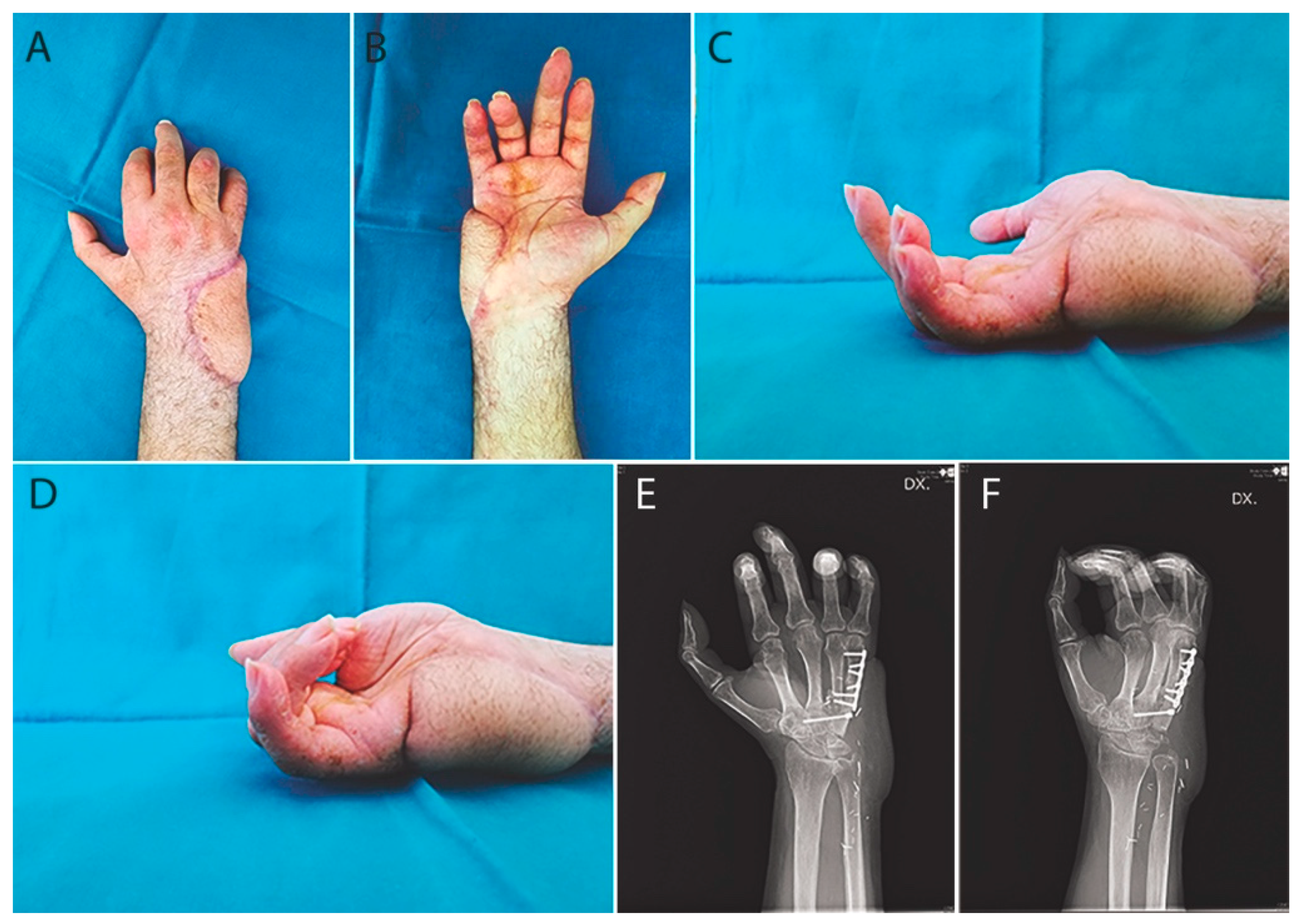
The skin paddle matched the wound without any tension and the fascia layer was used to wrap the nerve transplant. Donor site was sutured but a small central part was covered with a dermal substitute. The patient underwent early physiotherapy and, 3 months after soft tissue reconstruction, we performed an iliac crest graft to restore metacarpal bone. The wounds healed without any complication. No signs of infection were recorded. After 9 months, flap matching was satisfactory (Figure 5A–D).
Wrist ROM is improving, and the donor site is cosmetically acceptable both for the patient and the surgeon. Nine months after nerve reconstruction, Tinel sign is showing progress according to electromyography. Subjective nerve recovery was evidenced by Semmens Weinstein monofilament test showing a “diminished protective sensation” (purple filament touch detecTable 3.84–4.31) and two-point discrimination of 10 mm both static and moving. Any sensation of dysesthesia was denied by patient during wrist and fingers movements possible up to −30/+30 degrees for wrist and to medium level of flexion (1 cm from palm) and extension (lag of 25 degrees) for long fingers.

4. Discussion

Restoring a functional hand after high energy trauma, requires the replacement of all missing tissues according to a “like-with-like” concept trying to re-create a sensitive prehensile extremity. Soft tissue reconstruction can be performed in many ways but the risk to create a bulky hand is high if the flaps used are not extremely thin. Radial forearm flaps, anterolateral thigh flaps, superficial circumflex iliac perforator flaps or MSAP flaps are examples of widely used thin flaps [3,4,5]. MSAP flap was first described in 2001 by Cavadas and Hallok for both extremity and head and neck reconstructions [17,18] and now are widely used with many variations [18,19]. Literature data show that ulnar nerve injury most frequently results in hospital admission, when compared with median and radial nerve injuries and the functional outcome after repair through primary neurorrhaphy or grafting is relatively poor when compared to results after radial or median nerve repair [20]. Distal ulnar nerve lesions in zone 1 cause anesthesia of 4th and 5th fingers, intrinsic muscles impairment with the typical claw-like appearance of the hand [21]. The peripheral nerve regenerative capability has been recognized for more than a century; however, clinical and experimental evidence shows that such regeneration is often unsatisfactory, especially following severe lacerations: normal-to-good function is achieved only in approximately 50% of surgical cases [22]. Although repair within 1 week is the optimal approach for peripheral nerve injuries, a delayed repair is appropriate when an immediate repair is not possible due to other concomitant and complicating injuries [23] and, unfortunately, the longer the delay is, the worse the prognosis will be. The clinical advantage of a vascularized graft is still controversial, however there have been many clinical and experimental studies validating the superiority of nerve regeneration with a vascularized nerve graft, compared with conventional free nerve grafts in scarred and poor vascular beds. Despite this, nerve repair remains a difficult challenge [11,12,13]. One of the main complications in nerve surgery is perineural adherence involving the peripheral nerve system and this is more evident in mangled limbs [24]. In spite of the numerous anti-adherence devices and techniques have been developed in the last years to prevent the physiologic onset of perineural fibrosis after a surgical trauma, few reports are available in literature regarding the efficacy of these types of barriers in the peripheral neural system [15]. In some cases, ALT flap with femoral cutaneous nerve has been used in order to transfer a composite flap restoring both skin and nervous continuity, but in most cases this flap is quite thick compared to hand skin [1,2]. In other cases, just fascial free flaps have been used in order to reduce peripheral nerve scar tissue [25] without carrying any skin island. This way we tried to investigate the possibility to raise the sural nerve within the MSAP flap. This study was conducted to investigate vascular connection between nerve and MSAP pedicle, not just to create a “free innervated transplant”, but to evaluate the possibility to transfer a viable gliding tissue surrounding the nerve. A vascularized fascial surrounding a nerve in its original gliding tissue, offers the best possibility to reduce scar adherence. In our anatomical study we found in 57% of specimens a visible vascular network connecting the pedicle and the vasa nervorum system using liquid latex injection. The advantage in term of recovery of traditional nerve graft versus vascularized transfer is discussed controversially [11,12,13,14]. The main innovation of this composite flap is the possibility to raise the nerve within a viable fascia layer, used like a vascularized envelope. In the specimens without a visible network, the fascia layer still represents a viable tissue, surrounding the nerve with a potential reduction of the perineural scar. Moreover, the proximal harvesting of the flap allows to raise two different (separate or confluent) nerves.
We performed this flap in a complex clinical case trying to repair both cutaneous and nervous gap with the same flap. Skin paddle matched well the recipient area being thin and sufficiently large. The sural nerve branches within the flap offered the possibility to reconstruct two different nerves surrounded by a viable gliding tissue protecting the healthy sural nerve component from the wound. We found a potential bias in our anatomical study: the injection of popliteal artery instead of the medial sural one could lead to a potentially increased fascia perfusion from other collateral vessels. Nevertheless, the clinical application of this composite flap showed the complete viability of the entire skin paddle nourished only by the medial sural pedicle and consequently the viability of the underlying fascia.
This is the first report of a vascularized sural nerve graft included in a MSAP flap. Although a bigger cohort study and longer follow up are needed, in our experience the addition of a well-vascularized interpositional sural nerve graft could be considered for nerve reconstruction in complex cases, combining the skin coverage with the nervous reconstruction. In the MSAP flap, if skin is not needed, it may be used just as monitor skin paddle and the flap used as a pure fascia “nerve-through” flap. We hypothesize that the presence of gliding tissue will limit the presence of perineural scar.

5. Conclusions

We present a variation of the MSAP flap including a vascularized nerve graft within the fascia flap. In our study, we evidenced a direct vascular connection between the flap pedicle and the sural nerve in only 57% of the specimens. Moreover, the presence of microvascular connection in the fascia layer or in a dermal plane is always presumable, although it was not investigated with histological analysis. Even if there is no consensus on the superiority of vascularized nerve graft versus non-vascularized one, this modified MSAP flap allows to transfer a vascularized gliding tissue with a nerve graft, reducing the factors that lead to perineural scars and adherences.

Author Contributions

Conceptualization, P.P. and F.D.F.; methodology, P.P. and A.A.; validation, F.D.F. and M.R.; formal analysis, P.P. and A.A.; investigation, P.P.; resources, M.R.; data curation, P.P., A.C., P.P.P. and F.D.F.; writing—original draft preparation, P.P.; writing—review and editing, F.D.F.; supervision, M.R.; All authors have read and agreed to the published version of the manuscript.

Funding

This research received no external funding.

Institutional Review Board Statement

All procedures followed were in accordance with the ethical standards of the responsible committee on human experimentation (institutional and national) and with the Helsinki Declaration of 1975, as revised in 2008. The present study was approved from our local ethic committee (OR-2019-57).

Informed Consent Statement

Written informed consent has been obtained from the patient to publish this paper.

Data Availability Statement

The datasets generated during and/or analysed during the current study are available from the corresponding author upon reasonable request.

Acknowledgments

Authors are grateful to Giuseppina Caraglia, mother-tongue expert, University of Naples “Vanvitelli” to perform English revision.

Conflicts of Interest

The authors declare no conflict of interest.

References

  1. Koshima, I.; Nanba, Y.; Tsutsui, T.; Takahashi, Y.; Kawai, A. Vascularized femoral nerve graft with anterolateral thigh true perforator flap for massive defects after cancer ablation in the upper arm. J. Reconstr. Microsurg. 2003, 19, 299–302. [Google Scholar]
  2. Iida, T.; Nakagawa, M.; Asano, T.; Fukushima, C.; Tachi, K. Free vascularized lateral femoral cutaneous nerve graft with anterolateral thigh flap for reconstruction of facial nerve defects. J. Reconstr. Microsurg. 2006, 22, 343–348. [Google Scholar] [CrossRef] [PubMed]
  3. Narushima, M.; Kaji, N.; Yamamoto, T.; Yoshimatsu, H.; Hara, H.; Kikuchi, K.; Araki, J.; Yamashita, S.; Koshima, I. Superficial circumflex iliac artery pure skin perforator-based superthin flap for hand and finger reconstruction. J. Plast. Reconstr. Aesthet. Surg. 2016, 69, 827–834. [Google Scholar] [CrossRef] [PubMed]
  4. Hsu, K.C.; Tsai, W.H.; Ting, P.S.; Hsueh, J.H.; Chen, L.W.; Lin, Y.S. Comparison between anterolateral thigh, radial forearm, and peroneal artery flap donor site thickness in Asian patients—A sonographic study. Microsurgery 2017, 37, 655–660. [Google Scholar] [CrossRef]
  5. Balan, J.R. Medial sural artery perforator free flap for the reconstruction of leg, foot and ankle defect: An excellent option. ANZ J. Surg. 2018, 88, E132–E136. [Google Scholar] [CrossRef] [PubMed]
  6. Lan, C.Y.; Tien, H.Y.; Lin, Y.T.; Hsu, C.C.; Lin, C.H.; Chen, S.H. Prognosis of traumatic ulnar nerve injuries: A systematic review. Ann. Plast. Surg. 2019, 82, S45–S52. [Google Scholar] [CrossRef]
  7. Sunderland, S. The anatomy and physiology of nerve injury. Muscle Nerve 1990, 13, 771–784. [Google Scholar] [CrossRef]
  8. Hoke, A. A (heat) shock to the system promotes peripheral nerve regeneration. J. Clin. Investig. 2011, 121, 4231–4234. [Google Scholar] [CrossRef]
  9. Evriviades, D.; Jeffery, S.; Cubison, T.; Lawton, G.; Gill, M.; Mortiboy, D. Shaping the military wound: Issues surrounding the reconstruction of injured servicemen at the Royal Centre for Defence Medicine. Philos. Trans. R. Soc. Lond. B Biol. Sci. 2011, 366, 219–230. [Google Scholar] [CrossRef] [Green Version]
  10. Moore, A.M.; Wagner, I.J.; Fox, I.K. Principles of nerve repair in complex wounds of the upper extremity. Semin. Plast. Surg. 2015, 29, 40–47. [Google Scholar] [CrossRef] [PubMed] [Green Version]
  11. Koshima, I.; Harii, K. Experimental study of vascularized nerve grafts: Morphometric study of axonal regeneration of nerves transplanted into silicone tubes. Ann. Plast. Surg. 1985, 14, 235–243. [Google Scholar] [CrossRef] [PubMed]
  12. Kanaya, F.; Firrell, J.; Tsai, T.M.; Breidenbach, W.C. Functional results of vascularized versus nonvascularized nerve grafting. Plast. Reconstr. Surg. 1992, 89, 924–930. [Google Scholar] [CrossRef] [PubMed]
  13. Ozcan, G.; Shenaq, S.; Mirabi, B.; Spira, M. Nerve regeneration in a bony bed: Vascularized versus nonvascularized nerve grafts. Plast. Reconstr. Surg. 1993, 91, 1322–1331. [Google Scholar] [CrossRef]
  14. Mackinnon, S.E.; Kelly, L.; Hunter, D.A. Comparison of regeneration across a vascularized versus conventional nerve graft: Case report. Microsurgery 1988, 9, 226–234. [Google Scholar] [CrossRef] [PubMed]
  15. Cherubino, M.; Pellegatta, I.; Crosio, A.; Valdatta, L.; Geuna, S.; Gornati, R.; Tos, P. Use of human fat grafting in the prevention of perineural adherence: Experimental study in athymic mouse. PLoS ONE 2017, 12, e0176393. [Google Scholar] [CrossRef] [PubMed] [Green Version]
  16. Campodonico, A.; Pangrazi, P.; De Francesco, F.; Riccio, M. Reconstruction of a long defect of the median nerve with a free nerve conduit flap. Arch. Plast. Surg. 2020, 47, 187–193. [Google Scholar] [CrossRef] [PubMed] [Green Version]
  17. Hallock, G.G. Anatomic basis of the gastrocnemius perforator-based flap. Ann. Plast. Surg. 2001, 47, 517–522. [Google Scholar] [CrossRef]
  18. Cavadas, P.C.; Sanz-Giménez-Rico, J.R.; De la Càmara, G.A.; Navarro-Monzonìs, A.; Soler-Nomdedeu, S.; Martìnez-Soriano, F. The medial sural artery perforator free flap. Plast. Reconstr. Surg. 2001, 108, 1609–1617. [Google Scholar] [CrossRef]
  19. Wong, M.Z.; Wong, C.H.; Tan, B.K.; Chew, K.Y.; Tay, S.C. Surgical anatomy of the medial sural artery perforator flap. J. Reconstr. Microsurg. 2012, 28, 555–560. [Google Scholar] [CrossRef] [Green Version]
  20. Tapp, M.; Wenzinger, E.; Tarabishy, S.; Ricci, J.; Herrera, F.A. The epidemiology of upper extremity nerve injuries and associated cost in the US Emergency Departments. Ann. Plast. Surg. 2019, 83, 676–680. [Google Scholar] [CrossRef]
  21. Dy, C.J.; Mackinnon, S.E. Ulnar neuropathy: Evaluation and management. Curr. Rev. Musculoskelet. Med. 2016, 9, 178–184. [Google Scholar] [CrossRef] [PubMed]
  22. Pfister, B.J.; Gordon, T.; Loverde, J.R.; Kochar, A.S.; Mackinnon, S.E.; Cullen, D.K. Biomedical engineering strategies for peripheral nerve repair: Surgical applications, state of the art, and future challenges. Crit. Rev. Biomed. Eng. 2011, 39, 81–124. [Google Scholar] [CrossRef]
  23. Griffin, M.F.; Malahias, M.; Hindocha, S.; Khan, W.S. Peripheral nerve injury: Principles for repair and regeneration. Open Orthop. J. 2014, 8, 199–203. [Google Scholar]
  24. Wang, M.L.; Rivlin, M.; Graham, J.G.; Beredjiklian, P.K. Peripheral nerve injury, scarring, and recovery. Connect. Tissue Res. 2019, 60, 3–9. [Google Scholar] [CrossRef]
  25. Yamamoto, R.; Motomiva, M.; Sakurai, K.; Sekiguchi, H.; Funakoshi, T.; Iwasaki, N. Application of free temporoparietal fascial flap for recurrent neural adhesion of superficial radial nerve—A case report. Microsurgery 2015, 35, 489–493. [Google Scholar] [CrossRef] [PubMed]
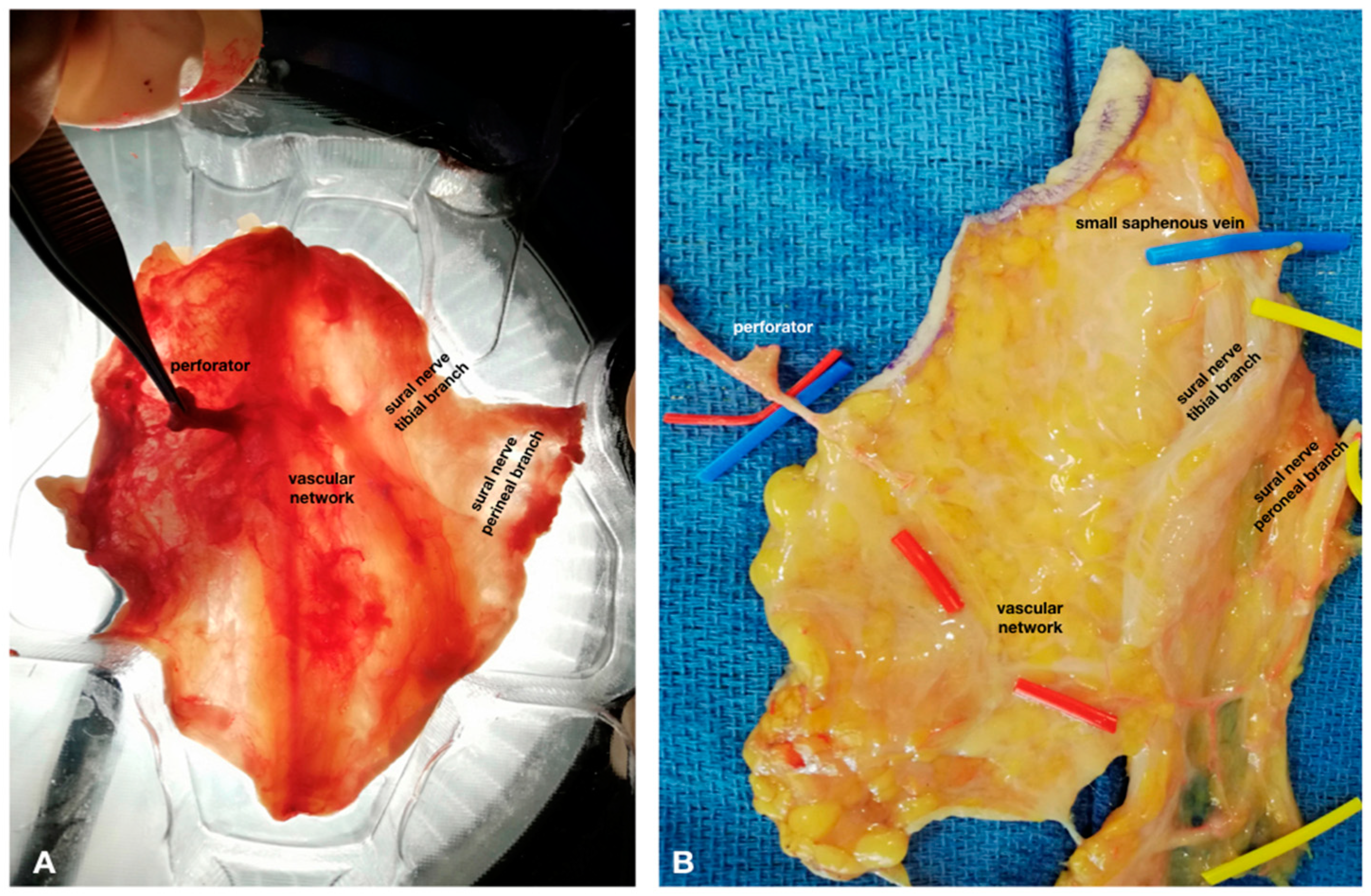
Figure 1. (A) De-epithelized specimen’s flap and backlight study: macroscopic vascular network connection; (B) Specimen’s flap: volar view. In both (A,B) the proximal part of the flap are in the upper side of the figures.
Traumacare 01 00002 g001
Figure 2. (A) Hand wound: dorsal view with extensor tendon exposure; (B) Hand wound: volar view; (C) TC 3D bone lesion; (D) 2 month post-operative, dorsal view; (E) 2 month post-operative, volar view; (F) X-ray post-operative control.
Traumacare 01 00002 g002
Figure 3. (A) Intra-operative view: sensitive nerve and ulnar artery distal stump; (B) Flap design to include sural nerve; (C) MSAP flap with pedicle and peroneal and tibial sural branch: dorsal view; (D) MSAP flap with pedicle and peroneal and tibial sural branch: volar view.
Traumacare 01 00002 g003
Figure 4. (A) Intra-operative view: tibial and peroneal sural branch; (B) Intra-operative view: nerves and perforators after suture; (C) Intra-operative view: tibial and peroneal sural branch sutured with sensitive branches for 4th and 5th finger; (D) 1-month post-operative: donor site with dermal substitute.
Traumacare 01 00002 g004
Figure 5. 6 months after bone graft (A) Frontal view; (B) Dorsal view; (C) Lateral view open palm; (D) Lateral view finger flexion; (E) X-ray frontal view; (F) X-ray lateral view.
Traumacare 01 00002 g005
Publisher’s Note: MDPI stays neutral with regard to jurisdictional claims in published maps and institutional affiliations.