Article

Lactobacillus acidophilus: Boosting Immunity Through IL-6 Induction

by
Isaac Oluseun Adejumo
Department of Animal Science, University of Ibadan, Ibadan 200005, Nigeria
Biologics 2025, 5(4), 29; https://doi.org/10.3390/biologics5040029
Submission received: 15 August 2025 / Revised: 13 September 2025 / Accepted: 17 September 2025 / Published: 29 September 2025

Abstract

Background and Objectives: Probiotics are live microorganisms that promote health when consumed in adequate amounts, ensure the balance of bacterial composition in the digestive system, and suppress harmful pathogenic bacteria, with overall implications for animal and human health, welfare and performance. However, a lot remains unclear about their functional mechanisms. Materials and Methods: In this study, 14 uncharacterized proteins of Lactobacillus acidophilus were analyzed for subcellular localization, structural and safety profiling and interleukin-6-(IL-6)-inducing potential. Results: Aliphatic index scores were generally high, ranging between 138.39 (LBA1705) and 78.39 (LBA1825). The instability index scores were less than 40 for all the query proteins except for LBA0995. All the proteins produced immunogenic IL-6-inducing peptides, except for LBA0037, LBA1825 and LBA1788. Conclusions: The findings provide insight into understanding the functional mechanism of probiotic Lactobacillus, laying a strong foundation for more experimental studies.

1. Introduction

Animal and human nutrition is confronted with many challenges. The rising cost of feed ingredients is a major challenge confronting animal production [1], while diseases remain the main challenge for animal production and human existence. Over the years, several attempts have been made to overcome these challenges, including the use of antibiotics, although the use of antibiotics in feed is no longer widely accepted owing to the potential risk of antibiotic resistance as well as the concerns of food safety arising from contamination or residual effects of chemicals and drugs in animal foods [2,3]. This has necessitated the shift in attention to probiotics to enhance growth performance in livestock, especially poultry, as well as to eliminate the risk of health hazards of antimicrobial drugs and vaccines [4].
Probiotics are live microorganisms that, when administered in adequate amounts, confer a health benefit on the host [5,6]. They promote health and ensure the balance of bacterial composition in the digestive system, leading to the suppression of harmful pathogenic bacteria [7,8], with overall implications for animal health, welfare and performance. The documented benefits of probiotics include combating enteric pathogenic bacteria, improving growth performance, modulating the intestinal microflora, improving immune responses and improving feed conversion efficiency [9,10]. A high proportion of probiotic bacteria in the intestinal tract can form colonies that may suppress pathogenic bacteria, thereby protecting the intestinal tract from invasion by pathogenic bacteria [11,12]. The benefits probiotics offer have placed pressure on their demands for use in animal nutrition and as human supplements, for cosmetic and therapeutic purposes.
Among the lactic acid bacteria, Lactobacillus has been proposed as the main candidate strain for probiotic potential [13,14]. A recent study reported that bacterial isolates belonging to Lactobacillus plantarum and Bacillus subtilis were the most promising probiotic agents in chicken nutrition among others tested [15], although further studies to assess the safety and protective benefits of these probiotic candidates are recommended [15].
Improved nutrient absorption and digestibility, enterotoxin neutralization, improved immune responses, reduced gastrointestinal colonization by foodborne pathogenic organisms (Campylobacter, Salmonella, Clostridium and Escherichia coli) and improved growth performance are some of the benefits of Lactobacillus probiotic strains [16,17,18]. However, to ensure the safety and sustainability of probiotics in nutrition and therapeutic applications, it is necessary to unravel most of the doubts associated with the constituents of the microorganism. In addition, many proteins from the Lactobacillus acidophilus genome that may play important roles and assist in the biological understanding of the organism are uncharacterized. Uncharacterized proteins are proteins that are predicted to be expressed in an organism but to which no proper function has been assigned or known [19]. Uncharacterized proteins have not been assigned any functions experimentally because they have not been studied or their functions are not yet known.
A lot remains unclear about the characteristics, safety profile and functional mechanisms of probiotics, owing in part to many of their proteins being uncharacterized or hypothetical. Understanding the proteins of the Lactobacillus acidophilus genome will shed more light on its functional mechanisms, safety profile and sustainable applications in animal and human nutrition and therapeutic and cosmetic products. Therefore, this study was carried out to stimulate interests in and lay foundations for in vivo functional characterization of these proteins in order to better understand their functional mechanisms, thereby ensuring their sustainable applications in animal and human nutrition and in cosmetic and therapeutic products. Investigating the selected uncharacterized proteins of a reference genome of Lactobacillus acidophilus may provide insights that may find applications in the animal nutrition, health, food production and nutraceutical industries.

2. Materials and Methods

2.1. Sequence Retrieval and Analysis of Protein Families

The reference genome of Lactobacillus acidophilus (CP005926.2) was retrieved from NCBI and the protein families were determined in Pathosystems Resource Integration Center (PATRIC). The protein families were sorted into different classes, and 14 uncharacterized proteins were randomly selected. The proteins are as follows: uncharacterized membrane protein YfhO-like (LBA1510), uncharacterized metal-dependent hydrolase YcfH (LBA0214), uncharacterized MFS-type transporter (LBA1705), uncharacterized MFS-type transporter (LBA1446), uncharacterized DUF1113 membrane protein family (LBA0037), uncharacterized protein CAC3725 (LBA1825), uncharacterized GabP-family amino acid permease LBA0995 (LBA0995), uncharacterized transcriptional regulator YozG, Cro/CI family (LBA1788), uncharacterized transporter YbxG (yifk), uncharacterized transporter YbxG (LBA0899), uncharacterized UPF0750 membrane protein SPy2155 (LBA1209), uncharacterized UPF0750 membrane protein YpjC (LBA0972), uncharacterized substrate:H+ symporter, LctP family (LctP) and uncharacterized UPF0721 integral membrane protein (LBA0338). Their nucleotide and amino acid sequences were retrieved from PATRIC. The selected proteins are presented in Table 1.

2.2. Blast and Subcellular Localization Analysis

The retrieved sequences of the selected protein were evaluated for similarity check using NCBI BLAST (BLAST+ 2.17.0) (https://blast.ncbi.nlm.nih.gov/Blast.cgi#alnHdr_1854278943, accessed on 6 March 2024). The subcellular localization of the query proteins was analyzed using PSORTb v3.0.2, https://www.psort.org/psortb/, accessed on 30 April 2024 [20].

2.3. Structural Determination, Physico-Chemical Properties and Protein–Protein Network

The secondary structures and three-dimensional tertiary structures of the query proteins were determined using the SOPMA server [21] and Alphafold 2.0, https://alphafold.ebi.ac.uk/, accessed on 11 March 2024 [22], respectively. SWISS-MODEL Interactive Workspace (https://swissmodel.expasy.org/assess, accessed on 12 March 2024) [23] was used to evaluate the obtained three-dimensional tertiary structures. ExPASy ProParam, www.web.expassy.org/protparam, accessed on 28 March 2024 [24] was used for the analysis of the physicochemical properties of the query proteins. Protein–protein network interactions of the query proteins were analyzed using STRING, a database of known and predicted protein interactions, drawn from genomic context, high-throughput experiments, conserved coexpression and previous PubMed literature [25]. The default thresholds were used.

2.4. Allergenicity, Immunogenicity, and Toxicity Analyses

Allergenicity evaluation of the query proteins used in this study was performed with AllerCatPro 2.0 and https://allercatpro.bii.a-star.edu.sg/, accessed on 13 March 2024 [26], while immunogenicity and toxicity evaluations of the query proteins of the reference genome were performed using VaxiJen 3.0, https://www.ddg-pharmfac.net/vaxijen3/, accessed on 12 March 2024 [27] and ToxinPred, https://webs.iiitd.edu.in/raghava/toxinpred2/, accessed on 15 March 2024 [28]. AllerCatPro 2.0 is a version of the previously established AllerCatPro method (AllerCatPro 1.7) [29]. The tool predicts protein allergenicity potential using the similarity of both amino acid sequences and 3D structures to the most comprehensive datasets (4979 protein allergens, 162 low allergenic proteins and 165 autoimmune allergens) of reliable proteins associated with allergenicity from the WHO/IUIS, COMPARE, FARRP, UniProtKB and Allergome databases [26]. The default thresholds were used.

2.5. Analysis of Active Binding Site of Query Proteins, Gene Expression and Multiple Sequence Alignment

CASTp 3.0, http://sts.bioe.uic.edu/castp/, accessed on 25 March 2024 [30] was used to determine the active binding sites of the query proteins used in the study. The CASTp server is a recognized bioinformatic tool used for identifying the identities of all interior cavities, surface pockets and cross channels in protein structures. It also provides a detailed delineation of all atoms participating in their formation [30]. IL-6-inducing potential of selected proteins was evaluated using il6pred (https://webs.iiitd.edu.in/raghava/il6pred/disp1.php?ran=36676, accessed on 29 July 2024). The top 10 based on their prediction values were further evaluated for immunogenicity [27]. Gene expressions of IL-6 across various conditions in Homo sapiens (platform: HS_AFFY_U133PLUS_2-0) and Mus musculus (platform: MM_AFFY_430_2-1) was performed using GENEVESTIGATOR. Multiple sequence alignment of the query proteins was analyzed using Clustal, https://www.ebi.ac.uk/, accessed on 29 July 2024 and a phylogenetic tree was constructed.

3. Results

The features, descriptions and designations of the query proteins of the reference genome of Lactobacillus acidophilus used in this study are presented in Table 1. The amino acid length ranged between 69 (LBA1788) and 859 (LBA1510). The BLAST results revealed that the selected proteins are similar, showing almost 100% identity across different Lactobacillus acidophilus strains.

3.1. Physicochemical Properties of Query Proteins

The atomic formulae of the selected proteins were C4693H7164N1136O1222S17 (LBA1510), C1326H2055N349O391S7 (LBA0214), C507H792N110O115S3 (LBA1705), C2448H3927N603O652S23 (LBA1446), C1462H2229N363O366S9 (LBA0037), C444H734N130O120S9 (LBA1825), C2635H3966N622O656S31 (LBA0995), C343H574N88O103S5 (LBA1788), C2361H3654N554O598S16 (yifk), C2397H3700N566O607S19 (LBA0899), C1546H2369N385O400S14 (LBA1209), C1524H2429N389O399S14 (LBA0972), C2446H3978N586O657S26 (LCTP) and C1313H2034N298O320S10 (LBA0338) (Table 2). The total negatively charged residues ranged between 4 (LBA1705) and 56 (LBA1510). The total number of positively charged residues ranged between 7 (LBA1705) and 78 (LBA1510). LBA1510 (14232) has the largest total number of atoms while LBA1788 (1113) has the lowest total number of atoms (Table 2).
The theoretical isoelectric points ranged between 5.61 (LBA0214) and 9.80 (LBA1825). LBA1510 has the highest amino acid length (859) and molecular weight (99,596.40) while LBA1788 has the lowest amino acid length (69) and molecular weight (7739.16) (Table 3).
The aliphatic index scores were generally high, ranging between 138.39 (LBA1705) and 78.39 (LBA1825). LBA1510, LBA1705, LBA1446, LBA0037, LBA0995, yifk, LBA0899, LBA1209, LBA0972, ltcP and LBA0338 had positive GRAVY values while negative values were obtained for the remaining proteins. The instability index scores were less than 40 for all the query proteins except for LBA0995, which had a value of 42.10 (Table 3). Only LBA0995 was observed to be unstable in nature. The estimated half-life was the same for all the proteins, 30 h for mammalian reticulocytes in vitro, >20 h for yeast in vivo and >10 h for Escherichia coli, in vivo (Table 4). The extinction coefficient (EC) was highest for LBA1510 (150580), followed by LBA0995 (119080) while LBA1788 (1615) had the lowest value.

3.2. Amino Acid Compositions of Query Proteins

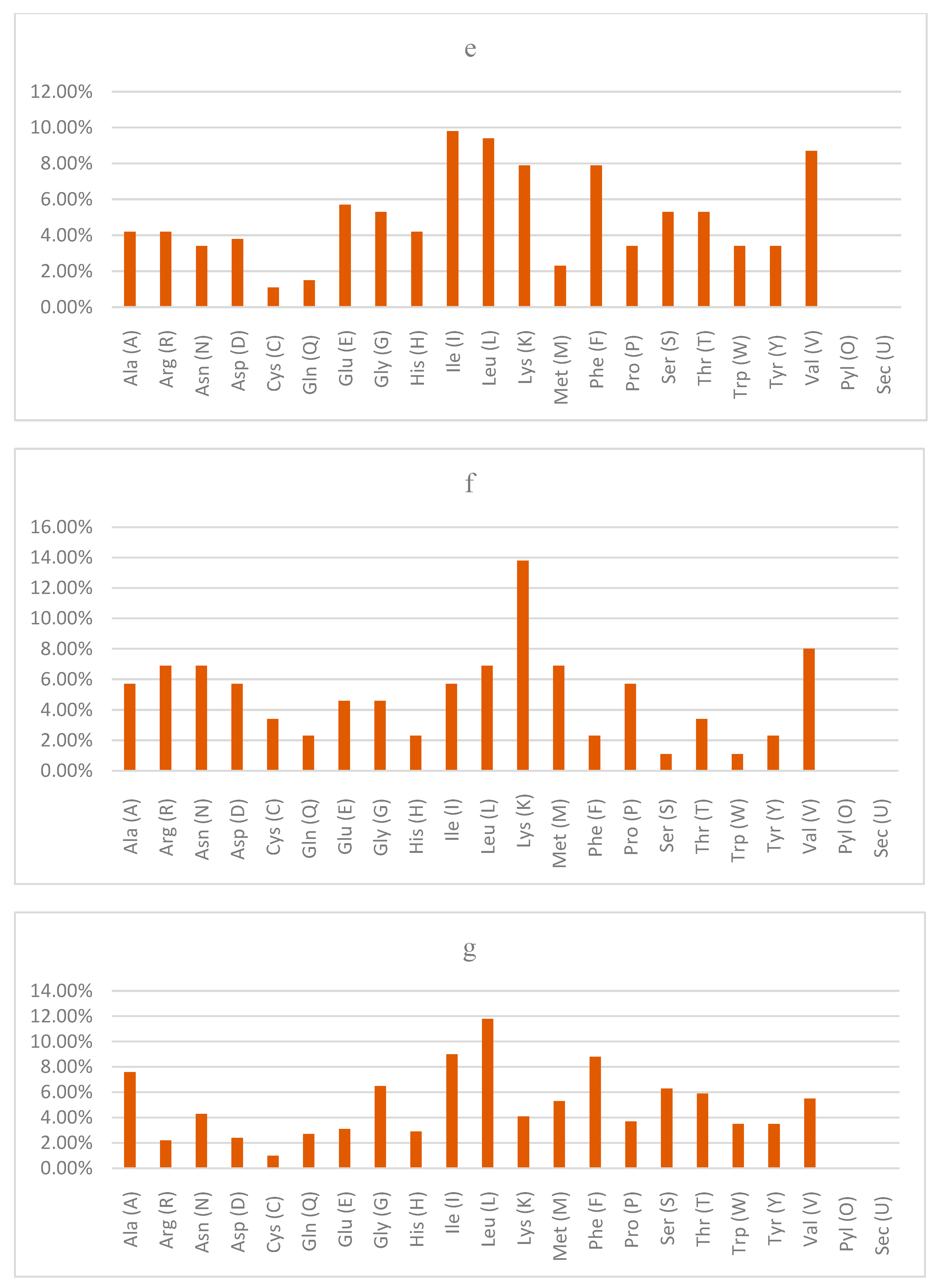
The amino acid compositions of the query proteins are shown in Figure 1. The prominent amino acids for LBA1510 are Leu, Ile and Phe, while the least prominent ones included His and Trp (Figure 1a). The most prominent amino acids of LBA0214 include Val, Lys and Leu while the least prominent amino acids included Try and Cys (Figure 1b). Leu and Ile are the most prominent amino acids of LBA1705, while Trp and Gln are the least prominent amino acids (Figure 1c). Leu and Ile, Gly and Ala, and Cys and Trp are the most and least prominent amino acids of LBA1446, respectively (Figure 1d). The most and least prominent amino acids of LBA0037 are Ile and Leu, and Cys and Gln, respectively (Figure 1e). The most prominent amino acids of LBA1825 are Lys and Val while the least prominent are Ser and Trp (Figure 1f). Leu and Ile were the most prominent amino acids, while Cys and Arg are the least prominent amino acids in LBA0995 (Figure 1g).
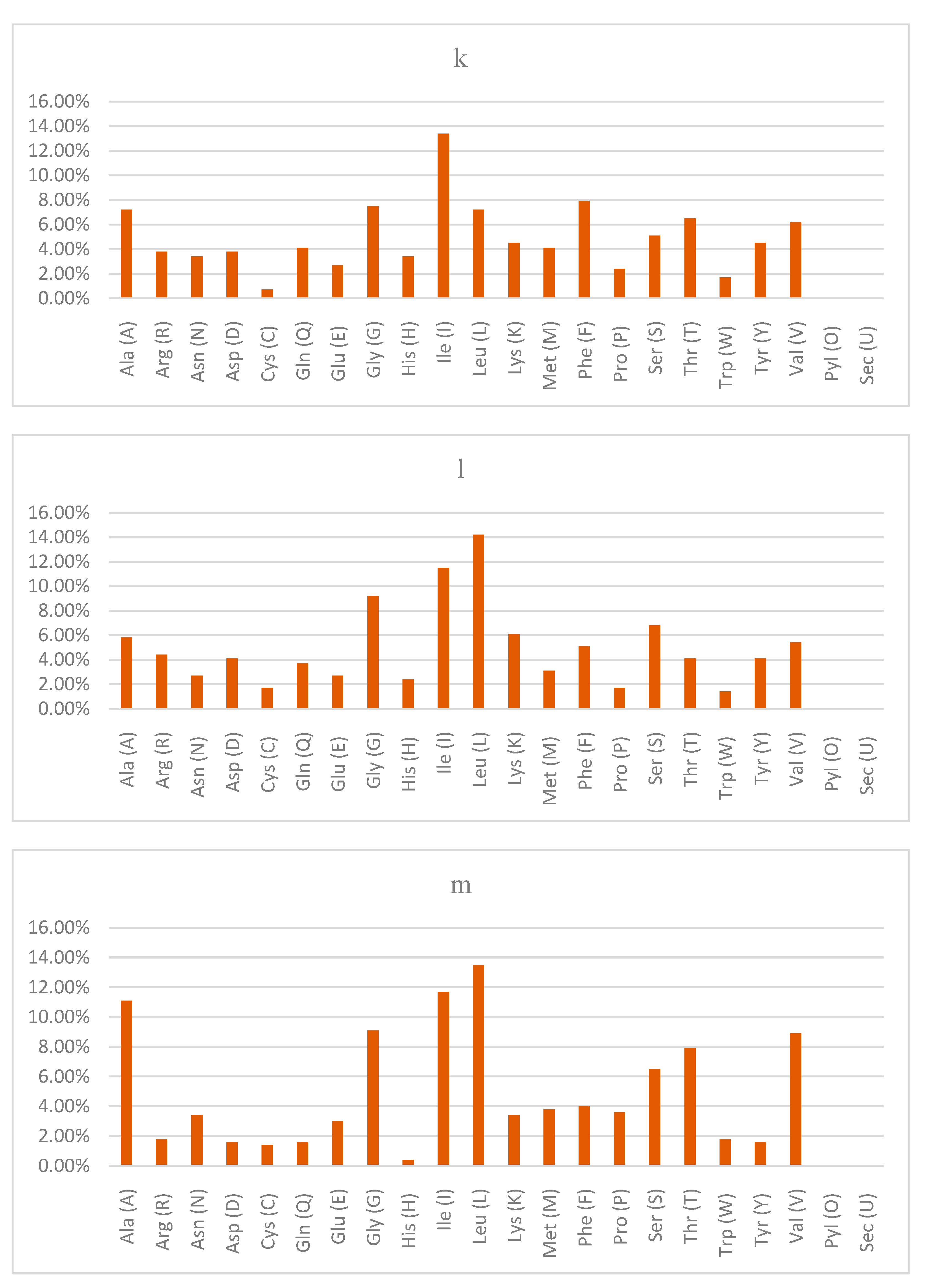
Figure 1h shows that Leu and Lys are the most prominent amino acids of LBA1788, while Asn, Gln, His and Tyr are the least prominent. Ile and Leu were the most abundant in yifk (Figure 1i) and LBA0899 (Figure 1j), while Gln and Asp were the least abundant in yifk, and Gln and His (LBA0899) were the least abundant amino acids. The most prominent amino acids were as follows: Ile, Phe, Gly and Ala for LBA1209 (Figure 1k); Leu, Ile and Gly for LBA0972 (Figure 1l); Leu, Ile and Ala for LctP (Figure 1m); and Ile, Leu and Ala for LBA0338 (Figure 1n).

3.3. Sub-Cellular Localization and Properties of the Secondary Structure of Query Proteins

The sub-cellular localization is presented in Table 5. With the exception of LBA1825, whose localization is not known, and LBA1788 whose localization s cytoplasmic, the rest of the proteins are found in the cytoplasmic membrane.
The properties of the secondary structure of the query proteins of the reference genome of the probiotic Lactobacillus acidophilus are presented in Table 6. Figure 2 shows a chart showing the percentages of alpha helices, extended strands, beta turns and random coils of the proteins of the reference genome of Lactobacillus acidophilus, respectively.
The alpha helix scores (%) ranged between 28.74 (LBA1825) and 64.15 (LBA0037), the extended strand values ranged between 14.49% (LBA1788) and 26.44% (LBA1825), the beta turn (%) values ranged between 1.51 (LBA0037) and 11.59 (LBA1788) and random coil values (%) ranged between 20 (LBA1788) and 40 (LBA1825).

3.4. Three-Dimensional Structure of Query Proteins

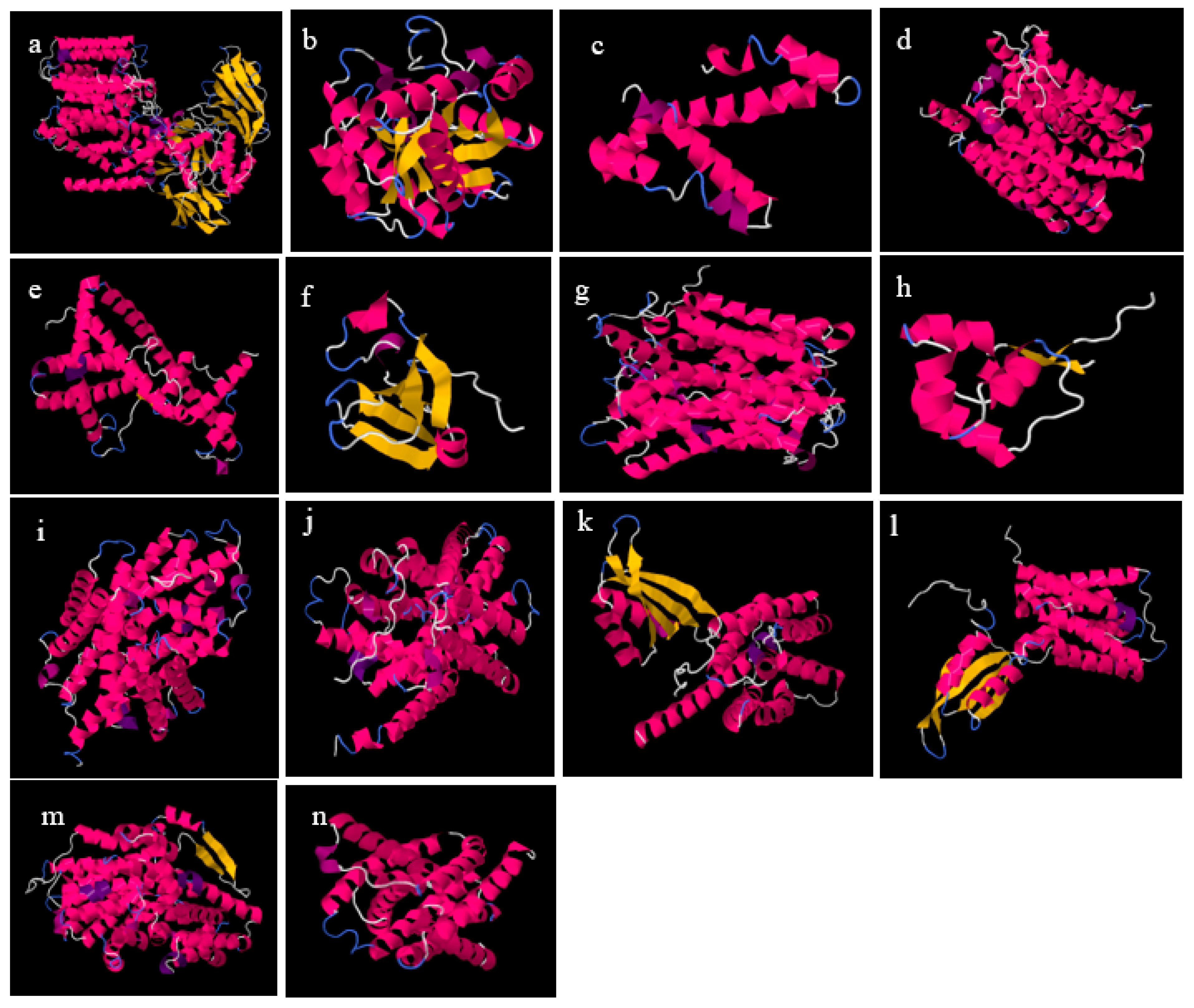
The three-dimensional tertiary structures and Ramachandran plots as well as terms in the SWISS-MODEL Server for the query proteins of the reference genome of Lactobacillus acidophilus are presented in Figure 3 and Figure 4 and Table 5. No experimental structure was found for any of the proteins in question and the probable biological functions for the proteins were as follows: undefined catalytic activity (LBA1510, LBA0214, LBA0037, LBA1825 and LBA1788); no probable function was available for LBA1705, LBA1446, LBA0995, yifk, LBA0899, LBA1209, LBA0972 and LBA0338; and for LctP, uptake of L-lactate across the membrane was predicted as was the possibility of transporting D-lactate and glycolate.
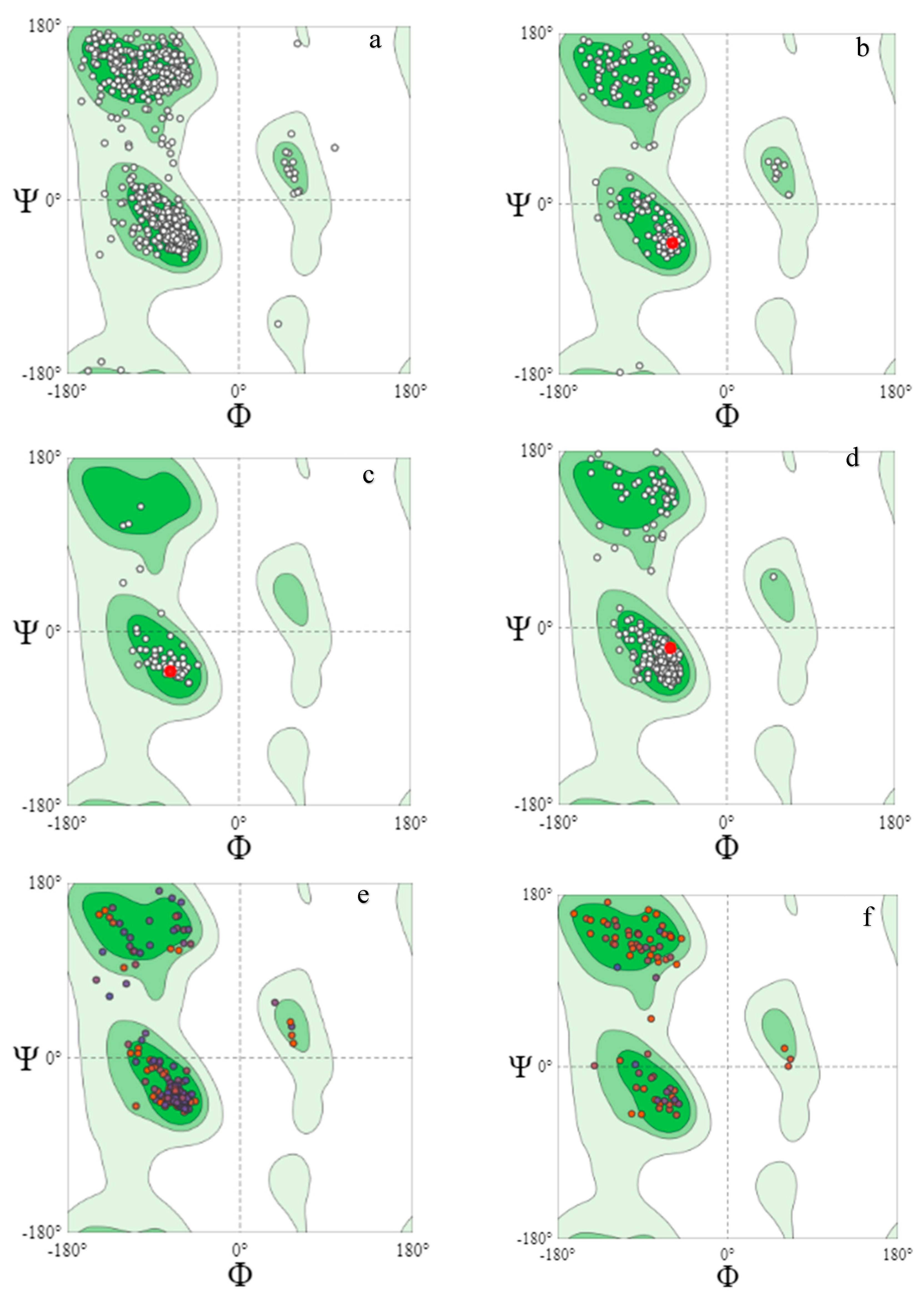
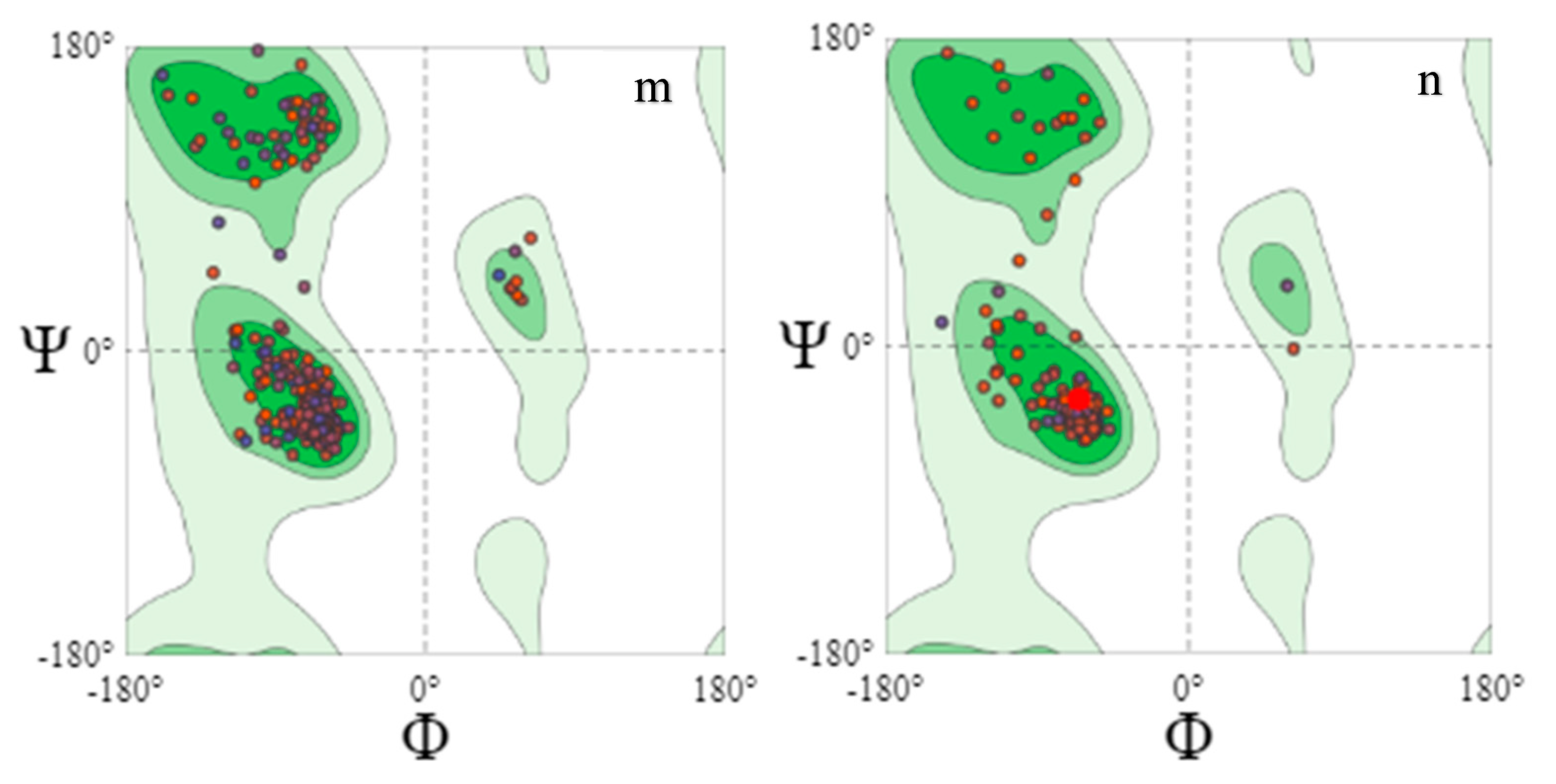
The Ramachandran plots validated the three-dimensional structures obtained (Figure 4). No transmembrane segment was found in LBA0214, LBA1825 or LBA1788. The favored area of Ramachandran (%) ranged between 99.59 (LBA1446) and 92.31 (LBA1705) (Table 7). The Q Mean scores ranged between −3.39 (LBA1705) and 0.84 (LBA0214), the torsion angle energy values ranged between −3.23 (LBA1446) and 0.46 (LBA1788), the solvation energy values ranged between −0.42 (LBA1825) and 4.28 (LBA1446) and the C_b interaction energy values ranged between −2.61 (LBA1510) and 2.17 (LBA0214), as shown in Table 5.

3.5. Protein–Protein Interactions of Query Proteins

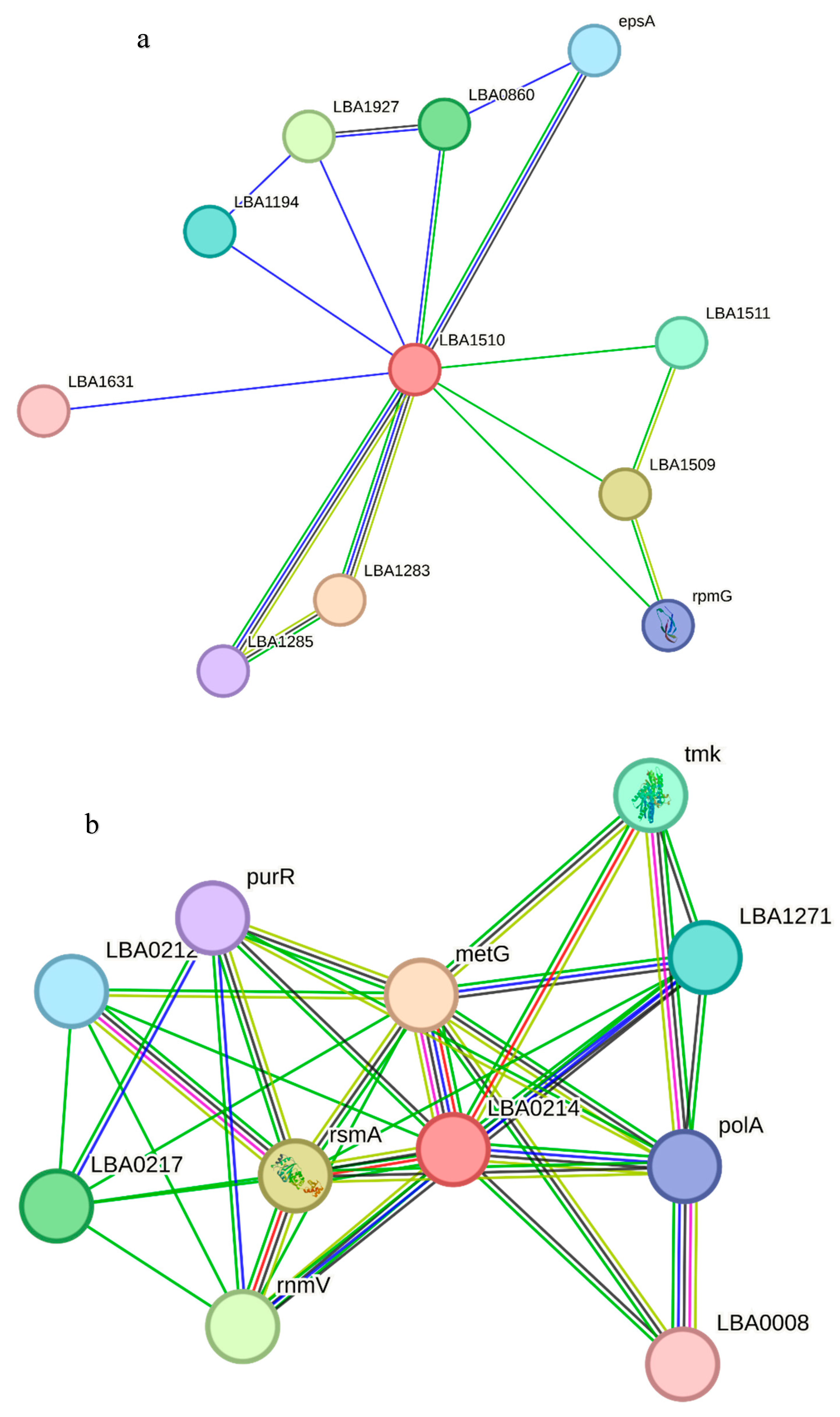
The predicted protein–protein interaction network for the query proteins of the reference genome of the Lactobacillus acidophilus is presented in Figure 5. Protein scores less than 0.5 were not included in this analysis. The functional partners with their corresponding confidence scores for LBA1510 included LBA1283 (0.765), LBA1509 (0.738), LBA1927 (0.671), LBA0860 (0.618), LBA1511 (0.523) and LBA1194 (517) (Figure 5a). For LBA0214, the functional partners and corresponding confidence scores were metG (0.980), rsmA (0.968), mmV (0.904), LBA0217 (0.783), tmk (0.722), LBA1271 (0.596), LBA0212 (0.562), polA (0.562), purR (0.543) and LBA0008 (0.521), as shown in Figure 5b.
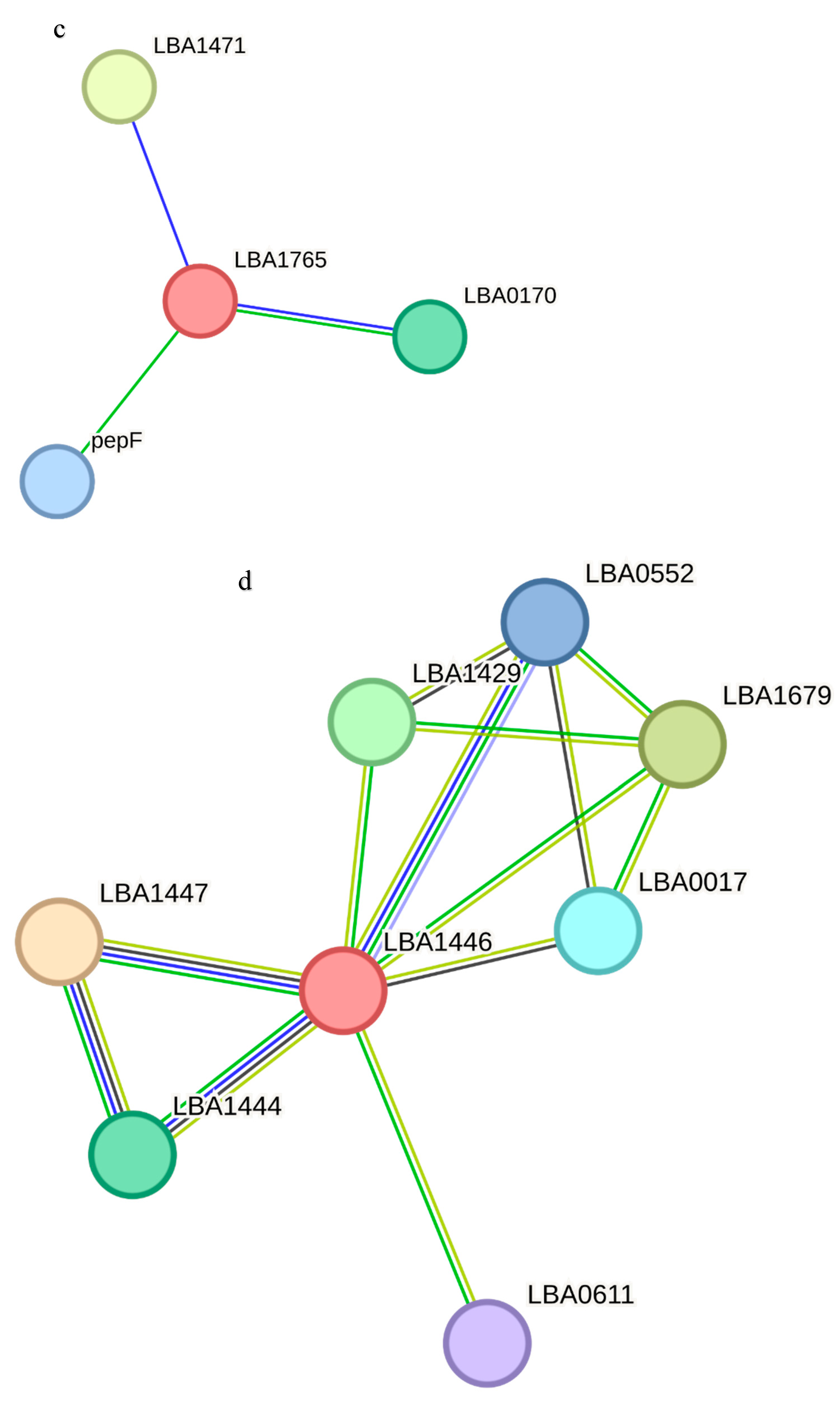
The predicted functional partners of LBA1705 include LBA1471 (0.754), LBA0170 (0.570) and pepF (0.559) (Figure 5c). LBA1447 (0.920), LBA1679 (0.831), LBA1429 (0.830), LBA1444 (0.785), LBA0017 (0.534) and LBA0552 (0.524) are the functional partners of LBA1446 (Figure 5d). LBA0036 (0.548) and pbpX-2 (0.528) are the functional partners of LBA0037 as revealed in Figure 5e.
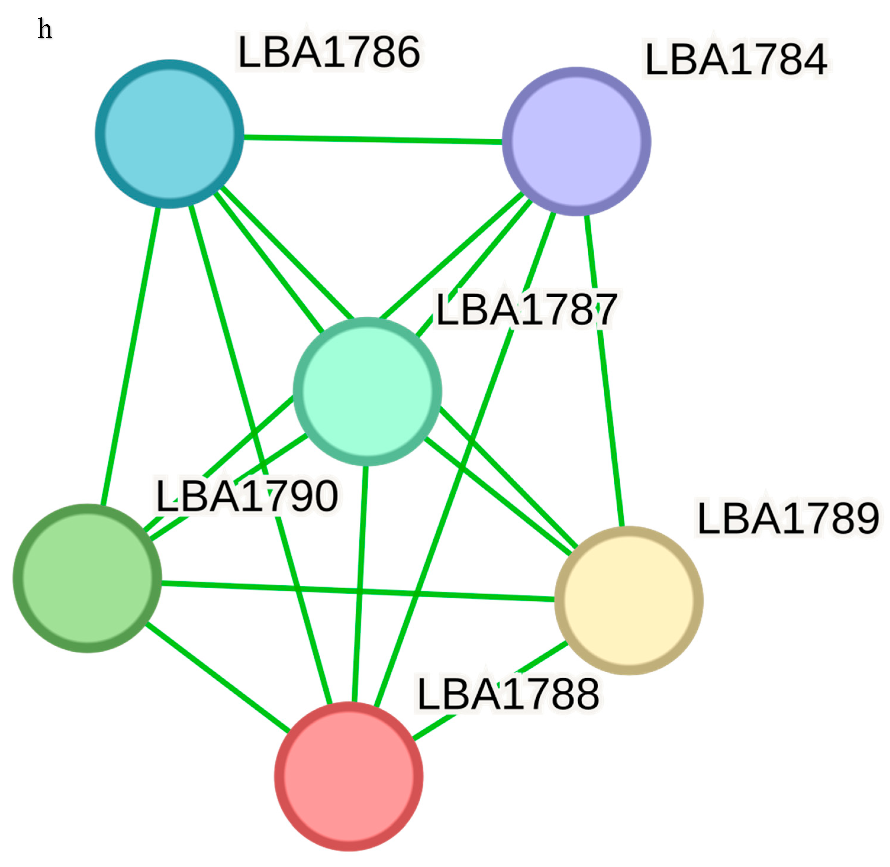
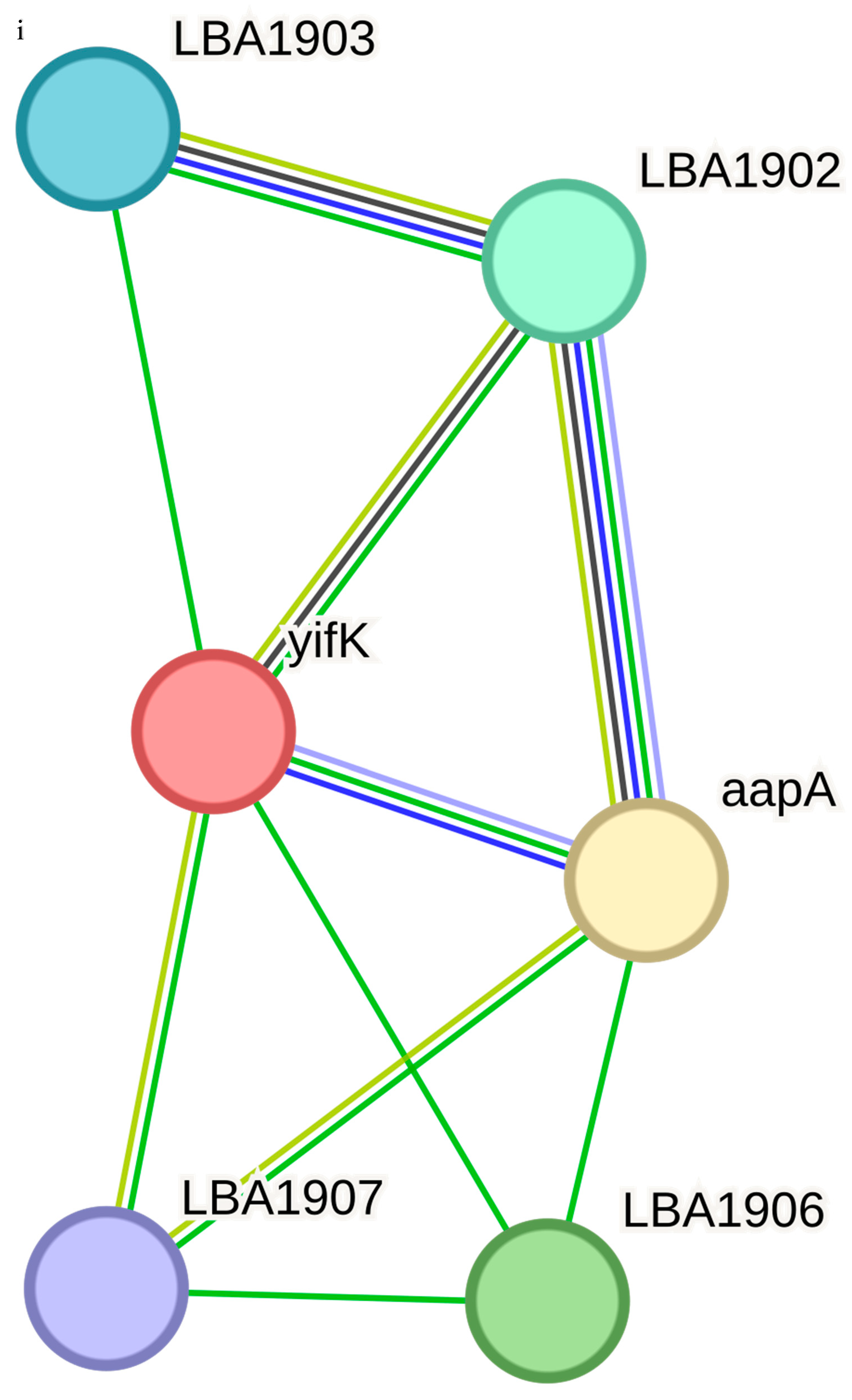
The functional partners of LBA1825 included gldB (0.913), ychF (0.909), parA (0.869), parB (0.863), parB-2 (0.863), LBA1823 (0.790), LBA0418 (0.689), LBA1258 (0.629) and LBA1830 (0.572) (Figure 5f). The functional partners of LBA0995 included LBA0096 (0.843), LBA0997 (0.711), fabG (0.565) and pepD (0.526) (Figure 5g). LBA1789 (0.800), LBA1790 (0.773) and LBA1787 (0.508) are the functional partners of LBA1788 (Figure 5h). Only aapA was the functional partner of yifk (Figure 5i) based on the specified condition, with a confidence score of 0.710. Both thyA (0.536) and dfrA (0.507) are functional partners of LBA0899 (Figure 5j).
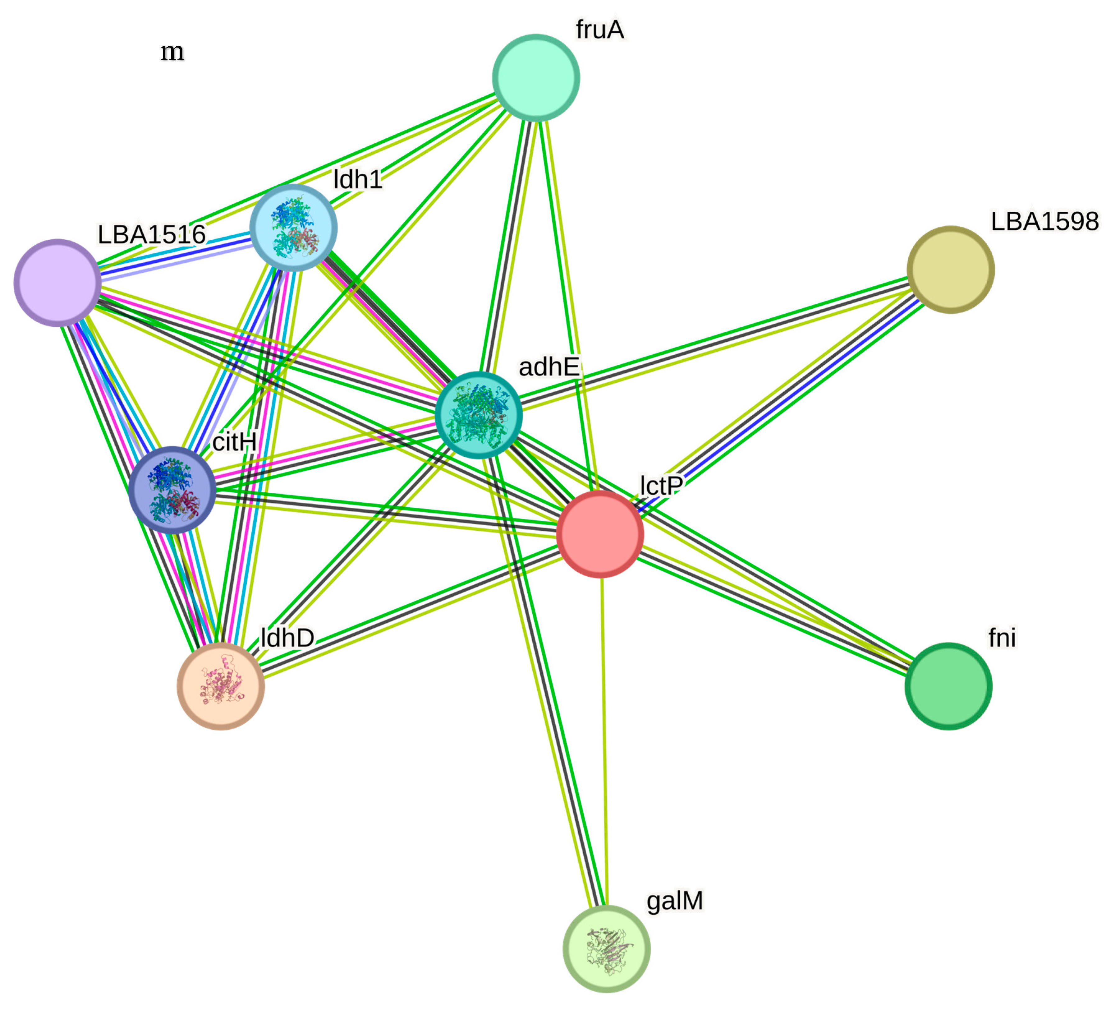
Based on the specified conditions, msrA was the functional partner of LBA1209 (Figure 5k) and its confidence score is 0.602. LBA0426 (0.883) and cca (0.611) are the functional partners of LBA0972 (Figure 5l). The functional partners of LctP include ldhD (0.807), LBA1598 (0.741), galM (0.702), fni (0.669), fruA (0.609), adhE (0.542), ldh1 (0.511) and citH (0.511), as shown in Figure 5m. For LBA0338, the following were detected: LBA0339 (0.688), LBA0340 (0.587), LBA0337 (0.574), pheS (0.540), LBA0336 (0.505) and htrA (0.502) (Figure 5n).

3.6. Active Site Analysis of the Query Proteins

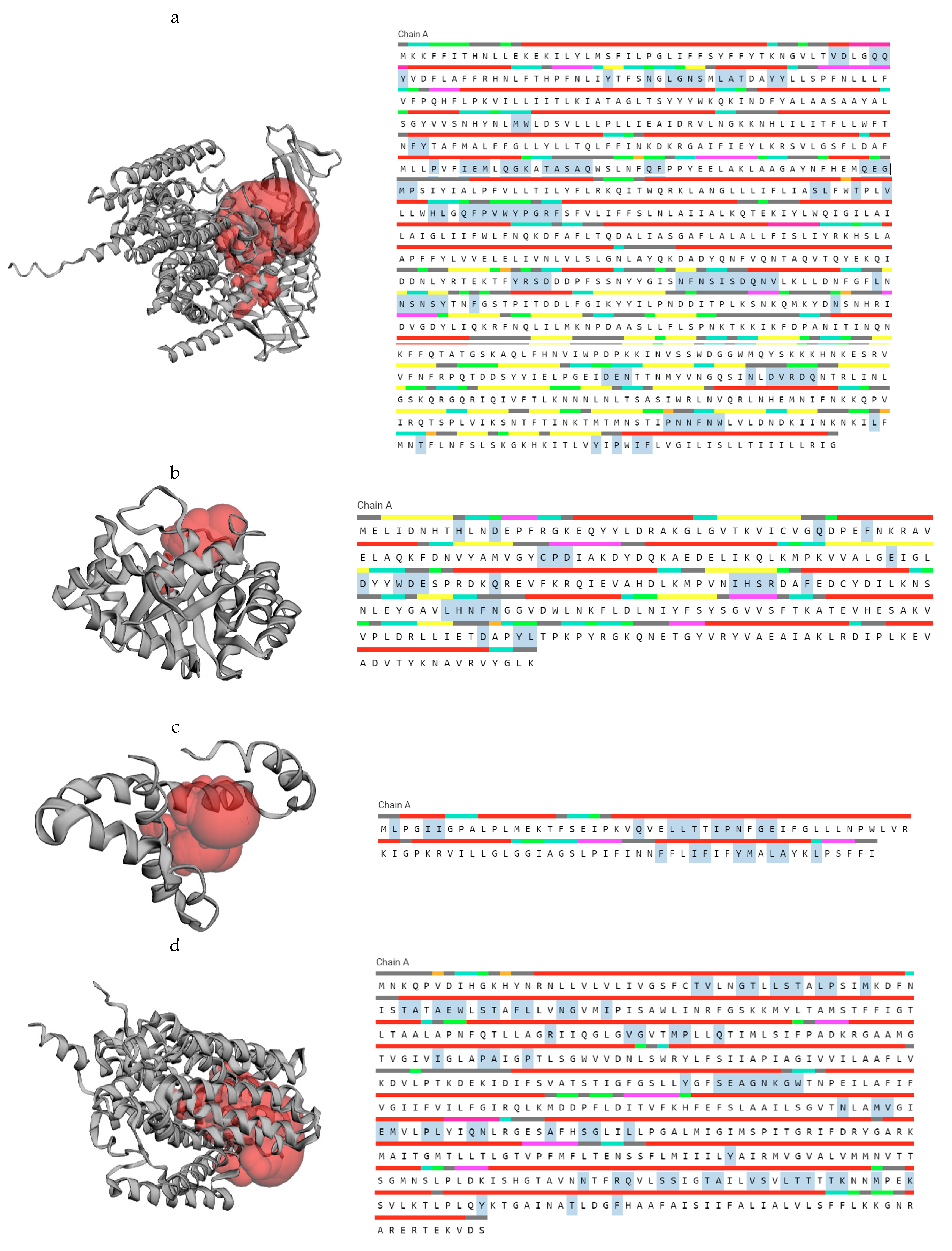
Images representing the results of the active site analysis of query proteins of the Lactobacillus acidophilus reference genome are presented in Figure 6. There are 98 amino acid residues in the active site (shaded) of LBA1510 (Figure 6a). Figure 6b shows the 26 amino acid residues (His, Asp, Gln, Phe, Cys, Pro, Asp, Glu, Asp, Trp, Asp, Glu, Gln, Ile, His, Ser, Arg, Phe, Leu, His, Asn, Phe, Asn, Asp, Try and Leu) in the active site (shaded) of LBA0214. For LBA1705, there are 20 amino acid residues (Leu, Ile, Ile, Gln, Leu, Leu, Thr, Ile, Pro, Asn, Gly, Glu, Phe, Ile, Phe, Try, Met, Leu, Ala and Leu) in its active site (shaded) as revealed in Figure 6c.
Figure 6d shows the 74 amino acid residues (Thr, Val, Gly, Thr, Leu, Ser, Thr, Leu, Pro, Met, Thr … Phe) in the active site (shaded) of LBA1446. Figure 6e reveals the 51 amino acid residues (Try, Ile, Gly, Trp, Glu, Try, Cys…Leu) in the active site (shaded) of LBA0037. The 11 amino acid residues (Leu, Leu, Arg, Met, Lys, Met, Ile, Val, Arg, Pro and Phe) in the active site of LBA1825 (shaded) are shown in Figure 6f.
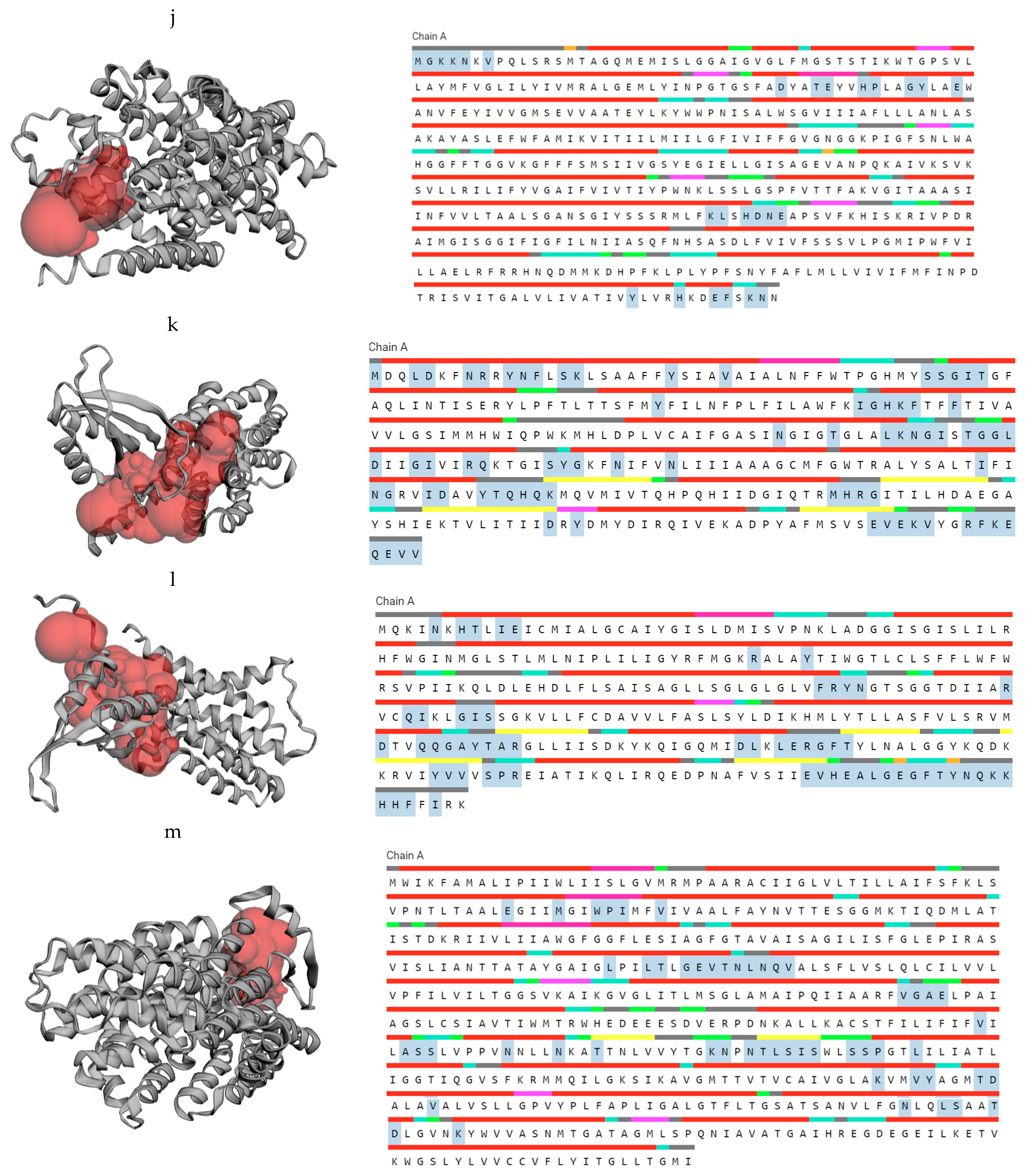
There are 89 amino acid residues (Ile, Glu, Thr, Ile, Asn, Phe… Lys) in the active site (shaded) of LBA0995 (Figure 6g). There are seven amino acid residues (Ile, Asn, Leu, Gly, Val, Arg and Thr) in the active site (shaded) of LBA1788 (Figure 6h). The 35 amino acid residues (Ala, Lys, Lys, Ala, Pro, Glu…Pro) in the active site of yifk are shaded (Figure 6i). The 26 amino acid residues (Met, Gly, Lys, Lys, Asn, Val…Asn) in the active site (shaded) of LBA0899 are shown in Figure 6j.
There are 72 amino acid residues (Met, Leu, Asp, Asn, Arg, Try, Asn, Phe, Ser, Lys, Ser, Ser, Gly…Val) in the active site (shaded) of LBA1209 (Figure 6k). There are 60 amino acid residues (Asn, His, Thr, Ile, Glu, Arg, Try, Phe…Ile) in the active site (shaded) of LBA0972 (Figure 6l). Figure 6m shows the 53 amino acid residues (Glu, Met, Trp, Pro, Ile, Val, Leu, Leu, Thr, Gly, Glu, Val, Thr, Asn, Leu…Lys) in the active site (shaded) of LctP. Figure 6n indicates that there are eight amino acid residues (Ile, Ile, Gly, Lys, Lys, Arg, Ile and Ala) in the active site (shaded) of LBA0338.

3.7. Immunogenicity, Allergenicity and Toxicity Evaluations of Query Proteins

Table 8 shows the results of the immunogenicity, allergenicity and toxicity analyses of the query proteins of the reference genome of Lactobacillus acidophilus. All the query proteins were non-antigenic, non-allergic and non-toxic.

3.8. IL-6-Inducing Peptides and Gene Expression Analyses

The proteins were scanned for regions producing IL-6-inducing peptides and the selected immunogenic IL-6-inducing peptides are presented in Table 9.
The importance of these regions is that they may be altered, amended or removed for vaccine development or for therapeutic purposes. In this study, all the proteins produce peptides which induce IL-6, suggesting their potential for effective therapeutic purposes in addition to being effective feed additives and food supplements. Immunogenic IL-6-inducing peptides from proteins of the L. acidophilus reference genome according to their IL-6-inducing prediction values and immunogenicity values when the nonimmunogenic ones (LBA0037, LBA1825) and non-IL-6-inducing peptides from LBA1788 have been removed are presented in Figure 7.
Gene expression levels of IL-6 across various conditions in Homo sapiens and Mus musculus are shown in Supplementary Figure S1. Il-6 is upregulated in urinary bladder, anterior prostate, blood vessel, liver, lung, synovium and breast in Homo sapiens. In Mus musculus, IL-6 is regulated in extraocular muscles, gonadal adipose tissue, bone marrow, lung, skeletal muscle and aorta among others. According to cancer categories in Homo sapiens, Il-6 is upregulated by prostate, neoplasm, malignant stroma and malignant lymphoma NOS. On top of the list of the genes positively co-expressed with IL-6, according to cancer categories in Mus musculus, are the following: paired related homeobox 2 (Prrx2), twist basic helix-loop-helix transcription factor 1 (Twist1), EH-domain containing 2 (Ehd2), brain derived neurotrophic factor (Bdnf), 5-hydroxytryptamine (serotonin) receptor 1B (Htr1b) and microtubule associated serine/threonine kinase family (Mast4).
Multiple sequence alignment of the query protein sequences was analyzed using Clustal Omega, https://www.ebi.ac.uk/, accessed on 29 July 2024 and a phylogenetic tree indicating the relationship existing among the proteins (Figure 8).

4. Discussion

Previous authors have documented the importance of exploring hypothetical proteins in different organisms of interest [31,32,33,34,35,36,37]. However, this is the first study in this area to focus on the structural and functional relationship, safety profile and IL-6-inducing capacity of uncharacterized proteins of probiotic Lactobacillus acidophilus for sustainable applications in animal and human nutrition as well as for therapeutic and cosmetics purposes.
The importance of proteins as the ‘building blocks’ for the body cannot be over-emphasized. Proteins play significant roles in the structural formation and functions of organisms, performing key roles in the cell, including protecting the body from pathogens and carrying out chemical reactions. However, proteins’ discrete native structure influences its roles, exposes several channels, receptors and binding sites thereby controlling how they bind and interact with other molecules as well as how they form complexes used for regulatory and structural functions. A protein attains its functional native structure and reaches its final form (three-dimensional structure) by folding. Amino acid composition influences protein classification into different folding types, structural groups and functions. The physicochemical properties of a protein encoded in its amino acid determine its folding.
The physicochemical properties usually used in characterizing uncharacterized proteins include the molecular weight, theoretical isoelectric point (pI), amino acid composition, instability index, aliphatic index and grand average of hydropathicity (GRAVY) [24]. In this study, the results of the physicochemical properties of the query proteins of the Lactobacillus acidophilus reference genome revealed that LBA1446 had the highest pI. LBA0214 and LBA1788 had values less than 7.00 while the remaining query proteins had values greater than 7.00, indicating whether they are basic or acidic.
A protein has no net charge at the isoelectric point [24]. At that point, the protein is not repelled or attracted by charged molecules. In a polar solvent, a charged molecule seems to be readily soluble. A protein with a pH above the pI is negatively charged and will not be attracted by a negatively charged surface, thereby decreasing its solubility. The pI plays an important role in protein purification, representing the pH during which solubility is minimal. The pI also talks about the basic or acidic nature of a protein. The pI of the amino acid and acidic protein will be lower and vice versa for a basic protein. Molecular weight is also useful in protein purification and separation.
The extinction coefficient has been defined as a measurement of how strongly a protein absorbs light at a given wavelength. Two values are usually produced by ProtParam for proteins measured in water at 280 nm. The first indicates that the computed value is based on the assumption that all cysteine residues appear as half cystines, that is, all pairs of Cys residues form cystines, while the second value is based on the assumption that no cysteine appears as half cystine, that is, the assumption that all Cys residues are reduced [24,35]. In this study, the extinction coefficient values are very high for LBA1510, LBA0214, LBA1446, LBA0037, LBA0995, yifk, LBA0899, LBA1209, LBA0972, LBA099513 and LBA0338 and high for LBA1705 and LBA1825, except for LBA1788. The high extinction coefficient may be an indication of the presence of a high concentration of Cys, Trp and Tyr in the query proteins of Lactobacillus acidophilus, except for LBA1788, which does not contain Trp residues.
In this study, the half-life estimation was the same for all the query proteins. The estimated half-life is a prediction of the time it takes for half of the amount of protein in a cell to disappear after its synthesis in the cell, relying on the “N-end rule”, which relates the half-life of a protein to the identity of its N-terminal residue, in this case, for humans (in vitro), yeast (in vivo) and Escherichia coli (in vivo) [24].
An instability index less than 40 indicates that the protein is stable while a value above 40 indicates that the protein may be unstable [24]. In the present study, all the query proteins had instability index values less than 40, which implies that they are stable, except for LBA0995, which may be regarded as unstable because it has an instability index greater than 40. The aliphatic index of a protein can be described as the relative volume occupied by aliphatic side chains, that is, alanine, isoleucine, leucine and valine, which may be regarded as a positive factor for the increase in thermostability of globular proteins [24].
The importance of a protein thermostability cannot be over-emphasized. A thermostable protein is preferred for industrial applications and food processing. The ability to withstand extreme industrial and preservation conditions in food processing and storage is beneficial for probiotic bacteria. Hence, the ability of probiotic bacteria to withstand harsh environmental conditions, having its proteins being thermostable is an advantage to food, cosmetic and therapeutic industries benefiting from the health-promoting probiotics in the era of climate change. Proteins with high aliphatic indices are thermostable under a wide temperature range. In this study, the aliphatic indices are generally high for all the query proteins, although the values for LBA1825 and LBA0214 are not as high as the values obtained for the other query proteins. The high aliphatic index values obtained by the query proteins of the Lactobacillus acidophilus reference genome used in this study indicate that the proteins are stable over a wide temperature range.
The importance of the instability index is mainly in the storage of proteins in the correct solvent. Highly stable proteins are easy to store. For instance, an insulin monomer with an instability index of 43 is considered unstable and may macroscopically aggregate in aqueous solution during storage, which may consequently result in decline in biological activities.
The calculation of the grand average of hydropathy value for a peptide or protein is based on the sum of hydropathy values of all the amino acids, divided by the number of residues in the sequence [24,38]. In the present study, only LBA0214, LBA1825 and LBA1788 obtained negative GRAVY values, which is an indication of being hydrophilic, while the other query proteins had positive GRAVY values, which implies that they are hydrophobic [39]. Another importance of GRAVY index is that it is used to determine whether a protein can be visualized on 2D gel. Proteins with GRAVY score greater than 0.4 are usually difficult to view on 2D gel because it lies outside the solubility range.
Proteins intended for use as food products or for use in consumer products are expected to be analyzed for their allergenic reaction potential before they are introduced into the market to determine and mitigate the risk of inducing an immediate type I (IgE-mediated) allergic response. Allergenicity or allergenic potential in this study is defined as the potential of a protein to cause or elicit immediate type (IgE-mediated) allergic reactions in humans [40]. It is not certain whether probiotics fed to animals or offered for commercial sale are subjected to allergenic potential evaluations before they are introduced into the market. To ascertain the safety and sustainability of these probiotics, their safety profile including toxicity, allergenicity and antigenicity may be necessary, revealing the novelty of this study.
Bioinformatics tools such as AllerCatPro 2.0, VaxiJen ToxinPred [26,27,28], for quick and cost-saving approaches, have been recommended. The findings from the present study revealed that none of the query proteins of the Lactobacillus acidophilus reference genome studied were antigenic, allergenic or toxic for human or animal consumption.
The benefits of computational prediction of bacterial protein subcellular localization include the provision of rapid and cost-effective approaches for gaining insight into protein function, verifying experimental results, annotating newly sequenced bacterial genomes, detecting potential cell surface or secreted drug targets, and identifying microbial biomarkers. One of the most precise tools recommended for computational prediction of bacterial protein subcellular localization is PSORTb version 2.0 [41,42], which generates prediction results for five major locations for Gram-negative bacteria (cytoplasmic, inner membrane, periplasmic, outer membrane and extracellular) and four locations for Gram-positive bacteria (cytoplasmic, cytoplasmic membrane, cell wall and extracellular). However, the PSORTb version 3.0 used in this study is an updated version of 2.0 [20] with more features. In this study, LBA1788 is cytoplasmic, LBA1510, LBA0214, LBA1705, LBA1446, LBA0037, LBA0995 and yifk-LBA0338 are found in the cytoplasmic membrane while the subcellular localization of LBA1825 is not known.
The functional associations of the query proteins of the Lactobacillus acidophilus reference genome are presented in the Results section (Figure 6). LBA1283 is a glycosil transferase, LBA1509 is a penicillin-binding protein and LBA1927 and LBA0860 are hypothetical proteins. metG is a met-rRNA synthetase that is not only required for the elongation of protein synthesis but also for the initiation of all mRNA translation. rsmA is a dimethyladenosine transferase, and mmV is a putative promase-like substance that is required for the correct processing of both the 5′ and 3′ ends of the 5S rRNA precursor. LBA0217 is a COG4466-uncharacterized protein conserved in bacteria while tmk is a thymidylate (dTMP) kinase; the phosphorylation of dTMP results in the formation of dTDP in both the de novo and salvage pathways of dTTP synthesis. LBA1471 is a putative multidrug efflux permease, LBA0170 is a putative 6-pyruvoyl-tetrahydropterin synthase and pepF is an oligopeptidase, endopeptidase F.
LBA1447 is a hypothetical protein, LBA1679 is an ABC transporter permease protein, LBA1429 is a putative transporter-membrane protein, LBA1444 is a transcriptional regulator family, MerR; LBA0017 is a putative general stress response and COG3237 is an uncharacterized protein conserved in bacteria belonging to the UPF0337 family. LBA0036 is a collagen-binding protein Cne precursor while pbpX-2 is a putative penicillin-binding protein. gldB is a glucose-inhibited division protein B that specifically methylates the N7 position of a guanine in 16S rRNA, ychF is a GTP-binding protein and parA, parB and parB-2 are chromosome-portioning proteins. Both parB and parB-2 are predicted transcriptional regulators of COG1475 and belong to the Par family.
LBA0996 is a COG1982 arginine–lysine–ornithine decarboxylase, LBA0997 is an aluminum resistance protein, fabG is a 3-oxoacyl-(acyl-carrier protein) reductase and pepD is an aminoacyl-histidine dipeptidase and PepD is a carnosinase. LBA1789 and LBA1787 are hypothetical proteins while LBA1790 is a vacuolar sorting receptor protein homologueg of the PV72-cucur bit. The only functional associate of yifk, aapA is an amino acid permease. The functional associate of LBA0899, thyA is a thymidylate synthase, and dfrA is a dihydrofolate reductase, which is a key enzyme in folate metabolism.
LBA0426 is a hypothetical protein, while cca is a tRNA nucleotidytransferase, polyA. LBA1598 is a glycolate oxidase, L-lactate dehydrogenase, cytochrome-type (lldD), ldhD is a D-lactate-dehydrogenase that belongs to the D-isomer specific 2-hydroxyacid dehydrogenase family, galM is a galactose-1-epimerase, mutarotase and fni are isopentenyl diphosphate isomerases, involved in the biosynthesis of isoprenoids. LBA0339, the functional associate of LBA0338, is a phosphoglycerate mutase, COG0406 fructose-2,6-bisphosphatase, which belongs to the phosphoglycerate mutase family. LBA0340 is a cation efflux protein that is a cation diffusion facilitator (CDF) transporter (TC 2.A. 4) Family. LBA0337 is a hypothetical protein while pheS is a phenylalanyl-tRNA synthetase, beta subunit, SyfB and COG0073 EMAP domain.
The Ramachandran plot, a graphical image of the dihedral angles φ (phi) and ψ (psi) in a protein’s backbone, indicates the combinations which are sterically permissible and favored for protein structure formation. The Ramachandran plots validated the structures obtained as the majority of the residues fell in the (B, A, L) areas. The favored Ramachandran areas (%) ranged between 92.31 and 99.59.
IL-6 is mainly produced by T lymphocytes, B lymphocytes, dendritic cells, microglia, fibroblasts, keratinocytes, mast cells, mesangial cells and vascular endothelial cells, binding to either soluble IL-6 receptors (sIL-6R) or the membrane-bound IL-6 receptors (mIL-6R) [43]. It is a pleiotropic cytokine which plays several key roles in the body, such as influencing embryonic development, acute phase reactant pathways, B and T lymphocytes, synovial inflammation, blood–brain barrier permeability and hematopoiesis [44]. It is known to play key functions in the normal, adaptive and innate autoimmunity. It is primarily expressed when tumor necrosis factor-alpha (TNFa) and interleukin 1 b (IL-1b) are activated. Prostaglandins, other cytokines, stress response, activated toll-like receptors (TLRs), and adipokines can also enhance its synthesis [45]. When dysregulated, IL-6 can contribute to inflammatory cascades, indicating pathophysiology of many autoimmune conditions.
At the initial phase of inflammation, after IL-6 is produced, it moves to the liver (via the bloodstream), which is followed by the quick induction of an extensive range of acute phase proteins including serum amyloid A (SAA), C-reactive protein (CRP), a1-antichymotrypsin fibrinogen and haptoglobin [46]. IL-6 also reduces albumin, transferrin and fibronectin production. A persistent high-level concentration of SAA may result in several chronic inflammatory conditions with a serious complication through amyloid A amyloidosis generation [47], leading to deposition of amyloid fibril and progressive deterioration in organs [48].
In addition, IL-6 regulates serum zinc and iron levels by regulating their transporters. It initiates the production of hepcidin that hinders the iron transporter ferroportin 1 action on the gut, thereby lowering the iron levels in the serum [49], indicating the importance of IL-6–hepcidin axis in regulating hypoferremia and anemia associated with chronic inflammation. In the bone marrow, IL-6 promotes the maturation of megakaryocytes, thereby releasing platelets [50]. Changes in acute phase protein levels and red blood cell and platelet counts can be used for determining the severity of inflammation in clinical laboratory examinations [48].
The IL-6 axis has been successfully modulated (therapeutically), resulting in the approval of multiple therapeutic agents currently being investigated [44]. IL-6 has been suggested to have potential applications in neuro-inflammatory conditions (not yet FDA approved) based on its ability to form complexes with other immunosuppressive medications (azathioprine and mycophenolate mofetil) [44].
IL-6 has been reported to be produced by senescent cells and is involved in senescence-induced inflammation, age-dependent pathologies and cancer [51]. It has been described as a crucial factor in inflammation, cancer and autoimmunity, performing its roles primarily via the IL-6–signal transducer, and acting as an activator of the transcription 3 (STAT3) pathway [45,52,53]. In addition, the IL-6 amplifier (IL-6 Amp) is an amplification mechanism of IL-6, growth factors, chemokine and cytokine production via a synergic interaction between STAT3 and nuclear factor-kappa B (NF-κB), which also performs important functions in inflammatory conditions such as cancer, autoimmunity and cytokine storm syndromes [54,55,56].
IL-6 inhibitors are now used clinically for conditions including rheumatoid arthritis (RA), Castleman’s disease and considered for treating COVID-19 [57,58] despite the involvement of many cytokines in inflammation-related diseases, based on the understanding that IL-6 is the main stimulator of STAT3 in inflammation. STA3 performs crucial functions in inflammation and oncogenesis together with NF-κB, expressing IL-6 as a target, indicating STAT3 and NF-κB as the basis of the IL-6 Amp. Injury, infection, obesity, stressors, senescence, smoking, pre-neoplastic mutation and cell death are factors which may activate the IL-6 Amp [59]. The author further noted the importance of developing highly specific and effective small peptides against diseases with a requirement for high-resolution structural analysis of the ligand-receptor complex.
IL-6 is produced by various cell types such as epithelial, immune, liver, nerve, kidney and cartilage. It has been shown to play key regulatory functions in liver regeneration, cancer development, immunological regulation and inflammation [60]. The dual roles of IL-6 have been established. IL-6 has a unique characteristic (dual signaling mechanism) referred to as classical signaling and trans-signaling. The pathway arises through the membrane-bound IL-6 receptor, while the trans-pathway is triggered through the soluble IL-6 receptor. IL-6 standard or trans-signaling ligand–receptor complex formation results in the stimulation of several intracellular signaling pathways [60].
A recent study reported an increased IL-6 linked with long COVID-19 [61]. The authors noted that IL6 levels in the serum were significantly raised in patients after COVID-19 infection in both long COVID-19 and the acute stage. Cytokine storms may cause the increase during the acute infection, but different dynamic changes in IL-6 and anti-bodies during the long COVID-19 phase may result in a decline. IL-6 was also significantly increased in patients with severe malaria compared with those in patients with non-severe malaria, demonstrating that IL-6 might be a candidate marker for severe malaria [62]. The importance of IL-6 as a cytokine that facilitates a wide variety of biological functions upon binding to its receptor cannot be overstated. However, its excessive production and dysregulation of IL-6R signaling may result in several inflammatory diseases [63].
Studies are on-going to provide potent and health-promoting alternatives to synthetic antibiotics in animal and human nutrition without any adverse effect to animals’ and human’s well-being [64,65,66,67,68,69,70], underscoring the importance of the present study, which attempted to unravel the functional mechanisms of one of such alternatives. Different analyses were carried out to characterize and investigate the safety profiles of query proteins in the L. acidophilus reference genome. The findings of the study revealed that none of the query proteins were allergenic or toxic, attesting to safety profile of LAB probiotics. All the query proteins are stable under a wide range of temperatures, indicating that they can withstand industrial processing and that climate change may not easily affect LAB probiotics. LBA0214, LBA1825 and LBA1788 are hydrophilic while the remaining ones are hydrophobic. Only LBA0995 has been determined to not be stable in nature, indicating that most of the proteins are stable, which is an advantage for their preservation and functionality. LBA1788 is cytoplasmic, while the rest are found in the cytoplasmic membrane. All the proteins produce peptides which induce IL-6 except LBA0037, LBA1825 and LBA1788, which are either non-immunogenic or non-IL-6-inducing. The immunogenic IL-6-inducing peptides are potential candidates for vaccine development and therapeutic purpose, further confirming the health-benefits of LAB probiotics in animal and human nutrition. The findings in this study are based on in silico analysis and a part of other preliminary studies that mainly aim at identifying and screening promising candidates for the next phases of the study focused on adopting an experimental approach (wet lab) to validate the claims in this preliminary study. Similar proposed studies are strongly recommended from other interested researchers. IL-6 is known to have dual roles: it contributes to host defense and mucosal immunity, but excessive or chronic induction is implicated in pathology. Therefore, any potential benefit of IL-6 induction must be context-dependent and carefully balanced against its pathogenic effects, highlighting the importance of downstream validation to assess physiological relevance.

Supplementary Materials

The following supporting information can be downloaded at: https://www.mdpi.com/article/10.3390/biologics5040029/s1, Figure S1. Gene expression levels of IL-6 and positively co-expressed genes with IL-6 in Homo sapiens and Mus musculus across various conditions: a. a scatter plot (Log2 scale) showing IL-6 expression across various anatomical parts and cell types in Homo sapiens; b. a scatter plot (Log2 scale) showing IL-6 expression across various anatomical parts and cell types in Mus musculus; c. a heatmap showing IL-6 expression (linear scale) in Homo sapiens according to various cancer categories; and d. a circular view of positively co-expressed genes with IL-6 (top 50) according to cancer categories in Mus musculus.

Funding

This research received no external funding.

Institutional Review Board Statement

Not applicable.

Informed Consent Statement

Not applicable.

Data Availability Statement

The datasets used in this manuscript are included or are available in public databases and appropriate links are provided where necessary.

Acknowledgments

Olawumi Adejumo’s support during data curation is appreciated.

Conflicts of Interest

The author declares no conflicts of interest.

Abbreviations

The following abbreviations are used in this manuscript:
GRAVYGrand average of hydropathicity
MWmolecular weight
AIaliphatic index
IIinstability index
IL-6interleukin-6

References

  1. Zampiga, M.J.; Flees, A.; Meluzzi, S.D.; Sirri, F. Application of omics technologies for a deeper insight into quali-quantitative production traits in broiler chickens: A review. J. Anim. Sci. Biotechnol. 2018, 61, 61. [Google Scholar] [CrossRef]
  2. Jadhav, K.; Sharma, K.S.; Katoch, S.; Sharma, V.K.; Mane, B.G. Probiotics in broiler poultry feeds: A review. Int. J. Anim. Vet. Sci. 2015, 2, 4–16. [Google Scholar]
  3. Vieco-Saiz, N.; Belguesmia, Y.; Raspoet, R.; Auclair, E.; Gancel, F.; Kempf, I.; Drider, D. Benefits and inputs from LAB and their bacteriocins as alternatives to antibiotic growth promoters during food-animal production. Front. Microbiol. 2019, 10, 57–65. [Google Scholar] [CrossRef]
  4. Mahfuz, S.U.; Nahar, M.J.; Chen, M.; Zhang, G.; Zhongjun, L.; Hui, S. Inclusion of probiotic on chicken performance and immunity: A review. Int. J. Poult. Sci. 2017, 16, 328–335. [Google Scholar] [CrossRef]
  5. FAO/WHO. Guidelines for the Evaluation of Probiotics in Food; FAO: Paris, France, 2002; pp. 1–11.
  6. Hill, C.; Guarner, F.; Reid, G.; Gibson, G.R.; Merenstein, D.J.; Pot, B.; Morelli, L.; Canani, R.B.; Flint, H.J.; Salminen, S.; et al. Expert consensus document. The International Scientific Association for Probiotics and Prebiotics consensus statement on the scope and appropriate use of the term probiotic. Nat. Rev. Gastroenterol. Hepatol. 2014, 11, 506–514. [Google Scholar] [CrossRef] [PubMed]
  7. Ashrafuzzaman, M.; Momtaz, F.; Foysal, J.; Ali, H.; Rahman, M.M. Optimization of probiotic activity of Lactobacillus acidophilus characterized from commercial yoghurt. Int. J. Biosci. 2015, 7, 94–99. [Google Scholar]
  8. Anadon, A.M.; Larrañaga, R.M.; Ares, I.; Martínez, M. Probiotics: Safety and toxicity considerations. Nutraceuticals 2016, 65, 1081–1105. [Google Scholar]
  9. Dhama, K.; Verma, V.; Sawant, P.M.; Tiwari, R.; Vaid, R.K.; Chauhan, R.S. Applications of probiotics in poultry: Enhancing immunity and beneficial effects on production performances and health—A review. J. Immunol. Immunopathol. 2011, 13, 1–19. [Google Scholar]
  10. Denli, M.; Demirel, R. Replacement of antibiotics in poultry diets. CAB Rev. 2018, 13, 35. [Google Scholar] [CrossRef]
  11. Shang, Y.S.; Kumar, S.; Oakley, B.; Woo, K.K. Chicken gut microbiota: Importance and detection technology. Front. Vet. Sci. 2018, 5, 254. [Google Scholar] [CrossRef]
  12. Sugiharto, Y.T.; Isroli, I.; Widiastuti, W.; Wahyuni, H.I. Hematological parameters and selected intestinal microbiota populations in the Indonesian indigenous crossbred chickens fed basal diet supplemented with multi-strain probiotic preparation in combination with vitamins and minerals. Vet. World 2018, 11, 874–882. [Google Scholar] [CrossRef]
  13. Noohi, N.; Papizadeh, M.; Rohani, M.; Talebi, M.; Pourshafie, M.R. Screening for probiotic characters in lactobacilli isolated from chickens revealed the intra-species diversity of Lactobacillus brevis. Anim. Nutr. 2021, 7, 119–126. [Google Scholar] [CrossRef] [PubMed]
  14. Sirisopapong, M.; Shimosato, T.; Okrathok, S.; Khempaka, S. Assessment of lactic acid bacteria isolated from the chicken digestive tract for potential use as poultry probiotics. Anim. Biosci. 2023, 3, 1209–1220. [Google Scholar] [CrossRef]
  15. Kassa, G.; Alemayehu, D.; Andualemy, B. Isolation identification and molecular characterization of probiotic bacteria from locally selected Ethiopian free-range chickens gastrointestinal tract. Poult. Sci. 2024, 103, 03311. [Google Scholar] [CrossRef]
  16. Kizerwetter-Świda, M.; Binek, M. Assessment of potentially probiotic properties of Lactobacillus strains isolated from chickens. Pol. J. Vet. Sci. 2016, 19, 15–20. [Google Scholar] [CrossRef]
  17. Al-Khalaifa, H.; Al-Nasser, A.; Al-Surayee, T.; Al-Kandari, S.; Al-Enzi, N.; Al-Sharrah, T.; Ragheb, G.; Al-Qalaf, S.; Mohammed, A. Effect of dietary probiotics and prebiotics on the performance of broiler chickens. Poult. Sci. 2019, 98, 4465–4479. [Google Scholar] [CrossRef]
  18. Ahmad, R.; Yu, Y.-H.; Hsiao, F.S.-H.; Su, C.-H.; Liu, H.-C.; Tobin, I.; Zhang, G.; Cheng, Y.-H. Influence of heat stress on poultry growth performance, intestinal inflammation, and immune function, and potential mitigation by probiotics. Animals 2022, 12, 2297. [Google Scholar] [CrossRef] [PubMed]
  19. Suravajhala, P.; Benso, A.; Valadi, J.K. Annotation and curation of uncharacterized proteins: Systems biology approaches. Front. Genet. 2015, 6, 224. [Google Scholar] [CrossRef] [PubMed]
  20. Yu, N.Y.; Wagner, J.R.; Laird, M.R.; Melli, G.; Rey, S.; Lo, R.; Dao, P.; Sahinalp, S.C.; Ester, M.; Foster, L.J.; et al. PSORTb 3.0: Improved protein subcellular localization prediction with refined localization subcategories and predictive capabilities for all prokaryotes. Bioinformatics 2010, 26, 1608–1615. [Google Scholar] [CrossRef]
  21. Geourjon, C.; Deleage, G. SOPMA: Significant improvements in protein secondary structure prediction by consensus prediction from multiple alignments. Comput. Appl. Biosci. 1995, 11, 681–684. [Google Scholar] [CrossRef]
  22. Kim, G.; Lee, S.; Karin, E.L.; Kim, H.; Moriwaki, Y.; Ovchinnikov, S.; Steinegger, M.; Mirdita, M. Easy and accurate protein structure prediction using ColabFold. Nat. Protoc. 2025, 20, 620–642. [Google Scholar] [CrossRef]
  23. Waterhouse, A.; Bertoni, M.; Bienert, S.; Studer, G.; Tauriello, G.; Gumienny, R.; Heer, F.T.; De Beer, T.A.P.; Rempfer, C.; Bordoli, L.; et al. SWISS-MODEL: Homology modelling of protein structures and complexes. Nucleic Acids Res. 2018, 46, W296–W303. [Google Scholar] [CrossRef]
  24. Gasteiger, E. The Proteomics Protocols Handbook; Walker, J.M., Ed.; Humana Press: Totowa, NJ, USA, 2005; pp. 571–607. [Google Scholar]
  25. Szklarczyk, D.; Franceschini, A.; Wyder, S.; Forslund, K.; Heller, D.; Huerta-Cepas, J.; Simonovic, M.; Roth, A.; Santos, A.; Tsafou, K.P.; et al. STRING v10: Protein-protein interaction networks, integrated over the tree of life. Nucleic Acids Res. 2015, 43, D447–D452. [Google Scholar] [CrossRef]
  26. Nguyen, M.N.; Krutz, N.L.; Limviphuvadh, V.; Lopata, A.L.; Gerberick, G.F.; Maurer-Stroh, S. AllerCatPro 20: A web server for predicting protein allergenicity potential. Nucleic Acids Res. 2022, 50, W36–W43. [Google Scholar] [CrossRef]
  27. Dimitrov, I.; Zaharieva, N.; Doytchinova, I. Bacterial immunogenicity prediction by machine learning methods. Vaccines 2020, 8, 709. [Google Scholar] [CrossRef]
  28. Sharma, N.; Naorem, L.D.; Jain, S.; Raghava, G.P.S. ToxinPred2: An improved method for predicting toxicity of proteins. Brief. Bioinform. 2022, 23, 1–12. [Google Scholar] [CrossRef]
  29. Maurer-Stroh, S.; Krutz, N.L.; Kern, P.S.; Gunalan, V.; Nguyen, M.N.; Limviphuvadh, V.; Eisenhaber, F.; Gerberick, G.F.; Valencia, A. AllerCatPro-prediction of protein allergenicity potential from the protein sequence. Bioinformatics 2019, 35, 3020–3027. [Google Scholar] [CrossRef]
  30. Tian, W.; Chen, C.; Lei, X.; Zhao, J.; Liang, J. CASTp 3.0: Computed atlas of surface topography of proteins. Nucleic Acids Res. 2018, 46, W363–W367. [Google Scholar] [CrossRef] [PubMed]
  31. Kolker, E.; Makarova, K.S.; Shabalina, S.; Picone, A.F.; Purvine, S.; Holzman, T.; Cherny, T.; Armbruster, D.; Munson, R.S., Jr.; Kolesov, G.; et al. Identification and functional analysis of ‘hypothetical’ genes expressed in Haemophilus influenza. Nucleic Acids Res. 2004, 32, 2353–2361. [Google Scholar] [CrossRef] [PubMed]
  32. Mazandu, G.K.; Mulder, N.J. Function prediction and analysis of Mycobacterium tuberculosis hypothetical proteins. Int. J. Mol. Sci. 2012, 13, 7283–7302. [Google Scholar] [CrossRef] [PubMed]
  33. Mohan, R.; Venugopal, S. Computational structure and functional analysis of hypothetical proteins of Staphylococcus aureus. Bioinformation 2012, 8, 722–728. [Google Scholar] [CrossRef]
  34. Park, S.J.; Son, W.S.; Lee, B.J. Structural analysis of hypothetical proteins from Helicobacter pylori: An approach to estimate functions of unknown or hypothetical proteins. Int. J. Mol. Sci. 2012, 13, 7109–7137. [Google Scholar] [CrossRef]
  35. Enany, S. Structural and functional analysis of hypothetical and conserved proteins of Clostridium tetani. J. Infect. Public Health 2014, 7, 296–307. [Google Scholar] [CrossRef] [PubMed]
  36. Rahman, F.; Hasan, R.; Biswas, M.S.; Shathi, J.H.; Hossain, F.; Yeasmin, A.; Abedin, M.Z.; Hossain, T. A bioinformatics approach to characterize a hypothetical protein Q6S8D9_SARS of SARS-CoV. Genom. Inform. 2023, 21, e3. [Google Scholar] [CrossRef]
  37. Adejumo, I.O. Hypothetical proteins of chicken-isolated Limosilactobacillus reuteri subjected to in silico analyses induce IL-2 and IL-10. Genes Nutr. 2024, 19, 21. [Google Scholar] [CrossRef] [PubMed]
  38. Kyte, J.; Doolittle, R.F. A simple method for displaying the hydropathic character of a protein. J. Mol. Biol. 1982, 157, 105–132. [Google Scholar] [CrossRef] [PubMed]
  39. Fernandez-Fernandez, A.D.; Corpas, F.J. In silico analysis of Arabidopsis thaliana peroxisomal 6-phosphogluconate dehydrogenase. Scientifica 2016, 1, 3482760. [Google Scholar]
  40. Blackburn, K.; N’jai, A.U.; Dearman, R.J.; Kimber, I.; Gerberick, G.F. Respiratory allergenic potential of plant-derived proteins: Understanding the relationship between exposure and potency for risk assessments. Crit. Rev. Toxicol. 2015, 45, 799–811. [Google Scholar] [CrossRef]
  41. Gardy, J.L. PSORTb 2.0: Expanded prediction of bacterial protein subcellular localization and insights gained from comparative proteome analysis. Bioinformatics 2005, 21, 617–623. [Google Scholar] [CrossRef]
  42. Gardy, J.L.; Brinkman, F.S.L. Methods for predicting bacterial protein subcellular localization. Nat. Rev. Microbiol. 2006, 4, 741–751. [Google Scholar] [CrossRef]
  43. Kamimura, D.; Ishihara, K.; Hirano, T. IL-6 signal transduction and its physiological roles: The signal orchestration model. Rev. Physiol. Biochem. Pharmacol. 2003, 149, 1–38. [Google Scholar] [CrossRef]
  44. Grebenciucova, E.; VanHaerents, S. Interleukin 6: At the interface of human health and disease. Front. Immunol. 2023, 14, 1255533. [Google Scholar] [CrossRef]
  45. Hunter, C.A.; Jones, S.A. IL-6 as a keystone cytokine in health and disease. Nat. Immunol. 2015, 16, 448–457. [Google Scholar] [CrossRef]
  46. Heinrich, P.C.; Castell, J.V.; Andus, T. Interleukin-6 and the acute phase response. Biochem. J. 1990, 265, 621–636. [Google Scholar] [CrossRef]
  47. Gillmore, J.D.; Lovat, L.B.; Persey, M.R.; Pepys, M.B.; Hawkins, P.N. Amyloid load and clinical outcome in AA amyloidosis in relation to circulating concentration of serum amyloid A protein. Lancet 2001, 358, 24–29. [Google Scholar] [CrossRef]
  48. Tanaka, T.; Narazaki, M.; Kishimoto, T. IL-6 in inflammation, immunity, and disease. Cold Spring Harb. Perspect. Biol. 2014, 6, a016295. [Google Scholar] [CrossRef]
  49. Nemeth, E.; Rivera, S.; Gabayan, V.; Keller, C.; Taudorf, S.; Pedersen, B.K.; Ganz, T. IL-6 mediates hypoferremia of inflammation by inducing the synthesis of the iron regulatory hormone hepcidin. J. Clin. Investig. 2004, 113, 1271–1276. [Google Scholar] [CrossRef]
  50. Ishibashi, T.; Kimura, H.; Shikama, Y.; Uchida, T.; Kariyone, S.; Hirano, T.; Kishimoto, T.; Takatsuki, F.; Akiyama, Y. Interleukin-6 is a potent thrombopoietic factor in vivo in mice. Blood 1989, 74, 1241–1244. [Google Scholar] [CrossRef]
  51. Kuilman, T.; Michaloglou, C.; Vredeveld, L.C.; Douma, S.; van Doorn, R.; Desmet, C.J.; Aarden, L.A.; Mooi, W.J.; Peeper, D.S. Oncogene-induced senescence relayed by an interleukin dependent inflammatory network. Cell 2008, 133, 1019. [Google Scholar] [CrossRef]
  52. Grivennikov, S.; Karin, E.; Terzic, J.; Mucida, D.; Yu, G.-Y.; Vallabhapurapu, S.; Scheller, J.; Rose-John, S.; Cheroutre, H.; Eckmann, L.; et al. IL-6 and Stat3 are required for survival of intestinal epithelial cells and development of colitis-associated cancer. Cancer Cell 2009, 15, 103. [Google Scholar] [CrossRef]
  53. Yu, H.; Pardoll, D.; Jove, R. STATs in cancer inflammation and immunity: A leading role for STAT3. Nat. Rev. Cancer 2009, 9, 798. [Google Scholar] [CrossRef]
  54. Grivennikov, S.I.; Karin, M. Dangerous liaisons: STAT3 and NF-kappaB collaboration and crosstalk in cancer. Cytokine Growth Factor Rev. 2010, 21, 11. [Google Scholar] [CrossRef]
  55. Yoon, S.; Woo, S.U.; Kang, J.H.; Kim, K.; Shin, H.-J.; Gwak, H.-S.; Park, S.; Chwae, Y.-J. NF-κB and STAT3 cooperatively induce IL-6 in starved cancer cells. Oncogene 2012, 31, 3467. [Google Scholar] [CrossRef]
  56. Hirano, T.; Murakami, M. COVID-19: A new virus, but a familiar receptor and cytokine release syndrome. Immunity 2020, 52, 731. [Google Scholar] [CrossRef]
  57. Jones, G.; Sebba, A.; Gu, J.; Lowenstein, M.B.; Calvo, A.; Gomez-Reino, J.J.; A Siri, D.; Tomšič, M.; Alecock, E.; Woodworth, T.; et al. Comparison of tocilizumab monotherapy versus methotrexate monotherapy in patients with moderate to severe rheumatoid arthritis: The AMBITION study. Ann. Rheum. Dis. 2010, 69, 88. [Google Scholar] [CrossRef]
  58. Xu, X.; Han, M.; Li, T.; Sun, W.; Wang, D.; Fu, B.; Zhou, Y.; Zheng, X.; Yang, Y.; Li, X.; et al. Effective treatment of severe COVID-19 patients with tocilizumab. Proc. Natl Acad. Sci. USA 2020, 117, 10970. [Google Scholar] [CrossRef]
  59. Hirano, T. IL-6 in inflammation, autoimmunity and cancer. Int. Immunol. 2020, 33, 127–148. [Google Scholar] [CrossRef]
  60. Taylor, P.C.; Feist, E.; Pope, J.E.; Nash, P.; Sibilia, J.; Caporali, R.; Balsa, A. What have we learnt from the inhibition of IL-6 in RA and what are the clinical opportunities for patient outcomes? Ther. Adv. Musculoskelet. Dis. 2024, 16, 1759720X241283340. [Google Scholar] [CrossRef]
  61. Yin, J.; Agbana, Y.L.; Sun, Z.; Fei, S.; Zhao, H.; Zhou, X.; Chen, J.; Kassegne, K. Increased interleukin-6 is associated with long COVID-19: A systematic review and meta-analysis. Infect. Dis. Poverty 2023, 12, 43. [Google Scholar] [CrossRef]
  62. Wilairatana, P.; Mala, W.; Milanez, G.D.J.; Masangkay, F.R.; Kotepui, K.U.; Kotepui, M. Increased interleukin-6 levels associated with malaria infection and disease severity: A systematic review and meta-analysis. Sci. Rep. 2022, 12, 5982. [Google Scholar] [CrossRef]
  63. Zhang, Z.; Wang, P.; Xiong, Q.; Xu, S.; Kang, D.; He, Z.; Yao, C.; Jian, G. Advancements in the study of IL-6 and its receptors in the pathogenesis of gout. Cytokine 2024, 182, 156705. [Google Scholar] [CrossRef]
  64. Adebiyi, F.G.; Ologhobo, A.D.; Adejumo, I.O. Efficacy of Allium sativum as growth promoter, immune booster and cholesterol-lowering agent on broiler chickens. Asian J. Anim. Sci. 2017, 11, 202–213. [Google Scholar] [CrossRef]
  65. Ologhobo, A.D.; Akangbe, E.; Adejumo, I.O.; Ere, R.; Agboola, B. Haematological and histological evaluation of African mistletoe (Viscum albium) leaf meal as feed additive for broilers. Annu. Res. Rev. Biol. 2017, 153, 1–7. [Google Scholar] [CrossRef]
  66. Adejumo, I.O.; Adetunji, C.O.; Olopade, C.O.; George, K.O. Effect of Moringa oleifera leaf meal and differently processed seed meal as a replacement for synthetic antibiotics in broiler diet. J. Anim. Prod. Res. 2016, 28, 200–206. [Google Scholar]
  67. Ologhobo, A.D.; Adejumo, I.O. Haematological response and serum biochemical profile of broiler finishers fed with oxytetracycline and stonebreaker (Phyllanthus amarus) leaf meal. Biotechnol. J. Int. 2015, 7, 51–56. [Google Scholar] [CrossRef]
  68. Adejumo, I.O. Immunotherapy potential of animal-sourced probiotic bacteria. Biologics 2025, 5, 17. [Google Scholar] [CrossRef]
  69. Adejumo, I.O.; Adebiyi, O.A. Peptides from hypothetical proteins of Lactobacillus acidophilus induce IL-4 and IL-10. Biomed. Res. Ther. 2025, 12, 7191–7206. [Google Scholar] [CrossRef]
  70. Adejumo, I.O.; Adebiyi, O.A. IL-4-inducing hypothetical proteins of chicken-isolated Limosilactobacillus reuteri. Biology, 2025; in press. [Google Scholar]
Figure 1. Charts showing amino acid compositions of query uncharacterized proteins as predicted for LBA1510 (a), LBA0214 (b), LBA1705 (c), LBA1446 (d), LBA0037 (e), LBA1825 (f), LBA0995 (g), LBA1788 (h), YIFK (i), LBA0899 (j), LBA1209 (k), LBA0972 (l), LctP (m) and LBA0338 (n).
Biologics 05 00029 g001aBiologics 05 00029 g001bBiologics 05 00029 g001cBiologics 05 00029 g001dBiologics 05 00029 g001eBiologics 05 00029 g001f
Figure 2. Chart showing the proportions in percentage of alpha helix, extended strand, beta turn and random coil of query uncharacterized proteins of reference genome of Lactobacillus acidophilus..
Biologics 05 00029 g002
Figure 3. Three-dimensional tertiary structures of query uncharacterized proteins of reference genome of Lactobacillus acidophilus for LBA1510 (a), LBA0214 (b), LBA1705 (c), LBA1446 (d), LBA0037 (e), LBA1825 (f), LBA0995 (g), LBA1788 (h), yifk (i), LBA0899 (j), LBA1209 (k), LBA0972 (l), LctP (m) and LBA0338 (n).
Biologics 05 00029 g003
Figure 4. Ramachandran plots of query uncharacterized proteins as predicted for LBA1510 (a), LBA0214 (b), LBA1705 (c), LBA1446 (d), LBA0037 (e), LBA1825 (f), LBA0995 (g), LBA1788 (h), YIFK (i), LBA0899 (j), LBA1209 (k), LBA0972 (l), LctP (m) and LBA0338 (n).
Biologics 05 00029 g004aBiologics 05 00029 g004bBiologics 05 00029 g004c
Figure 5. Protein–protein interaction network of query uncharacterized proteins as predicted by STRING for QUP1 (a), LBA0214 (b), LBA1705 (c), LBA1446 (d), LBA0037 (e), LBA1825 (f), LBA0995 (g), LBA1788 (h), YIFK (i), LBA0899 (j), LBA1209 (k), LBA0972 (l), LctP (m) and LBA0338 (n).
Biologics 05 00029 g005aBiologics 05 00029 g005bBiologics 05 00029 g005cBiologics 05 00029 g005dBiologics 05 00029 g005eBiologics 05 00029 g005fBiologics 05 00029 g005gBiologics 05 00029 g005hBiologics 05 00029 g005i
Figure 6. Active binding sites of query uncharacterized proteins for LBA1510 (a), LBA0214 (b), LBA1705 (c), LBA1446 (d), LBA0037 (e), LBA1825 (f), LBA0995 (g), LBA1788 (h), YIFK (i), LBA0899 (j), LBA1209 (k), LBA0972 (l), LctP (m) and LBA0338 (n). The red spots denote the active sites while chain A graphics show the amino residues.
Biologics 05 00029 g006aBiologics 05 00029 g006bBiologics 05 00029 g006cBiologics 05 00029 g006d
Figure 7. Immunogenic IL-6-inducing peptides from uncharacterized proteins of L. acidophilus reference genome: (a). according to their IL-6-inducing prediction values and (b). according to their immunogenicity values.
Biologics 05 00029 g007
Figure 8. Phylogenetic tree of the L. acidophilus query proteins: (a) a tree view and (b) a radial view; generated from multiple sequence alignment of their sequences.
Biologics 05 00029 g008
Table 1. Protein family identities, description and statistics of query uncharacterized proteins.
DesignationDescriptionAA MeanStd Dev
LBA1510Uncharacterized membrane protein YfhO-like8590
LBA0214Uncharacterized metal-dependent hydrolase YcfH2550
LBA1705Uncharacterized MFS-type transporter930
LBA1446Uncharacterized MFS-type transporter4900
LBA0037Uncharacterized DUF1113 membrane protein family2650
LBA1825Uncharacterized protein CAC3725870
LBA0995Uncharacterized GabP-family amino acid permease LBA09954900
LBA1788Uncharacterized transcriptional regulator YozG, Cro/CI family690
yifkUncharacterized transporter YbxG (peg.1837)4603
LBA0899Uncharacterized transporter YbxG (peg.869)4630
LBA1209Uncharacterized UPF0750 membrane protein SPy21552920
LBA0972Uncharacterized UPF0750 membrane protein YpjC2950
LctPUncharacterized substrate:H+ symporter, LctP family5040
LBA0338Uncharacterized UPF0721 integral membrane protein2520
AA = amino acid; std = standard deviation.
Table 2. Physicochemical properties of query uncharacterized proteins.
Proteins−R
(ASP + Glu)
+R
(Arg + Lys)
# AtomsAtomic Number
LBA1510567814,232C:4693, H:7164, N:1136, O:1222, S:17
LBA021441354128C:1326, H:2055, N:349, O:391, S:7
LBA1705471527C:507, H:792, N:110, O:115, S:3
LBA144625367653C:2448, H:3927, N:603, O:652, S:23
LBA003725324429C:1462, H:2229, N:363, O:366, S:9
LBA18259181437C:444, H:734, N:130, O:120, S:9
LBA099527317910C:2635, H:3966, N:622, O:656, S:31
LBA178811111113C:343, H:574, N:88, O:103, S:5
yifk24317183C:2361, H:3654, N:554, O:598, S:16
LBA089922327289C:2397, H:3700, N:566, O:607, S:19
LBA120919244714C:1546, H:2369, N:385, O:400, S:14
LBA097220314755C:1524, H:2429, N:389, O:399, S:14
LctP23267693C:2446, H:3978, N:586, O:657, S:26
LBA03386153975C:1313, H:2034, N:298, O:320, S:10
−R (ASP + Glu) = total number of negatively charged residues (ASP + Glu); +R (Arg + Lys) = total number of positively charged residues (Arg + Lys); # atoms = total number of atoms.
Table 3. Physicochemical properties of query uncharacterized proteins (continued).
ProteinspI#AAMWAIGRAVYII
LBA15109.4385999,596.40110.700.18331.82
LBA02145.6125529,366.4387.88−0.48436.26
LBA17059.609310,364.71138.390.97836.99
LBA14469.6649052,976.14121.200.72327.99
LBA00379.2126531,035.53104.380.17234.85
LBA18259.808710,102.0578.39−0.54631.49
LBA09958.6449055,848.11104.710.55342.10
LBA17886.55697739.16104.49−0.09030.46
yifk9.2545749,881.30122.040.81621.95
LBA08999.4946350,768.31117.490.77526.15
LBA12099.1029233,198.00105.210.40629.50
LBA09729.4329533,034.24121.970.44724.35
LctP8.4150552,941.58135.281.03628.50
LBA03389.6125227,435.00128.101.04523.94
pI = theoretical isoelectric point, #AAs = number of amino acids; MW = molecular weight; AI = aliphatic index; GRAVY = Grand average of hydropathicity; II = instability index.
Table 4. Physicochemical properties of query uncharacterized proteins (continued).
Estimated Half-Life
ProteinsECEC **Mammalian Reticulocytes, In VitroYeast,
In Vivo
Escherichia coli, In Vivo
LBA1510150,580 30 h>20 h>10 h
LBA021436,455 +36,33030 h>20 h>10 h
LBA17058480 30 h>20 h>10 h
LBA144639,420 +39,42030 h>20 h>10 h
LBA003763,035 +62,91030 h>20 h>10 h
LBA18258605 +848030 h>20 h>10 h
LBA0995119,080 +118,83030 h>20 h>10 h
LBA17881615 *+149030 h>20 h>10 h
yifk83,310 30 h>20 h>10 h
LBA089974,830 30 h>20 h>10 h
LBA120946,995 +46,87030 h>20 h>10 h
LBA097240,130 +39,88030 h>20 h>10 h
LctP61,795 +61,42030 h>20 h>10 h
LBA033851,005 +50,88030 h>20 h>10 h
ECs = Extinction coefficients, measured in units of M−1 cm−1 at 280 nm measured in water. * This protein does not contain any Trp residues. + Assuming all pairs of Cys residues form cystines. ** Assuming all pairs of Cys residues are reduced.
Table 5. Subcellular localization results of query uncharacterized proteins.
Subcellular Localization Scores
ProteinCytoplasmicCytoplasmic Membrane Cell WallExtracellularPrediction
LBA15100.0010.000.000.00Cytoplasmic membrane
LBA02149.970.000.010.02Cytoplasmic membrane
LBA17050.329.550.120.01Cytoplasmic membrane
LBA14460.0010.000.000.00Cytoplasmic membrane
LBA00370.0010.000.000.00Cytoplasmic membrane
LBA18252.502.502.502.50Unknown
LBA09950.0010.000.000.00Cytoplasmic membrane
LBA17887.501.150.620.73Cytoplasmic
yifk0.0010.000.000.00Cytoplasmic membrane
LBA08990.0010.000.000.00Cytoplasmic membrane
LBA12090.0010.000.000.00Cytoplasmic membrane
LBA09720.0010.000.000.00Cytoplasmic membrane
LctP0.0010.000.000.00Cytoplasmic membrane
LBA03380.0010.000.000.00Cytoplasmic membrane
Table 6. Properties of secondary structure of query uncharacterized proteins.
Secondary Structure Elements (%)
ProteinsAH310 HelixPi helBBESBTBRRCASOS
LBA151041.560.000.000.0019.564.770.0034.110.000.00
LBA021445.880.000.000.0018.434.710.0030.980.000.00
LBA170539.780.000.000.0023.665.380.0031.180.000.00
LBA144651.840.000.000.0018.785.310.0024.080.000.00
LBA003764.150.000.000.0013.581.510.0020.750.000.00
LBA182528.740.000.000.0026.444.600.0040.230.000.00
LBA099552.450.000.000.0014.695.100.0027.760.000.00
LBA178853.620.000.000.0014.4911.590.0020.290.000.00
yifk50.550.000.000.0017.946.350.0025.160.000.00
LBA089948.600.000.000.0017.067.130.0027.210.000.00
LBA120946.920.000.000.0023.977.530.0021.580.000.00
LBA097242.030.000.000.0023.398.470.0026.100.000.00
LctP56.150.000.000.0015.085.750.0023.020.000.00
LBA033858.330.000.000.0017.862.780.0021.030.000.00
AH = Alpha helix; BB = Beta bridge; ES = Extended strand; BT = Beta turn; BR = Bend region; RC = Random coil; ASs = Ambiguous states; OSs = Other states.
Table 7. MolProbity, QMEAN, solvation energy and torsion angle energy scores for query uncharacterized proteins.
ProteinsTransmembrane Segment PredictionQMEAN ScoreC_b Interaction EnergyAll Atoms Pairwise EnergySolvation EnergyTorsion Angle EnergyRamachandran Favored (%)Ramachandran Outliers (%)
LBA1510P−1.64−2.610.131.01−1.5297.550.58, A560 PRO, A535 ASN, A43 VAL, A9 ASN, A8 HIS
LBA0214NP0.842.171.681.78−0.2599.210.00
LBA1705P−3.39−1.85−0.40−2.16−2.2292.311.10 A2 LEU
LBA1446P−1.490.850.944.28−3.2399.590.00
LBA0037P−0.560.621.802.44−1.7098.480.00
LBA1825NP−0.92−0.06−0.81−0.42−0.7392.940.00
LBA0995P−2.35−2.250.592.16−2.8197.750.41, A6 GLU, A8 ILE
LBA1788NP0.75−0.170.340.840.4697.010.00
yifkP−1.69−0.540.632.22−2.4196.480.00
LBA0899P−1.27−0.940.452.53−1.9997.400.22, A3 LYS
LBA1209P−1.061.311.731.49−1.9996.901.03, A291 VAL, A142 GLY, A204 HIS
LBA0972P−1.79−0.040.891.89−2.6196.250.68, A293 ILE, A285 ASN
LctPP−0.890.410.863.32−2.1698.610.00
LBA0338P−1.91−0.230.462.75−3.0197.200.00
NP = not predicted; P = predicted, QMEAN = Qualitative Model Energy Analysis.
Table 8. Immunogenicity, allergenicity and toxicity evaluation of query uncharacterized proteins.
ProteinAllergenicityAntigenicityToxicity
LBA1510Non-antigenicNon-allergenicNon-toxic
LBA0214Non-antigenicNon-allergenicNon-toxic
LBA1705Non-antigenicNon-allergenicNon-toxic
LBA1446Non-antigenicNon-allergenicNon-toxic
LBA0037Non-antigenicNon-allergenicNon-toxic
LBA1825Non-antigenicNon-allergenicNon-toxic
LBA0995Non-antigenicNon-allergenicNon-toxic
LBA1788Non-antigenicNon-allergenicNon-toxic
yifkNon-antigenicNon-allergenicNon-toxic
LBA0899Non-antigenicNon-allergenicNon-toxic
LBA1209Non-antigenicNon-allergenicNon-toxic
LBA0972Non-antigenicNon-allergenicNon-toxic
LctPNon-antigenicNon-allergenicNon-toxic
LBA0338Non-antigenicNon-allergenicNon-toxic
Table 9. Selected immunogenic IL-6-inducing peptides from uncharacterized proteins from L. acidophilus reference genome.
SourceStartSequenceIL-6 ScorePredictionImmunogenicity Score
LBA1510175LNGKKNHLILITFLL0.54IL-6 inducer1.2859
LBA151085YYLLSPFNLLLFVFP0.43IL-6 inducer1.9628
LBA1510321LLLIFLIASLFWTPL0.53IL-6 inducer1.7229
LBA1510176NGKKNHLILITFLLW0.46IL-6 inducer1.6838
LBA021464CPDIAKDYDQKAEDE0.21IL-6 inducer1.3600
LBA021463YCPDIAKDYDQKAED0.14IL-6 inducer1.2623
LBA021468AKDYDQKAEDELIKQ0.13IL-6 inducer1.2104
LBA021465PDIAKDYDQKAEDEL0.22IL-6 inducer1.1326
LBA170574FFLIFIFYMALAYKL0.31IL-6 inducer2.9050
LBA170573NFFLIFIFYMALAYK0.26IL-6 inducer2.4815
LBA170575FLIFIFYMALAYKLP0.25IL-6 inducer2.4695
LBA170572NNFFLIFIFYMALAY0.27IL-6 inducer2.1912
LBA1446461SIIFALIALVLSFFL0.36IL-6 inducer1.5321
LBA1446462IIFALIALVLSFFLK0.33IL-6 inducer1.4800
LBA1446286VGIEMVLPLYIQNLR0.26IL-6 inducer1.4267
LBA1446285MVGIEMVLPLYIQNL0.27IL-6 inducer1.2747
LBA099552WIFLLFAYVIPYALM0.29IL-6 inducer2.5476
LBA099553IFLLFAYVIPYALMC0.29IL-6 inducer2.0570
LBA0995423LIMGWWCLIFTFICA0.29IL-6 inducer1.9308
LBA0995422ALIMGWWCLIFTFIC0.28IL-6 inducer1.8432
ylfk413FALIMLVVIVIFMFI0.24IL-6 inducer2.1249
ylfk376MVAWFVILLAELRFR0.29IL-6 inducer1.5889
ylfk123SDWIAGIIIILFLLI0.23IL-6 inducer1.4489
ylfk377VAWFVILLAELRFRR0.25IL-6 inducer1.2896
LBA0899415FAFLMLLVIVIFMFI0.33IL-6 inducer2.6087
LBA0899414YFAFLMLLVIVIFMF0.29IL-6 inducer2.4398
LBA0899416AFLMLLVIVIFMFIN0.28IL-6 inducer1.9764
LBA0899375LPGMIPWFVILLAEL0.28IL-6 inducer1.4713
LBA120969MYFILNFPLFILAWF0.29IL-6 inducer3.0628
LBA120968FMYFILNFPLFILAW0.29IL-6 inducer3.0552
LBA120971FILNFPLFILAWFKI0.3IL-6 inducer2.7935
LBA120970YFILNFPLFILAWFK0.28IL-6 inducer2.5148
LBA097282TIWGTLCLSFFLWFW0.34IL-6 inducer2.3249
LBA097286TLCLSFFLWFWRSVP0.3IL-6 inducer2.1876
LBA097287LCLSFFLWFWRSVPI0.35IL-6 inducer2.1674
LBA097283IWGTLCLSFFLWFWR0.41IL-6 inducer2.1239
LctP3IKFAMALIPIIWLII0.2IL-6 inducer2.3257
LctP5FAMALIPIIWLIISL0.18IL-6 inducer1.9856
LctP1MWIKFAMALIPIIWL0.18IL-6 inducer1.9148
LctP4KFAMALIPIIWLIIS0.17IL-6 inducer1.9062
LBA0338130PMWLKIVYLVALLIM0.28IL-6 inducer1.2871
LBA0338129QPMWLKIVYLVALLI0.29IL-6 inducer1.1688
LBA033866ELKGHWKETWFYTIF0.38IL-6 inducer1.0227
LBA0338131MWLKIVYLVALLIMG0.29IL-6 inducer0.8849
Disclaimer/Publisher’s Note: The statements, opinions and data contained in all publications are solely those of the individual author(s) and contributor(s) and not of MDPI and/or the editor(s). MDPI and/or the editor(s) disclaim responsibility for any injury to people or property resulting from any ideas, methods, instructions or products referred to in the content.