Article

Oleuropein Is a Stimulator of Melanocyte Dendricity: Potential for Treatment of Hypopigmentation

1
Department of Biochemistry and Cell Biology, Stony Brook University, Stony Brook, NY 11794-5215, USA
2
Department of Biomedical Engineering, Stony Brook University, Stony Brook, NY 11794-5281, USA
Biologics 2025, 5(2), 8; https://doi.org/10.3390/biologics5020008
Submission received: 22 December 2024 / Revised: 10 February 2025 / Accepted: 28 February 2025 / Published: 22 March 2025
(This article belongs to the Section Natural Products)

Abstract

:
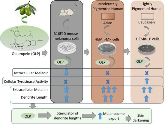
Background/Objectives: Oleuropein (OLP), the key bioactive in olive leaf extracts, has demonstrated various biological benefits. We previously reported on the pro-melanogenic action with increased dendricity of a patented olive leaf extract (Benolea®) that was standardized to 16–24% OLP. In this study, purified OLP was evaluated to identify if it might be the bioactive responsible for the stimulating effects on melanocytes. Moreover, previous studies on OLP have never reported the effects on melanocyte dendricity or melanin export in the medium. Methods: Herein, the effect of OLP on melanogenesis was first evaluated using the B16F10 cell model and validated using the physiological model of normal human melanocytes from Caucasian (lightly pigmented; LP) and Asian (moderately pigmented; MP) skin. The effects of OLP on melanin export in LP and MP cells were indirectly evaluated by dendricity indices. Results: OLP lowered the intracellular melanin content in B16F10 cells by 26.36%, 24.48%, and 27.71% at 100, 150, and 200 µg/mL (all p < 0.01), respectively, with no effect on the intracellular melanin contents of LP or MP cells. OLP treatment did not influence tyrosinase activity in B16F10 cells or MP cells but significantly enhanced the activity in LP cells. The measurement of extracellular melanin showed significantly higher levels for all three cells, although the levels were considerably higher in MP cells, after the adjustment for OLP autoxidation observed in the cell-free system, which caused melanin-like brown coloration. Furthermore, OLP induced morphological alterations of extended dendrites of B16F10 cells that were retained in LP and MP cells. The quantitation of the dendricity of cells treated with OLP at 200 μg/mL revealed that the total dendrite length was increased by 35.24% (p < 0.05) in LP cells and by 58.45% (p < 0.001) in MP cells without any change in the dendrite number. Conclusions: This is the first study to demonstrate the novel finding that OLP possesses a hitherto unreported unique capacity to stimulate melanocyte dendricity, hence establishing the efficacy for use in increasing human pigmentation. Our findings show significance, with a potential application of the compound OLP for addressing human hypopigmentation disorders in clinical settings or for cosmetic uses related to sunless tanning.

Graphical Abstract

1. Introduction

Melanin is a polymeric pigment that plays a crucial role in determining the coloration of skin and hair. It is produced in melanosomes within specialized cells known as melanocytes and transferred to adjacent keratinocytes via dendrites [1,2]. An insufficient synthesis or export of melanin can cause hypopigmentation disorders, which comprise leukoderma, vitiligo, post-burn hypopigmentation [3], and post-inflammatory hypopigmentation [4], that are marked by light-colored patches, producing uneven skin tone [5]. For many fair-skinned Westerners, a tanned complexion is seen as esthetically desirable and contributes to a psychological sense of well-being. The presence of these hypopigmentation disorders not only results in esthetic concerns but can also contribute to psychological distress and a decline in the overall quality of life [6]. Current strategies to increase skin pigmentation through alternatives to natural sun exposure include chemical tanning, most often produced by the application of dihydroxyacetone (DHA), an active ingredient used as a browning agent in spray-on tanning lotions, or indoor tanning achieved by regular exposure to artificial UV light sources [7]. However, both the use of DHA and indoor tanning have disadvantages associated with an increased risk of DNA damage [8] and skin cancer [9]. For the purpose of stimulating pigmentation, it is desired to have compounds that can target subsequent phases in the melanin formation pathway as the melanin synthesized within melanocytes is exported via dendrites to neighboring keratinocytes [10]. The dendrites of melanocytes serve as crucial structural elements for the effective export of melanosomes to keratinocytes, as a single melanocyte in the skin interacts with approximately 30 to 40 neighboring keratinocytes via its dendrites within the epidermis [11]. Furthermore, within the hair structure, the proportion of melanocytes to keratinocytes is 1:1 in the basal layer, situated close to the dermal papilla, while throughout the entire hair bulb, this ratio shifts to 1:5 [12,13]. Melanocytes increase their dendritic numbers to engage with multiple nearby keratinocytes, while also elongating their dendrites to convey pigment to various keratinocytes located in the suprabasal layers of the skin or hair. The enhancement of dendricity as an indicator of melanosome export stimulation is becoming a promising focus in developing pigmentation stimulators aimed at treating hypopigmentation. Multiple earlier investigations [14,15,16,17,18,19], along with our prior studies [20,21,22], have revealed various compounds that exhibit promise for addressing hypopigmentation through the promotion of melanosome export by enhancing the dendricity of melanocytes.
Oleuropein (OLP) is a naturally occurring glycoside that is most abundant in leaves of the olive (Olea europaea L.) plant compared to the pressed oil or whole fruit [23] and is accountable for the acrid flavor of olives [24]. OLP is categorized within the secoiridoids class of phenolic compounds, consisting of three structural units: elenolic acid, hydroxytyrosol, and a glucose moiety [25]. The beneficial effects of OLP have been extensively reviewed [26]. OLP has demonstrated multiple pharmacological benefits, such as anticancer activity [27,28], antiaging activity [29], antioxidant activity [30], anti-psoriatic activity [31], antifungal activity [32], antifibrotic activity [33], and neuroprotective activity [34]. Furthermore, OLP was shown to promote healing in aged mice [35] and a burn wound model [36], possess anti-inflammatory activity in an atopic dermatitis model [37], stimulate hair growth in mice [38], and diminish skin injury and cancer due to chronic UVB radiation in mice [39]. OLP-rich olive leaf extracts included in hydrogel face masks showed efficacy in mitigating the damaging impact of UVA radiation on the skin [40]. Natural ingredients and plant-based extracts have been utilized to address hypopigmentation concerns. Repigmentation therapies utilize UV alongside psoralen compounds derived from the Psoralen corylifolia L. plant, including 8-methoxypsoralen and 5-methoxypsoralen [41]. These compounds have the potential to stimulate the production of melanin; however, they are not without adverse effects of acute hepatitis and erythema and can also contribute to skin cancer [42,43]. Therefore, evaluating novel plant-derived compounds that exhibit enhanced melanogenesis activity is essential, as well as minimizing toxicity.
Our previous study [22] reported the pro-melanogenic effect of a patented dry olive leaf extract (DOLE; Benolea®) standardized to 16–24% OLP. However, other studies have reported the anti-melanogenic capacity of olive leaf extracts that were standardized to 12% OLP [44] or 15% OLP [39]. So far, the impact of purified OLP on melanogenesis has yielded inconsistent findings, with some studies indicating pro-melanogenic effects while others suggest anti-melanogenic effects that were dependent on cell type and OLP concentration. For example, a recent study [45] demonstrated that OLP at a remarkably low concentration of 2 µM increased melanin production in primary human melanocytes and OLP at 5 µM increased tyrosinase, TRP-1, and TRP-2 gene expression in B16F10 cells following a 48 h treatment. Nonetheless, another recent study [46] revealed that OLP at concentrations of 3 mM or higher suppressed tyrosinase activity and melanin synthesis in B16 cells and a zebrafish model. OLP at 350 μg/mL (~648 µM) suppressed melanin content by 41.6% in B16 melanoma cells, though the specific subtype of B16 cells was not detailed [47]. Moreover, in all the aforementioned reports, only the effects of OLP on melanin production within cells were reported, while the effects of OLP on dendricity-related melanin export were not investigated. In our earlier research [22], although we did not examine purified OLP concurrently, our findings on a patented dry olive leaf extract (DOLE) standardized to 16–24% OLP demonstrated that this extract promoted extracellular melanin production and dendricity without influencing intracellular melanin levels in B16F10 cells and primary human melanocytes from the skin of Caucasian (HEMn-LP) and Asian (HEMn-MP) people. Herein, we have expanded our studies to investigate the effects of purified OLP on melanogenesis using B16F10 cells, HEMn-LP, and HEMn-MP cells. Compared to prior reports on OLP, our study is novel as it examines the effects of purified OLP on melanin production as well as export via dendritic changes across a concentration spectrum of 10–200 µg/mL (equivalent to 19–370 µM) in primary human melanocytes from two different ethnicities (Asian and Caucasian) that, to date, has not been investigated. Our findings reveal that OLP potently stimulates melanosome export in B16F10 cells. We have further confirmed our findings in normal human melanocytes from moderately and lightly pigmented skin, representing Caucasian and Asian phenotypes.

2. Materials and Methods

2.1. Materials

Oleuropein (OLP) with a percentage purity of 98% (as provided by the supplier), L-3,4-dihydroxyphenylalanine (L-DOPA), and mushroom tyrosinase were purchased from Sigma-Aldrich (St. Louis, MO, USA). The OLP powder was reconstituted using sterile Hank’s buffered salt saline (HBSS) and stored at −20 °C until use. Dulbecco’s modified Eagle medium (DMEM), antibiotic cocktail (penicillin–streptomycin), and TrypLE Express enzyme (1×) were purchased from Gibco (Thermo Fisher Scientific, Waltham, MA, USA). Heat-inactivated fetal bovine serum (FBS) was procured from R&D Systems Inc. (Minneapolis, MN, USA). The bicinchoninic acid (BCA) assay kit, melanocyte basal medium 254, and growth supplement (HMGS) were acquired from Thermo Fisher Scientific Inc.

2.2. Cell Culture

B16F10 mouse melanoma cells were obtained from ATCC (Manassas, VA, USA) and cultured using DMEM supplemented with 10% heat-inactivated FBS and 1% antibiotics (penicillin–streptomycin). Primary human epidermal melanocytes (HEMn) from moderately pigmented (MP) and lightly pigmented (LP) neonatal foreskin donors were purchased from Thermo Fisher Scientific Inc. (Cascade Biologics™, Portland, OR, USA) and cultivated using medium 254 supplemented with 1% HMGS and 1% antibiotics. All cells were cultivated in a humidified incubator at 37 °C with 5% CO2.

2.3. MTS Cytotoxicity Assay

Various concentrations of OLP were initially tested to identify noncytotoxic concentration ranges before proceeding with the melanogenesis assays. Briefly, 1 × 10⁴ B16F10 cells were inoculated in each well of a 96-well plate and grown for 24 h, after which the culture medium was changed to a new medium that contained OLP at concentration ranges of 10–200 µg/mL, and the cells were maintained for a duration of 48 h. To evaluate the cytotoxicity of OLP on primary melanocytes, 2 × 10⁴ HEMn-LP or HEMn-MP cells per well were cultivated in a 96-well plate for 24 h, following which OLP was introduced at varying concentrations, and the cultures were sustained for an additional 48 h. Upon completion of the treatments, the medium was replaced with 100 μL of new medium with 20 μL of MTS reagent. The 96-well culture plate was incubated for 40 min (for B16F10 cells) or 90 min (for HEMn cells), after which the optical density of the aliquots was recorded at 490 nm using a Versamax® microplate reader (Molecular Devices, LLC.: San Jose, CA, USA).

2.4. Extracellular and Intracellular Melanin Assay

We measured the effects of OLP on the intracellular levels and the levels released into the culture medium (extracellular) using the experimental method described in our earlier study [22]. Briefly, 1 × 10⁵ B16F10 cells were plated in each well of a 12-well plate, and varying concentrations of OLP were added after 24 h. Subsequently, the cell monolayers were maintained for 48 h. In experiments with primary human melanocytes, 1.5 × 10⁵ HEMn-MP cells/well or 2.3 × 10⁵ HEMn-LP cells/well were similarly cultured and treated with OLP. Subsequently, the culture medium was centrifuged, and the absorbance was measured at 475 nm for the extracellular melanin. In the absence of cells, OLP was found to undergo autoxidation, resulting in the formation of melanin-like brown particles. Consequently, the absorbances of the culture medium with varying concentrations of OLP for B16F10 cells (Figure S1A) and HEMn cells (Figure S1B) in a cell-free system were also estimated and subtracted from the extracellular melanin absorbances in the cellular system to correct for the autoxidation (Supplementary method). These results of adjusted extracellular melanin were displayed as a percentage of the control group. For the determination of intracellular melanin, the cells in the wells underwent the solubilization procedure using the hot NaOH lysis method. The intracellular and extracellular melanin absorbances were adjusted to the total protein content and presented as relative melanin as a percentage of the control.

2.5. Cellular Tyrosinase Activity

Following a 48 h treatment with a range of OLP concentrations, the tyrosinase activity of B16F10 cells, HEMn-LP cells, and HEMn-MP cells was measured in accordance with the method described in our earlier study [22]. Briefly, cell lysate supernatants were mixed with DOPA substrate and measured kinetically at 475 nm. Values were adjusted to the protein content and reported as a percentage of the control group.

2.6. Cell-Free Tyrosinase Activity

A purified mushroom tyrosinase enzyme with L-DOPA substrate was used to examine OLP’s direct effects on enzyme activity using the method outlined in our prior report [48]. Briefly, 80 µL of OLP at various concentrations was dispensed to 96-well plates. Next, 100 µL of L-DOPA substrate solution and 20 µL of enzyme were added. A final substrate concentration of 3 mM and enzyme concentration of 3.5 µg/mL were achieved in the well. Absorbance at 475 nm was measured every 30 s for 30 min, and the kinetic slopes were used to compare the tyrosinase activity to the control.

2.7. Quantitation of Dendricity in Human Melanocytes

The experimental details of the culture of HEMn-LP and HEMn-MP cells, along with the corresponding measurements of dendricity indices—total dendrite length, dendrite number, and average dendrite length—were elucidated after a 48 h treatment with OLP, following the methodology outlined in our previous study [22]. All the dendrites of a cell were traced from the cell center to the tip and added to calculate the total dendrite length with the use of image analysis software (NIS Elements 5.0). Dendrites per cell were recorded manually from images. The total dendritic length divided by the number of dendrites yielded the average dendrite length. The folders of raw images from each well of the two groups (control and OLP-200 µg/mL) were analyzed in a blinded manner.

2.8. Statistical Analysis

The data for two groups were compared using Student s t-test, whereas for the comparison of multiple groups (>2), a one-way analysis of variance (ANOVA) with Dunnett’s post hoc test was used. All the analyses were performed using GraphPad Prism software (version 10.0, San Diego, CA, USA). Statistical significance was established at p < 0.05. All data are presented as mean ± SD.

3. Results

3.1. Effects of OLP in B16F10 Cells

OLP (structure in Figure 1A) over the concentration range 10–200 μg/mL was noncytotoxic to B16F10 cells; the values of viability of B16F10 cells after treatment with OLP at 0, 10, 25, 50, 100, 150, and 200 µg/mL were 100.27 ± 0.17%, 103.94 ± 5.97%, 98.48 ± 5.03%, 97.43 ± 2.66%, 98.74 ± 9.94%, 104.37 ± 5.68%, and 107.69 ± 7.56%, respectively, and not significantly different from the control (Figure 1B). Accordingly, these concentrations were selected in subsequent experiments.
The B16F10 cells after the OLP treatment showed a lighter color at OLP concentrations ≥ 100 µg/mL (photo panel; Figure 2A). This was confirmed by the spectrophotometric method, where intracellular melanin was significantly lowered by 26.36%, 24.48%, and 27.71% at OLP concentrations of 100, 150, and 200 µg/mL, respectively (Figure 2A). However, the cell culture medium after treatment with OLP at higher concentration ranges was visibly darker than that of the untreated control (photo panel, Figure 2B). The results of the spectrophotometric estimation of extracellular melanin confirmed that OLP increased the release of melanin into the culture medium (photo panel; Figure 2B) with significant increases of 33%, 36.45%, and 38.03% noted for OLP concentrations of 50, 100, and 200 μg/mL, respectively (Figure 2B). The photomicrographs showed elongated dendrites of B16F10 cells that were treated with higher concentrations of 100–200 µg/mL OLP (Figure 2C). Collectively, the findings suggest that OLP facilitated the transfer of melanin from cells into the surrounding medium.
The results of the effect of OLP on tyrosinase enzyme activity showed that OLP did not affect cellular tyrosinase activity significantly at any concentration (Figure 3A) but increased cell-free tyrosinase activity for the concentration range 25–200 µg/mL; significant increases of 15.71%, 19.19%, 26.24%, 24.34%, and 23.67% were obtained at concentrations of 25, 50, 100, 150, and 200 µg/mL, respectively (Figure 3B). This result confirms that OLP has a direct stimulatory effect on the activity of tyrosinase enzyme.

3.2. Effects of OLP in HEMn-LP Cells

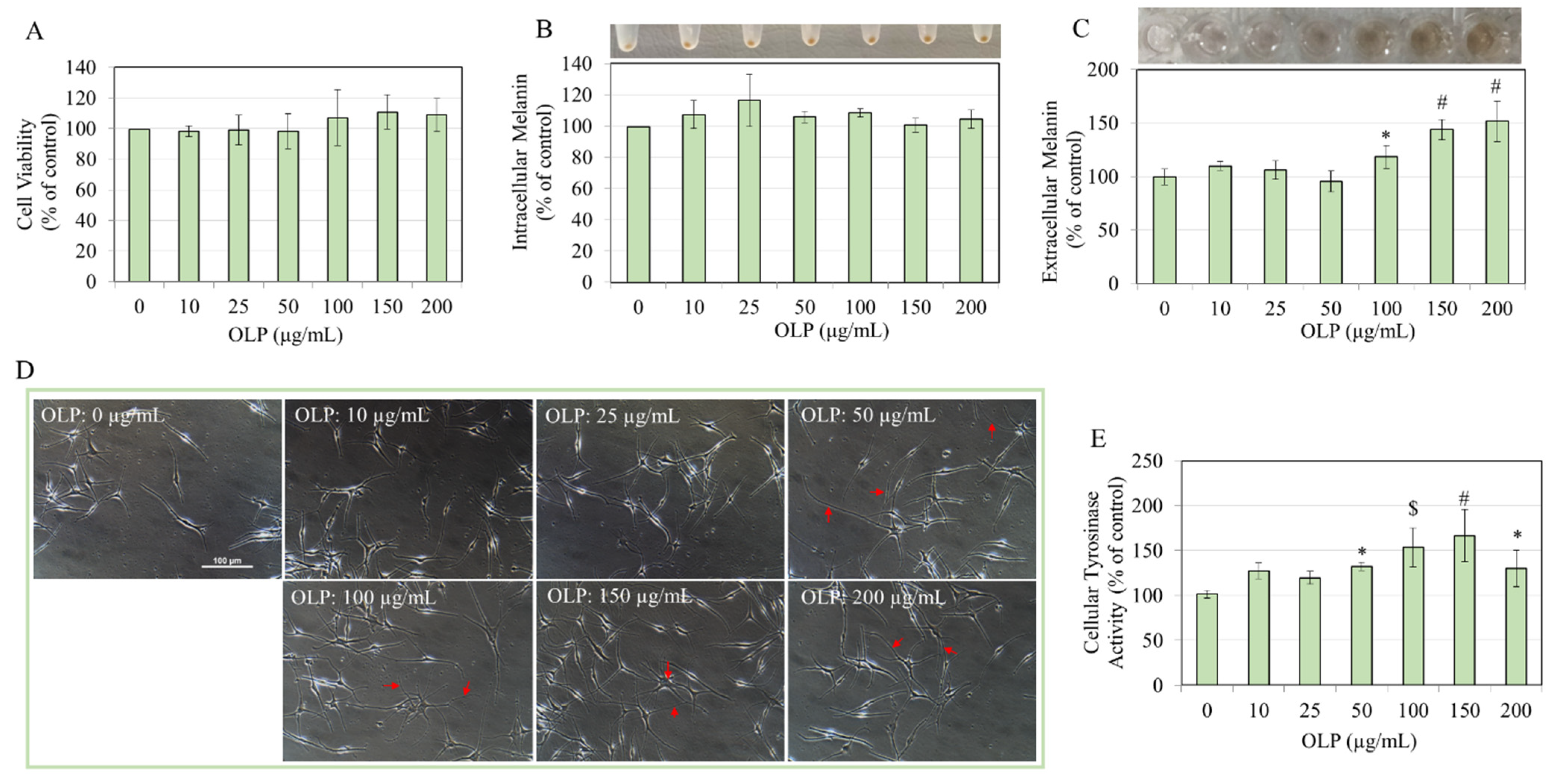
Similarly to B16F10 cells, OLP was noncytotoxic to HEMn-LP cells over the entire concentration range between 10 and 200 µg/mL (Figure 4A). LP cells treated with OLP showed no change in intracellular melanin levels at any concentration, as could be seen visually or after the quantitation of the melanin contents (Figure 4B).
The culture media of cells treated with OLP at increasing concentrations were much darker than the untreated control (panel, Figure 4C). Estimating the relative extracellular melanin levels corroborated our visual findings as we obtained a significantly robust enhancement of extracellular melanin with increases of 18.81%, 44.36%, and 52.04% at OLP concentrations of 100, 150, and 200 µg/mL, respectively (Figure 4C).
The morphology of LP cells after treatment with various concentrations of OLP showed a marked distinction from the untreated control group; OLP-treated cells showed extended dendrites that were visible at OLP concentrations ≥ 50 µg/mL onwards (Figure 4D). Interestingly, the effect of the OLP treatment on the tyrosinase activity of LP cells showed a stimulatory effect, with the activity significantly increased by 30.63%, 52.15%, 65.22%, and 28.90% at OLP concentrations of 50, 100, 150, and 200 µg/mL, respectively (Figure 4E).
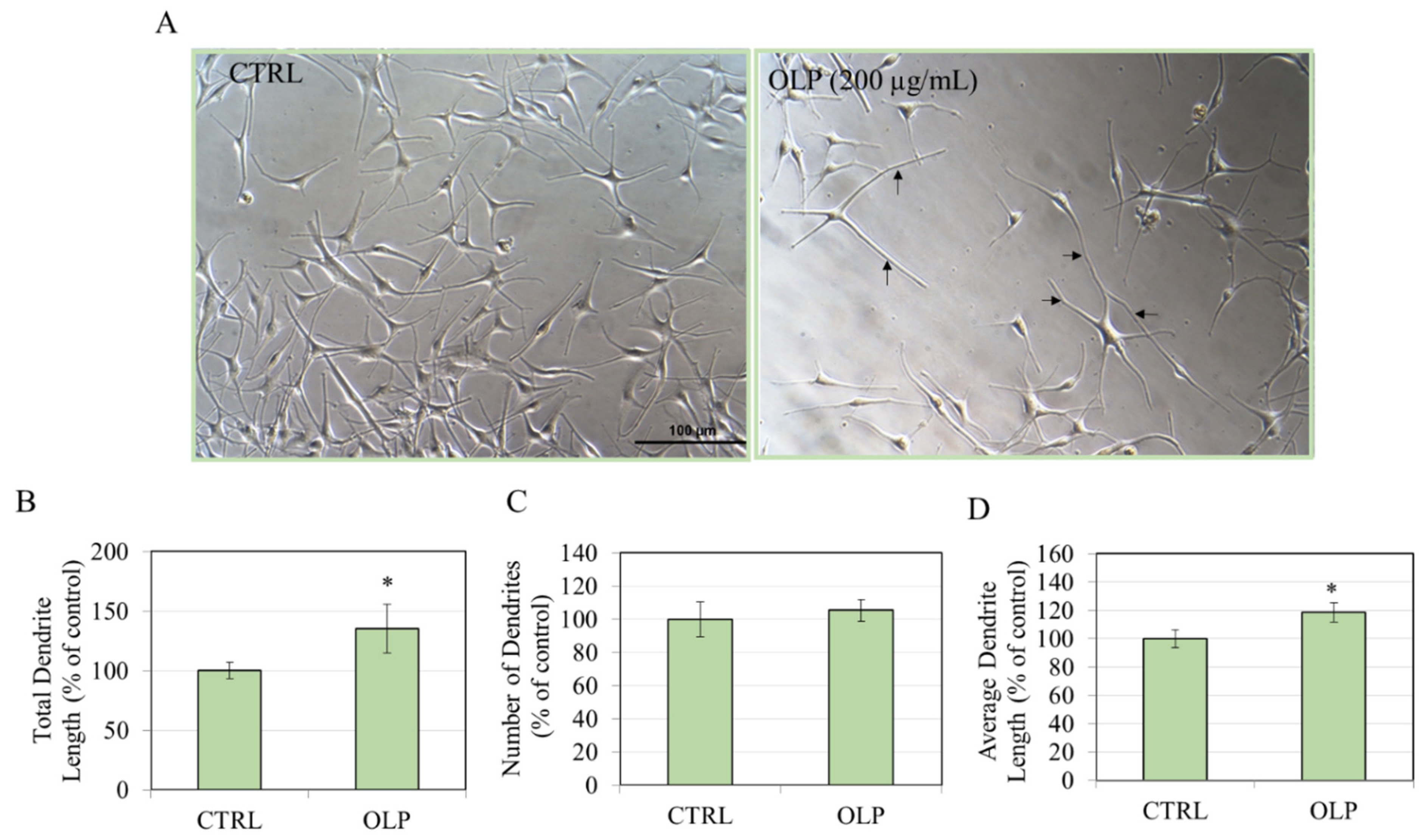
Given that OLP at 200 µg/mL exhibited the greatest effect on extracellular melanin, we quantified the dendricity of cells at this concentration. In particular, the lengths and numbers of melanocyte dendrites, which signify cellular processes involved in the export of melanosomes, were measured. Cells treated with OLP at 200 µg/mL exhibited dramatically extended dendrites (Figure 5A) that was corroborated by the quantitation; a significant increase in the total dendrite length by 35.24% was achieved (Figure 5B) compared to the control, although there was no increase in the dendrite number (Figure 5C). The average dendrite length was significantly increased by 18.62% (Figure 5D).

3.3. Effects of OLP in HEMn-MP Cells

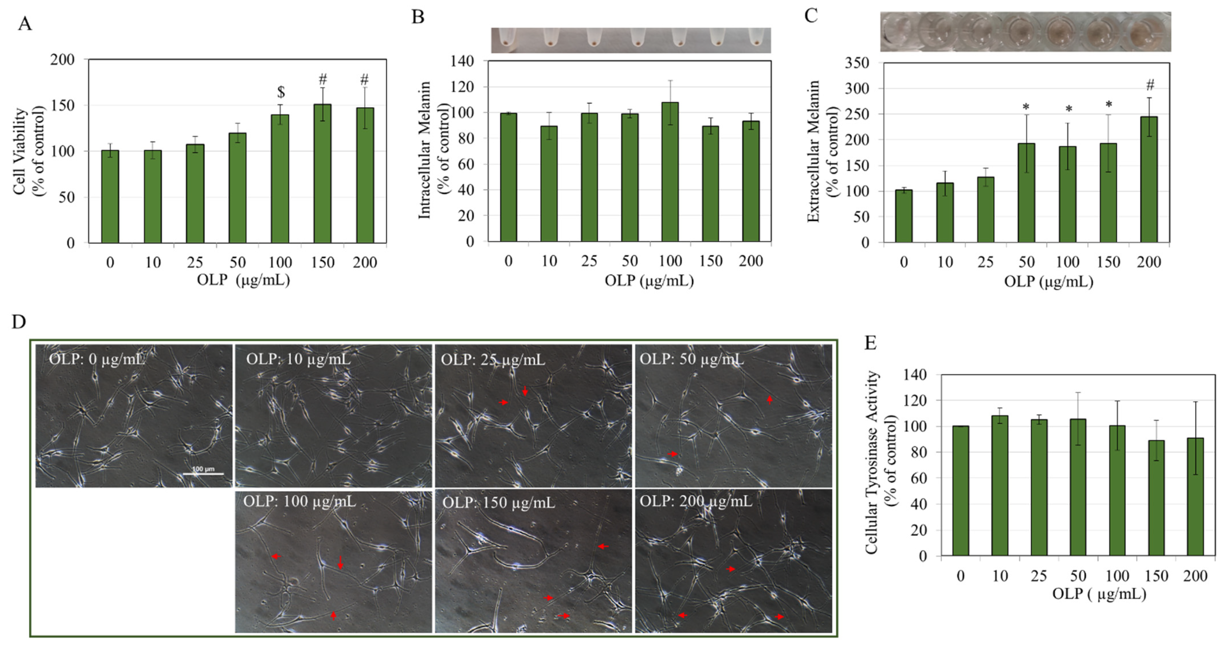
OLP continued to show no cytotoxicity even to HEMn-MP cells over the concentration range of 10–200 μg/mL for the 48 h duration (Figure 6A). Interestingly, the cell viability was significantly enhanced by 39.12%, 50.31%, and 46.43% at OLP concentrations of 100, 150, and 200 µg/mL, respectively (Figure 6A). Like LP cells, OLP did not affect cellular melanin levels at any concentration, as seen by the pellets and subsequent melanin quantification (Figure 6B). Consistent with our results on LP cells, the culture medium of MP cells was darker after treatment with OLP at elevated doses, suggesting melanin release into the medium (panel, Figure 6C). OLP significantly increased extracellular melanin by 91.25%, 86.03%, 91.77%, and 143.16% at 50, 100, 150, and 200 µg/mL, respectively. Morphological alterations resulting from OLP treatment were apparent in MP cells starting from the concentration of 25 µg/mL onwards, exhibiting significantly elongated dendrites compared to the control group (Figure 6D). The effects of OLP on tyrosinase activity in cellular lysates of MP cells did not demonstrate significant differences relative to the untreated control (Figure 6E).
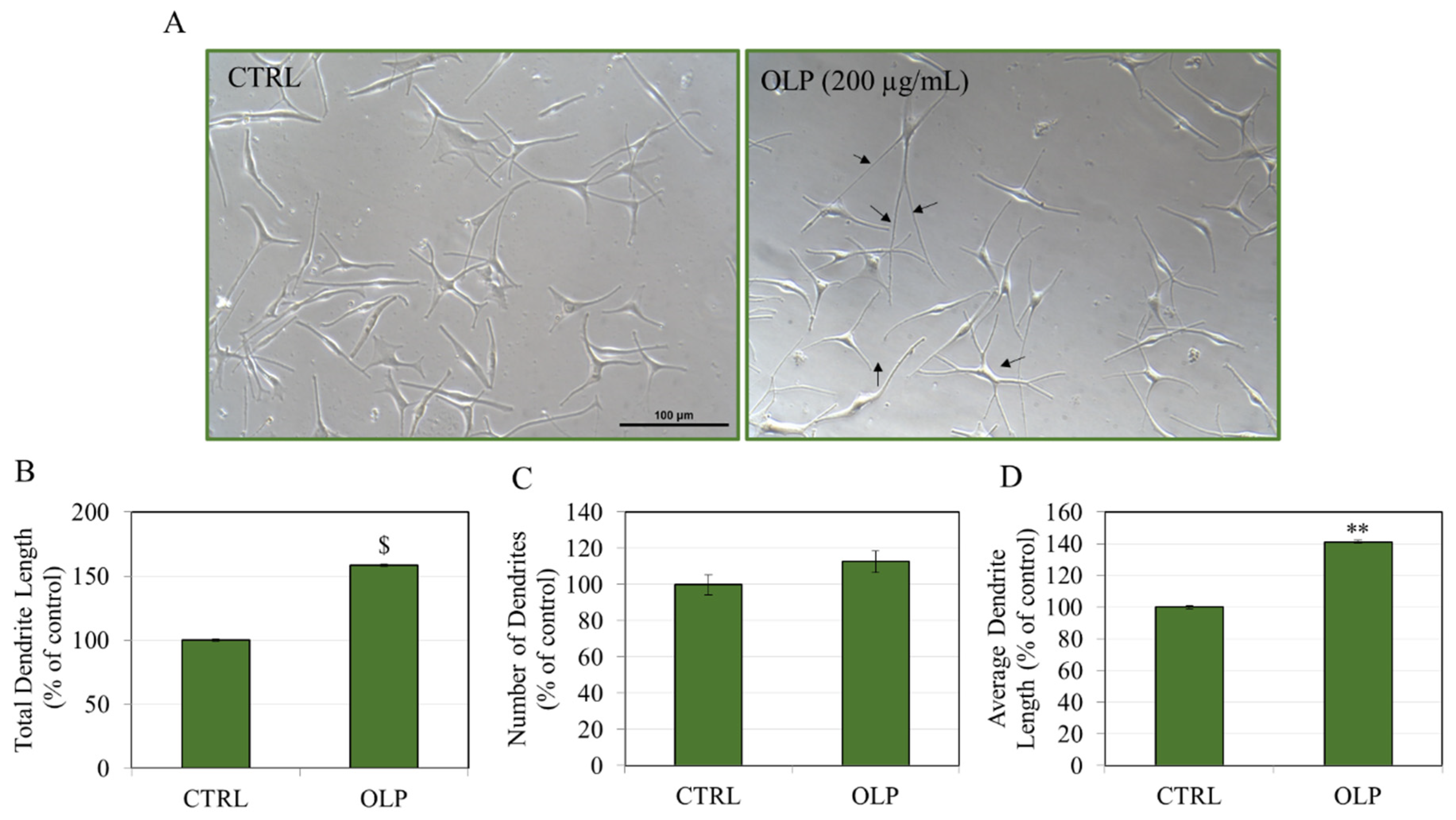
MP cells subjected to OLP at a concentration of 200 µg/mL displayed highly elongated dendrites, a characteristic absent in the control group (Figure 7A). The quantification of dendricity indices showed that OLP at 200 µg/mL exhibited a significant elongation of the total dendrite lengths of HEMn-MP cells by 58.45% (Figure 7B), without altering the dendrite numbers (Figure 7C). The average dendrite length was significantly increased by 41.08% (Figure 7D). These results suggest that OLP exerts a greater impact on stimulating dendricity in MP cells than LP cells. Moreover, it only stimulates dendritic elongation without influencing dendrite number.
Taken together, these results reveal that OLP had no effect on the intracellular melanin of LP or MP cells but concurrently elevated the extracellular melanin in both cells, with higher levels in MP cells. The tyrosinase activity was not altered by OLP treatment in MP cells but significantly enhanced in LP cells. The efficiency of OLP in stimulating extracellular melanin and increasing dendritic lengths was superior in MP melanocytes compared to LP melanocytes. The exact reason for this phenomenon is currently unknown, and it remains unclear whether it is a donor-dependent occurrence, arises from the greater melanin content and tyrosinase activity of MP cells relative to LP cells, or is associated with our utilization of a lower cell density for MP cells in comparison to LP cells.

4. Discussion

The occurrence of the browning of olive fruits upon harvest due to wounding from bark is a well-known occurrence [49]. Since OLP has phenolic dihydroxy groups similar to catechol, it can be oxidized, and such a reaction to induce browning has been reported before for secoiridoids [50]. Several previous studies have shown that OLP is readily oxidized to OLP quinone brown products by both enzymatic and non-enzymatic processes [49,51,52]. Notably, we also found that OLP induced a brown coloring in the culture medium in the absence of cells, attributed to the formation of melanin-like OLP dimers resulting from its autoxidation in the presence of air. Consequently, the extracellular melanin results were corrected for OLP’s autoxidation and still indicated significantly elevated levels of extracellular melanin, confirming that OLP promotes the release of melanosomes from melanocyte dendrites into the culture medium without inducing toxicity. Moreover, the results revealed a higher autoxidation of OLP in the HEMn culture medium, which contains only 0.5% serum (Figure S1A), as opposed to the B16F10 DMEM culture medium that contains 10% serum, indicating that the presence of serum may decrease the rate of autoxidation (Figure S1B). Our results of increased tyrosinase activity under the DOPA substrate confirm that OLP can directly stimulate the tyrosinase activity. Prior research on the impact of OLP on melanogenesis has shown concentration-dependent contradictory outcomes [45,46,47]. For instance, OLP at a concentration of 350 μg/mL (~648 µM) led to a 41.6% decrease in melanin content in B16 cells [47], while in another study, OLP at much higher concentrations ≥3 mM suppressed tyrosinase activity and intracellular melanin production in B16 cells and zebrafish, which was correlated to the decrease in the levels of melanogenesis cascade proteins (TRP-1, TRP-2, and MITF) [46]. However, in a separate report [45], OLP at low concentration of 5 µM enhanced the gene expression of tyrosinase, TRP-1, and TRP-2 in B16F10 cells, whereas OLP at 2 µM augmented melanin production in primary human melanocytes, correlating with increased protein levels and gene expression of TRP-1, TRP-2, and MITF. Furthermore, the studies previously mentioned focused exclusively on the impact of OLP on cellular melanin production, overlooking the critical factor of dendricity-related melanin export. We noted that B16F10 melanoma cells treated with OLP concentrations of 100, 150, and 200 µg/mL had intracellular melanin suppressions of 26.36%, 24.48%, and 27.71%, respectively. These results of diminished intracellular melanin are in a similar concentration range to a prior study [47] that also showed that OLP, albeit at a higher concentration of 350 μg/mL, suppressed melanin content by 41.6% in the absence of any cytotoxicity in B16 melanoma cells, although the subtype of B16 cells was not specified. Moreover, this study [47] did not report the effects of OLP on extracellular melanin in B16 cells. Our study showed that in addition to diminishing intracellular melanin, OLP significantly stimulates extracellular melanin, which confirms that the action of OLP is that of a pro-melanogenic compound. To evaluate the influence of a substance on melanin levels, it is imperative to examine both the production of melanin and its secretion. For example, our findings in B16F10 cells indicated that OLP at concentrations ≥100 μg/mL suppressed intracellular melanin levels while simultaneously increasing extracellular levels, thereby confirming that OLP predominantly promotes melanin export. Furthermore, in HEMn-LP cells and HEMn-MP cells, OLP at concentrations ≥100 μg/mL and ≥50 μg/mL, respectively, increased extracellular melanin levels without any change in intracellular melanin. The impact of a 150 µg/mL OLP concentration on B16F10 cells’ extracellular melanin levels exhibited a non-linear response in comparison to the concentrations of 100 and 200 µg/mL. Currently, the explanation behind this response remains unclear; however, B16F10 is an immortalized mouse melanoma cell line, and a comparable non-linear effect at a specific concentration was also observed in our prior study [53], wherein an analog, demethoxycalebin-A1, of the natural compound calebin A, repressed cellular melanin non-linearly as concentrations increased in B16F10 cells.
Our results of OLP bear a similarity to the action of methylquercetin derivatives that were shown to enhance dendrite formation without influencing melanin production in Caucasian melanocytes, although the authors did not provide data on extracellular melanin [54]. Interestingly, these derivatives showed the enhancement of extracellular melanin in B16F10 mouse cells in a previous study by the same authors [17]. In addition, our results of increased dendricity by OLP also exhibit a striking similarity to the results of a prior study, which reported increased melanocyte dendricity in the absence of increased melanin synthesis by analogs of a natural molecule, piperine [19]. The distinction between our results on OLP and earlier studies on piperine’s effects on pigmentation merits consideration. The authors showed that piperine and its analogs at a concentration of 10 µM stimulated pigmentation by solely increasing melanocyte proliferation in Melan-a cells, although the intracellular melanin was unaffected, but cells exhibited enlarged dendrite lengths as well as the number of dendrites [19]. Our results obtained in primary human melanocytes showed a similar profile, except OLP did not have any appreciable effect on the proliferation of LP cells (although it increased the proliferation of MP cells), and the concentration at which we obtained these effects is considerably higher; OLP at a concentration of 100 µg/mL (185 µM) onwards stimulated dendricity with no effects at concentrations lower than that. Moreover, OLP selectively increases dendrite lengths without affecting the number of dendrites per cell. Melan-a cells are immortalized cells and hence show intrinsic differences from normal melanocytes. Piperine was shown to stimulate pigmentation better than UVR alone, and the combination of both elicited an even pigmentation pattern in a sparsely pigmented mice strain [55], although the authors discussed the limitation of the photostability of piperine as it underwent photoisomerization upon UVA irradiation, leading to the loss of melanocyte stimulatory activity [56]. Based on this limitation, the use of piperine was recommended before the application of UVA to avoid light exposure. A prior study [57] showed that OLP could decompose upon irradiation with UVC light into elenolic acid and hydroxytyrosol, although the stability of OLP under UVA light has not been documented yet. Studies to explore the photostability of OLP and whether its effects on melanocyte dendricity are retained post-UVA irradiation are worthy of further investigation.
Our research has demonstrated that OLP uniquely stimulates dendrite length, which partially accounts for the observed increase in melanin export in MP and LP cultures. A scheme illustrating this is shown in Figure 8. Substances that trigger toxicity in primary melanocytes may also lead to an elevation in extracellular melanin; however, OLP exhibited no cytotoxic effects at the concentrations that prompted an increase in extracellular melanin. This discovery reveals that OLP enhances the transport of melanin through the elongation of dendrites while leaving the intracellular melanin levels unchanged. Furthermore, our results of the stimulation of melanosome export by OLP in LP cells are an added advantage since fair skin is typically known to exhibit a reduced capacity to tan based on the Fitzpatrick skin type classification (skin types I and II) [58]. EPI64 is a GTPase-activating protein that inactivates Rab27A. Rab27A is a protein found on mature melanosomes that regulates the actin-based transport of melanosomes [59,60]. Slp-2a is a Rab27A-binding protein that participates in cell elongation via its interaction with the plasma membrane and melanosomes [61]. The vacuolar protein sorting 9-ankyrin-repeat protein (VARP) is essential for dendritic elongation and the promotion of enzyme trafficking in melanogenesis [62,63]. A previous study [17] indicated that the extracellular melanin-enhancing and dendricity-stimulating activities of methyl quercetin derivatives were facilitated via the inhibition of the EPI64 protein. Another compound, kaempferol, was shown to enhance dendritic lengths in B16F10 cells, with the authors noting that it facilitated the actin-based movement of melanosomes by increasing the expression of Rab27A, RhoA, and Cdc42 proteins in B16F10 cells [15]. Olive leaf extract at 100 µg/mL and 1000 µg/mL concentrations increased the rate of actin polymerization in renal cells, therefore modifying cytoskeletal remodeling [64]. We propose that the process by which OLP enhances the elongation of cell dendrite lengths may be attributed to an increased expression or activation of proteins such as Slac-2a, VARP, Rab27A, RhoA, or Cdc42, or alternatively, to the downregulation of EPI64 expression. Additional investigation is required to clarify these hypotheses.
Our findings of diminished intracellular melanin in B16F10 melanoma cells are in marked contrast to human melanocytes, where OLP did not affect intracellular melanin at any concentration. Nevertheless, in both cells, OLP stimulated extracellular melanin and dendricity. The differences between species (mouse vs. human) and phenotype (cancer cells vs. normal cells) might account for the differences in intracellular melanin levels for OLP treatments in normal human melanocytes and B16F10 mouse melanoma cells. The comparison of the results of olive leaf extract (DOLE) standardized to 16–24% OLP in our previous study [22] and the results of purified OLP of this study is as follows. In our earlier study [22], DOLE showed no effects on intracellular melanin in B16F10 cells over the concentration range of 10–200 µg/mL but stimulated extracellular melanin by 56.70%, 45.53%, and 59.70% at concentrations of 100, 150, and 200 µg/mL, respectively. Considering a mean OLP content of 20% in DOLE, the equivalent amounts of OLP in DOLE concentrations of 100, 150, and 200 µg/mL will be 20, 30, and 40 µg/mL, respectively. Based on the results of our current study in B16F10 cells, OLP lowered intracellular melanin at concentrations ≥100 µg/mL and stimulated extracellular melanin by 33%, 36.45%, and 59.70% only at 50, 100, and 200 µg/mL, respectively. Therefore, it can be concluded that the higher amounts of extracellular melanin in DOLE did not originate from OLP since purified OLP at those equivalent concentrations did not alter extracellular melanin, as noted in the current study. Furthermore, in HEMn-LP cells, the comparison of the results of DOLE from our prior study [22] and the current study shows that DOLE enhanced extracellular melanin in HEMn-LP cells by 49.97% and 66.30% at DOLE concentrations of 150 and 200 µg/mL (equivalent to 30 and 40 µg/mL OLP), but OLP only stimulated extracellular melanin at concentrations ≥100 µg/mL, as seen in this study. Moreover, in HEMn-MP cells, the comparison of the results of DOLE from our prior study [22] and the current study shows that DOLE enhanced extracellular melanin in HEMn-MP cells by 121.65% and 112.37% at DOLE concentrations of 150 and 200 µg/mL (equivalent to 30 and 40 µg/mL OLP), but OLP stimulated extracellular melanin by 91.25%, 86.03%, 91.77%, and 143.16% at OLP concentrations of 50, 100, 150, and 200 µg/mL, respectively. This shows that the effects of the extract compared to purified OLP at the same concentration on the extracellular melanin are greater, suggestive of the synergistic effects of other constituents present in the extract. Intriguingly, the stimulation of dendrite lengths by DOLE, as observed in our previous study, demonstrated a greater impact on dendricity and extracellular melanin in HEMn-MP cells when contrasted with HEMn-LP cells. This finding parallels the OLP dendricity data in the current study; however, direct comparison is not feasible. In our prior research, increased dendrite lengths were quantified for DOLE exclusively at 200 µg/mL (~40 µg/mL OLP), whereas in this study, dendricity was quantified solely at the maximum OLP concentration of 200 µg/mL. Nonetheless, the examination of HEMn cell micrographs (Figure 4D and Figure 6D) does reveal that OLP can stimulate dendrite lengths at concentrations of ≥25 µg/mL, thus suggesting that OLP in the extract does contribute to the unique ability to result in the elongation of dendrites.
OLP significantly increased tyrosinase activity in LP cells while having no effect on MP cells at the same concentrations. This effect aligns with the results from our earlier study [22], in which OLP-containing DOLE showed differing results: an elevation in tyrosinase activity in LP cells at a DOLE concentration of 200 µg/mL, while MP cells exhibited an inhibitory response at the same concentration. The explanation for the biphasic response of OLP on cell tyrosinase activity likely arises from the specific effects of OLP related to the type of cells, which depend on the baseline tyrosinase activities present in those cells. LP cells demonstrate a 5.65-fold lower basal tyrosinase activity when compared to MP cells (Figure S2). As a result, OLP, shown to directly boost tyrosinase activity in a cell-free setting, could potentially have a more significant impact on LP cells. On the other hand, given that MP cells inherently exhibit elevated tyrosinase activity, the potential impact of OLP in further enhancing that activity may be constrained. Moreover, analyzing the expression levels of both protein and mRNA for tyrosinase could provide valuable insights into this phenomenon.
The influence of OLP on the melanogenesis process was also investigated using a contact coculture model that mimicked in vivo keratinocyte–melanocyte communication [65,66]. Similarly, OLP at 200 µg/mL was evaluated for any cytotoxicity using the human keratinocytes (HaCaT cells). The values of HaCaT cell viabilities after treatment with OLP at 0, 10, 25, 50, 100, 150, and 200 µg/mL were 103.26 ± 8.35%, 102.39 ± 12.92%, 96.81 ± 2.55%, 96.23 ± 1.54%, 100.72 ± 5.39%, 98.99 ± 13.32%, and 103.70 ± 23.17%, respectively (Figure S3), which show no cytotoxicity over the concentration range of 10–200 µg/mL. The visual observation of cocultures treated with OLP at 200 µg/mL showed a darker appearance (Figure S4A). Interestingly, the results in HEMn-LP cocultures showed that OLP at 200 µg/mL enhanced intracellular melanin by 51.18% (Figure S4B). In HEMn-MP cocultures, OLP at 200 µg/mL enhanced intracellular melanin by 37.17% (Figure S4C). Taken together, these results suggest that keratinocytes contribute to enhancing melanogenesis in cocultures, with the results more pronounced for cocultures of LP cells than MP cells. However, we cannot unequivocally conclude that in cocultures, OLP stimulated melanin synthesis within melanocytes (based on the values of higher intracellular melanin) since the possibility that part of the large amounts of melanin secreted was taken up by keratinocytes and might have contributed to the increased intracellular melanin levels (especially since the intracellular melanin was reported in the cocultured cell pellet as a whole, without separation of the individual cells) cannot be ruled out. Our speculation of melanin phagocytosed from the culture medium is plausible as keratinocytes can phagocytose melanin in the culture medium [67,68]. Increased intracellular melanin in cocultures included both cells analyzed together since they were in contact coculture and so were not amenable to separation and individual analysis for melanin levels. Our result of the cocultures of cells should be interpreted with caution owing to limitations. An oxidative stress stimulus was absent in our cocultures. However, a future study to explore the effects of OLP in a more realistic scenario, such as that of vitiligo, which is accompanied by oxidative stress, should be undertaken. For example, a model with ROS-challenged keratinocytes cocultured with melanocytes has been reported [69]. Moreover, the keratinocytes employed in our coculture system were immortalized cells that are not truly physiological; the use of primary cultures of keratinocytes for future studies would be a better model. Moreover, the cell type, coculture ratio, coculture medium, confluency and duration, and coculture type can all have effects, and diverse methods to assay for melanosome transfer in cocultures have been described. Contact cocultures were employed since melanocyte–keratinocyte cross-talk mimics in vivo conditions where melanocytes contact keratinocytes. The isolation of keratinocytes from contact cocultures and further measurements of exported melanin in their cellular compartments necessitate the use of fluorescence markers and flow cytometry, which was not conducted in this study. Future studies incorporating specialized assays to estimate melanosome transfer are necessary to establish if OLP can enhance melanosome exportation to keratinocytes via these elongated dendrites. It is worthwhile to note that our findings of increased intracellular melanin in MP and LP cocultures were absent in monocultures, suggestive of the role of keratinocytes in contributing to the stimulating effects on intracellular melanin levels. A similar observation was found in another study [70], where a compound, manassantin B, was shown to suppress melanosome export, but the trend was similar as the intracellular melanin level was diminished only in cocultures and unchanged in HEMn monocultures.
OLP did not exhibit cytotoxicity to primary human dermal fibroblasts (Figure S5), one of the other important cells that surround melanocytes in the dermis and participate in regulating melanogenesis [71], at any concentration tested for the period examined in melanogenesis assays, corroborating that it influences the process of melanogenesis without inducing cytotoxicity to fibroblasts. Our study’s noncytotoxicity of OLP up to high concentrations of 200 µg/mL (~370 µM) is in line with prior studies [72,73] that have demonstrated OLP’s lack of cytotoxic effects even at high concentrations. For example, OLP was noncytotoxic to human foreskin fibroblasts up to 400 µg/mL after a 48 h treatment, with the IC50 value being 644 µg/mL [72]. In another study [73], OLP concentrations up to 100 mM exhibited no signs of cytotoxicity to human gingival fibroblasts and neutrophils. Moreover, in our study, since we did not assess OLP at concentrations exceeding 200 µg/mL in HEMn cells, we cannot determine the cytotoxicity range in these cells. Nevertheless, a prior study [74] reported that IC50 values of OLP in HEMn cells (from unspecified donor type) were 793.95 µg/mL, indicating a high tolerability of OLP to primary melanocytes.
Oleuropein aglycone (OLPA; structure shown in Figure S6A) is generated from OLP following the removal of its glucose moiety. Several prior studies have reported favorable biological effects of OLPA in addition to OLP [75,76]. As a preliminary screening, OLPA was also examined in HEMn-LP cells after a 48 h treatment. Unexpectedly, OLPA was found to be highly cytotoxic, significantly lowering cell viability by 12.07%, 21.10%, and 28.69% at 20, 35, and 50 µM, respectively (Figure S6B). The noncytotoxic concentration of 10 µM failed to reveal any distinct morphological changes in dendrites (Figure S6C) or the extracellular culture medium that lacked any brown coloration (Figure S6D). Moreover, the cellular melanin contents were unaltered, as observed visually (Figure S6E) and after quantitation (Figure S6F). These results show that the noncytotoxicity and dendrite elongating capacity of OLP are abolished after its deglycosylation. A prior study [77] showed that two vanilloid compounds exhibited alterations in their extracellular melanin inhibitory action upon deglycosylation. Specifically, 6-shogaol aglycone exhibited diminished extracellular melanin inhibitory action, while 6-gingerol aglycone functioned as an extracellular melanin inhibitor despite its glycosylated counterpart lacking such an ability. Therefore, our findings regarding the absence of an extracellular melanin stimulatory impact of OLPA compared to OLP are not unexpected. The increased cytotoxicity of OLPA relative to OLP may be attributed to its enhanced lipophilicity after eliminating the glucose moiety. The enhanced lipophilicity contributes to a higher permeability of OLPA within cells, resulting in cytotoxic effects. This result agrees with a prior report [73] that also showed that OLPA exerted several-fold greater cytotoxicity to human cells of buccal mucosa than OLP.
While olive leaf extracts and related products are the primary dietary sources of OLP for humans, the OLP content exhibits significant variability (76.6–204.4 mg of OLP per liter), and the plasma concentrations of OLP in volunteers after OLE consumption are notably low [78,79]. The oral administration of 204.4 mg of OLP (as OLE) to two volunteers (mean age 21.5 years) resulted in mean peak plasma concentrations of 109.5 ng/mL (after 4 h) and 146.9 ng/mL (after 2 h) [78]. A separate study [79] involving older participants (mean age 42.6 years) demonstrated that the oral administration of OLE in either capsule or liquid form (both containing 76.6 mg OLP) resulted in much lower peak OLP plasma concentrations of 0.60 ng/mL (reached after 40 min) and 3.55 ng/mL (reached after 20 min), respectively. Furthermore, the aforementioned studies indicated the bioavailability of OLP when consumed as extracts, therefore precluding the determination of the concentration of OLP attained in human plasma upon the administration of the pure compound. The low plasma concentrations seen after the oral administration of OLP are ascribed to decreased oral bioavailability due to many parameters, including gender, age, inter-individual variance, and OLP formulation [78,79]. These reports suggest that systemic OLP concentrations resulting from the regular diet are far lower than the levels required for the melanogenic impact found in cell cultures (50–250 µg/mL), as noted in our study. Nonetheless, a topical application of OLP may provide greater concentrations in the melanocyte milieu compared to those achieved after oral ingestion. Previous investigations have shown the diffusion of OLP by Franz cell skin diffusion assays, indicating its potential for topical formulation usage [31,40,80]. A ufasome formulation with OLP at a concentration of 100 µM (54 µg/mL) released 75% of the encapsulated OLP after 24 h, as assessed in dynamic Franz cell studies with a cellulose membrane [80]. A separate study [31] demonstrated that a water-in-oil (W/O) microemulsion containing OLP at a concentration of 25 mg per 1 mL (46.28 mM), when administered to rat skin, indicated that OLP functioned as a topical agent. Furthermore, the same formulation applied to human volunteers with plaque psoriasis over an 8-week period resulted in an improvement in the condition, although the influence of the skin melanin index was not reported [31]. In another study [40], 31% of the OLP (which was present at 20% in OLE) was identified on the skin membrane two hours after the application of a cosmetic gel-based mask, suggesting its potential applicability for skin usage. We do not expect dietary supplementation of OLP to produce the desired pro-melanogenic effects; however, the previously discussed topical application may yield better results. At present, there is no known evidence indicating any potential safety issues associated with the topical application of OLP in the cosmetic industry. A previous study [81] indicated that the dietary supplementation of high doses of OLP in rats enhanced protein synthesis influenced by hormonal regulation. Nonetheless, this does not imply that OLP may lead to negative outcomes in humans through ingestion or topical use, particularly since multiple studies have shown the safety of olive extracts when taken orally [82,83]. For instance, the oral intake of olive fruit extracts by participants demonstrated no adverse effects and was considered safe for human consumption [82]. In other studies, the oral consumption of OLP-containing olive leaf extracts (OLEs) demonstrated no signs of genotoxicity or mutagenicity [83].
The remarkable antioxidant activity of OLP and olive sources has been well established [30,84] and provides an advantage when considering using OLP as an adjuvant with NB-UVB irradiation. Several antioxidant compounds are used with UVB therapy as oxidative stress is a hallmark of hypopigmentation disorders. A combination of tetrahydrocurcumin with UVB irradiation increased pigmentation in a vitiligo model [85]. Topically applied OLP increased hair growth by promoting hair follicle number and diameter in mouse models; the authors attributed the effects to the stimulation of the Wnt/B-catenin pathway [38], as this signaling pathway activates melanogenesis [86]. Moreover, as OLP has shown beneficial effects on the stimulation of hair follicles, its likelihood of aiding repigmentation in vitiligo is higher. OLP at 20 µM increased protein and mRNA levels of KGF and HGF in skin tissue; these factors are known melanogens and have been shown to promote melanosome transfer [87]. Thus, we speculate that these findings provide further impetus for the use of OLP for stimulating repigmentation for vitiligo patients. An advantage of OLP is that it is a colorless powder that can be incorporated into cosmetic formulations without any limitations on excessive coloration. However, the solubility of OLP poses a constraint to its use in delivery vehicles. Nonetheless, OLP has been successfully delivered for the topical treatment of psoriasis via a microemulsion nanocarrier formulation [31], thus showing promise in its ability to be delivered to the skin to treat hypopigmentation.
Owing to the antioxidant characteristics of olive phenolics, OLP is used in cosmetic products under the INCI (International Nomenclature of Cosmetic Ingredients) name of Olea europaea leaf extract. Nonetheless, the concentrations used in cosmetic formulations may vary from those used in our experiments, and numerous products either do not disclose the concentration of OLP or may contain OLP at higher concentrations than those utilized in our assays. Moreover, other bioactive compounds are often included in these cosmetic formulations, which may explain the observed effects on melanogenesis rather than OLP alone. In a prior clinical study [88], participants used a face cream containing OLP (SuperHeal™ O-Live Cream), which showed effectiveness in reducing facial hyperpigmented spots and promoting skin regeneration. However, the skin lightening effect of this cream likely originated from its additional components, such as green tea extract, grapeseed extracts, hyaluronic acid, and ceramides [89], particularly since both grapeseed and green tea extracts have shown efficacy in suppressing pigmentation [90,91]. To date, we are unaware of any cosmetic product containing OLP that asserts the capability to induce skin tanning. Prior studies have revealed opposing effects of low and high concentrations of OLP on melanogenesis; specifically, low concentrations (2 µM) of OLP were found to stimulate melanin production in primary melanocytes [45], while high concentrations (3 mM) of OLP inhibited melanin production in B16 cells [46]. Contrasting effects associated with concentration have also been seen with another compound, rosmarinic acid; a low dose (10 µM) decreased melanin synthesis, whereas a greater concentration (1 mM) increased it [92]. We propose the presence of a concentration range of low to intermediate levels, namely 50–200 µg/mL (corresponding to 93–370 µM), in which OLP enhances pigmentation via promoting dendricity. However, given the absence of in vivo investigations and clinical trials, it is premature to ascertain whether OLP will induce or inhibit pigmentation. Should the effectiveness of OLP in enhancing pigmentation via melanin export be validated in vivo, it might be advantageous to include it in cosmetic sunless tanning formulations for those desiring a darkened complexion. Moreover, it may be possible to include OLP into cosmetic serums for the treatment of hair graying by increasing melanin transfer, since hair cells, similar to skin cells, also produce melanin that is sent to precortical keratinocytes of the hair.
Given that our research was conducted in an in vitro setting using cell cultures, we cannot extend our findings to in vivo instances of skin darkening caused by OLP unless additional in vivo studies and preclinical investigations are performed. The limitation of this study is that the impact of OLP on melanosome export was not specifically investigated, which requires the use of specialized staining assays and flow cytometry, indicating a need for further research. Moreover, the assessment of protein levels related to melanosome export proteins was not conducted, which is essential for broadening mechanistic understanding. Our study demonstrated that, despite its autooxidation, OLP maintained its ability to stimulate melanosome secretion and enhance dendricity; however, the stability of OLP in delivery formulations and its capacity to stimulate pigmentation were not investigated, as they were beyond this study’s scope, though they warrant exploration in future research. A further limitation is that the results of our study were obtained from experiments involving a single donor for Caucasian and a single donor for Asian populations, which may introduce donor-dependent bias; therefore, incorporating cells from additional donors is crucial to validate the findings concerning the differential effects of OLP in LP/MP cells. The limitations associated with reliance on in vitro models must be acknowledged. The findings of our study were derived from two-dimensional in vitro cultures of melanocyte or keratinocyte monolayers, which do not accurately reflect the intricate environment of in vivo conditions characterized by interactions among various cell types. The implementation of a 3D cell culture with a skin tissue equivalent is essential to validate our novel results that OLP, at the employed dosage ranges, may increase pigmentation by promoting dendritic extension to facilitate melanin export.

5. Conclusions

In summary, our results show that OLP decreased intracellular melanin levels in B16F10 cells with no effect in LP or MP cells while simultaneously increasing extracellular melanin in all cell types, demonstrating enhanced efficacy in MP cells. OLP treatment did not alter tyrosinase activity in B16F10 or MP cells but significantly enhanced activity in LP cells. OLP augmented the dendritic lengths of B16F10, LP, and MP cells, with 200 µg/mL OLP elongating dendrite lengths by a greater degree in MP cells than LP cells, without altering the dendrite number. These results illustrate the novel finding of the pro-melanogenic effect of OLP by selectively stimulating melanosome secretion and dendrite length without affecting the melanin content within the human melanocytes. Consequently, OLP might be a potential candidate for sunless tanning or for the repigmentation of gray hair in cosmetic applications, as well as being utilized for the treatment of skin hypopigmentation disorders in clinical contexts. Nonetheless, it should be emphasized that follow-up studies to evaluate the stimulatory activity of OLP on melanosome export in a 3D skin tissue model or in clinical settings will be necessary to validate the pro-melanogenic effect of OLP.

Supplementary Materials

The following supporting information can be downloaded at: https://www.mdpi.com/article/10.3390/biologics5020008/s1, Figure S1: Absorbances of the culture medium of (A) B16F10 cells and (B) HEMn cells (LP, DP) measured at 475 nm after incubation with varying OLP concentrations (10–200 µg/mL) at 37 °C for 48 h; Figure S2: Tyrosinase activities under basal (no treatment) conditions for HEMn-LP and HEMn-MP cells; Figure S3: Cell viability of human keratinocytes (HaCaT) after a 48-hour treatment with varying doses of OLP; Figure S4: (A) Images of cocultures of HEMn-LP cells with HaCaT cells before and after treatment with OLP (200 µg/mL); estimation of intracellular melanin in (B) HEMn-LP cocultures and (C) HEMn-MP cocultures; Figure S5: The viability of primary human dermal fibroblasts was assessed following a 48 h treatment with varying doses of OLP; Figure S6: (A) Structure of oleuropein aglycone (OLPA), (B) viability of HEMn-LP cells after a 48 h treatment with OLPA concentrations of 10, 20, 35, and 50 µM, (C) morphology of cells before and after treatment with 10 µM OLPA; photos of (D) culture medium and (E) cell pellets of HEMn-LP cells after treatment with 10 µM OLPA and quantitation of (F) relative melanin content.

Funding

This work was funded, in part, through funds from the Research Foundation for The State University of New York (85184-1155067).

Institutional Review Board Statement

Not applicable.

Informed Consent Statement

Not applicable.

Data Availability Statement

Data are available upon reasonable request from the corresponding author.

Acknowledgments

The author acknowledges Sanford R. Simon (Department of Biochemistry and Cell Biology, Stony Brook University) for access to resources.

Conflicts of Interest

The author declares no conflict of interest.

References

  1. Costin, G.-E.; Hearing, V.J. Human skin pigmentation: Melanocytes modulate skin color in response to stress. FASEB J. 2007, 21, 976–994. [Google Scholar]
  2. Benito-Martínez, S.; Salavessa, L.; Raposo, G.; Marks, M.S.; Delevoye, C. Melanin transfer and fate within keratinocytes in human skin pigmentation. Integr. Comp. Biol. 2021, 61, 1546–1555. [Google Scholar] [PubMed]
  3. Lupon, E.; Berkane, Y.; Bertheuil, N.; Cetrulo, C.L.; Vaillant, C.; Chaput, B.; Camuzard, O.; Lellouch, A.G. Nonsurgical treatment of postburn hypopigmentation: A literature review. J. Burn Care Res. 2024, 45, 601–607. [Google Scholar] [CrossRef]
  4. Rao, M.; Young, K.; Jackson-Cowan, L.; Kourosh, A.; Theodosakis, N. Post-inflammatory Hypopigmentation: Review of the etiology, clinical manifestations, and Treatment options. J. Clin. Med. 2023, 12, 1243. [Google Scholar] [CrossRef] [PubMed]
  5. Speeckaert, R.; Caelenberg, E.V.; Belpaire, A.; Speeckaert, M.M.; Geel, N.v. Vitiligo: From Pathogenesis to Treatment. J. Clin. Med. 2024, 13, 5225. [Google Scholar] [CrossRef] [PubMed]
  6. Dabas, G.; Vinay, K.; Parsad, D.; Kumar, A.; Kumaran, M. Psychological disturbances in patients with pigmentary disorders: A cross-sectional study. J. Eur. Acad. Dermatol. Venereol. 2020, 34, 392–399. [Google Scholar]
  7. Rajatanavin, N.; Suwanachote, S.; Kulkollakarn, S. Dihydroxyacetone: A safe camouflaging option in vitiligo. Int. J. Dermatol. 2008, 47, 402–406. [Google Scholar] [CrossRef]
  8. Petersen, A.B.; Wulf, H.C.; Gniadecki, R.; Gajkowska, B. Dihydroxyacetone, the active browning ingredient in sunless tanning lotions, induces DNA damage, cell-cycle block and apoptosis in cultured HaCaT keratinocytes. Mutat. Res./Genet. Toxicol. Environ. Mutagen. 2004, 560, 173–186. [Google Scholar]
  9. Levine, J.A.; Sorace, M.; Spencer, J.; Siegel, D.M. The indoor UV tanning industry: A review of skin cancer risk, health benefit claims, and regulation. J. Am. Acad. Dermatol. 2005, 53, 1038–1044. [Google Scholar] [CrossRef]
  10. Ando, H.; Niki, Y.; Ito, M.; Akiyama, K.; Matsui, M.S.; Yarosh, D.B.; Ichihashi, M. Melanosomes are transferred from melanocytes to keratinocytes through the processes of packaging, release, uptake, and dispersion. J. Investig. Dermatol. 2012, 132, 1222–1229. [Google Scholar] [CrossRef]
  11. Fitzpatrick, T.B.; Breathnach, A. The epidermal melanin unit system. Dermatol. Wochenschr. 1963, 147, 481–489. [Google Scholar]
  12. Shah, A.N.; Marfatia, R.K.; Saikia, S.S. A study of noncultured extracted hair follicle outer root sheath cell suspension for transplantation in vitiligo. Int. J. Trichology 2016, 8, 67–72. [Google Scholar]
  13. Tobin, D.J.; Paus, R. Graying: Gerontobiology of the hair follicle pigmentary unit. Exp. Gerontol. 2001, 36, 29–54. [Google Scholar] [PubMed]
  14. Cao, Y.; Lv, J.; Tan, Y.; Chen, R.; Jiang, X.; Meng, D.; Zou, K.; Pan, M.; Tang, L. Tribuloside acts on the PDE/cAMP/PKA pathway to enhance melanogenesis, melanocyte dendricity and melanosome transport. J. Ethnopharmacol. 2024, 323, 117673. [Google Scholar] [CrossRef] [PubMed]
  15. Tang, H.; Yang, L.; Wu, L.; Wang, H.; Chen, K.; Wu, H.; Li, Y. Kaempferol, the melanogenic component of Sanguisorba officinalis, enhances dendricity and melanosome maturation/transport in melanocytes. J. Pharmacol. Sci. 2021, 147, 348–357. [Google Scholar] [PubMed]
  16. Lv, J.; Fu, Y.; Gao, R.; Li, J.; Kang, M.; Song, G.; Yun, C. Diazepam enhances melanogenesis, melanocyte dendricity and melanosome transport via the PBR/cAMP/PKA pathway. Int. J. Biochem. Cell Biol. 2019, 116, 105620. [Google Scholar] [CrossRef]
  17. Yamauchi, K.; Mitsunaga, T.; Inagaki, M.; Suzuki, T. Quercetin derivatives regulate melanosome transportation via EPI64 inhibition and elongate the cell shape of B16 melanoma cells. Biomed. Pharmacother. 2015, 70, 206–212. [Google Scholar] [CrossRef]
  18. Jeon, S.; Kim, N.-H.; Koo, B.-S.; Lee, H.-J.; Lee, A.-Y. Bee venom stimulates human melanocyte proliferation, melanogenesis, dendricity and migration. Exp. Mol. Med. 2007, 39, 603–613. [Google Scholar]
  19. Venkatasamy, R.; Faas, L.; Young, A.R.; Raman, A.; Hider, R.C. Effects of piperine analogues on stimulation of melanocyte proliferation and melanocyte differentiation. Bioorganic Med. Chem. 2004, 12, 1905–1920. [Google Scholar] [CrossRef]
  20. Goenka, S. Cyclocurcumin, a Minor Curcuminoid, is a Novel Candidate for Hypopigmentary Skin Disorders with Melanogenesis-Stimulating Capacity. Drugs Drug Candidates 2024, 3, 410–436. [Google Scholar] [CrossRef]
  21. Goenka, S. Effects of a standardized hydrogenated extract of curcumin (curowhite™) on melanogenesis: A pilot study. Nutraceuticals 2023, 3, 421–437. [Google Scholar] [CrossRef]
  22. Goenka, S.; Simon, S.R. A novel pro-melanogenic effect of standardized dry olive leaf extract on primary human melanocytes from lightly pigmented and moderately pigmented skin. Pharmaceuticals 2021, 14, 252. [Google Scholar] [CrossRef] [PubMed]
  23. Barbaro, B.; Toietta, G.; Maggio, R.; Arciello, M.; Tarocchi, M.; Galli, A.; Balsano, C. Effects of the olive-derived polyphenol oleuropein on human health. Int. J. Mol. Sci. 2014, 15, 18508–18524. [Google Scholar] [CrossRef] [PubMed]
  24. Omar, S.H. Oleuropein in olive and its pharmacological effects. Sci. Pharm. 2010, 78, 133–154. [Google Scholar] [CrossRef]
  25. Nediani, C.; Ruzzolini, J.; Romani, A.; Calorini, L. Oleuropein, a bioactive compound from Olea europaea L., as a potential preventive and therapeutic agent in non-communicable diseases. Antioxidants 2019, 8, 578. [Google Scholar] [CrossRef] [PubMed]
  26. Hassen, I.; Casabianca, H.; Hosni, K. Biological activities of the natural antioxidant oleuropein: Exceeding the expectation—A mini-review. J. Funct. Foods 2015, 18, 926–940. [Google Scholar]
  27. Gonçalves, M.; Aiello, A.; Rodríguez-Pérez, M.; Accardi, G.; Burgos-Ramos, E.; Silva, P. Olive oil components as novel antioxidants in neuroblastoma treatment: Exploring the therapeutic potential of oleuropein and hydroxytyrosol. Nutrients 2024, 16, 818. [Google Scholar] [CrossRef]
  28. Yılmaz, G.; Özdemir, F. Novel anti-tumor strategy for breast cancer: Synergistic role of oleuropein with paclitaxel therapeutic in MCF-7 cells. Anti-Cancer Agents Med. Chem. 2024, 24, 224–234. [Google Scholar]
  29. Li, H.; He, H.; Liu, C.; Akanji, T.; Gutkowski, J.; Li, R.; Ma, H.; Wan, Y.; Wu, P.; Li, D. Dietary polyphenol oleuropein and its metabolite hydroxytyrosol are moderate skin permeable elastase and collagenase inhibitors with synergistic cellular antioxidant effects in human skin fibroblasts. Int. J. Food Sci. Nutr. 2021, 73, 460–470. [Google Scholar]
  30. Visioli, F.; Bellomo, G.; Galli, C. Free radical-scavenging properties of olive oil polyphenols. Biochem. Biophys. Res. Commun. 1998, 247, 60–64. [Google Scholar] [CrossRef]
  31. El-Gogary, R.I.; Ragai, M.H.; Moftah, N.; Nasr, M. Oleuropein as a novel topical antipsoriatic nutraceutical: Formulation in microemulsion nanocarrier and exploratory clinical appraisal. Expert Opin. Drug Deliv. 2021, 18, 1523–1532. [Google Scholar] [CrossRef] [PubMed]
  32. Zoric, N.; Kopjar, N.; Bobnjaric, I.; Horvat, I.; Tomic, S.; Kosalec, I. Antifungal activity of oleuropein against Candida albicans-the in vitro study. Molecules 2016, 21, 1631. [Google Scholar] [CrossRef]
  33. Bharathy, H.; Fathima, N.N. Exploiting oleuropein for inhibiting collagen fibril formation. Int. J. Biol. Macromol. 2017, 101, 179–186. [Google Scholar] [CrossRef] [PubMed]
  34. Achour, I.; Arel-Dubeau, A.M.; Renaud, J.; Legrand, M.; Attard, E.; Germain, M.; Martinoli, M.G. Oleuropein Prevents Neuronal Death, Mitigates Mitochondrial Superoxide Production and Modulates Autophagy in a Dopaminergic Cellular Model. Int. J. Mol. Sci. 2016, 17, 1293. [Google Scholar] [CrossRef] [PubMed]
  35. Mehraein, F.; Sarbishegi, M.; Aslani, A. Evaluation of effect of oleuropein on skin wound healing in aged male BALB/c mice. Cell J. 2014, 16, 25–30. [Google Scholar]
  36. Ahmadi, I.; Foruozandeh, H.; Yekke, F. Evaluation the healing potential of oleuropein on second-degree burn wounds in a rat model. Jundishapur J. Nat. Pharm. Prod. 2022, 17, e114568. [Google Scholar] [CrossRef]
  37. Huang, W.-C.; Liou, C.-J.; Shen, S.-C.; Hu, S.; Chao, J.; Huang, C.-H.; Wu, S.-J. Oleuropein attenuates inflammation and regulates immune responses in a 2, 4-dinitrochlorobenzene-induced atopic dermatitis mouse model. Asian Pac. J. Allergy Immunol 2022, 10, 1–12. [Google Scholar]
  38. Tong, T.; Kim, N.; Park, T. Topical application of oleuropein induces anagen hair growth in telogen mouse skin. PLoS ONE 2015, 10, e0129578. [Google Scholar] [CrossRef]
  39. Kimura, Y.; Sumiyoshi, M. Olive leaf extract and its main component oleuropein prevent chronic ultraviolet B radiation-induced skin damage and carcinogenesis in hairless mice. J. Nutr. 2009, 139, 2079–2086. [Google Scholar] [CrossRef]
  40. Busto, F.; Licini, C.; Luccarini, A.; Damiani, E.; Mattioli-Belmonte, M.; Cometa, S.; De Giglio, E. Oleuropein-rich gellan gum/alginate films as innovative treatments against photo-induced skin aging. Molecules 2023, 28, 4352. [Google Scholar] [CrossRef]
  41. Bansal, S.; Sahoo, B.; Garg, V. Psoralen–narrowband UVB phototherapy for the treatment of vitiligo in comparison to narrowband UVB alone. Photodermatol. Photoimmunol. Photomed. 2013, 29, 311–317. [Google Scholar] [PubMed]
  42. Hussain, I. The safety of medicinal plants used in the treatment of vitiligo and hypermelanosis: A systematic review of use and reports of harm. Clin. Cosmet. Investig. Dermatol. 2021, 14, 261–284. [Google Scholar] [PubMed]
  43. Stern, R.S.; Lange, R. Non-melanoma skin cancer occurring in patients treated with PUVA five to ten years after first treatment. J. Investig. Dermatol. 1988, 91, 120–124. [Google Scholar] [PubMed]
  44. Lueder, M. The Active Ingredients Mixture of Olives Provides Skin Whitening and Age Spot Reduction. Cosmet. Sci. Technol. 2011, 1–6. [Google Scholar]
  45. Cho, J.; Bejaoui, M.; Tominaga, K.; Isoda, H. Comparative analysis of olive-derived phenolic compounds’ pro-melanogenesis effects on B16F10 cells and epidermal human melanocytes. Int. J. Mol. Sci. 2024, 25, 4479. [Google Scholar] [CrossRef]
  46. Wang, H.; Chen, J.; Hu, J.; Si, J.; Xie, Y.; Wei, J.; Liu, Y.; Pei, D. Tyrosinase inhibitor screened from Olea europaea L. leaves: Identification, molecular docking analysis and molecular mechanisms. Ind. Crops Prod. 2024, 210, 118112. [Google Scholar]
  47. Kishikawa, A.; Ashour, A.; Zhu, Q.; Yasuda, M.; Ishikawa, H.; Shimizu, K. Multiple biological effects of olive oil by-products such as leaves, stems, flowers, olive milled waste, fruit pulp, and seeds of the olive plant on skin. Phytother. Res. 2015, 29, 877–886. [Google Scholar]
  48. Goenka, S.; Simon, S.R. Inhibitory effects of the bioactive thermorubin isolated from the fungus thermoactinomyces antibioticus on melanogenesis. Cosmetics 2020, 7, 61. [Google Scholar] [CrossRef]
  49. Goupy, P.; Fleuriet, A.; Amiot, M.J.; Macheix, J.J. Enzymic browning, oleuropein content, and diphenol oxidase activity in olive cultivars (Olea europaea L.). J. Agric. Food Chem. 1991, 39, 92–95. [Google Scholar]
  50. Konno, K.; Hirayama, C.; Yasui, H.; Nakamura, M. Enzymatic activation of oleuropein: A protein crosslinker used as a chemical defense in the privet tree. Proc. Natl. Acad. Sci. USA 1999, 96, 9159–9164. [Google Scholar]
  51. Gentile, L.; Uccella, N.A.; Sivakumar, G. Soft-MS and computational mapping of oleuropein. Int. J. Mol. Sci. 2017, 18, 992. [Google Scholar] [CrossRef] [PubMed]
  52. Segovia-Bravo, K.A.; Jarén-Galán, M.; García-García, P.; Garrido-Fernández, A. Browning reactions in olives: Mechanism and polyphenols involved. Food Chem. 2009, 114, 1380–1385. [Google Scholar] [CrossRef]
  53. Goenka, S.; Nagabhushanam, K.; Majeed, M. Study of the Effects of Novel Analogs of Calebin-A on Melanogenesis. Drugs Drug Candidates 2024, 3, 471–487. [Google Scholar] [CrossRef]
  54. Yamauchi, K.; Mitsunaga, T. Methylquercetins stimulate melanin biosynthesis in a three-dimensional skin model. J. Nat. Med. 2018, 72, 563–569. [Google Scholar] [CrossRef]
  55. Faas, L.; Venkatasamy, R.; Hider, R.; Young, A.; Soumyanath, A. In vivo evaluation of piperine and synthetic analogues as potential treatments for vitiligo using a sparsely pigmented mouse model. Br. J. Dermatol. 2008, 158, 941–950. [Google Scholar] [CrossRef] [PubMed]
  56. Soumyanath, A.; Venkatasamy, R.; Joshi, M.; Faas, L.; Adejuyigbe, B.; Drake, A.F.; Hider, R.C.; Young, A.R. UV irradiation affects melanocyte stimulatory activity and protein binding of piperine. Photochem. Photobiol. 2006, 82, 1541–1548. [Google Scholar] [CrossRef]
  57. Longo, E.; Morozova, K.; Scampicchio, M. Effect of light irradiation on the antioxidant stability of oleuropein. Food Chem. 2017, 237, 91–97. [Google Scholar] [CrossRef]
  58. Sachdeva, S. Fitzpatrick skin typing: Applications in dermatology. Indian J. Dermatol. Venereol. Leprol. 2009, 75, 93. [Google Scholar] [CrossRef]
  59. Fukuda, M. Rab27 effectors, pleiotropic regulators in secretory pathways. Traffic 2013, 14, 949–963. [Google Scholar] [CrossRef]
  60. Jordens, I.; Westbroek, W.; Marsman, M.; Rocha, N.; Mommaas, M.; Huizing, M.; Lambert, J.; Naeyaert, J.M.; Neefjes, J. Rab7 and Rab27a control two motor protein activities involved in melanosomal transport. Pigment Cell Res. 2006, 19, 412–423. [Google Scholar] [CrossRef]
  61. Kuroda, T.S.; Fukuda, M. Rab27A-binding protein Slp2-a is required for peripheral melanosome distribution and elongated cell shape in melanocytes. Nat. Cell Biol. 2004, 6, 1195–1203. [Google Scholar] [CrossRef] [PubMed]
  62. Ohbayashi, N.; Yatsu, A.; Tamura, K.; Fukuda, M. The Rab21-GEF activity of Varp, but not its Rab32/38 effector function, is required for dendrite formation in melanocytes. Mol. Biol. Cell 2012, 23, 669–678. [Google Scholar] [CrossRef] [PubMed]
  63. Tamura, K.; Ohbayashi, N.; Maruta, Y.; Kanno, E.; Itoh, T.; Fukuda, M. Varp Is a Novel Rab32/38-binding Protein That Regulates Tyrp1 Trafficking in Melanocytes. Mol. Biol. Cell. 2009, 20, 2900–2908. [Google Scholar] [CrossRef]
  64. Ranieri, M.; Di Mise, A.; Difonzo, G.; Centrone, M.; Venneri, M.; Pellegrino, T.; Russo, A.; Mastrodonato, M.; Caponio, F.; Valenti, G. Green olive leaf extract (OLE) provides cytoprotection in renal cells exposed to low doses of cadmium. PLoS ONE 2019, 14, e0214159. [Google Scholar]
  65. Shin, Y.-H.; Seo, Y.-K.; Yoon, H.-H.; Song, K.-Y.; Park, J.-K. Effect of keratinocytes on regulation of melanogenesis in culture of melanocytes. Biotechnol. Bioprocess Eng. 2012, 17, 203–210. [Google Scholar] [CrossRef]
  66. Chen, N.; Hu, Y.; Li, W.H.; Eisinger, M.; Seiberg, M.; Lin, C.B. The role of keratinocyte growth factor in melanogenesis: A possible mechanism for the initiation of solar lentigines. Exp. Dermatol. 2010, 19, 865–872. [Google Scholar]
  67. Yan, X.; Wang, T.-M.; Ming, Y.-C.; Yeh, Y.-M.; Chen, T.-Y.; Pang, J.-H.S. Melanin uptake reduces cell proliferation of human epidermal keratinocytes. J. Cosmet. Dermatol. Sci. Appl. 2015, 5, 300–310. [Google Scholar]
  68. Potter, B.; Medenica, M. Ultramicroscopic phagocytosis of synthetic melanin by epidermal cells in vivo. J. Invest. Dermatol. 1968, 51, 300–303. [Google Scholar] [CrossRef]
  69. Barygina, V.; Becatti, M.; Lotti, T.; Moretti, S.; Taddei, N.; Fiorillo, C. ROS-challenged keratinocytes as a new model for oxidative stress-mediated skin diseases. J. Cell. Biochem. 2019, 120, 28–36. [Google Scholar]
  70. Chang, H.; Choi, H.; Joo, K.M.; Kim, D.; Lee, T.R. Manassantin B inhibits melanosome transport in melanocytes by disrupting the melanophilin–myosin Va interaction. Pigment Cell Melanoma Res. 2012, 25, 765–772. [Google Scholar]
  71. Wang, Y.; Viennet, C.; Robin, S.; Berthon, J.-Y.; He, L.; Humbert, P. Precise role of dermal fibroblasts on melanocyte pigmentation. J. Dermatol. Sci. 2017, 88, 159–166. [Google Scholar] [CrossRef] [PubMed]
  72. Asghariazar, V.; Vahidian, F.; Karimi, A.; Abbaspour-Ravasjani, S.; Mansoori, B.; Safarzadeh, E. The role of oleuropein, derived from olives, in human skin fibroblast cells: Investigating the underlying molecular mechanisms of cytotoxicity and antioxidant and anti-Inflammatory activities. Int. J. Clin. Pract. 2024, 2024, 8827501. [Google Scholar] [CrossRef]
  73. Babich, H.; Visioli, F. In vitro cytotoxicity to human cells in culture of some phenolics from olive oil. Il Farm. 2003, 58, 403–407. [Google Scholar] [CrossRef]
  74. Yang, J.; Wu, S.; Xu, H.; Yan, Y.; Ju, B.; Zhu, D.; Liang, X.; Hu, J. Inhibitory effects of phenylethanoid glycosides on melanin synthesis in cultured human epidermal melanocytes. Int. J. Clin. Exp. Med. 2016, 9, 18019–18025. [Google Scholar]
  75. Chaari, A. Inhibition of human islet amyloid polypeptide aggregation and cellular toxicity by oleuropein and derivatives from olive oil. Int. J. Biol. Macromol. 2020, 162, 284–300. [Google Scholar] [CrossRef]
  76. Xu, F.; Li, Y.; Zheng, M.; Xi, X.; Zhang, X.; Han, C. Structure properties, acquisition protocols, and biological activities of oleuropein aglycone. Front. Chem. 2018, 6, 239. [Google Scholar] [CrossRef] [PubMed]
  77. Yamauchi, K.; Natsume, M.; Yamaguchi, K.; Batubara, I.; Mitsunaga, T. Structure-activity relationship for vanilloid compounds from extract of Zingiber officinale var. rubrum rhizomes: Effect on extracellular melanogenesis inhibitory activity. Med. Chem. Res. 2019, 28, 1402–1412. [Google Scholar] [CrossRef]
  78. Lemonakis, N.; Mougios, V.; Halabalaki, M.; Skaltsounis, A.L.; Gikas, E. A novel bioanalytical method based on UHPLC-HRMS/MS for the quantification of oleuropein in human serum. Application to a pharmacokinetic study. Biomed. Chromatogr. 2016, 30, 2016–2023. [Google Scholar] [CrossRef]
  79. De Bock, M.; Thorstensen, E.B.; Derraik, J.G.; Henderson, H.V.; Hofman, P.L.; Cutfield, W.S. Human absorption and metabolism of oleuropein and hydroxytyrosol ingested as olive (Olea europaea L.) leaf extract. Mol. Nutr. Food Res. 2013, 57, 2079–2085. [Google Scholar] [CrossRef]
  80. Cristiano, M.C.; Froiio, F.; Mancuso, A.; Cosco, D.; Dini, L.; Di Marzio, L.; Fresta, M.; Paolino, D. Oleuropein-laded ufasomes improve the nutraceutical efficacy. Nanomaterials 2021, 11, 105. [Google Scholar] [CrossRef]
  81. Oi-Kano, Y.; Kawada, T.; Watanabe, T.; Koyama, F.; Watanabe, K.; Senbongi, R.; Iwai, K. Oleuropein supplementation increases urinary noradrenaline and testicular testosterone levels and decreases plasma corticosterone level in rats fed high-protein diet. J. Nutr. Biochem. 2013, 24, 887–893. [Google Scholar] [PubMed]
  82. Liamin, M.; Lara, M.P.; Michelet, O.; Rouault, M.; Quintela, J.C.; Le Bloch, J. Olive juice dry extract containing hydroxytyrosol, as a nontoxic and safe substance: Results from pre-clinical studies and review of toxicological studies. Toxicol. Rep. 2023, 10, 245–260. [Google Scholar]
  83. Clewell, A.E.; Béres, E.; Vértesi, A.; Glávits, R.; Hirka, G.; Endres, J.R.; Murbach, T.S.; Szakonyiné, I.P. A comprehensive toxicological safety assessment of an extract of Olea europaea L. leaves (bonolive™). Int. J. Toxicol. 2016, 35, 208–221. [Google Scholar] [CrossRef] [PubMed]
  84. Speroni, E.; Guerra, M.; Minghetti, A.; Crespi-Perellino, N.; Pasini, P.; Piazza, F.; Roda, A. Oleuropein evaluated in vitro and in vivo as an antioxidant. Phytother. Res. 1998, 12, S98–S100. [Google Scholar]
  85. Asawanonda, P.; Klahan, S.-O. Tetrahydrocurcuminoid cream plus targeted narrowband UVB phototherapy for vitiligo: A preliminary randomized controlled study. Photomed. Laser Surg. 2010, 28, 679–684. [Google Scholar]
  86. Park, S.; Choi, H.; Kim, Y.J. Inhibition of isorhamnetin on β-Catenin/Tcf signaling and β-catenin-activated melanogenesis. J. Basic Appl. Sci. 2013, 9, 401–409. [Google Scholar]
  87. Cardinali, G.; Ceccarelli, S.; Kovacs, D.; Aspite, N.; Lotti, L.V.; Torrisi, M.R.; Picardo, M. Keratinocyte growth factor promotes melanosome transfer to keratinocytes. J. Investig. Dermatol. 2005, 125, 1190–1199. [Google Scholar]
  88. Wanitphakdeedecha, R.; Ng, J.N.C.; Junsuwan, N.; Phaitoonwattanakij, S.; Phothong, W.; Eimpunth, S.; Manuskiatti, W. Efficacy of olive leaf extract–containing cream for facial rejuvenation: A pilot study. J. Cosmet. Dermatol. 2020, 19, 1662–1666. [Google Scholar]
  89. SuperHeal® O-Live Cream 60g, Phyto-C Skin Care. Available online: https://www.phyto-c.com/products/superheal-o-live-cream-1?srsltid=AfmBOor19IiUJv-LxNVzbC6Rsp0ExipXO_d8HrRQe9WJFFNTLA6UvkVB (accessed on 15 January 2025).
  90. Kim, Y.C.; Choi, S.Y.; Park, E.Y. Anti-melanogenic effects of black, green, and white tea extracts on immortalized melanocytes. J. Vet. Sci. 2015, 16, 135–143. [Google Scholar]
  91. Yamakoshi, J.; Otsuka, F.; Sano, A.; Tokutake, S.; Saito, M.; Kikuchi, M.; Kubota, Y. Lightening effect on ultraviolet-induced pigmentation of guinea pig skin by oral administration of a proanthocyanidin-rich extract from grape seeds. Pigment Cell Res. 2003, 16, 629–638. [Google Scholar]
  92. Oliveira, K.B.; Palú, É.; Weffort-Santos, A.M.; Oliveira, B.H. Influence of rosmarinic acid and Salvia officinalis extracts on melanogenesis of B16F10 cells. Rev. Bras. Farmacogn. 2013, 23, 249–258. [Google Scholar]
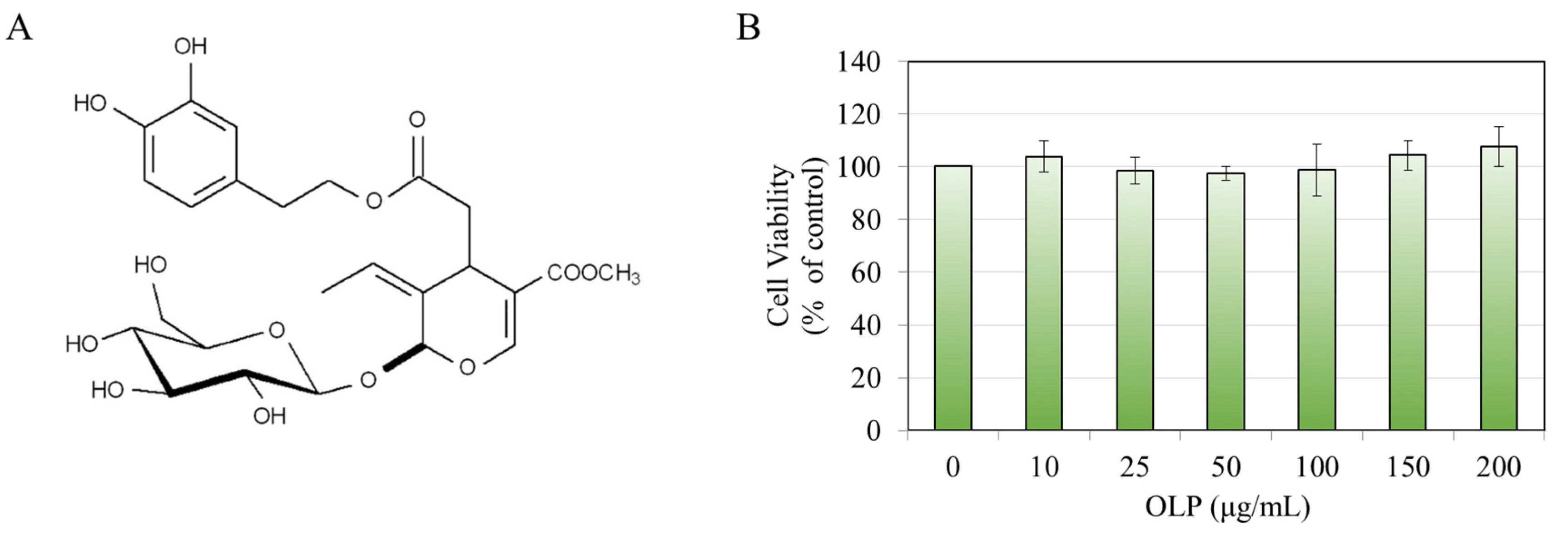
Figure 1. (A) Chemical structure of oleuropein (OLP). (B) B16F10 cell viability after a 48 h treatment with OLP (0–200 µg/mL). Data are mean ± SD of at least three independent experiments.
Biologics 05 00008 g001
Figure 2. (A) Relative cellular melanin content and (B) extracellular melanin content in B16F10 cells after a 48 h treatment with OLP (0–200 µg/mL); the corresponding photos of cell pellets and culture medium are shown above respective plots; (C) microscopic images of B16F10 cells subjected to various OLP concentrations showing the formation of elongated dendrites (red arrows in images) for higher concentrations; data for (A) are mean ± SD of triplicates (n = 3) from one representative experiment of two separate experiments, while data for (B) are mean ± SD of values from two independent experiments (n = 5); * p < 0.05, ** p < 0.01.
Biologics 05 00008 g002
Figure 3. Tyrosinase activity of (A) B16F10 cells and in (B) cell-free system using mushroom tyrosinase after treatment with OLP (10–200 µg/mL); data for (A) are mean ± SD of data combined from two independent experiments (n = 5), and data for (B) are mean ± SD of triplicates (n = 3); ** p < 0.01, $ p < 0.001, and # p < 0.0001.
Biologics 05 00008 g003
Figure 4. (A) Viability of HEMn-LP cells after a 48 h treatment with OLP (0–200 µg/mL). (B) Relative intracellular melanin and (C) extracellular melanin contents in HEMn-LP cells after treatment with OLP (0–200 µg/mL); the corresponding photos of cell pellets and culture medium are shown above the respective plots; (D) representative phase-contrast images of HEMn-LP cells treated with varying OLP concentrations; red arrows indicate the extended dendrites; (E) tyrosinase activity of HEMn-LP cells treated with various OLP concentrations; data for (A,B) are mean ± SD of three independent experiments (n = 3), while data for (CE) are mean ± SD of values from two independent experiments (n = 5–6); * p < 0.05, $ p < 0.001, and # p < 0.0001.
Biologics 05 00008 g004
Figure 5. (A) Micrographs of HEMn-LP cells before and after a 48 h treatment with 200 µg/mL OLP; black arrows denote extended dendrites. Quantitation of dendritic parameters: (B) total dendrite length; (C) dendrite number; and (D) average dendrite length; * p < 0.05.
Biologics 05 00008 g005
Figure 6. (A) HEMn-MP cell viability after a 48 h treatment with OLP (0–200 µg/mL). (B) Relative intracellular melanin and (C) extracellular melanin contents in HEMn-MP cells after treatment with OLP (0–200 µg/mL) with the corresponding photos shown above the respective plots; (D) phase-contrast images of HEMn-MP cells treated with OLP (0–200 µg/mL); the extended dendrites are shown by red arrows; (E) tyrosinase activity of HEMn-MP cells after OLP treatment; data for (B,E) are mean ± SD of three independent experiments (n = 3), while data for (A,E) are mean ± SD of values from two independent experiments (n = 5); * p < 0.05, $ p < 0.001, and # p < 0.0001.
Biologics 05 00008 g006
Figure 7. (A) Micrographs of HEMn-MP cells before and after a 48 h treatment with 200 µg/mL OLP; black arrows denote extended dendrites. Quantitation of (B) total dendrite length; (C) dendrite number; and (D) average dendrite length; ** p < 0.01 and $ p < 0.001.
Biologics 05 00008 g007
Figure 8. Scheme illustrating the capacity of OLP to act as dendrite elongator in melanocytes to augment melanosome export to keratinocytes.
Biologics 05 00008 g008
Disclaimer/Publisher’s Note: The statements, opinions and data contained in all publications are solely those of the individual author(s) and contributor(s) and not of MDPI and/or the editor(s). MDPI and/or the editor(s) disclaim responsibility for any injury to people or property resulting from any ideas, methods, instructions or products referred to in the content.