Review

Unveiling Novel Insights in Helminth Proteomics: Advancements, Applications, and Implications for Parasitology and Beyond

1
Department of Pathobiology, Faculty of Veterinary Medicine, Ferdowsi University of Mashhad, Mashhad 9177948974, Iran
2
WOAH and NRL for Echinococcosis, Animal Health, Istituto Zooprofilattico Sperimentale della Sardegna, 07100 Sassari, Italy
3
Department of Clinical Sciences, Faculty of Veterinary Medicine, Shahrekord University, Shahrekord 8818634141, Iran
4
Department of Pathobiology, Faculty of Veterinary Medicine, Science and Research Branch, Islamic Azad University, Tehran 1477893855, Iran
*
Authors to whom correspondence should be addressed.
Biologics 2024, 4(3), 314-344; https://doi.org/10.3390/biologics4030020
Submission received: 19 July 2024 / Revised: 27 August 2024 / Accepted: 14 September 2024 / Published: 19 September 2024

Abstract

:
Helminths have developed intricate mechanisms to survive and evade the host’s immune responses. Hence, understanding the excretory-secretory products (ESPs) by helminths is crucial for developing control tools, including drug targets, vaccines, and potential therapies for inflammatory and metabolic disorders caused by them. Proteomics, the large-scale analysis of proteins, offers a powerful approach to unravel the complex proteomes of helminths and gain insights into their biology. Proteomics, as a science that delves into the functions of proteins, has the potential to revolutionize clinical therapies against parasitic infections that have developed anthelminthic resistance. Proteomic technologies lay a framework for accompanying genomic, reverse genetics, and pharmacokinetic approaches to provide more profound or broader coverage of the cellular mechanisms that underlie the response to anthelmintics. With the development of vaccines against helminth infections, proteomics has brought a major change to parasitology. The proteome of helminths can be analyzed comprehensively, revealing the complex network of proteins that enable parasite survival and pathogenicity. Furthermore, it reveals how parasites interact with hosts’ immune systems. The current article reviews the latest advancements in helminth proteomics and highlights their valuable contributions to the search for anthelminthic vaccines.

Graphical Abstract

1. Introduction

Proteins are vital parts of living organisms and are the main components of physiological metabolic pathways in cells [1]. They have diverse functions, including catalyzing biochemical reactions, providing structural support, regulating cellular processes, and participating in signaling networks [2,3]. The field of proteomics aims to characterize the entire protein complement of a cell line, tissue, or organism on a large scale [4]. This comprehensive approach provides a deeper understanding of cellular biology and has far-reaching implications in medicine, agriculture, and environmental science [5,6,7].
The term “proteomics” initially referred to the large-scale analysis of gene products, specifically proteins [8]. However, as technology has advanced and the research has progressed, the definition of proteomics has expanded to encompass a more inclusive approach [9]. Today, proteomics integrates protein studies with other genetic readouts, such as mRNA analysis, genomics, and yeast two-hybrid analysis [10]. This multidimensional approach allows for a complete and systems-level understanding of biological processes, unraveling complex interactions and networks within cells and organisms [11]. Like in several fields, within parasitology, proteomics has emerged as a powerful tool for investigating the intricate biology of helminths [12].
Helminths are multicellular parasitic organisms that infect many hosts, including humans, and may cause zoonotic diseases [13]. They are typically classified into three classes, i.e., nematodes (roundworms), trematodes (flukes), and cestodes (tapeworms). They have adapted to live in diverse niches within the human body, including organs with a filter function such as the liver, lungs, gastrointestinal tract, and blood vessels [14]. To facilitate their penetration, migration, and establishment within the host, helminths secrete a wide range of molecules [15]. These molecules play crucial roles in various aspects of the parasite life cycle, including host invasion, nutrient acquisition, immune evasion, and modulation of host immune responses [16,17]. Some helminths have been implicated in influencing the evolution of an entire arm of T helper (Th) cell immunity, known as the Th2 pathway [18]. The host induces this response as an immune defense to repair the damage caused by these large parasites. These adaptations and interactions between helminths and their hosts contribute to the long-term survival of helminths within the hostile environment of the host, often persisting for years or even decades [19].
Each developmental stage of a helminth species secretes hundreds or even thousands of proteins, often with limited conservation between stages [20]. This diversity in protein secretion reflects the different niches that each stage must navigate and adapt to during its life cycle. Parasitic helminth proteins have become a focal point of research efforts to develop control tools, including drug targets and vaccines [21]. Additionally, the unique protein repertoire of helminths has recently emerged as a potential source of drugs for treating inflammatory and metabolic disorders prevalent in populations free of helminth infections [22].
An important factor in the host–parasite interaction is the excretory-secretory products (ESPs) of helminths [23,24]. ESPs play a significant role in persistence and pathogenicity. The parasite secretes various proteins, enzymes, and other molecules that stimulate the host’s immune response [25,26,27]. These molecules facilitate tissue invasion and create favorable conditions for the parasite to survive [28,29]. An ESP may suppress or manipulate the host’s immune system to allow the parasite to evade detection and continue to exist in the body [30,31]. They also contribute to the host tissues’ degradation and assimilation of nutrients, which are essential for the parasite’s growth, reproduction, and nutrient uptake [32,33]. It is crucial to study ESPs not only to unravel the complex biology of helminths but also to develop new strategies for preventing and treating parasitic diseases [34,35]. These products could be of value in disrupting parasites’ life cycle and reducing the burden of parasitic infections, which remain a significant public health concern in many parts of the world due to targeting them [36,37,38,39].
This article reviews the latest advancements in proteomics related to helminths and how proteomics has provided invaluable tools and data for addressing the urgent need for anthelminthic vaccines. Moreover, it delves into the cutting-edge techniques and methodologies employed in helminth proteomics, highlighting their contributions to understanding the biology and pathogenicity of helminths. Finally, the current review discusses how helminth proteomics has advanced our understanding of host–parasite interactions, shedding light on the mechanisms employed by helminths to modulate and evade the host immune response.

2. Methods

Several international databases, namely PubMed, Scopus, Google Scholar, and Web of Science, were used to retrieve articles relevant to helminth proteomics, published between 2000 and 2023 in peer-reviewed international journals. Some of the keywords used for our comprehensive and systematic literature search were “Proteomics”, “Nematodes”, “Heligmosomoides”, “Ascaris”, “Toxocara”, “Nippostrongylus”, “Ancylostoma”, “Haemonchus”, “Trichostrongylus”, “Trichinella”, “Trematodes”, “Fasciola”, Dicrocoelium”, “Schistosoma”, “Echinostoma”, “Cestodes”, “Echinococcus”, “Taenia”, and “Hymenolepis”, alone and in combination with each other. Following an initial search, duplicates were removed, and the remaining articles were screened based on their titles and abstracts. A thorough review of full-text articles that met the inclusion criteria was conducted. In addition, (1) reference lists of the selected articles and (2) publications that cited retrieved articles were examined for any additional relevant studies missed during the initial search. The selected studies were analyzed for information regarding helminth proteomics, advancements, applications, and implications for parasitology.

3. Proteomics in Nematodes

Several researchers have conducted the proteomic analysis of nematodes using mass spectrometry in the last decade [40]. Investigations of these parasites are usually based on studies of their effects on humans and economies. Anthelmintics is the most common parasite control method [41,42,43]. Because anthelmintics have a short duration of action and hosts can release parasite eggs that contaminate the environment, reinfection is likely to happen soon. Proteomic exploration of nine distinct nematode species is described in the text and illustrated in Figure 1 (Figure 1).
This Figure illustrates the proteomic exploration of nine distinct nematode species—Trichinella spiralis, Trichostrongylus colubriformis, Ascaris suum, Haemonchus contortus, Heligmosomoides polygyrus, Ancylostoma caninum, Nippostrongylus brasiliensis, Toxocara cati, and Toxocara canis. Advanced mass spectrometry techniques have been employed to reveal the intricate protein landscapes of these nematodes. The names of the identified antigens associated with each nematode species are listed below, providing insight into the specific proteins added during the proteomic analysis.

3.1. Heligmosomoides polygyrus

The nematode Heligmosomoides polygyrus (Figure 1) belongs to the family Trychostrongylidaeand, and males and females can be distinguished by their morphology. In rodents, H. polygyrus (Figure 1), previously called Nematospiroides dubius, resides naturally in the intestinal tract [26]. Likewise, it is practical to study pathology and immune reactions to gastrointestinal parasite infections using the murine nematode H. polygyrus [44]. ESPs identified 209 proteins, including allergen V5/Tpx-1-related proteins, retinol- and fatty acid-binding proteins, homologs of vitellogenins, and globins [45]. These findings also point to the possibility that multiple immunomodulators may exist. These include galectins, peroxiredoxins, macrophage migration inhibitory factors, C-type lectins, glutathione S-transferases, and cysteine protease inhibitors. When cultured together or separately, a comparison was also made between the ESP compositions of male and female nematodes at the L4 stage [46]. A mass spectrometric method was used to detect proteins. Mixed larval cultures containing sexual pheromones generated 258 proteins, while pure male cultures produced 172 proteins and pure female cultures had 160 proteins. Nematodes exposed to sex pheromones produce many proteins with immune-modulating effects, including TGF-β mimic 9, Val proteins, HpARI, and acetyl-cholinesterases. In the ESP derived from mixed cultures, TGF mimics 6 and 7 were also detected, along with galectin.

3.2. Ascaris suum

Human and pig ascariasis occurs due to the helminths of the genus Ascaris [47]. In humans (especially in children), Ascaris lumbricoides is the most prevalent internal macro-parasite, while for pigs, A. suum (Figure 1) is the most prevalent [48]. It is estimated that 1.2 billion people worldwide are infected, with children living in tropical and subtropical regions having the highest occurrence rates [49]. Affected children often exhibit growth impairment, nutrient deficiencies, intellectual disabilities, and poor academic performance [50]. In pigs affected by Ascaris, the infection causes increased feed conversion efficiency and lower growth rates [51]. Moreover, liver lesions caused by larval migration called “milk spots” result in substantial economic losses due to the necessity of eliminating these livers in abattoirs [52].
A slaughterhouse collection of naturally infected pigs concluded that A. suum worms were found in adult form in both sexes. After dissection, male and female worms’ intestines, reproductive components, and cuticles were collected, stored, and thawed at −80 °C. For 28 to 30 days, A. suum eggs were cultured in 0.1% potassium dichromate (K2Cr2O7) at 25 °C from female worms’ uteruses. Following the embryonation of 90% of the eggs, the infectious L3s were hatched, as described by Urban and Douvres [53]. A Baermannization process was then applied to separate the larvae from fragments of eggshell and other debris. Gavage experimentation with the larvated eggs of A. suum was performed on two groups of two pigs. Five hundred thousand eggs were inoculated into the pigs of group one, and they were euthanized seven days post-infection (PI) for lung larvae sampling (L3-lung). In comparison, 30,000 eggs were inoculated into pigs in group two, which were euthanized 14 days PI to collect intestinal larvae (L4). Using a Baermannization process, the L4 and L3 lungs were isolated from the contents of the small intestine and the lung tissue of the host. Using gel digestion, LC-MS/MS, and tryptic digestion of ESPs, a total of 106 proteins were discovered, of which 20 were found during the L3 stage of the egg, 45 during the L3 stage of the lung, and 58 during the L4 stage [54]. There were 15 proteins discovered in at least two sets of ESPs, though most were stage-specific. ESPs from three distinct larval stages contain a serpin-like protein and a 14-3-3-like protein. Interestingly, ESPs from L4 appear rich in metabolic enzymes, especially glycosyl hydrolases. Furthermore, enzymatic assays showed that extracts from the gut of adult nematodes had the most glycosidase activities.

3.3. Toxocara canis

Toxocara canis (Figure 1) is a nematode that infects the Canidae family [55]. When accidentally consuming eggs from contaminated soil, humans become infected with T. canis [56]. The transmission of infections can also occur via ingestion of larvae present in raw meat from paratenic hosts and from embrocated eggs on vegetables that have not been washed [57]. Ocular and visceral larva migrants, neuro-toxocariasis, and covert parasitic infection are clinical manifestations distinguished by the organs the parasite infects [58]. By administering pyrantel pamoate at a dose of 15 mg/kg to naturally infected four- to eight-week-old dogs, female T. canis adults were collected from their small intestines. A culture of T. canis larvae was then conducted. A total of 582 somatic proteins were found in the larval extract of T. canis larvae, along with 64 ESPs. A complex pattern of bands was observed in T. canis extract proteins. Larval extracts contained mostly proteins ranging from 10 to 97 kDa, while those in ESPs ranged between 12 and 175 kDa [59]. SDS-PAGE analysis of ESP antigens revealed a variety of band patterns, including a single 35 kDa band, 32, 55, 70, 120, and 400 kDa bands, and bands comprising 29 to 125 kDa and 28 to 280 kDa. The carbohydrate composition of ESPs, one-dimensional and two-dimensional SDS-PAGEs, protease sensitivity analyses, and immunoblotting demonstrated at least 20 proteins.
A total of 870 ESPs were predicted to be present in the T. canis secretome after the nematode genome was sequenced [60]. There are, however, 732 proteins whose functions are unclear. Several proteins have known functions, including lectins of the C-type and glycoprotein complexes. Among the seven identified secreted proteins (extracellular category), mucin TES–120 (T.can15190) and C-type lectin TES–32 (T.can00022) are the two most dominant and abundant [61,62]. Signal P analysis contributed complementary information for specific extracellular proteins without a signal peptide.

3.4. Toxocara cati

One of the most prevalent felid intestinal helminths, Toxocara cati (Figure 1) has profound epidemiological implications worldwide [63,64]. An accidental human infection can occur by consuming embrocated eggs from a paratenic host [65]. During human development, larvae cannot fully develop into adults and can survive in the tissues for several months to years [66]. A sedimentation technique was used to examine stray cats’ fecal samples to determine whether they were infected with T. cati [67]. In a recent study, 363 proteins were identified in crude somatic extracts of adult T. cati [68]. Most of these proteins are involved in metabolic and energy processes; some are involved in motor activity, some are engaged in cytoskeletons, some are implicated in ATP binding, and others are interested in nuclear and mitochondrial function. In contrast, 158 of the proteins are unknown. The study focuses on 34 proteins, most of which have already been confirmed to be present in helminth parasites [69]. This extract contains diverse metabolic and glycolytic enzymes, including triose-phosphate isomerases and enolases, and other protein groups such as (1) proteins that bind to ATP, such as heat shock proteins, myosin, and actin; (2) some proteins that involve the action of transaminases, particularly the enzymes aspartate aminotransferase and alanine aminotransferase; (3) motor proteins that contain calcium-transporting ATPase and acetyl-coenzyme A; (4) other proteins that contribute to phosphor pyruvate hydratase action, including paramyosin; (5) proteins such as the antigens OV-17 and OV-16, with unknown functions; and (6) a 28 kDa Trypsin that comes from the host.

3.5. Nippostrongylus brasiliensis

The nematode Nippostrongylus brasiliensis (Figure 1) infects rodents, especially rats, through their gastrointestinal tract [70]. The parasite’s simple life cycle and capability to be used in animal models make it one of the most frequently studied parasites [71]. This parasite has a life cycle similar to human hookworms, particularly Ancylostoma duodenale and Necator americanus, which mature sexually after five molting stages [72]. N. brasiliensis larvae and adults exhibit 52 and 261 proteins, respectively, in their ESPs [73]. The proteins detected in the ESPs are also different from those found in the Nippostrongylus brasiliansis somatic extract (NEx). Consequently, they are more likely to be caused by accurate secretory production than by leakage of intracellular and structural proteins from damaged or deceased parasites. L3 and mature worm ESPs were analyzed by LC-MS/MS, and 313 proteins were found. Most adult and L3 ESPs were stage-specific, and only 13 proteins shared CAP domains. Atacin metalloprotease and CAP domain protein family’s SCP (Sperm-Coating Protein)/TAPS (Tpx-1/Ag5/PR-1/Sc7) (SCP/TAPS) were notably abundant. Several of these protein families are found in nematodes, some of which are found to facilitate larval migration and immune evasion, both of which are crucial to nematode survival. As assessed by phylogenetics and overall gene similarity analyses, N. brasiliensis is the most conserved model nematode concerning human hookworms. Based on these findings, human hookworm infestations can be investigated using N. brasiliensis in a feasible animal model.

3.6. Ancylostoma caninum

Ancylostoma caninum (Figure 1) is a parasitic nematode worm that primarily infects dogs, causing a condition known as canine hookworm disease [74]. These small, thread-like worms inhabit the small intestine of their hosts, where they feed on blood, leading to symptoms such as anemia and gastrointestinal disturbances. A. caninum is a significant concern in veterinary medicine, and effective prevention and treatment strategies are crucial to protect the health of infected dogs [75]. Adult A. caninum was obtained from euthanized stray dogs for ESP preparation and then cultured and extracted. Proteins with MWs ranging from 250 kDa to less than 5 kDa were discovered by conducting LC-MS/MS analysis on ESPs. Most ESPs were found to have a size of 5 to 15 kDa based on staining intensity. Nevertheless, groups of proteins were apparent between 25 and 37 kDa and 75 to 100 kDa. The ESPs of A. caninum contained 105 proteins [76]. These proteases include cysteine, aspartic, and metalloproteases, which are the three major categories of proteases found in nematodes. One of the C-type lectins identified in this study uniquely targets galactoside. A noteworthy 28% of the detected proteins showed similarities with activation-associated secreted proteins, a group of proteins with a cysteine-rich structure found in the sterol carrier protein/Tpx-1/Ag5/PR-1/Sc-7 (TAPS) family [77]. Thirty-four of these proteins were detected, suggesting that they play a significant role in the relationship between the host and the parasite. There are also proteins related to hyaluronidases, transthyretins, and lysozyme-like proteins. A comprehensive understanding of hookworm infection biology has been gained by identifying a set of proteins deemed crucial for the parasitic lifestyle.

3.7. Haemonchus contortus

Haemonchus contortus (Figure 1) is a parasitic nematode commonly called the barber’s pole worm due to its distinctive red and white spiral appearance [78]. This blood-feeding parasite primarily infects ruminant animals, including sheep and goats, causing significant economic losses in the livestock industry [79]. H. contortus infestations can lead to symptoms such as anemia, weight loss, and decreased productivity in affected animals [80]. Effective management and control measures are essential for preventing the detrimental impact of this parasitic worm on livestock health and productivity.
The excretory-secretory products of H. contortus have been identified as containing seven cysteine proteases (CBLs) with similar characteristics to cathepsin B [81,82,83]. The localization of active CBLs was observed using 2D gel electrophoresis zymography and biotinylated inhibitors. The clustered expressed sequence tags also encoded three novel CBLs encoding GCP7, AC-4, HMCP1, and HMCP2 [84].

3.8. Trichostrongylus colubriformis

Trichostrongylus colubriformis (Figure 1) is a parasitic nematode commonly found in the digestive tracts of small ruminant animals, particularly sheep and goats [85]. This gastrointestinal parasite can cause significant damage to the mucosal lining of the host’s intestines, leading to symptoms such as weight loss, diarrhea, and reduced growth rates [86]. Controlling and managing T. colubriformis infestations are crucial for maintaining the health and productivity of these livestock animals in the agricultural industry [87]. An analysis of immunoreactive proteins was conducted with sheep IgG from T. colubriformis L3s. The separation of proteins using two-dimensional electrophoresis identified immunoreactive protein spots following mass spectrometry-based proteomic analysis of plasma from sheep resistant to T. colubriformis [88]. A total of 28 immune targets were pinpointed, including enolase, a kinase, an aspartyl protease inhibitor, a galectin, glycolytic enzymes, a chaperone protein, and a phosphatase. Moreover, structural muscle proteins such as myosin, paramyosin, DIM-1, and calponin can also be found within. The cytoplasmically expressed proteins of T. colubriformis suggest that a wide range of antigens are involved in immune responses against this parasite.

3.9. Trichinella spiralis

It has been shown that Trichinella spiralis (Figure 1) nematode infections correlate with profound yet stereotypical pathological transformations of the epithelium [89]. Researchers discovered proteins involved in stress response, metabolic adaptation, and development [90,91,92]. Essential proteins that contribute to parasite survival and immune evasion were identified in a proteomic analysis of ESPs from adult T. spiralis worms [93]. The parasite can interfere with host proteolytic processes to evade immune responses with protease inhibitors. Protease inhibitors are essential in establishing successful infections, as this study shows. An analysis of the proteomics profile of T. spiralis muscle larvae under host-induced conditions revealed the parasite’s adaptation strategies within the host [94,95,96]. The proteins related to stress response, metabolic adaptation, and development have been identified [97]. As a result of the detection of anaerobic metabolism-related proteins, T. spiralis has adapted to the hypoxic conditions of the host’s muscle tissue [98]. A parasite with immunomodulatory proteins and antigens can manipulate the host immune system, facilitating its survival and growth. Proteins involved in tissue invasion and immune modulation were discovered from excretory-secretory products of T. spiralis intestinal larvae (L3) [99,100]. The parasite penetrated and migrated through host tissues via enzymes associated with tissue degradation [101]. Additionally, T. spiralis’ sophisticated mechanisms to evade host immune responses and construct a favorable environment within its host are evident from identifying immunomodulatory proteins such as galectins. References [102,103,104,105,106] are cited in the Supplementary Materials Table S1.

4. Proteomics in Trematodes

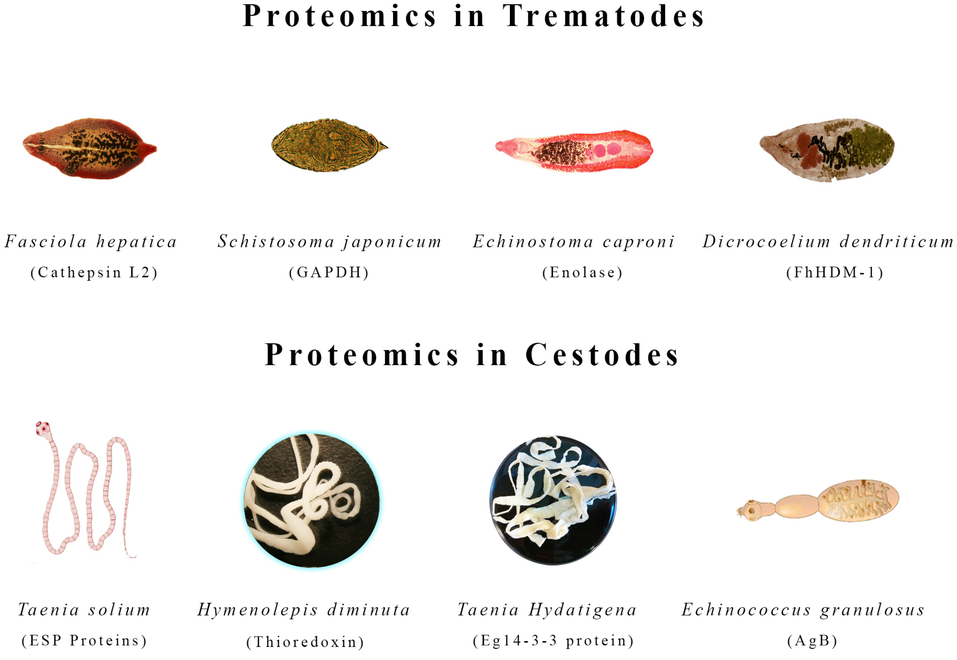
Millions of people worldwide are infected with trematodes, but monotherapies are used to treat the infections because commercial vaccines are unavailable [107]. Due to the emergence of drug-resistant fluke populations, identifying immunogenic vaccine candidates and new drug targets requires a comprehensive understanding of host interactions and parasite biology [108,109]. Molecular information about host–parasite interactions is limited at present. Therefore, proteomic studies could identify diagnostic biomarkers that could be used for early disease detection, as well as new vaccine targets. A vital tool for this is mass spectrometry-based proteomics. In food-borne trematodiasis, proteins involved in parasite pathogenesis can be identified using proteomic techniques [110,111]. In addition to being diagnostic biomarkers, these proteins may be used in vaccine development. Figure 2 describes the proteins related to eight different helminth species (Figure 2).
This Figure reports the proteins related to eight different helminth species of the class Cestodes, comprising Taenia solium, Taenia hydatigena, Echinococcus granulosus, and Hymenolepis diminuta, along with the class of Trematodes, encompassing Fasciola hepatica, Dicrocoelium dendriticum, Schistosoma japonicum, and Echinostoma caproni. The visual compilation signifies the distinct proteomic landscapes of each helminth, providing a foundational understanding of these parasitic organisms. Below each entity, the names of identified antigens are presented, indicating the proteins added during the proteomic analysis. This Figure is a concise reference, offering a snapshot of the varied protein compositions within trematodes and cestodes, thereby facilitating further research and comprehension of the intricate host–parasite relationships.

4.1. Fasciola hepatica

Larvae of Fasciola hepatica (Figure 2) infect mammalian hosts by passing through the intestinal walls of the duodenum, moving across the peritoneum, and penetrating the liver [112,113]. The parasites migrate through and feed on liver tissue, causing intensive damage to the tissue before moving to the bile ducts for maturation and egg production [114]. Using proteomics to examine proteins secreted by different phases of F. hepatica, including infective larvae, immature flukes, and adult F. hepatica, a recent study discovered that proteases are regulated as F. hepatica develops [115,116]. Parasites encounter host macromolecules and tissues while passing through host tissues [117]. Several proteases play an important role in activating larvae and allowing their penetration into the intestinal wall, including FhCL3 and cathepsin B. FhCL1, FhCL5, and FhCL2 are required for liver feeding and penetration [118,119,120]. In particular, FhCL2 can self-cleave substrates containing proline residues and facilitate the degradation of interstitial collagen when parasites migrate through tissues. Another protein named FhSAP-2 is involved in the lysis of red blood cells and penetration by Fasciola and other helminths. Cathepsin L enzymes are also believed to be involved in blood digestion, especially hemoglobin [121,122,123,124]. Aside from proteases, parasites secrete a variety of antioxidants, which are also tightly regulated as they migrate through host tissues. The F. hepatica proteases are secreted through the classical endoplasmic reticulum/Golgi pathway, whereas antioxidants are thought to be secreted through non-classical trans-tegumental pathways.

4.2. Dicrocoelium dendriticum

In ruminants, dicrocoeliosis is caused by Dicrocoelium dendriticum (Figure 2), which remains an uncontrollable hepatic parasite whose immunological diagnosis and treatment are unsatisfactory [125,126]. In the life cycle of this Digenea, different species of mollusks and ants act as primary and secondary intermediate hosts [127]. Two methods were used to treat the samples before isoelectric focusing to concentrate proteins and remove contaminants that may inhibit two-dimensional electrophoresis. Among the proteins, metabolic enzymes constituted the largest group. These proteins are the glycolytic enzymes: aldehyde dehydrogenase, enolase, triose phosphate isomerase, glyceraldehyde-3-phosphate dehydrogenase (GAPDH), 3-retinal dehydrogenase, fructose-bisphosphate aldolase, a putative aldehyde dehydrogenase family member, transketolase, and malate dehydrogenase, all of which are essential for parasite energy production [128]. Other important functions of these proteins include modulating the interaction between parasites and hosts, making them attractive targets for vaccines or therapies. There has also been an identification of motor and structure proteins in D. dendriticum, such as actin, which exists in two forms in both extracts, and paramyosin, which appears in ES. Exosomes from D. dendriticum have a distinct proteomic profile compared to those from F. hepatica and E. caproni [129]. This suggests that exosomes may represent the mechanism by which these proteins are present in their respective ESPs. D. dendriticum and F. hepatica parasites commonly inhabit the same parasitized animals and share the same location in the liver [130,131]. A protein identified in D. dendriticum exosomes, MF6p (also called MF6p/FhHDM-1), has recently been described as a potential diagnostic candidate. It has been demonstrated that this protein is a heme-binding protein, suggesting a role in detoxification and heme uptake in the liver of F. hepatica, a protein involved in immunomodulation [132].

4.3. Schistosoma japonicum

Mortality and morbidity rates associated with Schistosomiasis are among the highest of all human helminthiases [133]. With 779 million at risk and 200 million infected, Schistosomiasis remains a serious public health issue [134]. An early stage in Schistosoma japonicum’s (Figure 2) complex life cycle in vertebrates is the schistosomulum [135,136]. Vaccines are primarily used to provide immunity at this stage [137,138]. ESPs play a major role in parasite–host interactions, and so far, the ESP composition of Schistosomula of S. japonicum has not been characterized [139]. Using mass spectrometry, 20 protein spots were identified, belonging to five categories: energy metabolism-associated enzymes (glyceraldehydes-3-phosphate dehydrogenase), structural and motor proteins (actin), signaling transduction pathway-associated molecules (14-3-3 protein), the 70 kDa heat shock protein (HSP70) family, and other functional proteins (20S proteasome) [140].
Using tandem mass spectrometry/tandem mass spectrometry liquid chromatography, 713 unique ESPs were identified from the 14-day schistosomula of S. japonicum [141]. Pathway analysis and gene ontology revealed that the identified proteins have roles in degradation, oxidation–reduction, response to stimulus, carbohydrate metabolism, biological regulation, and binding. Based on flow cytometry analysis, LPS-activated macrophages were inhibited from expressing MHCII and CD86 by thioredoxin peroxidase. Fibrin, paramyosin, prosaposin, and 89 eggshell proteins such as antigen SM22.6 (A12), the alpha subunit of the G protein, calcium-binding protein Sj66, and flavoprotein (Fp) also seem to exhibit antigenic polymorphisms due to candidate single-nucleotide polymorphisms (SNPs). SNPs may represent schistosome populations’ capability to modify, parry, or attenuate host immune responses during mammalian infection. Transcriptomic and proteomic approaches have identified a wide range of mammalian-like receptors, such as the vasopressin-activated calcium-mobilizing receptor, the dioxin receptor, receptor tyrosine phosphatase (gamma and delta), insulin receptor protein kinase RTK-2, the feline leukemia virus receptor (subtype-B), and purinergic receptor P2X (ligand-gated ion channel, 4). Thus, the parasite can receive various cytokine signals and hormones from its mammalian host and endogenous signals from the parasite. In addition to motor-associated and cytoskeleton proteins, chaperones, extracellular matrix molecules, and enzymes involved in redox homeostasis, antigenic mimicry may be used to evade immune responses. This strategy has long been predicted (along with others, including the masking of host antigens) to explain the chronic nature of schistosome infections. Several immune-associated molecules, such as cyclophilin B, immunophilin, the HLA-B associated transcript 1, and endoplasmin (gp96), may also play a part in parasite growth and immunity by modulating the mammalian adaptive and innate immune systems [142].

4.4. Echinostoma caproni

A trematode of the Echinostomatidae family called Echinostoma caproni (Figure 2) infects the definitive host without presenting tissue phases. After infection, the metacercariae exocyst in the duodenum and the juvenile worms migrate to the posterior third of the intestine, attaching to the mucosa. E. caproni can cause infection in various hosts, although its compatibility for worm survival and development varies greatly among rodent species [143,144]. These characteristics make the E. caproni rodent system particularly suitable for revealing aspects of host-specific factors that regulate the course of intestinal helminth infections [145]. The ESPs of adult E. caproni worms remain poorly understood, although these host–parasite models are widely used [146]. E. caproni adult worm ESPs were initially characterized by Toledo et al. (2004) [147]. As a result of proteomic studies, we have identified proteins in the ESPs of E. caproni, including enolase and heat shock protein 70, which appear to play an important role in the host–parasite interaction [148,149].
Furthermore, a recent study showed that post-translational modifications, such as glycosylation, may contribute to the antigenicity of several molecules in the ESPs of E. caproni [150]. There are six proteins identified that participate in glucose metabolism, of which five (aldolase, enolase, GAPDH, phosphoglycerate kinase, and triose phosphate isomerase) are glycolytic and one (phosphoenolpyruvate carboxykinase) can contribute to glucose synthesis. Several studies are reporting their existence in the helminth tegument and extracellular matrix. Aldolase, enolase, and GAPDH, among other glycolytic enzymes, can bind plasminogen, facilitating host invasion and migration [151]. As a result, E. caproni infections significantly erode and destroy the host villi. Additionally, it has been shown that Glycolytic enzymes identified in E. caproni’s secretome (GAPDH and triose phosphate isomerase) may participate in the glutathione redox cycle and anti-oxidative processes. In the absence of enzymes normally involved in hydrogen peroxide detoxification, pyruvate can perform this function. A further benefit of NADH produced during glycolysis is its ability to reduce oxidized glutathione. Additionally, enolase and aldolase are immunodominant antigens in E. caproni infections. This suggests that they are secreted or exposed on the surface during host–parasite interactions. In contrast, aldolase evokes IgA responses, although different subclasses of Ig recognize enolase. As part of gluconeogenesis, phosphoenolpyruvate carboxykinase may play a role in parasite adaptations to host energy environments (Figure 2) (Table S2).

5. Proteomics in Cestodes

5.1. Echinococcus granulosus

Cystic Echinococcosis (CE) is an infection caused by the larval stage or metacestode of the tapeworm Echinococcus granulosus (Figure 2) that can affect both humans and animals [152]. This infection can cause cysts in the liver, lung, and other organs [153]. If untreated, the disease can result in serious and even fatal complications. Several reports describe E. granulosus spreading worldwide with great medical and economic impacts [154]. The disease is more prevalent in areas where livestock are reared and dogs are owned [155]. Several regions worldwide are particularly endemic to this disease, including Central Asia, South America, Africa, and the Mediterranean [156,157]. During the life cycle of E. granulosus, there are two main hosts: definitive and intermediate [158]. As definitive hosts, canids, such as dogs, serve as carriers, while intermediate hosts include sheep, cattle, and goats [159,160]. As the tapeworm eggs are transmitted from the definitive host into the environment, humans can become infected when they accidentally ingest these eggs from contaminated food or water sources or during close contact with infected animals [161]. The cyst is unilocular and filled with fluid as a fully developed hydatid cyst of E. granulosus [162]. Externally, no cellular laminated layer is supported by a germinal layer, which is then surrounded by fibrous adventitial tissue produced by the host [163]. The protoscoleces (PSC) are produced by asexual reproduction by the germinal layer and are subsequently ingested by the definitive host and develop into adult worms [164].

5.1.1. Protoscolex Proteins

Several proteins were identified in the metacestode expressed by the PSC during infection. The list includes chaperones, detoxification enzymes, immunomodulatory proteins, and protease inhibitors [165]. These proteins may play an important role in the parasite’s life cycle, allowing it to survive in its host and evade its immune system. Furthermore, they may interfere with the host’s response to the parasite when the cyst ruptures and PSCs reveal themselves within the host cavity, promoting their survival and development into secondary cysts [166,167]. Through two-dimensional electrophoresis (2-DE) immunoblotting, 14 PSC proteins were identified in the serum of CE patients [168]. Several proteins, such as EgcMDH, HSP70, P-29, grp78, and EgTPx, have been previously identified as antigens from E. granulosus infections in humans [169].

5.1.2. Germinal Layer and Hydatid Cyst Fluid Proteins

Some of the proteins discovered, such as paramyosin and tetraspanin, could be used in vaccine development as they are involved in immunoregulatory events between the host and the parasite during infection [170,171]. An important role played by helminth paramyosins in the host immune response is their ability to bind complement components and immunoglobulins and secrete components of the cellular immune response [172,173]. In addition to interacting with a variety of cell surface molecules, such as the major histocompatibility complex (MHC) and Fragment Crystallizable receptor (Fcr), tetraspanins may also function or modulate their signaling, thereby acting as ligand receptors for host ligands to modulate immunity [174]. It appears that tetraspanins play a significant role in helminth biology. Their roles in invasion, immune evasion, and modulation make them attractive targets for novel intervention strategies to combat these parasitic worms. As a result of AgB and EgTeg, a Th2-polarized microenvironment is elicited and maintained, resulting in chronic E. granulosus infection [175]. Nevertheless, the biological significance of Ag5 is still unclear. It contains a highly conserved glycosaminoglycan-binding motif that focuses Ag5 to host–parasite interactions.

5.2. Taenia solium

Around the world, Taenia solium (Figure 2) cysticerci are a major cause of epilepsy and seizures [176]. Taeniid eggs are released into the ecospheres of infected individuals’ gastrointestinal tracts. Cysticercosis is caused by the metacestode larvae of T. solium [177]. There is a greater risk of taeniasis caused by T. solium in areas with poor sanitation facilities and low socioeconomic privilege [178]. It is widespread across Latin America, Africa, Southeast Asia, and some Pacific islands [179,180]. ESPs are specifically important for T. solium because they enable the parasite to penetrate the host, establish and maintain an infection while being attacked by the host’s immune system, and incorporate metabolites from the host to obtain nutrition [181,182]. Its most important function is to evade the immune system of the host. ESPs can also be used for therapeutic purposes such as vaccination and immunomodulation [183]. Several oncospheric proteins were identified to participate in adhesion, detoxification, protein folding, and proteolysis [184]. There are many strategies developed by T. solium metacestodes to deal with its host’s innate and adaptive immune systems, many of which are similar to those developed by other helminths, including protease production and protease inhibitors, preventing complement formation, and steering the host immune response in a non-inflammatory manner [185,186]. ESPs are responsible not only for evading the immune system but also for acquiring nutrients. In parasite ESPs, for example, many hydrophobic ligand-binding proteins indicate this fact [187]. In addition to the functions listed above, ESPs also participate in signaling, defense against oxygen-mediated killing mechanisms, various binding processes, and multiple other functions [188,189]. Furthermore, many ESPs are capable of having multiple functions as well. Extensive research has been conducted on ESPs since these cells play such a significant role in parasite survival. They have been extensively studied to diagnose, vaccinate, and develop antiparasitic drugs [190]. It has also been shown that ESP levels are a good indicator of parasite viability since they decrease relatively rapidly when the metacestodes enter a stage of degeneration/calcification, in contrast to antibody levels [191]. Proteins from activated oncospheres and immunogenic proteins have also been reported as part of the proteome of T. solium. The role of these proteins in the complex immuno-pathogenesis of the disease is not completely understood. Further research is needed to determine these proteins’ exact mechanism of action and their role in the disease. Apart from the fact that these are still early efforts, proteomic changes associated with the tissue localization of cysts remain uncharacterized. These changes may be essential to understanding the cysts’ tissue preference.

5.3. Taenia hydatigena

The canine tapeworm Taenia hydatigena (Figure 2), a global parasite spreading worldwide, causes great economic loss to the food industry [192]. To complete its entire life cycle, T. hydatigena requires two different hosts. Adults reside in the intestines of dogs, wolves, and foxes, while larvae mostly parasitize intermediate hosts such as pigs and sheep [193]. The intermediate hosts are generally infected upon digestion of food or water contaminated with eggs expelled by infected dogs as feces [194]. As soon as the eggs enter the host’s digestive tract, the oncospheres become activated and then move through the liver to the intestine, where they are further developed. As a result of dogs consuming infected offal or organs, the larvae will develop into adult worms in their intestines, thus completing their life cycle [195].
In analyzing the proteomes of T. hydatigena cysts with scolex, referred to as CS, and cysts without scolex, referred to as CWS, 764 proteins were detected, of which 664 were identified in CS, 412 in CWS, and 312 in both [196]. Based on comparative analysis, it was found that CS had greater levels of proteins associated with growth and development. In comparison, CWS had higher levels of proteins constituting scaffolds and protective extracellular matrix proteins. Western blotting confirmed that the five selected proteins are more abundant in CWS than in CS, which agrees with the sequencing data. Western blotting is a technique used to detect the presence of proteins in a sample by separating them according to their size and shape and then transferring them onto a membrane. The membrane is then probed with antibodies specific to the protein of interest, and the proteins are visualized with an appropriate detection reagent. By analyzing a particular protein’s localization, we can better understand how it contributes to the organism’s development, metabolism, or response to its environment [197]. One of the proteins identified in the study, 14-3-3, regulates parasite growth and development by binding to signaling proteins. Additionally, 14-3-3 has been utilized as a vaccine antigen in the fight against parasitic diseases due to its promising properties [198]. As a result, 14-3-3 is expressed in both CS and CWS, which makes it a promising vaccine candidate or drug target for preventing T. hydatigena infections. Paramyosin is highly expressed throughout the body and may be a good drug target for T. hydatigena or even a vaccine antigen [199].

5.4. Hymenolepis diminuta

The zoonotic cestode Hymenolepis diminuta (Figure 2) parasitizes the small intestines of rodents [200]. The human body can become infected with hymenolepiasis by ingesting insects (intermediate hosts) infected with cestode cysticercoids, accidentally entering the tapeworm’s life cycle [201]. Humans are considered accidental hosts, as the disease is not directly transmissible from person to person. The symptoms of hymenolepiasis depend on the number and location of parasites in the body [202]. Symptoms include abdominal discomfort, nausea, diarrhea, and itching in the perianal region [203]. Treatment typically consists of anti-parasitic drugs and supportive therapy. Hymenolepiasis is a neglected tropical disease mostly associated with poverty and poor hygiene. Recent findings indicate that H. diminuta ESPs are replete with immunogenic proteins, including crucial molecules involved in host–parasite interactions. A recent study conducted by Zawistowska-Deniziak et al. (2017), which analyzed the polarization of human macrophages in H. diminuta, confirmed both the immune modulatory properties and the complexity of host–parasite interactions [204].
In vertebrate hosts, cysticercoids are the first proteins to cause an immunological reaction, which makes them highly useful for diagnostics. As long as these proteins are present at the adult stage, antibodies will be present throughout the infection cycle. Data derived from these studies provide insight into which proteins offer the best chances for diagnosis or drug development and which proteins may be key to host–parasite interactions [205]. Other proteins have been identified that participate in a wide range of biological activities [206,207]. In both consecutive developmental stages of H. diminuta, thioredoxin was found, suggesting a common means of evading host immunity. In both developmental stages, there is an abundance of a protein called 14-3-3, considered a good candidate for vaccines and commonly observed as a strong immunogen [202].
There have been several proteins identified in the cysticercoid of H. diminuta and adult samples that are considered vaccine candidates, including tegumental membrane proteins such as enolase, calpain, GAPDH, and heat shock protein [208,209]. The protein oncospheres were found to be characteristic of the adult stage of H. diminuta, resulting in the identification of the oncosphere protein Tso22a (troponin) and the oncosphere antigen, both of which are present in the gravid proglottids of H. diminuta. LC-MS/MS was used to identify 36 cross-reacting protein spots in rat serum. References [210,211] are cited in the Supplementary Materials Tables S3 and S4.

6. Anthelmintic Resistance

Anthelmintic use is widespread, which has led to the development of resistance in parasites, thus rendering drugs ineffective [212]. Developing better strategies for controlling helminths is imperative, along with obtaining more knowledge about how anthelmintics function, which might instigate new therapeutic approaches [213]. The lack of comprehensive and properly annotated genomic and proteomic databases has been a major hurdle in studying host–hookworm interactions over the past few decades [214]. Mass spectrometers with increased sensitivity and developments in sequencing platforms have offered useful information [215].
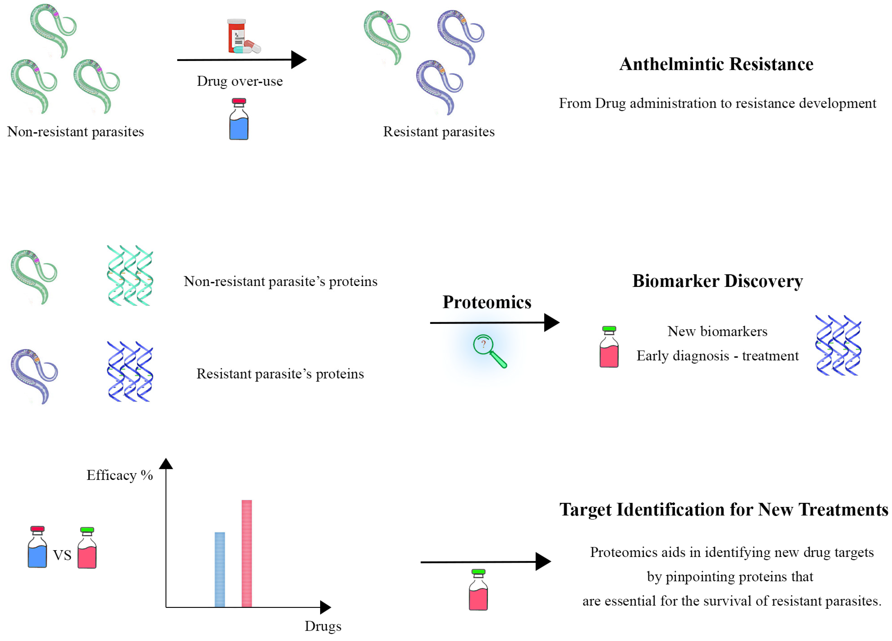
Proteomics, as a science that delves into the functions of proteins, has the potential to revolutionize clinical therapies against parasitic infections that have developed anthelminthic resistance [216]. The emergence of drug-resistant strains threatens the availability of anthelmintics for treating parasitic infections [217]. However, integrating proteomics into various fields shows novel findings and potential improvements, offering a beacon of hope in the battle against drug resistance [218].
Proteomic technologies lay a framework for accompanying genomic, reverse genetics, and pharmacokinetic approaches to provide a more profound or broader coverage of the cellular mechanisms that underlie the response to anthelmintics [219]. This is coupled with identifying biomarker panels associated with the emergence of anthelmintic resistance [220].
By comparing the expression and modification levels of proteins in drug-sensitive versus resistant parasite strains, proteomics offers a means to examine such complex molecular mechanisms [221]. The alteration in transport proteins could change drug uptake or efflux, eventually decreasing its efficacy [222]. Moreover, metabolic enzymes might undergo a modification to generate an inactive drug metabolite or stress response proteins could be upregulated, which would allow the cell to better cope with the drug’s effects [223]. These proteins can provide insight into the mechanisms of resistance and influence efforts to treat patients by targeting these mechanisms directly [224].
The ability of proteomics to elucidate the molecular interactions and pathways that are affected in resistant strains is beneficial for creating novel anthelmintic drugs [220]. Knowing how resistance changes the biology of a parasite enables scientists to identify vulnerabilities that can be blocked or circumvented by new medicines [225]. This information is also critical to the design of combinatorial therapies, which are often essential for fully circumventing resistance on a system-level basis rather than single-agent treatments [226]. The development of anthelmintic resistance is mainly through inheritance due to the existence of resistance genes [41]. Due to this collective functioning of multiple genes, resistance can be polymorphic and cannot be ascribed to one single gene [227]. Hence, it is very important to study drug resistance to monitor and control its evolution and delay the accumulation of genes conferring drug resistance [41].
Based on a study, Ivermectin significantly modulated the expression levels of 4528 genes in susceptible strains and 3038 genes in resistant strains, demonstrating that it was not just capable of modulating gene (or protein) expression but also exerts a more significant regulatory effect on susceptible H. contortus strains [228]. Studies also provided evidence that reducing or inhibiting the expression of a proteolytic exoenzyme in the muscle larvae stage may be beneficial to the survival and growth of T. spiralis parasites under albendazole stress, which is involved in parasite adjustment against drug stresses. It has serine-type endopeptidase activity and plays a role in several processes, such as cellular responses to salt and collagen, as well as cuticulin-based cuticle development or the positive regulation of synaptic growth at the neuromuscular junction [229]. This marks a major step forward in clinical therapeutics by using proteomics to assess anthelmintic-resistant infections [230].
Proteomic techniques provide a proactive means to monitor resistance emergence and spread in the parasite population [231]. This surveillance through proteomics is critical, empowering us to monitor emergent resistance changes and adapt treatment recommendations as needed [232]. By monitoring trends in resistance, we can ensure that treatment guidelines remain efficacious overall, facilitating better disease management and helping to reduce resistance [233]. Resistance strain proteins essential for survival or virulence can be potential targets for specific drug development [234]. For example, an inhibitor could escape resistance if a protein required for the parasite to survive is found in resistant parasites because it has been overexpressed or mutated [235]. This intervention could enable the discovery of better therapies to hit parasite resistance pathways more directly [236].
Additionally, systems-level proteomics analysis establishes key mechanistic links to pathways associated with resistance and enables the discovery of biomarkers involved in response [237]. A collection of biomarkers could be used to develop diagnostic tests that rapidly and accurately find resistant strains [238]. The identification of such biomarkers of resistance at an early stage would enable us to change treatments, and patients could be given the best-known treatment as soon as possible [239,240]. Such proactive steps facilitate better disease management and thus help to reduce resistance [229].
Proteomics offers a universal platform by elucidating resistance mechanisms and identifying novel therapeutic targets and biomarkers for diagnostics and personalized treatment [241]. Furthermore, this integrated approach will not only increase our ability to design successful antiparasitic drugs but also boost current programs aimed at controlling and eventually eliminating the scourge of drug-resistant parasitosis [242] (Figure 3).

7. Vaccine Production

With the development of vaccines against helminth infections, proteomics has brought about a major change in parasitology. The proteome of helminths can be analyzed comprehensively, revealing the complex network of proteins that enables parasite survival and pathogenicity. Furthermore, it reveals how parasites interact with hosts’ immune systems. Identifying novel vaccine targets may better explain how these preparations can neutralize these parasites. Several potential vaccine and diagnostic targets have been identified through helminth biology. For instance, a proteomic analysis of S. mansoni schistosomula has identified several proteins that could serve as vaccine antigens, including glucose transport protein-1 (SGTP1), tetraspanin 2 (TSP-2), and calpain [243].
The ability of proteomics to identify antigens that are both highly immunogenic and essential to the parasite’s life cycle has been one of the most significant contributions of proteomics to vaccine development. In several studies on E. granulosus, the causative agent of cystic echinococcosis, proteomics has been utilized to identify key proteins such as Eg14-3-3, EgTPx, and EgEF-1β. Infecting and surviving within its definitive host, dogs require these proteins, which are critical to disease transmission. These recombinant proteins have been demonstrated to stimulate significant immune responses in dogs, resulting in partial protection against infection with E. granulosus when vaccinated. This partial protection may mean that these proteins, when combined, could enhance the overall efficacy of a vaccine, providing a strategy for breaking the cycle of parasite transmission [244].
Additionally, proteomic analyses have been instrumental in characterizing the extracellular vesicles (EVs) secreted by helminths, which have emerged as the potential vaccine targets of the future. As a result of the proteins contained in these vesicles, helminths can manipulate the host’s immune response, thereby enabling their survival [174].
In Sheng and colleagues’ study, exosome-like vesicles (ELVs) from the liver fluke Fasciola gigantica, responsible for fascioliasis, were analyzed using proteomics [245]. Within these vesicles, they identified several proteins involved in immunoevasion and pathogenicity, among other characteristics. Vaccines targeting these proteins could disrupt the parasite’s ability to modulate the host’s immune system, inducing protective immunity and preventing infection.
In addition to identifying vaccine targets for single species, proteomics supports the development of cross-reactive vaccines that could target several helminth species, especially in areas where multiple helminths co-infect hosts and control efforts are complicated. Researchers have identified antigens with significant cross-reactivity in intestinal protein extracts of Mecistocirrus digitatus and Haemonchus contortus, two gastrointestinal nematodes. As a result of these findings, vaccines developed for one species may provide partial protection against other species, thereby simplifying and reducing vaccination costs in endemic regions [246].
Furthermore, proteomics has facilitated the development of multi-epitope vaccines by combining conserved antigens identified across various helminth species to develop vaccines. In Taenia species research, proteomic data have been integrated with bioinformatics and immunoinformatics, resulting in the identification of conserved proteins that can be combined into a single vaccine. With a single immunization strategy, these multi-epitope vaccines may induce a broad and robust immune response that protects a range of helminth infections [247].
Through techniques such as enzymatic shaving, proteomics can be innovatively applied to vaccine development to identify surface-exposed proteins. By using this method, previously unknown proteins have been discovered on Schistosoma species’ outer surface and tegument, which interacts directly with the host’s immune system. Some of these proteins play crucial roles in immune evasion, allowing the parasite to avoid detection and destruction by the host’s immune system. Vaccines designed to target these surface-exposed proteins enhance the host immune response, increasing the likelihood that the host will recognize and eliminate parasites. This approach allows helminths to survive within their hosts using mechanisms specifically targeted in vaccine development [248].
Using proteomics, Sotillo et al. have gained an understanding of Schistosoma species, the source of schistosomiasis, which highlights the potential of this approach for vaccine development [249]. This research aims to identify new proteins essential to parasite survival, development, and immune evasion, particularly those present on parasite surfaces or secreted during infection. The researchers have identified key antigens that could serve as vaccine candidates by profiling proteins expressed during different stages of life. Developing targeted and effective vaccines against schistosomiasis and other helminth infections is expected to be advanced by combining proteomic analysis with immunological studies.
Miles et al. have identified E. granulosus, the parasite responsible for cystic echinococcosis, as a potential vaccine candidate [250]. They have studied tegumental proteins, which play a crucial role in host–parasite interactions and are important targets for vaccine development. Researchers identified nine novel tegumental proteins as potential vaccine targets by analyzing protoscolex-stage (PSEx) protein composition using mass spectrometry, including proteins involved in immune modulation and parasite survival. Gene Ontology analysis was also used to predict these proteins’ biological processes, molecular functions, and cellular locations. As a result of this approach, 14 peptide sequences were identified, and their antigenicity predictions, epitope mapping, and structural analysis were used to verify that the peptides would be immunogenic, nontoxic, and non-allergenic. As a result of the discovery of these peptides and proteins as potential vaccine candidates, proteomics has demonstrated its role in advancing the development of targeted vaccines against Echinococcus granulosus and other helminth infections.
Significant advances have been made in developing vaccines against helminth infections through proteomics analysis of H. polygyrus [251]. According to a study, immunomodulatory ESPs, particularly those produced during L4, are significant in triggering sterile immunity. The researchers identified specific proteins, such as those with Sushi and ShK/SXC domains, which are highly expressed in L4 ES and are associated with immune modulation by comparing the proteomes of adult, L4, and egg stages. Additionally, antigens such as VAL-1 and ACE-1 were identified as major vaccine targets, demonstrating their importance for protective immunity against these intestinal nematodes. As a result of this study, new vaccine candidates are likely to be identified by elucidating proteins involved in host–parasite interactions and immune responses.
Using proteomics and bioinformatics, Sharma et al. developed a targeted vaccine against Taenia species, which cause significant health burdens in developing countries [247]. Taenia spp. cysts contain 451 conserved proteins essential for survival and pathogenicity. The proteins were then meticulously screened for antigenic epitopes, leading to the construction of a multi-epitope chimera vaccine. Immuno-informatics is crucial in developing resource-efficient and cost-effective vaccines for neglected tropical diseases caused by soil- and water-transmitted helminths. Taenia infections upregulate TLR4 receptors, which the product interacts with effectively. PSIPRED, I-TASSER, and molecular docking simulations assessed the vaccine’s structural integrity and immune response. The immune simulation results were promising, showing an increase in immunoglobulins (IgM, IgG1, and IgG2) and cytokines (TGF-b, IL-2, and IFN-g), along with increased populations of B- and T-cells, indicating a strong humoral and cellular immune response. Based on these findings, the vaccine will likely offer a broad level of protection against multiple strains of Taenia spp., addressing a critical need for effective vaccines in regions with these infections. Despite immuno-informatics’ shortcomings, the study emphasizes that animal models for validation can be difficult to select and that predicted epitopes may not be accessible in vivo. However, the research offers significant advancements in proteomics to identify and develop helminth vaccine candidates. Vaccines against Taenia species and other helminth species may be developed due to this study, which will contribute to tackling these prevalent and debilitating worldwide diseases. Despite the need for experimental validation, the findings provide a solid basis for further research.
As stated by Newton et al., vaccine development against helminth infections relies heavily on proteomics [252]. Proteomics has demonstrated that several promising antigens can induce protective immunity in sheep by identifying key proteins involved in parasite life cycles and host–pathogen interactions. Based on these findings, vaccine candidates have been selected for further testing. Proteomic data should be combined with genomic and transcriptomic data for enhanced antigen discovery precision. In addition to improving vaccine efficacy, this integrated approach addresses the challenges associated with replicating native antigens with recombinant versions. Even though this study emphasizes proteomics’ potential to develop effective and durable vaccines for livestock and humans, it also emphasizes improving knowledge of natural immunity mechanisms.
Rehman et al. investigated a potential multi-epitope vaccine for Schistosomiasis using core proteomics, subtractive proteomics, and immuno-informatics [253]. This study used advanced proteomics and bioinformatics to identify vaccine candidates for schistosomiasis. This study identified essential proteins conserved across schistosome species by combining core proteomics with subtractive proteomics and immuno-informatics. These proteins were screened as vaccine targets based on antigenicity, non-homology to human proteins, and localization. In this study, epitopes, regions that trigger immune responses, were predicted using immuno-informatics tools to successfully construct a multi-epitope vaccine. This integrated approach will likely induce a broad immune response, potentially targeting multiple schistosome species. Combining proteomics with computational tools to accelerate vaccine development offers a promising strategy for schistosomiasis and other helminth infections.
As discussed in Mutapi’s study, proteomics has been applied to research on helminth infection and vaccination [254]. Several key proteins were discovered to participate in parasite life cycles, immune evasion, and host interactions. As a result of their ability to elicit an immune response in the host, proteins were identified as potential vaccine candidates using experimental models and proteomic analysis. As a result of these findings, vaccines targeting specific stages of helminth life cycles can be developed, thus increasing vaccination efficacy. However, proteomics can help identify novel antigens that can lead to developing more effective and targeted vaccines against helminth infections in different species despite the challenges of translating these findings into human infections. Additionally, this study emphasizes the significance of vaccines in combating parasites.
As a result of the application of proteomics to helminth vaccine research, novel vaccination targets have been identified, allowing a deeper understanding of the proteins critical to parasite survival and immunity. Proteomics has enhanced human biology understanding and enabled researchers to develop vaccines that could significantly reduce their prevalence [255]. As proteomics technologies advance, their role in developing effective and targeted vaccines is expected to become even more central, offering renewed hope for controlling and potentially eradicating helminthiases in humans and animals (Figure 4).

8. Challenges and Future Prospects

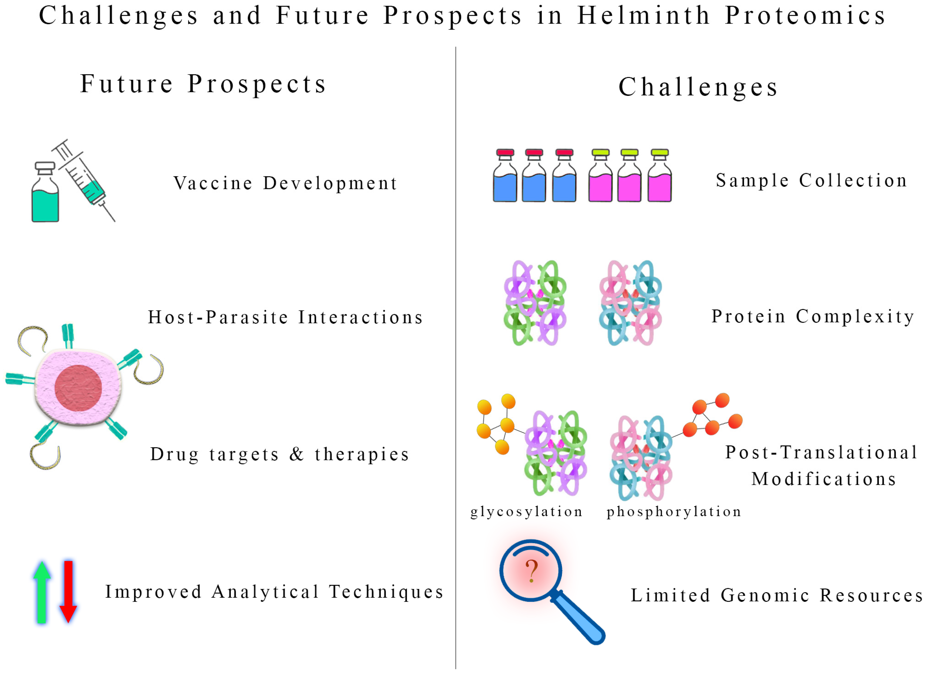
The host’s tissues may harbor helminths, making obtaining sufficient quantities of pure helminth proteins for analysis challenging. The isolation and purification of helminth proteins from the host’s complex milieu require meticulous techniques to ensure accurate results. Helminth proteomes are vast and highly complex, comprising various proteins with varying abundance levels. Identifying and quantifying low-abundance proteins become particularly difficult due to the presence of highly abundant host proteins in the samples.
Helminth proteins often undergo Post-Translational Modifications (PTMs), such as glycosylation and phosphorylation, significantly affecting their function and interaction with the host. Analyzing and characterizing PTMs in helminth proteins requires specialized methodologies and expertise. Many helminths have large and complex genomes, leading to difficulty assembling and annotating their proteomes accurately. Bioinformatics tools must be optimized to handle the vast data generated during proteomic analysis. Even with careful sample preparation, host contamination remains a concern in helminth proteomics. Host-derived proteins can mask or interfere with the identification of low-abundance helminth proteins. Limited genomic data are available for some helminth species, hindering the comprehensive analysis of their proteomes. More effort is needed to expand genomic resources for a wider range of helminths.
Helminth proteomics offers an invaluable resource for identifying potential vaccine candidates. Unraveling the proteins involved in host–parasite interactions and immune evasion can aid in developing anthelminthic vaccines. Understanding the proteomes of helminths may reveal essential drug targets that can disrupt their survival and reproduction. Proteomics can contribute to developing novel therapeutic strategies to combat helminth infections effectively. Helminth proteomics sheds light on the complex interplay between these parasites and their hosts. A deeper understanding of these interactions may lead to innovative strategies to modulate the host’s immune response, potentially preventing or ameliorating inflammatory and autoimmune diseases. Advancements in mass spectrometry and other proteomic technologies will enhance the sensitivity and accuracy of helminth proteomic analysis, enabling the identification of low-abundance and PTM-modified proteins.
Integrating proteomic data with genomic, transcriptomic, and metabolomic information can provide a more comprehensive view of helminth biology. Such multi-omics approaches will facilitate the identification of key regulatory pathways and virulence factors. The application of helminth proteomics may lead to the development of cost-effective diagnostics, vaccines, and therapeutics that can significantly improve public health, particularly in regions burdened by helminth infections (Figure 5).

9. Discussion

Three types of proteins related to helminths are identified: enzymes, cytoskeletal proteins, and proteins that bind to other proteins. Several major proteins were recognized, such as glycolytic and metabolic enzymes such as fructose-bisphosphate aldolase and aldehyde dehydrogenase. Glycolytic enzymes provide energy to parasites and modulate the interaction between the parasite and host, making them good candidates for vaccines and diagnostics. They inhibit parasite survival, migration, and evasion by acting on fructose-bisphosphate aldolase. In parasite infection, this enzyme may be responsible for pathogenicity and evasion. Enolase is found on the surface of nematodes as pathogens invade tissues. Inflammatory processes are prevented by glycosylated proteins, which encourage parasite invasion. Furthermore, glycosylated proteins can be involved in immune response, evasion, migration, pathology, and host survival.
It has been suggested that glycosylated proteins play a key role in initiating the immune system of parasites, which may elaborate on the pathogenicity of parasites. Proteins secreted by nematodes play a significant role in the host–parasite interaction. It was found that phosphoenolpyruvate carboxy kinase (GTP), Glyceraldehyde-3-phosphate dehydrogenase, and enolase were immunodominant proteins that regulate oxidative damage. Several important protein groups were identified as heat shock proteins, such as HSP60, HSP beta-1, HSP70, HSP varienr2, and HSP10. They function as protein chaperones and immune modulatory molecules found in helminths and play a vital role in parasite survival. Somatic extract proteins include HSP70, mitochondrial HSP60, and chitinases, which play an important role in the immune system. Parasite survival depends on evasion and modulation. Platyhelminthes also have HSP70, which plays an essential role in parasite survival and is a stress defense mechanism. Several studies have demonstrated that HSPs can stimulate IgG and IgM responses; therefore, they can be used as therapeutic agents for allergic and autoimmune diseases. Vaccines or anthelminthic therapies could be developed using HSPs because they are immunogenic.
Trematodes and Cestodes also contain ATP synthase subunit beta, another protein that may have been identified in nematodes. Galectins are soluble proteins that bind exclusively to sugars and exhibit the essential property of slowing down under decreasing conditions to maintain their activity when unable to bind to their targets. Galectins can enhance cell-to-cell adhesion. At the same time, some of them have robust biological properties, including the ability to induce apoptosis (scheduled cell death) and metabolic reactions, which can result in cellular activation and mitosis. In addition, Galectins modulate the cellular immune response and immune regulation and are associated with parasite–host interactions. In the immune response, galectins regulate immune cell functions.
The lectins can interact with glycoconjugates on the cell surface and in the extracellular matrix via carbohydrate interactions. Their action increases cell growth, promotes cell survival, modulates adhesions, and induces migration. Cells and tissues in helminths have 14-3-3 proteins within the cytoplasm, intracellular organelles, and the plasma membrane. The 14-3-3 proteins play a role in several processes essential to the survival of eukaryotic cells. These proteins play a role in metabolism as well as stress tolerance. Several studies have focused on the role of 14-3-3 proteins in parasite biology and immunology, making them an attractive target for further exploration of host–parasite relationships. In cell cycle regulation, cell survival, and differentiation, 14-3-3 proteins play an important role. Many other proteins have been identified as having metabolic functions, such as trans aldolase, sodium/potassium ATPase, calcium-transporting ATPase, aspartate aminotransferase, aspartate aminotransferase, glucose-6-phosphate-isomerase, glutathione peroxidase, ribonuclease, NADH dehydrogenase, malate dehydrogenase, the pyruvate dehydrogenase complex, glycogen synthase, pyruvate dehydrogenase, succinate dehydrogenase, acetyl-CoA, acetyltransferase A, phosphatidylinositol phosphatase, and nucleoside diphosphate kinase.
The structure and motor protein is one of the most essential molecules in helminths. The actin cytoskeleton plays a crucial role in the transfer of cells. The surface of several parasites contains actin. Moreover, paramyosin, a protein found in the muscles of many invertebrate animals, is also found in many helminths and is a core component of thick myosin filaments. It has been shown that paramyosin plays a critical role in various helminths and, therefore, can be considered a vaccine candidate or a target for anthelmintic therapies. Translated genomic and RNAseq data are unavailable for helminth proteins. Additionally, the characterization of proteins is vital to understanding host–parasite interactions, discovering drug targets, generating subunit vaccines against them, and creating immunodiagnostic kits in animals and humans, as well as the identification of new immunomodulatory methods for treating inflammatory diseases and new therapeutics for these diseases. More molecular investigations are needed to understand parasite–host relationships.

10. Conclusions

In conclusion, helminth proteomics represents a powerful and indispensable tool in unraveling the complex biology of parasitic organisms, their intricate interactions with the human host, and their pathogenicity. The future of helminth proteomics holds immense potential, particularly in developing vaccines and novel drug targets. The ability of proteomics to elucidate the molecular interactions and pathways that are affected in resistant strains is beneficial for creating novel anthelmintic drugs. Furthermore, proteomics has facilitated the development of multi-epitope vaccines by combining conserved antigens identified across various helminth species to develop vaccines.

Supplementary Materials

The following supporting information can be downloaded at: https://www.mdpi.com/article/10.3390/biologics4030020/s1, Table S1: Proteomics of Nematoda; Table S2: Proteomics of Trematoda; Table S3: Proteomics of Cestoda; Table S4: Function of proteins found in helminths.

Author Contributions

Conceptualization, H.B. and C.S.; software, S.S. and N.S.; validation, H.B., C.S., N.S. and S.S.; investigation, C.S., N.S., S.S., S.D., N.L., A.H. and F.H.; data curation, C.S., N.S., S.S., S.D., N.L., A.H. and F.H.; writing—original draft preparation, S.S.; writing—review and editing, C.S., N.S., S.S., S.D., N.L., A.H. and F.H.; visualization, H.B. and C.S.; supervision, H.B. and C.S.; project administration, H.B. and C.S.; funding acquisition, C.S. All authors have read and agreed to the published version of the manuscript.

Funding

This research received no external funding.

Institutional Review Board Statement

Not applicable.

Informed Consent Statement

Not applicable.

Data Availability Statement

The datasets generated during and/or analyzed during the current study are available from the corresponding author upon reasonable request.

Acknowledgments

The authors wish to thank Alireza Sazmand (Bu-Ali Sina University, Hamedan, Iran) for his critical comments. We thank the research deputy of the Ferdowsi University of Mashhad for supporting us.

Conflicts of Interest

The authors declare no conflicts of interest.

References

  1. Serin, Y.; Acar Tek, N. Effect of circadian rhythm on metabolic processes and the regulation of energy balance. Ann. Nutr. Metab. 2019, 74, 322–330. [Google Scholar] [CrossRef] [PubMed]
  2. Nechushtai, R.; Karmi, O.; Zuo, K.; Marjault, H.-B.; Darash-Yahana, M.; Sohn, Y.-S.; King, S.D.; Zandalinas, S.I.; Carloni, P.; Mittler, R. The balancing act of NEET proteins: Iron, ROS, calcium and metabolism. Biochim. Biophys. Acta Mol. Cell Res. 2020, 1867, 118805. [Google Scholar] [CrossRef] [PubMed]
  3. Kong, P.; Cui, Z.-Y.; Huang, X.-F.; Zhang, D.-D.; Guo, R.-J.; Han, M. Inflammation and atherosclerosis: Signaling pathways and therapeutic intervention. Signal Transduct. Target. Ther. 2022, 7, 131. [Google Scholar] [CrossRef] [PubMed]
  4. Dupree, E.J.; Jayathirtha, M.; Yorkey, H.; Mihasan, M.; Petre, B.A.; Darie, C.C. A critical review of bottom-up proteomics: The good, the bad, and the future of this field. Proteomes 2020, 8, 14. [Google Scholar] [CrossRef] [PubMed]
  5. Mergner, J.; Kuster, B. Plant proteome dynamics. Annu. Rev. Plant Biol. 2022, 73, 67–92. [Google Scholar] [CrossRef]
  6. Gajahin Gamage, N.T.; Miyashita, R.; Takahashi, K.; Asakawa, S.; Senevirathna, J.D.M. Proteomic applications in aquatic environment studies. Proteomes 2022, 10, 32. [Google Scholar] [CrossRef]
  7. Hood, L.E.; Omenn, G.S.; Moritz, R.L.; Aebersold, R.; Yamamoto, K.R.; Amos, M.; Hunter-Cevera, J.; Locascio, L.; Participants, W. New and improved proteomics technologies for understanding complex biological systems: Addressing a grand challenge in the life sciences. Proteomics 2012, 12, 2773–2783. [Google Scholar] [CrossRef]
  8. Zhou, H.; Ning, Z.; Starr, A.E.; Abu-Farha, M.; Figeys, D. Advancements in top-down proteomics. Anal. Chem. 2012, 84, 720–734. [Google Scholar] [CrossRef]
  9. O’Donnell, S.T.; Ross, R.P.; Stanton, C. The progress of multi-omics technologies: Determining function in lactic acid bacteria using a systems level approach. Front. Microbiol. 2020, 10, 3084. [Google Scholar] [CrossRef]
  10. Öztürk, M.; Freiwald, A.; Cartano, J.; Schmitt, R.; Dejung, M.; Luck, K.; Al-Sady, B.; Braun, S.; Levin, M.; Butter, F. Proteome effects of genome-wide single gene perturbations. Nat. Commun. 2022, 13, 6153. [Google Scholar] [CrossRef]
  11. Reitz, C.J.; Kuzmanov, U.; Gramolini, A.O. Multi-omic analyses and network biology in cardiovascular disease. Proteomics 2023, 23, 2200289. [Google Scholar] [CrossRef] [PubMed]
  12. Hotez, P.J.; Brindley, P.J.; Bethony, J.M.; King, C.H.; Pearce, E.J.; Jacobson, J. Helminth infections: The great neglected tropical diseases. J. Clin. Investig. 2008, 118, 1311–1321. [Google Scholar] [CrossRef] [PubMed]
  13. Ianiro, G.; Iorio, A.; Porcari, S.; Masucci, L.; Sanguinetti, M.; Perno, C.F.; Gasbarrini, A.; Putignani, L.; Cammarota, G. How the gut parasitome affects human health. Therap. Adv. Gastroenterol. 2022, 15, 17562848221091524. [Google Scholar] [CrossRef]
  14. Braseth, A.L.; Elliott, D.E.; Ince, M.N. Parasitic infections of the gastrointestinal track and liver. Gastroenterol. Clin. 2021, 50, 361–381. [Google Scholar] [CrossRef]
  15. Yeshi, K.; Ruscher, R.; Loukas, A.; Wangchuk, P. Immunomodulatory and biological properties of helminth-derived small molecules: Potential applications in diagnostics and therapeutics. Front. Parasitol. 2022, 1, 984152. [Google Scholar] [CrossRef]
  16. Stijlemans, B.; Caljon, G.; Van Den Abbeele, J.; Van Ginderachter, J.A.; Magez, S.; De Trez, C. Immune evasion strategies of Trypanosoma brucei within the mammalian host: Progression to pathogenicity. Front. Immunol. 2016, 7, 233. [Google Scholar] [CrossRef]
  17. Gómez-Arreaza, A.; Acosta, H.; Quiñones, W.; Concepción, J.L.; Michels, P.A.; Avilán, L. Extracellular functions of glycolytic enzymes of parasites: Unpredicted use of ancient proteins. Mol. Biochem. Parasitol. 2014, 193, 75–81. [Google Scholar] [CrossRef]
  18. Jackson, J.A.; Friberg, I.M.; Little, S.; Bradley, J.E. Review series on helminths, immune modulation and the hygiene hypothesis: Immunity against helminths and immunological phenomena in modern human populations: Coevolutionary legacies? Immunology 2009, 126, 18–27. [Google Scholar] [CrossRef] [PubMed]
  19. McSorley, H.J.; Maizels, R.M. Helminth infections and host immune regulation. Clin. Microbiol. Rev. 2012, 25, 585–608. [Google Scholar] [CrossRef]
  20. Sotillo, J.; Toledo, R.; Mulvenna, J.; Loukas, A. Exploiting helminth–host interactomes through big data. Trends Parasitol. 2017, 33, 875–888. [Google Scholar] [CrossRef]
  21. Gazzinelli-Guimarães, A.C.; Gazzinelli-Guimarães, P.; Weatherhead, J.E. A historical and systematic overview of Ascaris vaccine development. Parasitology 2021, 148, 1795–1805. [Google Scholar] [CrossRef] [PubMed]
  22. You, H.; Jones, M.K.; Gordon, C.A.; Arganda, A.E.; Cai, P.; Al-Wassiti, H.; Pouton, C.W.; McManus, D.P. The mRNA vaccine technology era and the future control of Parasitic infections. Clin. Microbiol. Rev. 2023, 36, e00241-21. [Google Scholar] [CrossRef] [PubMed]
  23. Okakpu, O.K.; Dillman, A.R. Review of the role of parasitic nematode excretory/secretory proteins in host immunomodulation. J. Parasitol. 2022, 108, 199–208. [Google Scholar] [CrossRef]
  24. Joshi, P.; Mishra, P.K.K. Functional diversity of the excretory/secretory proteins of nematode parasites. Acta Parasitol. 2022, 67, 619–627. [Google Scholar] [CrossRef]
  25. Sadr, S.; Ghiassi, S.; Lotfalizadeh, N.; Simab, P.A.; Hajjafari, A.; Borji, H. Antitumor mechanisms of molecules secreted by Trypanosoma cruzi in colon and breast cancer: A review. Anti-Cancer Agents Med. Chem. 2023, 23, 1710–1721. [Google Scholar] [CrossRef] [PubMed]
  26. Sadr, S.; Borji, H. Echinococcus granulosus as a promising therapeutic agent against triple-negative breast cancer. Curr. Cancer Ther. Rev. 2023, 19, 292–297. [Google Scholar] [CrossRef]
  27. Lotfalizadeh, N.; Sadr, S.; Morovati, S.; Lotfalizadeh, M.; Hajjafari, A.; Borji, H. A potential cure for tumor-associated immunosuppression by Toxoplasma gondii. Cancer Rep. 2024, 7, e1963. [Google Scholar] [CrossRef]
  28. Marcilla, A.; Trelis, M.; Cortés, A.; Sotillo, J.; Cantalapiedra, F.; Minguez, M.T.; Valero, M.L.; Sánchez del Pino, M.M.; Muñoz-Antoli, C.; Toledo, R. Extracellular vesicles from parasitic helminths contain specific excretory/secretory proteins and are internalized in intestinal host cells. PLoS ONE 2012, 7, e45974. [Google Scholar] [CrossRef]
  29. Sadr, S.; Yousefsani, Z.; Simab, P.A.; Alizadeh, A.J.R.; Lotfalizadeh, N.; Borji, H. Trichinella spiralis as a potential antitumor agent: An update. World’s Vet. J. 2023, 13, 65–74. [Google Scholar] [CrossRef]
  30. Wangchuk, P.; Kouremenos, K.; Eichenberger, R.M.; Pearson, M.; Susianto, A.; Wishart, D.S.; McConville, M.J.; Loukas, A. Metabolomic profiling of the excretory–secretory products of hookworm and whipworm. Metabolomics 2019, 15, 101. [Google Scholar] [CrossRef]
  31. Gomez-Fuentes, S.; Morales-Ruiz, V.; López-Recinos, D.; Guevara-Salinas, A.; Arce-Sillas, A.; Rodríguez, J.; Parada-Colin, C.; Adalid-Peralta, L. Biological role of excretory–secretory proteins in endemic parasites of Latin America and the Caribbean. J. Helminthol. 2020, 94, e53. [Google Scholar] [CrossRef]
  32. Wu, F.; Chen, X.; Du, Z.; Chen, Y.; Tong, D.; Zhang, J.; Yang, Y.; Ma, G.; Du, A. Proteomic differences between extracellular vesicles and extracellular vesicle-depleted excretory/secretory products of barber’s pole worm. Parasit. Vectors 2024, 17, 17. [Google Scholar] [CrossRef]
  33. Vanhamme, L.; Souopgui, J.; Ghogomu, S.; Ngale Njume, F. The functional parasitic worm secretome: Mapping the place of Onchocerca volvulus excretory secretory products. Pathogens 2020, 9, 975. [Google Scholar] [CrossRef]
  34. Hotterbeekx, A.; Perneel, J.; Vieri, M.K.; Colebunders, R.; Kumar-Singh, S. The secretome of filarial nematodes and its role in host-parasite interactions and pathogenicity in onchocerciasis-associated epilepsy. Front. Cell. Infect. Microbiol. 2021, 11, 662766. [Google Scholar] [CrossRef]
  35. Pan, W.; Shen, Y.; Han, X.; Wang, Y.; Liu, H.; Jiang, Y.; Zhang, Y.; Wang, Y.; Xu, Y.; Cao, J. Transcriptome profiles of the protoscoleces of Echinococcus granulosus reveal that excretory-secretory products are essential to metabolic adaptation. PLoS Negl. Trop. Dis. 2014, 8, e3392. [Google Scholar] [CrossRef]
  36. Huang, S.-Y.; Yue, D.-M.; Hou, J.-L.; Zhang, X.-X.; Zhang, F.-k.; Wang, C.-R.; Zhu, X.-Q. Proteomic analysis of Fasciola gigantica excretory and secretory products (FgESPs) interacting with buffalo serum of different infection periods by shotgun LC-MS/MS. Parasitol. Res. 2019, 118, 453–460. [Google Scholar] [CrossRef]
  37. Sánchez-López, C.M.; Trelis, M.; Bernal, D.; Marcilla, A. Overview of the interaction of helminth extracellular vesicles with the host and their potential functions and biological applications. Mol. Immunol. 2021, 134, 228–235. [Google Scholar] [CrossRef]
  38. White, R.R.; Artavanis-Tsakonas, K. How helminths use excretory secretory fractions to modulate dendritic cells. Virulence 2012, 3, 668–677. [Google Scholar] [CrossRef] [PubMed]
  39. van der Ree, A.M.; Mutapi, F. The helminth parasite proteome at the host–parasite interface–informing diagnosis and control. Exp. Parasitol. 2015, 157, 48–58. [Google Scholar] [CrossRef] [PubMed]
  40. Marzano, V.; Pane, S.; Foglietta, G.; Levi Mortera, S.; Vernocchi, P.; Onetti Muda, A.; Putignani, L. Mass spectrometry based-proteomic analysis of Anisakis spp.: A preliminary study towards a new diagnostic tool. Genes 2020, 11, 693. [Google Scholar] [CrossRef] [PubMed]
  41. Fissiha, W.; Kinde, M.Z. Anthelmintic resistance and its mechanism: A review. Infect. Drug Resist. 2021, 14, 5403–5410. [Google Scholar] [CrossRef] [PubMed]
  42. Shalaby, H.A. Anthelmintics resistance; how to overcome it? Iran. J. Parasitol. 2013, 8, 18–32. [Google Scholar]
  43. Sadr, S.; Ahmadi Simab, P.; Niazi, M.; Yousefsani, Z.; Lotfalizadeh, N.; Hajjafari, A.; Borji, H. Anti-inflammatory and immunomodulatory effects of mesenchymal stem cell therapy on parasitic drug resistance. Expert Rev. Anti-Infect. Ther. 2024, 22, 435–451. [Google Scholar] [CrossRef] [PubMed]
  44. Zaiss, M.M.; Harris, N.L. Interactions between the intestinal microbiome and helminth parasites. Parasite Immunol. 2016, 38, 5–11. [Google Scholar] [CrossRef]
  45. Moreno, Y.; Gros, P.-P.; Tam, M.; Segura, M.; Valanparambil, R.; Geary, T.G.; Stevenson, M.M. Proteomic analysis of excretory-secretory products of Heligmosomoides polygyrus assessed with next-generation sequencing transcriptomic information. PLOS. Negl. Trop. Dis. 2011, 5, e1370. [Google Scholar] [CrossRef]
  46. Maruszewska-Cheruiyot, M.; Szewczak, L.; Krawczak-Wójcik, K.; Głaczyńska, M.; Donskow-Łysoniewska, K. The production of excretory-secretory molecules from Heligmosomoides polygyrus bakeri fourth stage larvae varies between mixed and single sex cultures. Parasit. Vectors 2021, 14, 106. [Google Scholar] [CrossRef]
  47. Zhou, C.; Chen, J.; Niu, H.; Ouyang, S.; Wu, X. Study on the population evolution of Ascaris lumbricoides and Ascaris suum based on whole genome resequencing. Vet. Parasitol. 2020, 279, 109062. [Google Scholar] [CrossRef]
  48. Eamsobhana, P.; Yong, H.-S.; Boonyong, S.; Wanachiwanawin, D.; Tungtrongchitr, A. Genetic diversity and identity of Ascaris worms from human and pig hosts in Thailand. Vet. Parasitol. Reg. Stud. Rep. 2022, 33, 100752. [Google Scholar] [CrossRef]
  49. Leung, A.K.; Leung, A.A.; Wong, A.H.; Hon, K.L. Human ascariasis: An updated review. Recent. Pat. Inflamm. Allergy. Drug. Discov. 2020, 14, 133–145. [Google Scholar] [CrossRef] [PubMed]
  50. Fauziah, N.; Aviani, J.K.; Agrianfanny, Y.N.; Fatimah, S.N. Intestinal parasitic infection and nutritional status in children under five years old: A systematic review. Trop. Med. Infect. Dis. 2022, 7, 371. [Google Scholar] [CrossRef]
  51. Roesel, K.; Dohoo, I.; Baumann, M.; Dione, M.; Grace, D.; Clausen, P.-H. Prevalence and risk factors for gastrointestinal parasites in small-scale pig enterprises in Central and Eastern Uganda. J. Parasitol. Res. 2017, 116, 335–345. [Google Scholar] [CrossRef]
  52. Vismarra, A.; Lenti, A.; Genchi, M.; Kramer, L.; Geldhof, P. Seroprevalence of Ascaris suum compared to milk spot prevalence at slaughter in Italian fattening pigs. Vet. Parasitol. Reg. Stud. Rep. 2023, 37, 100828. [Google Scholar] [CrossRef]
  53. Urban Jr, J.F.; Douvres, F.W. In vitro development of Ascaris suum from third-to fourth-stage larvae and detection of metabolic antigens in multi-well culture systems. J. Parasitol. 1981, 67, 800–806. [Google Scholar] [CrossRef]
  54. Wang, T.; Van Steendam, K.; Dhaenens, M.; Vlaminck, J.; Deforce, D.; Jex, A.R.; Gasser, R.B.; Geldhof, P. Proteomic analysis of the excretory-secretory products from larval stages of Ascaris suum reveals high abundance of glycosyl hydrolases. PLOS. Negl. Trop. Dis. 2013, 7, e2467. [Google Scholar] [CrossRef]
  55. Okulewicz, A.; Perec-Matysiak, A.; Buńkowska, K.; Hildebrand, J. Toxocara canis Toxocara cati and Toxascaris leonina in wild and domestic carnivores. Helminthologia 2012, 49, 3–10. [Google Scholar] [CrossRef]
  56. Schwartz, R.; Bidaisee, S.; Fields, P.J.; Macpherson, M.L.; Macpherson, C.N. The epidemiology and control of Toxocara canis in puppies. Parasite Epidemiol. Control 2022, 8, e00232. [Google Scholar] [CrossRef]
  57. Choi, D.; Lim, J.H.; Choi, D.-C.; Lee, K.S.; Paik, S.W.; Kim, S.-H.; Choi, Y.-H.; Huh, S. Transmission of Toxocara canis via ingestion of raw cow liver: A cross-sectional study in healthy adults. Korean. J. Parasitol. 2012, 50, 23–27. [Google Scholar] [CrossRef]
  58. Omonijo, A.; Kalinda, C.; Mukaratirwa, S. A systematic review and meta-analysis of canine, feline and human Toxocara infections in sub-Saharan Africa. J. Helminthol. 2020, 94, e96. [Google Scholar] [CrossRef]
  59. da Silva, M.B.; Oviedo, Y.; Cooper, P.J.; Pacheco, L.G.; Pinheiro, C.S.; Ferreira, F.; Briza, P.; Alcantara-Neves, N.M. The somatic proteins of Toxocara canis larvae and excretory-secretory products revealed by proteomics. Vet. Parasitol. 2018, 259, 25–34. [Google Scholar] [CrossRef] [PubMed]
  60. Sperotto, R.L.; Kremer, F.S.; Berne, M.E.A.; de Avila, L.F.C.; da Silva Pinto, L.; Monteiro, K.M.; Caumo, K.S.; Ferreira, H.B.; Berne, N.; Borsuk, S. Proteomic analysis of Toxocara canis excretory and secretory (TES) proteins. Mol. Biochem. Parasitol. 2017, 211, 39–47. [Google Scholar] [CrossRef] [PubMed]
  61. Raulf, M.-K.; Lepenies, B.; Strube, C. Toxocara canis and Toxocara cati somatic and excretory-secretory antigens are recognised by C-type lectin receptors. Pathogens 2021, 10, 321. [Google Scholar] [CrossRef] [PubMed]
  62. Zheng, W.-B.; Zou, Y.; Zhu, X.-Q.; Liu, G.-H. Toxocara “omics” and the promises it holds for medicine and veterinary medicine. J. Adv. Parasitol. 2020, 109, 89–108. [Google Scholar] [CrossRef]
  63. Torkan, S.; Ghandehari-Alavijeh, M.; Khamesipour, F. Survey of the prevalence of Toxocara cati in stray cats in Isfahan city, Iran by PCR method. Trop. Biomed. 2017, 34, 550–555. [Google Scholar] [PubMed]
  64. Zanzani, S.A.; Gazzonis, A.L.; Scarpa, P.; Berrilli, F.; Manfredi, M.T. Intestinal parasites of owned dogs and cats from metropolitan and micropolitan areas: Prevalence, zoonotic risks, and pet owner awareness in northern Italy. Biomed. Res. Int. 2014, 2014, 696508. [Google Scholar] [CrossRef]
  65. Maciag, L.; Morgan, E.R.; Holland, C. Toxocara: Time to let cati ‘out of the bag’. Trends. Parasitol. 2022, 38, 280–289. [Google Scholar] [CrossRef]
  66. Wu, T.; Bowman, D.D. Visceral larval migrans of Toxocara canis and Toxocara cati in non-canid and non-felid hosts. Adv. Parasitol. 2020, 109, 63–88. [Google Scholar] [CrossRef]
  67. Abbas, I.; Al-Araby, M.; Elmishmishy, B.; El-Alfy, E.-S. Gastrointestinal parasites of cats in Egypt: High prevalence high zoonotic risk. BMC. Vet. Res. 2022, 18, 420. [Google Scholar] [CrossRef]
  68. Soleyman, N.M.; Darnhofer, B.; Gruenberger, R.B.; Abnous, K.; Borji, H. Proteomic analysis of soluble protein extract of adult Toxocara cati. Comp. Immunol. Microbiol. Infect. Dis. 2020, 73, 101528. [Google Scholar] [CrossRef]
  69. Soleymani, N.; Grunberger, R.B.; Abnous, K.; Borji, H.; Vahdati, F. Identification and immunological characterization of somatic proteins from adults of Toxocara cati by proteomics technique. Iran. J. Parasitol. 2021, 16, 23. [Google Scholar] [CrossRef]
  70. Yeshi, K.; Creek, D.J.; Anderson, D.; Ritmejerytė, E.; Becker, L.; Loukas, A.; Wangchuk, P. Metabolomes and lipidomes of the infective stages of the gastrointestinal nematodes, Nippostrongylus brasiliensis and Trichuris muris. Metabolites 2020, 10, 446. [Google Scholar] [CrossRef]
  71. Thuma, N.; Döhler, D.; Mielenz, D.; Sticht, H.; Radtke, D.; Reimann, L.; Warscheid, B.; Voehringer, D. A newly identified secreted larval antigen elicits basophil-dependent protective immunity against N. brasiliensis infection. Front. Immunol. 2022, 13, 979491. [Google Scholar] [CrossRef] [PubMed]
  72. Loukas, A.; Hotez, P.J.; Diemert, D.; Yazdanbakhsh, M.; McCarthy, J.S.; Correa-Oliveira, R.; Croese, J.; Bethony, J.M. Hookworm infection. Nat. Rev. Dis. Primers 2016, 2, 16088. [Google Scholar] [CrossRef] [PubMed]
  73. Sotillo, J.; Sanchez-Flores, A.; Cantacessi, C.; Harcus, Y.; Pickering, D.; Bouchery, T.; Camberis, M.; Tang, S.-C.; Giacomin, P.; Mulvenna, J. Secreted proteomes of different developmental stages of the gastrointestinal nematode Nippostrongylus brasiliensis. Mol. Cell. Proteom. 2014, 13, 2736–2751. [Google Scholar] [CrossRef] [PubMed]
  74. Hawdon, J.M.; Wise, K.A. Ancylostoma caninum and other canine hookworms. In Dog Parasites Endangering Human Health; Springer: Berlin/Heidelberg, Germany, 2021; pp. 147–193. [Google Scholar] [CrossRef]
  75. Traub, R.J. Ancylostoma ceylanicum, a re-emerging but neglected parasitic zoonosis. Int. J. Parasitol. 2013, 43, 1009–1015. [Google Scholar] [CrossRef]
  76. Mulvenna, J.; Hamilton, B.; Nagaraj, S.H.; Smyth, D.; Loukas, A.; Gorman, J.J. Proteomics analysis of the excretory/secretory component of the blood-feeding stage of the hookworm, Ancylostoma caninum. Mol. Cell. Proteom. 2009, 8, 109–121. [Google Scholar] [CrossRef]
  77. Ju, C.; Feng, Z.; Brindley, P.J.; McManus, D.P.; Han, Z.; Peng, J.-x.; Hu, W. Our wormy world: Genomics, proteomics and transcriptomics in East and Southeast Asia. Adv. Parasitol. 2010, 73, 327–371. [Google Scholar] [CrossRef]
  78. Crilly, J.P.; Evans, M.; Tähepõld, K.; Sargison, N. Haemonchosis: Dealing with the increasing threat of the barber’s pole worm. Livestock 2020, 25, 237–246. [Google Scholar] [CrossRef]
  79. Adduci, I.; Sajovitz, F.; Hinney, B.; Lichtmannsperger, K.; Joachim, A.; Wittek, T.; Yan, S. Haemonchosis in sheep and goats, control strategies and development of vaccines against Haemonchus contortus. Animals 2022, 12, 2339. [Google Scholar] [CrossRef]
  80. Besier, R.; Kahn, L.; Sargison, N.; Van Wyk, J. Diagnosis, treatment and management of Haemonchus contortus in small ruminants. Adv. Parasitol. 2016, 93, 181–238. [Google Scholar] [CrossRef]
  81. Zheng, Y.; Young, N.D.; Song, J.; Gasser, R.B. Genome-Wide Analysis of Haemonchus contortus Proteases and Protease Inhibitors Using Advanced Informatics Provides Insights into Parasite Biology and Host–Parasite Interactions. Int. J. Mol. Sci. 2023, 24, 12320. [Google Scholar] [CrossRef]
  82. De Vries, E.; Bakker, N.; Krijgsveld, J.; Knox, D.P.; Heck, A.J.; Yatsuda, A.P. An AC-5 cathepsin B-like protease purified from Haemonchus contortus excretory secretory products shows protective antigen potential for lambs. Vet. Res. 2009, 40, 41. [Google Scholar] [CrossRef] [PubMed]
  83. Yatsuda, A.P.; Bakker, N.; Krijgsveld, J.; Knox, D.P.; Heck, A.J.; de Vries, E. Identification of secreted cysteine proteases from the parasitic nematode Haemonchus contortus detected by biotinylated inhibitors. Infect. Immun. 2006, 74, 1989–1993. [Google Scholar] [CrossRef] [PubMed]
  84. Jasmer, D.P.; Mitreva, M.D.; McCarter, J.P. mRNA sequences for Haemonchus contortus intestinal cathepsin B-like cysteine proteases display an extreme in abundance and diversity compared with other adult mammalian parasitic nematodes. Mol. Biochem. Parasitol. 2004, 137, 297–305. [Google Scholar] [CrossRef]
  85. Hosseinnezhad, H.; Sharifdini, M.; Ashrafi, K.; Atrkar Roushan, Z.; Mirjalali, H.; Rahmati, B. Trichostrongyloid nematodes in ruminants of northern Iran: Prevalence and molecular analysis. BMC. Vet. Res. 2021, 17, 371. [Google Scholar] [CrossRef]
  86. Tafere, A.; Terefe, G.; Mamo, G.; Kaba, T.; Shiferaw, J. A Comparative Study on Pathological Changes in the Small Intestine of Sheep and Goat Experimentally Infected with Trichostrongylus colubriformis. Vet. Med. 2022, 13, 213–233. [Google Scholar] [CrossRef]
  87. Kaba, T.; Terefe, G.; Waktole, H. Sheep and goat response to Trichostrongylus colubriformis infection based on egg output and worm burden. Trop. Anim. Health. Prod. 2023, 55, 177. [Google Scholar] [CrossRef]
  88. Kiel, M.; Josh, P.; Jones, A.; Windon, R.; Hunt, P.; Kongsuwan, K. Identification of immuno-reactive proteins from a sheep gastrointestinal nematode, Trichostrongylus colubriformis, using two-dimensional electrophoresis and mass spectrometry. Int. J. Parasitol. 2007, 37, 1419–1429. [Google Scholar] [CrossRef]
  89. Ortega-Pierres, G.; Vaquero-Vera, A.; Fonseca-Linan, R.; Bermudez-Cruz, R.; Argüello-García, R. Induction of protection in murine experimental models against Trichinella spiralis: An up-to-date review. J. Helminthol. 2015, 89, 526–539. [Google Scholar] [CrossRef] [PubMed]
  90. Gao, X.; Yang, Y.; Liu, X.; Xu, F.; Wang, Y.; Liu, L.; Yang, Y.; Liu, M.; Bai, X. Extracellular vesicles from Trichinella spiralis: Proteomic analysis and protective immunity. PLoS Negl. Trop. Dis. 2022, 16, e0010528. [Google Scholar] [CrossRef]
  91. Xu, J.; Pang, Z.; Zhang, J.; Xia, S.; Wang, R.; Zhang, Y.; Zhen, J.; Song, X.; Lin, L.; Sun, F. Regulatory effects of Trichinella spiralis and a serine protease inhibitor on the endoplasmic reticulum stress response of intestinal epithelial cells. Vet. Res. 2022, 53, 18. [Google Scholar] [CrossRef]
  92. Thawornkuno, C.; Nogrado, K.; Adisakwattana, P.; Thiangtrongjit, T.; Reamtong, O. Identification and profiling of Trichinella spiralis circulating antigens and proteins in sera of mice with trichinellosis. PLoS ONE 2022, 17, e0265013. [Google Scholar] [CrossRef] [PubMed]
  93. Yang, X.; Yang, Y.; Wang, Y.; Zhan, B.; Gu, Y.; Cheng, Y.; Zhu, X. Excretory/secretory products from Trichinella spiralis adult worms ameliorate DSS-induced colitis in mice. PLoS ONE 2014, 9, e96454. [Google Scholar] [CrossRef] [PubMed]
  94. Liu, R.D.; Cui, J.; Liu, X.L.; Jiang, P.; Sun, G.G.; Zhang, X.; Long, S.R.; Wang, L.; Wang, Z.Q. Comparative proteomic analysis of surface proteins of Trichinella spiralis muscle larvae and intestinal infective larvae. Acta Trop. 2015, 150, 79–86. [Google Scholar] [CrossRef] [PubMed]
  95. Wang, X.; Li, L.; Wei, X.; Wang, Y.; Zhang, H.; Shi, A.; Liu, T.; Yang, X.; Fang, Q. Proteomic analysis of the response of Trichinella spiralis muscle larvae to exogenous nitric oxide. PLoS ONE 2018, 13, e0198205. [Google Scholar] [CrossRef]
  96. Liu, J.; Zhang, N.; Li, W.; Li, L.; Yan, H.; Qu, Z.; Li, T.; Cui, J.; Yang, Y.; Jia, W. Proteomic analysis of differentially expressed proteins in the three developmental stages of Trichinella spiralis. Vet. Parasitol. 2016, 231, 32–38. [Google Scholar] [CrossRef]
  97. Ren, H.N.; Liu, R.D.; Song, Y.Y.; Zhuo, T.X.; Guo, K.X.; Zhang, Y.; Jiang, P.; Wang, Z.Q.; Cui, J. Label-free quantitative proteomic analysis of molting-related proteins of Trichinella spiralis intestinal infective larvae. Vet. Res. 2019, 50, 70. [Google Scholar] [CrossRef]
  98. Cui, J.; Liu, R.D.; Wang, L.; Zhang, X.; Jiang, P.; Liu, M.Y.; Wang, Z.Q. Proteomic analysis of surface proteins of Trichinella spiralis muscle larvae by two-dimensional gel electrophoresis and mass spectrometry. Parasites Vectors 2013, 6, 355. [Google Scholar] [CrossRef]
  99. Ren, H.N.; Zhuo, T.X.; Bai, S.J.; Bai, Y.; Sun, X.Y.; Liu, R.D.; Long, S.R.; Cui, J.; Wang, Z.Q. Proteomic analysis of hydrolytic proteases in excretory/secretory proteins from Trichinella spiralis intestinal infective larvae using zymography combined with shotgun LC-MS/MS approach. Acta Trop. 2021, 216, 105825. [Google Scholar] [CrossRef]
  100. Jiang, H.-Y.; Zhao, N.; Zhang, Q.-L.; Gao, J.-M.; Liu, L.-L.; Wu, T.-F.; Wang, Y.; Huang, Q.-H.; Gou, Q.; Chen, W. Intestinal microbes influence the survival, reproduction and protein profile of Trichinella spiralis in vitro. Int. J. Parasitol. 2016, 46, 51–58. [Google Scholar] [CrossRef]
  101. Wang, Z.Q.; Wang, L.; Cui, J. Proteomic analysis of Trichinella spiralis proteins in intestinal epithelial cells after culture with their larvae by shotgun LC–MS/MS approach. J. Proteom. 2012, 75, 2375–2383. [Google Scholar] [CrossRef]
  102. Janwan, P.; Intapan, P.M.; Laummaunwai, P.; Rodpai, R.; Wongkham, C.; Insawang, T.; Thanchomnang, T.; Sanpool, O.; Maleewong, W. Proteomic analysis identification of antigenic proteins in Gnathostoma spinigerum larvae. Exp. Parasitol. 2015, 159, 53–58. [Google Scholar] [CrossRef] [PubMed]
  103. Soblik, H.; Younis, A.E.; Mitreva, M.; Renard, B.Y.; Kirchner, M.; Geisinger, F.; Steen, H.; Brattig, N.W. Life cycle stage-resolved proteomic analysis of the excretome/secretome from Strongyloides ratti—Identification of stage-specific proteases. Mol. Cell. Proteom. 2011, 10, M111.010157. [Google Scholar] [CrossRef] [PubMed]
  104. Vo, D.; Le, K.; Nguyen, T.; Nguyen, H. Identification of excretory/secretory antigens produced by L2 stage larvae of Toxocara canis involving in induction of IgG response in mice by proteomics approach. In 6th International Conference on the Development of Biomedical Engineering in Vietnam (BME6); Vo Van, T., Nguyen Le, T., Nguyen Duc, T., Eds.; BME 2017. IFMBE Proceedings; Springer: Singapore, 2017; Volume 63, pp. 633–636. [Google Scholar] [CrossRef]
  105. Dea-Ayuela, M.; Bolás-Fernández, F. Two-dimensional electrophoresis and mass spectrometry for the identification of species-specific Trichinella antigens. Vet. Parasitol. 2005, 132, 43–49. [Google Scholar] [CrossRef] [PubMed]
  106. Robinson, M.W.; Connolly, B. Proteomic analysis of the excretory-secretory proteins of the Trichinella spiralis L1 larva, a nematode parasite of skeletal muscle. Proteomics 2005, 5, 4525–4532. [Google Scholar] [CrossRef] [PubMed]
  107. Else, K.J.; Keiser, J.; Holland, C.V.; Grencis, R.K.; Sattelle, D.B.; Fujiwara, R.T.; Bueno, L.L.; Asaolu, S.O.; Sowemimo, O.A.; Cooper, P.J. Whipworm and roundworm infections. Nat. Rev. Dis. Primers 2020, 6, 44. [Google Scholar] [CrossRef]
  108. Bennett, A.P.; Robinson, M.W. Trematode proteomics: Recent advances and future directions. Pathogens 2021, 10, 348. [Google Scholar] [CrossRef]
  109. McManus, D.P.; Dalton, J.P. Vaccines against the zoonotic trematodes Schistosoma japonicum, Fasciola hepatica and Fasciola gigantica. Parasitology 2006, 133, 43–61. [Google Scholar]
  110. Toledo, R.; Bernal, M.D.; Marcilla, A. Proteomics of foodborne trematodes. J. Proteom. 2011, 74, 1485–1503. [Google Scholar] [CrossRef]
  111. Sripa, B.; Kaewkes, S.; Intapan, P.M.; Maleewong, W.; Brindley, P.J. Food-borne trematodiases in Southeast Asia: Epidemiology, pathology, clinical manifestation and control. Adv. Parasitol. 2010, 72, 305–350. [Google Scholar] [CrossRef]
  112. Moazeni, M.; Ahmadi, A. Controversial aspects of the life cycle of Fasciola hepatica. Exp. Parasitol. 2016, 169, 81–89. [Google Scholar] [CrossRef]
  113. Lalor, R.; Cwiklinski, K.; Calvani, N.E.D.; Dorey, A.; Hamon, S.; Corrales, J.L.; Dalton, J.P.; De Marco Verissimo, C. Pathogenicity and virulence of the liver flukes Fasciola hepatica and Fasciola gigantica that cause the zoonosis Fasciolosis. Virulence 2021, 12, 2839–2867. [Google Scholar] [CrossRef] [PubMed]
  114. Cwiklinski, K.; Jewhurst, H.; McVeigh, P.; Barbour, T.; Maule, A.G.; Tort, J.; O’Neill, S.M.; Robinson, M.W.; Donnelly, S.; Dalton, J.P. Infection by the helminth parasite Fasciola hepatica requires rapid regulation of metabolic, virulence, and invasive factors to adjust to its mammalian host. Mol. Cell. Proteom. 2018, 17, 792–809. [Google Scholar] [CrossRef] [PubMed]
  115. Robinson, M.W.; Menon, R.; Donnelly, S.M.; Dalton, J.P.; Ranganathan, S. An integrated transcriptomics and proteomics analysis of the secretome of the helminth pathogen Fasciola hepatica: Proteins associated with invasion and infection of the mammalian host. Mol. Cell. Proteom. 2009, 8, 1891–1907. [Google Scholar] [CrossRef]
  116. Cwiklinski, K.; Robinson, M.W.; Donnelly, S.; Dalton, J.P. Complementary transcriptomic and proteomic analyses reveal the cellular and molecular processes that drive growth and development of Fasciola hepatica in the host liver. BMC Genom. 2021, 22, 46. [Google Scholar] [CrossRef]
  117. Robinson, M.W.; Tort, J.F.; Lowther, J.; Donnelly, S.M.; Wong, E.; Xu, W.; Stack, C.M.; Padula, M.; Herbert, B.; Dalton, J.P. Proteomics and phylogenetic analysis of the cathepsin L protease family of the helminth pathogen Fasciola hepatica: Expansion of a repertoire of virulence-associated factors. Mol. Cell. Proteom. 2008, 7, 1111–1123. [Google Scholar] [CrossRef]
  118. Stack, C.; Dalton, J.P.; Robinson, M.W. The phylogeny, structure and function of trematode cysteine proteases, with particular emphasis on the Fasciola hepatica cathepsin L family. Cysteine proteases of pathogenic organisms. In Cysteine Proteases of Pathogenic Organisms; Springer: Berlin/Heidelberg, Germany, 2011; pp. 116–135. [Google Scholar] [CrossRef]
  119. McVeigh, P.; Maule, A.G.; Dalton, J.P.; Robinson, M.W. Fasciola hepatica virulence-associated cysteine peptidases: A systems biology perspective. Microbes Infect. 2012, 14, 301–310. [Google Scholar] [CrossRef]
  120. Cwiklinski, K.; Donnelly, S.; Drysdale, O.; Jewhurst, H.; Smith, D.; Verissimo, C.D.M.; Pritsch, I.C.; O’Neill, S.; Dalton, J.P.; Robinson, M.W. The cathepsin-like cysteine peptidases of trematodes of the genus Fasciola. Adv. Parasitol. 2019, 104, 113–164. [Google Scholar] [CrossRef] [PubMed]
  121. Barbour, T.; Cwiklinski, K.; Lalor, R.; Dalton, J.P.; De Marco Verissimo, C. The zoonotic helminth parasite Fasciola hepatica: Virulence-associated cathepsin B and cathepsin L cysteine peptidases secreted by infective newly excysted juveniles (NEJ). Animals 2021, 11, 3495. [Google Scholar] [CrossRef]
  122. Corrales, J.L.; Cwiklinski, K.; Verissimo, C.D.M.; Dorey, A.; Lalor, R.; Jewhurst, H.; McEvoy, A.; Diskin, M.; Duffy, C.; Cosby, S.L. Diagnosis of sheep fasciolosis caused by Fasciola hepatica using cathepsin L enzyme-linked immunosorbent assays (ELISA). Vet. Parasitol. 2021, 298, 109517. [Google Scholar] [CrossRef]
  123. Collett, C.F.; Phillips, H.C.; Fisher, M.; Smith, S.; Fenn, C.; Goodwin, P.; Morphew, R.M.; Brophy, P.M. Fasciola hepatica cathepsin L zymogens: Immuno-proteomic evidence for highly immunogenic zymogen-specific conformational epitopes to support diagnostics development. J. Proteome Res. 2022, 21, 1997–2010. [Google Scholar] [CrossRef]
  124. De Marco Verissimo, C.; Jewhurst, H.L.; Tikhonova, I.G.; Urbanus, R.T.; Maule, A.G.; Dalton, J.P.; Cwiklinski, K. Fasciola hepatica serine protease inhibitor family (serpins): Purposely crafted for regulating host proteases. PLoS Negl. Trop. Dis. 2020, 14, e0008510. [Google Scholar] [CrossRef] [PubMed]
  125. Lucius, R. Dicrocoelium dendriticum. Trends Parasitol. 2022, 38, 1089–1090. [Google Scholar] [CrossRef] [PubMed]
  126. Kahl, A.; von Samson-Himmelstjerna, G.; Krücken, J.; Ganter, M. Chronic wasting due to liver and rumen flukes in sheep. Animals 2021, 11, 549. [Google Scholar] [CrossRef] [PubMed]
  127. Cézilly, F.; Thomas, F.; Médoc, V.; Perrot-Minnot, M.-J. Host-manipulation by parasites with complex life cycles: Adaptive or not? Trends. Parasitol. 2010, 26, 311–317. [Google Scholar] [CrossRef]
  128. Martínez-Ibeas, A.; González-Lanza, C.; Manga-González, M.Y. Proteomic analysis of the tegument and excretory–secretory products of Dicrocoelium dendriticum (Digenea) adult worms. Exp. Parasitol. 2013, 133, 411–420. [Google Scholar] [CrossRef] [PubMed]
  129. Montaner, S.; Galiano, A.; Trelis, M.; Martin-Jaular, L.; Del Portillo, H.A.; Bernal, D.; Marcilla, A. The role of extracellular vesicles in modulating the host immune response during parasitic infections. Front. Immunol. 2014, 5, 433. [Google Scholar] [CrossRef]
  130. Bernal, D.; Trelis, M.; Montaner, S.; Cantalapiedra, F.; Galiano, A.; Hackenberg, M.; Marcilla, A. Surface analysis of Dicrocoelium dendriticum. The molecular characterization of exosomes reveals the presence of miRNAs. J. Proteom. 2014, 105, 232–241. [Google Scholar] [CrossRef]
  131. Rojo-Vázquez, F.A.; Meana, A.; Valcárcel, F.; Martínez-Valladares, M. Update on trematode infections in sheep. Vet. Parasitol. 2012, 189, 15–38. [Google Scholar] [CrossRef]
  132. Di Maggio, L.S.; Tirloni, L.; Pinto, A.F.; Diedrich, J.K.; Yates III, J.R.; Benavides, U.; Carmona, C.; da Silva Vaz Jr, I.; Berasain, P. Across intra-mammalian stages of the liver fluke Fasciola hepatica: A proteomic study. Sci. Rep. 2016, 6, 32796. [Google Scholar] [CrossRef]
  133. Lo, N.C.; Bezerra, F.S.M.; Colley, D.G.; Fleming, F.M.; Homeida, M.; Kabatereine, N.; Kabole, F.M.; King, C.H.; Mafe, M.A.; Midzi, N.; et al. Review of 2022 WHO guidelines on the control and elimination of schistosomiasis. Lancet. Infec. Dis 2022, 22, E327–E335. [Google Scholar] [CrossRef]
  134. Xue, Q.; Deng, Y.; Liu, Y.; Wang, Y.; Hu, W.; Huang, Y.; Yang, K. A retrospective analysis of schistosomiasis related literature from 2011-2020: Focusing on the next decade. Acta Trop. 2023, 238, 106750. [Google Scholar] [CrossRef] [PubMed]
  135. Lima, R.R.; Lima, J.V.; Ribeiro, J.F.; Nascimento, J.B.; Oliveira, W.F.; Cabral Filho, P.E.; Fontes, A. Emerging biomedical tools for biomarkers detection and diagnostics in schistosomiasis. Talanta 2023, 265, 124900. [Google Scholar] [CrossRef] [PubMed]
  136. Lowe, C.; Ahmadabadi, Z.; Gray, D.; Kelly, M.; McManus, D.P.; Williams, G. Systematic review of applied mathematical models for the control of Schistosoma japonicum. Acta Trop. 2023, 241, 106873. [Google Scholar] [CrossRef] [PubMed]
  137. Liu, F.; Lu, J.; Hu, W.; Wang, S.-Y.; Cui, S.-J.; Chi, M.; Yan, Q.; Wang, X.-R.; Song, H.-D.; Xu, X.-N. New perspectives on host-parasite interplay by comparative transcriptomic and proteomic analyses of Schistosoma japonicum. PLoS Pathog. 2006, 2, e29. [Google Scholar] [CrossRef]
  138. Tritten, L.; Geary, T.G. Helminth extracellular vesicles in host–parasite interactions. Curr. Opin. Microbiol. 2018, 46, 73–79. [Google Scholar] [CrossRef]
  139. Liao, Q.; Yuan, X.; Xiao, H.; Liu, C.; Lv, Z.; Zhao, Y.; Wu, Z. Identifying Schistosoma japonicum excretory/secretory proteins and their interactions with host immune system. PLoS ONE 2011, 6, e23786. [Google Scholar] [CrossRef] [PubMed]
  140. Yang, L.-l.; Lv, Z.-y.; Hu, S.-m.; He, S.-j.; Li, Z.-y.; Zhang, S.-m.; Zheng, H.-q.; Li, M.-t.; Yu, X.-b.; Fung, M.-C. Schistosoma japonicum: Proteomics analysis of differentially expressed proteins from ultraviolet-attenuated cercariae compared to normal cercariae. Parasitol. Res. 2009, 105, 237–248. [Google Scholar] [CrossRef]
  141. Cao, X.; Fu, Z.; Zhang, M.; Han, Y.; Han, Q.; Lu, K.; Li, H.; Zhu, C.; Hong, Y.; Lin, J. Excretory/secretory proteome of 14-day schistosomula, Schistosoma japonicum. J. Proteom. 2016, 130, 221–230. [Google Scholar] [CrossRef]
  142. Han, Z.-G.; Brindley, P.J.; Wang, S.-Y.; Chen, Z. Schistosoma genomics: New perspectives on schistosome biology and host-parasite interaction. Annu. Rev. Genomics. Hum. Genet. 2009, 10, 211–240. [Google Scholar] [CrossRef]
  143. Cortés, A.; Muñoz-Antoli, C.; Sotillo, J.; Fried, B.; Esteban, J.; Toledo, R. Echinostoma caproni (T rematoda): Differential in vivo mucin expression and glycosylation in high-and low-compatible hosts. Parasit. Immunol. 2015, 37, 32–42. [Google Scholar] [CrossRef]
  144. Trelis, M.; Sotillo, J.; Monteagudo, C.; Fried, B.; Marcilla, A.; Esteban, J.G.; Toledo, R. Echinostoma caproni (Trematoda): Differential in vivo cytokine responses in high and low compatible hosts. Exp. Parasitol. 2011, 127, 387–397. [Google Scholar] [CrossRef] [PubMed]
  145. Eichenberger, R.M.; Sotillo, J.; Loukas, A. Immunobiology of parasitic worm extracellular vesicles. Immunol. Cell Biol. 2018, 96, 704–713. [Google Scholar] [CrossRef] [PubMed]
  146. Cortés, A.; Sotillo, J.; Muñoz-Antolí, C.; Trelis, M.; Esteban, J.G.; Toledo, R. Definitive host influences the proteomic profile of excretory/secretory products of the trematode Echinostoma caproni. Parasites Vectors 2016, 9, 185. [Google Scholar] [CrossRef]
  147. Toledo, R.; Espert, A.; Carpena, I.; Muñoz-Antoli, C.; Fried, B.; Esteban, J.G. Immunological characterization of somatic and excretory–secretory antigens of Echinostoma caproni (Trematoda: Echinostomatidae) in experimentally infected rats. Comp. Parasitol. 2004, 71, 42–48. [Google Scholar] [CrossRef]
  148. Sotillo, J.; Valero, L.; Sanchez Del Pino, M.; Fried, B.; Esteban, J.; Marcilla, A.; Toledo, R. Identification of antigenic proteins from Echinostoma caproni (Trematoda) recognized by mouse immunoglobulins M, A and G using an immunoproteomic approach. Parasite Immunol. 2008, 30, 271–279. [Google Scholar] [CrossRef]
  149. Higón, M.; Monteagudo, C.; Fried, B.; Esteban, J.; Toledo, R.; Marcilla, A. Molecular cloning and characterization of Echinostoma caproni heat shock protein-70 and differential expression in the parasite derived from low-and high-compatible hosts. Parasitology 2008, 135, 1469–1477. [Google Scholar] [CrossRef]
  150. Sotillo, J.; Valero, M.L.; Sanchez Del Pino, M.M.; Fried, B.; Esteban, J.G.; Marcilla, A.; Toledo, R. Excretory/secretory proteome of the adult stage of Echinostoma caproni. Parasitol. Res. 2010, 107, 691–697. [Google Scholar] [CrossRef]
  151. Toledo, R.; Esteban, J.G.; Fried, B. Chapter 3 recent advances in the biology of echinostomes. Adv. Parasitol. 2009, 69, 147–204. [Google Scholar] [CrossRef] [PubMed]
  152. Casulli, A.; Massolo, A.; Saarma, U.; Umhang, G.; Santolamazza, F.; Santoro, A. Species and genotypes belonging to Echinococcus granulosus sensu lato complex causing human cystic echinococcosis in Europe (2000–2021): A systematic review. Parasites Vectors 2022, 15, 109. [Google Scholar] [CrossRef]
  153. Tamarozzi, F.; Legnardi, M.; Fittipaldo, A.; Drigo, M.; Cassini, R. Epidemiological distribution of Echinococcus granulosus s.l. infection in human and domestic animal hosts in European Mediterranean and Balkan countries: A systematic review. PLoS Neg. Trop. Dis. 2020, 14, e0008519. [Google Scholar] [CrossRef]
  154. Karshima, S.N.; Ahmed, M.I.; Adamu, N.B.; Magaji, A.A.; Zakariah, M.; Mohammed, K. Africa-wide meta-analysis on the prevalence and distribution of human cystic echinococcosis and canine Echinococcus granulosus infections. Parasites Vectors 2022, 15, 357. [Google Scholar] [CrossRef] [PubMed]
  155. Pal, M.; Alemu, H.H.; Marami, L.M.; Garedo, D.R.; Bodena, E.B. Cystic Echincoccoosis: A comprehensive review on life cycle, epidemiology, pathogenesis, clinical spectrum, diagnosis, public health and economic implications, treatment, and control. Int. J. Clin. Exp. Med. Res. 2022, 6, 131–141. [Google Scholar] [CrossRef]
  156. Alshoabi, S.A.; Alkalady, A.H.; Almas, K.M.; Magram, A.O.; Algaberi, A.K.; Alareqi, A.A.; Hamid, A.M.; Alhazmi, F.H.; Qurashi, A.A.; Abdulaal, O.M. Hydatid disease: A radiological pictorial review of a great neoplasms mimicker. Diagnostics 2023, 13, 1127. [Google Scholar] [CrossRef] [PubMed]
  157. Gessese, A.T. Review on epidemiology and public health significance of hydatidosis. Vet. Med. Int. 2020, 2020, 8859116. [Google Scholar] [CrossRef]
  158. Tamarozzi, F.; Deplazes, P.; Casulli, A. Reinventing the wheel of Echinococcus granulosus sensu lato transmission to humans. Trends. Parasitol. 2020, 36, 427–434. [Google Scholar] [CrossRef]
  159. Woolsey, I.D.; Miller, A.L. Echinococcus granulosus sensu lato and Echinococcus multilocularis: A review. Res. Vet. Sci. 2021, 135, 517–522. [Google Scholar] [CrossRef]
  160. Ohiolei, J.A.; Yan, H.-B.; Odeniran, P.O.; Li, L.; Shumuye, N.A.; Qurishi, S.A.; Isaac, C.; Fu, B.-Q.; Jia, W.-Z. Echinococcus granulosus sensu lato in animal intermediate hosts: What is with the organ location? Vet. Parasitol. 2022, 304, 109695. [Google Scholar] [CrossRef]
  161. Serra, E.; Masu, G.; Chisu, V.; Cappai, S.; Masala, G.; Loi, F.; Piseddu, T. Environmental contamination by Echinococcus spp. eggs as a risk for human health in educational farms of Sardinia, Italy. Vet. Sci. 2022, 9, 143. [Google Scholar] [CrossRef]
  162. Zhang, X.; Gong, W.; Cao, S.; Yin, J.; Zhang, J.; Cao, J.; Shen, Y. Comprehensive analysis of non-coding RNA profiles of exosome-like vesicles from the protoscoleces and hydatid cyst fluid of Echinococcus granulosus. Front. Cell. Infect. Microbiol. 2020, 10, 316. [Google Scholar] [CrossRef]
  163. Wen, H.; Vuitton, L.; Tuxun, T.; Li, J.; Vuitton, D.A.; Zhang, W.; McManus, D.P. Echinococcosis: Advances in the 21st century. Clin. Microbiol. Rev. 2019, 32, 10–1128. [Google Scholar] [CrossRef]
  164. Debarba, J.A.; Monteiro, K.M.; Moura, H.; Barr, J.R.; Ferreira, H.B.; Zaha, A. Identification of newly synthesized proteins by Echinococcus granulosus protoscoleces upon induction of strobilation. PLoS Negl. Trop. Dis. 2015, 9, e0004085. [Google Scholar] [CrossRef] [PubMed]
  165. Monteiro, K.M.; de Carvalho, M.O.; Zaha, A.; Ferreira, H.B. Proteomic analysis of the Echinococcus granulosus metacestode during infection of its intermediate host. Proteomics 2010, 10, 1985–1999. [Google Scholar] [CrossRef] [PubMed]
  166. Diaz, A.; Casaravilla, C.; Barrios, A.A.; Ferreira, A.M. Parasite molecules and host responses in cystic echinococcosis. Parasite Immunol. 2016, 38, 193–205. [Google Scholar] [CrossRef]
  167. Cancela, M.; Paes, J.A.; Moura, H.; Barr, J.R.; Zaha, A.; Ferreira, H.B. Unraveling oxidative stress response in the cestode parasite Echinococcus granulosus. Sci. Rep. 2019, 9, 15876. [Google Scholar] [CrossRef]
  168. Wang, Y.; Cheng, Z.; Lu, X.; Tang, C. Echinococcus multilocularis: Proteomic analysis of the protoscoleces by two-dimensional electrophoresis and mass spectrometry. Exp. Parasitol. 2009, 123, 162–167. [Google Scholar] [CrossRef]
  169. Siracusano, A.; Delunardo, F.; Teggi, A.; Ortona, E. Host-parasite relationship in cystic echinococcosis: An evolving story. J. Immunol. Res. 2012, 639362. [Google Scholar] [CrossRef] [PubMed]
  170. Hewitson, J.P.; Maizels, R.M. Vaccination against helminth parasite infections. Expert Rev. Vaccines 2014, 13, 473–487. [Google Scholar] [CrossRef]
  171. Wu, Z.; Wang, L.; Li, J.; Wang, L.; Wu, Z.; Sun, X. Extracellular vesicle-mediated communication within host-parasite interactions. Front. Immunol. 2019, 9, 3066. [Google Scholar] [CrossRef]
  172. Zeghir-Bouteldja, R.; Touil-Boukoffa, C. Identification of proteins of laminated layer of Echinococcus granulosus: Interface among host and parasite. Veterinaria 2022, 71, 53–60. [Google Scholar] [CrossRef]
  173. Wang, Y.; Xiao, D.; Shen, Y.; Han, X.; Zhao, F.; Li, X.; Wu, W.; Zhou, H.; Zhang, J.; Cao, J. Proteomic analysis of the excretory/secretory products and antigenic proteins of Echinococcus granulosus adult worms from infected dogs. BMC Vet. Res. 2015, 11, 1–7. [Google Scholar] [CrossRef]
  174. Drurey, C.; Coakley, G.; Maizels, R.M. Extracellular vesicles: New targets for vaccines against helminth parasites. Int. J. Parasitol. 2020, 50, 623–633. [Google Scholar] [CrossRef] [PubMed]
  175. Makepeace, B.L.; Martin, C.; Turner, J.D.; Specht, S. Granulocytes in helminth infection-who is calling the shots? Curr. Med. Chem. 2012, 19, 1567–1586. [Google Scholar] [CrossRef]
  176. Schmidt, V.; O’Hara, M.-C.; Ngowi, B.; Herbinger, K.-H.; Noh, J.; Wilkins, P.P.; Richter, V.; Kositz, C.; Matuja, W.; Winkler, A.S. Taenia solium cysticercosis and taeniasis in urban settings: Epidemiological evidence from a health-center based study among people with epilepsy in Dar es Salaam, Tanzania. PLoS Negl. Trop. Dis. 2019, 13, e0007751. [Google Scholar] [CrossRef]
  177. Acosta Soto, L.; Parker, L.A.; Irisarri-Gutiérrez, M.J.; Bustos, J.A.; Castillo, Y.; Perez, E.; Muñoz-Antoli, C.; Esteban, J.G.; García, H.H.; Bornay-Llinares, F.J. Evidence for transmission of Taenia solium taeniasis/cysticercosis in a rural area of Northern Rwanda. Front. Vet. Sci. 2021, 8, 645076. [Google Scholar] [CrossRef]
  178. Sciutto, E.; Fragoso, G.; Fleury, A.; Laclette, J.P.; Sotelo, J.; Aluja, A.; Vargas, L.; Larralde, C. Taenia solium disease in humans and pigs: An ancient parasitosis disease rooted in developing countries and emerging as a major health problem of global dimensions. Microbes Infect. 2000, 2, 1875–1890. [Google Scholar] [CrossRef] [PubMed]
  179. Wu, H.-W.; Ito, A.; Ai, L.; Zhou, X.-N.; Acosta, L.P.; Willingham III, A.L. Cysticercosis/taeniasis endemicity in Southeast Asia: Current status and control measures. Acta Trop. 2017, 165, 121–132. [Google Scholar] [CrossRef] [PubMed]
  180. Ito, A.; Yanagida, T.; Nakao, M. Recent advances and perspectives in molecular epidemiology of Taenia solium cysticercosis. Infect. Genet. Evol. 2016, 40, 357–367. [Google Scholar] [CrossRef] [PubMed]
  181. Victor, B.; Kanobana, K.; Gabriël, S.; Polman, K.; Deckers, N.; Dorny, P.; Deelder, A.M.; Palmblad, M. Proteomic analysis of Taenia solium metacestode excretion-secretion proteins. Proteomics 2012, 12, 1860–1869. [Google Scholar] [CrossRef]
  182. Santivanez, S.; Hernandez-Gonzalez, A.; Chile, N.; Oleaga, A.; Arana, Y.; Palma, S.; Verastegui, M.; Gonzalez, A.; Gilman, R.; Garcia, H. Proteomic study of activated Taenia solium oncospheres. Mol. Biochem. Parasitol. 2010, 171, 32–39. [Google Scholar] [CrossRef]
  183. Kaur, R.; Arora, N.; Jamakhani, M.A.; Malik, S.; Kumar, P.; Anjum, F.; Tripathi, S.; Mishra, A.; Prasad, A. Development of multi-epitope chimeric vaccine against Taenia solium by exploring its proteome: An in silico approach. Expert Rev. Vaccines 2020, 19, 105–114. [Google Scholar] [CrossRef]
  184. Li, L.; He, W.; Fan, X.; Liu, M.; Luo, B.; Yang, F.; Jiang, N.; Wang, L.; Zhou, B. Proteomic analysis of Taenia solium cysticercus and adult stages. Front. Vet. Sci. 2023, 9, 934197. [Google Scholar] [CrossRef]
  185. Navarrete-Perea, J.; Isasa, M.; Paulo, J.A.; Corral-Corral, R.; Flores-Bautista, J.; Hernández-Téllez, B.; Bobes, R.J.; Fragoso, G.; Sciutto, E.; Soberón, X. Quantitative multiplexed proteomics of Taenia solium cysts obtained from the skeletal muscle and central nervous system of pigs. PLoS Negl. Trop. Dis. 2017, 11, e0005962. [Google Scholar] [CrossRef]
  186. Arora, N.; Prasad, A. Taenia solium proteins: A beautiful kaleidoscope of pro and anti-inflammatory antigens. Expert Rev. Proteom. 2020, 17, 609–622. [Google Scholar] [CrossRef]
  187. Lee, E.G.; Kim, S.H.; Bae, Y.A.; Chung, J.Y.; Suh, M.; Na, B.K.; Kim, T.S.; Kang, I.; Ma, L.; Kong, Y. A hydrophobic ligand-binding protein of the Taenia solium metacestode mediates uptake of the host lipid: Implication for the maintenance of parasitic cellular homeostasis. Proteomics 2007, 7, 4016–4030. [Google Scholar] [CrossRef]
  188. Kim, S.-H.; Bae, Y.-A.; Yang, Y.; Hong, S.-T.; Kong, Y. Paralogous proteins comprising the 150 kDa hydrophobic-ligand-binding-protein complex of the Taenia solium metacestode have evolved non-overlapped binding affinities toward fatty acid analogs. Int. J. Parasitol. 2011, 41, 1207–1215. [Google Scholar] [CrossRef]
  189. Kim, S.-H.; Bae, Y.-A.; Yang, H.-J.; Shin, J.-H.; Diaz-Camacho, S.P.; Nawa, Y.; Kang, I.; Kong, Y. Structural and binding properties of two paralogous fatty acid binding proteins of Taenia solium metacestode. PLoS Negl. Trop. Dis. 2012, 6, e1868. [Google Scholar] [CrossRef]
  190. Kaur, R.; Arora, N.; Rawat, S.S.; Keshri, A.K.; Singh, G.; Kumar, R.; Prasad, A. Recognition of immune reactive proteins as a potential multiepitope vaccine candidate of Taenia solium cysticerci through proteomic approach. J. Cell. Biochem. 2023, 124, 1587–1602. [Google Scholar] [CrossRef]
  191. Kaur, R.; Arora, N.; Rawat, S.S.; Keshri, A.K.; Sharma, S.R.; Mishra, A.; Singh, G.; Prasad, A. Vaccine for a neglected tropical disease Taenia solium cysticercosis: Fight for eradication against all odds. Expert Rev. Vaccines 2021, 20, 1447–1458. [Google Scholar] [CrossRef]
  192. Jarošová, J.; Antolová, D.; Iglodyová, A.; Königová, A.; Dolinská, M.U.; Víchová, B. Molecular identification of Taenia hydatigena from domestic and free-living animals in Slovakia, Central Europe. Parasitol. Res. 2022, 121, 1345–1354. [Google Scholar] [CrossRef]
  193. Ulziijargal, G.; Yeruult, C.; Khulan, J.; Gantsetseg, C.; Wandra, T.; Yamasaki, H.; Narankhajid, M. Molecular identification of Taenia hydatigena and Mesocestoides species based on copro-DNA analysis of wild carnivores in Mongolia. Int. J. Parasitol. Parasit. Wild Life 2020, 11, 72–82. [Google Scholar] [CrossRef]
  194. Velusamy, R.; Annamalai, L.; Vijayasarathi, M.K. Parasites in the gastrointestinal system of dogs and cats. In Organ-Specific Parasitic Diseases of Dogs and Cats; Elsevier: Amsterdam, The Netherlands, 2023; pp. 205–238. [Google Scholar] [CrossRef]
  195. Wang, S.; Liu, X.; Liu, Z.; Wang, Y.; Guo, A.; Huang, W.; Wang, Q.; Zhang, S.; Zhu, G.; Luo, X. The genome of the thin-necked bladder worm Taenia hydatigena reveals evolutionary strategies for helminth survival. Commun. Biol. 2021, 4, 1004. [Google Scholar] [CrossRef]
  196. Cai, M.; Li, Y.; He, G.; Guo, X.; Zhang, S.; Yan, L.; Zhang, J.; Ding, J. Comparative Proteomic Analysis of Different Parts of Taenia Hydatigena. Front. Vet. Sci. 2021, 8, 626579. [Google Scholar] [CrossRef]
  197. Zheng, Y. Proteomic analysis of Taenia hydatigena cyst fluid reveals unique internal microenvironment. Acta Trop. 2017, 176, 224–227. [Google Scholar] [CrossRef]
  198. Guo, X. Proteomics analysis of Hydatigera taeniaeformis metacestode stage. Front. Vet. Sci. 2020, 7, 474. [Google Scholar] [CrossRef]
  199. Knox, D.P. Development of vaccines against gastrointestinal nematodes. Parasitology 2000, 120, 43–61. [Google Scholar] [CrossRef]
  200. Panti-May, J.A.; Rodríguez-Vivas, R.I.; García-Prieto, L.; Servián, A.; Costa, F. Worldwide overview of human infections with Hymenolepis diminuta. Parasitol. Res. 2020, 119, 1997–2004. [Google Scholar] [CrossRef]
  201. Thompson, R. Neglected zoonotic helminths: Hymenolepis nana, Echinococcus canadensis and Ancylostoma ceylanicum. Clin. Microbiol. Infect. 2015, 21, 426–432. [Google Scholar] [CrossRef]
  202. Sulima-Celińska, A.; Kalinowska, A.; Młocicki, D. The Tapeworm Hymenolepis diminuta as an important model organism in the experimental parasitology of the 21st century. Pathogens 2022, 11, 1439. [Google Scholar] [CrossRef]
  203. Galoș, F.; Anghel, M.; Ioan, A.; Ieșanu, M.-I.; Boboc, C.; Boboc, A.A. Hymenolepis diminuta infection in a Romanian Child from an urban area. Pathogens 2022, 11, 322. [Google Scholar] [CrossRef]
  204. Zawistowska-Deniziak, A.; Basałaj, K.; Strojny, B.; Młocicki, D. New data on human macrophages polarization by Hymenolepis diminuta tapeworm-an in-vitro study. Front. Immunol. 2017, 8, 148. [Google Scholar] [CrossRef]
  205. Sulima, A.; Savijoki, K.; Bień, J.; Näreaho, A.; Sałamatin, R.; Conn, D.B.; Młocicki, D. Comparative proteomic analysis of Hymenolepis diminuta cysticercoid and adult stages. Front. Microbiol. 2018, 8, 2672. [Google Scholar] [CrossRef] [PubMed]
  206. Mazanec, H.; Koník, P.; Gardian, Z.; Kuchta, R. Extracellular vesicles secreted by model tapeworm Hymenolepis diminuta: Biogenesis, ultrastructure and protein composition. Int. J. Parasitol. 2021, 51, 327–332. [Google Scholar] [CrossRef] [PubMed]
  207. Sulima, A.; Bień, J.; Savijoki, K.; Näreaho, A.; Sałamatin, R.; Conn, D.B.; Młocicki, D. Identification of immunogenic proteins of the cysticercoid of Hymenolepis diminuta. Parasites Vectors 2017, 10, 577. [Google Scholar] [CrossRef] [PubMed]
  208. Bień, J.; Sałamatin, R.; Sulima, A.; Savijoki, K.; Conn, D.B.; Näreaho, A.; Młocicki, D. Mass spectrometry analysis of the excretory-secretory (ES) products of the model cestode Hymenolepis diminuta reveals their immunogenic properties and the presence of new ES proteins in cestodes. Acta Parasitol. 2016, 61, 429–442. [Google Scholar] [CrossRef]
  209. Młocicki, D.; Sulima, A.; Bień, J.; Näreaho, A.; Zawistowska-Deniziak, A.; Basałaj, K.; Sałamatin, R.; Conn, D.B.; Savijoki, K. Immunoproteomics and surfaceomics of the adult tapeworm Hymenolepis diminuta. Front. Immunol. 2018, 9, 2487. [Google Scholar] [CrossRef]
  210. Chemale, G.; van Rossum, A.J.; Jefferies, J.R.; Barrett, J.; Brophy, P.M.; Ferreira, H.B.; Zaha, A. Proteomic analysis of the larval stage of the parasite Echinococcus granulosus: Causative agent of cystic hydatid disease. PROTEOMICS Int. Ed. 2003, 3, 1633–1636. [Google Scholar] [CrossRef]
  211. Monteiro, K.M.; Lorenzatto, K.R.; de Lima, J.C.; Dos Santos, G.B.; Förster, S.; Paludo, G.P.; Carvalho, P.C.; Brehm, K.; Ferreira, H.B. Comparative proteomics of hydatid fluids from two Echinococcus multilocularis isolates. J. Proteom. 2017, 162, 40–51. [Google Scholar] [CrossRef]
  212. Blanco, M.G.; De Rosa, M.J.; Rayes, D. Anthelmintic drug discovery: Current situation and future perspectives. Front. Clin. Drug Res. Anti-Infect. 2021, 8, 209. [Google Scholar] [CrossRef]
  213. Riaz, A.; Bano, F.; Marescotti, M.; Saba, E.; Manzoor, Z. Anthelmintic Resistance. In Antiparasitic Drug Resistance in Veterinary Practice; CAB International 2024; Rizwan, H.M., Naeem, M.A., Younus, M., Sajid, M.S., Chen, X., Eds.; CAB International: Willingford, UK, 2024; pp. 41–57. [Google Scholar] [CrossRef]
  214. McVeigh, P. Post-genomic progress in helminth parasitology. Parasitology 2020, 147, 835–840. [Google Scholar] [CrossRef]
  215. Kim, E.; Park, S.; Park, H.; Choi, J.; Yoon, H.J.; Kim, J.-H. Determination of anthelmintic and antiprotozoal drug residues in fish using liquid chromatography-tandem mass spectrometry. Molecules 2021, 26, 2575. [Google Scholar] [CrossRef]
  216. Morales-Montor, J.; Del Río-Araiza, V.H.; Hernandéz-Bello, R. Parasitic Helminths and Zoonoses: From Basic to Applied Research; BoD–Books on Demand: Norderstedt, Germany, 2022. [Google Scholar]
  217. Malik, M.A.; Sajid, M.S.; Abbas, R.Z.; Aleem, M.T.; Anjum, F.R.; Khan, A.; Farhab, M.; Maqbool, M.; Zeeshan, M.; Hussain, K. Anthelmintic drug resistance in livestock: Current understanding and future trends. In Parasitic Helminths and Zoonoses-From Basic to Applied Research; IntechOpen: London, UK, 2022. [Google Scholar]
  218. Khodadadi, E.; Zeinalzadeh, E.; Taghizadeh, S.; Mehramouz, B.; Kamounah, F.S.; Khodadadi, E.; Ganbarov, K.; Yousefi, B.; Bastami, M.; Kafil, H.S. Proteomic applications in antimicrobial resistance and clinical microbiology studies. Infect. Drug Resist. 2020, 13, 1785–1806. [Google Scholar] [CrossRef] [PubMed]
  219. Rastogi, S.; Rastogi, P.; Mendiratta, N. Bioinformatics: Methods and Applications-Genomics, Proteomics and Drug Discovery; PHI Learning Pvt. Ltd.: New Delhi, India, 2022. [Google Scholar]
  220. Chagas, A.C.S.; Ribeiro, D.M.; Osório, H.; Abreu, A.A.; Okino, C.H.; Niciura, S.C.; Amarante, A.F.; Bello, H.J.; Melito, G.R.; Esteves, S.N. Molecular signatures of Haemonchus contortus infection in sheep: A comparative serum proteomic study on susceptible and resistant sheep breeds. Vet. Parasitol. 2024, 331, 110280. [Google Scholar] [CrossRef] [PubMed]
  221. Ibarra-Meneses, A.V.; Corbeil, A.; Wagner, V.; Beaudry, F.; do Monte-Neto, R.L.; Fernandez-Prada, C. Exploring direct and indirect targets of current antileishmanial drugs using a novel thermal proteomics profiling approach. Front. Cell. Infect. Microbiol. 2022, 12, 954144. [Google Scholar] [CrossRef]
  222. Ahire, D.; Kruger, L.; Sharma, S.; Mettu, V.S.; Basit, A.; Prasad, B. Quantitative proteomics in translational absorption, distribution, metabolism, and excretion and precision medicine. Pharmacol. Rev. 2022, 74, 771–798. [Google Scholar] [CrossRef]
  223. Seebacher, N.A.; Krchniakova, M.; Stacy, A.E.; Skoda, J.; Jansson, P.J. Tumour microenvironment stress promotes the development of drug resistance. Antioxidants 2021, 10, 1801. [Google Scholar] [CrossRef]
  224. Saracino, M.P.; Vila, C.C.; Baldi, P.C.; Gonzalez Maglio, D.H. Searching for the one (s): Using probiotics as anthelmintic treatments. Front. Pharmacol. 2021, 12, 714198. [Google Scholar] [CrossRef]
  225. Rahime, Ş.; Aqsa, F.; Salah-Ud-Din, K.; Shahanavaj, K. Parasitic infections: Immune responses and therapeutics. Chapter 1, Back to the future‒solutions for parasitic problems. In Parasitic Infections: Immune Responses and Therapeutics; Wiley: Hoboken, NJ, USA, 2023. [Google Scholar] [CrossRef]
  226. Zhang, C.; Liang, C.; Zhang, C.; Ma, Y. Symbiosis evolution model and behavior of multiple resource agents in the smart elderly care service ecosystem. Symmetry 2021, 13, 570. [Google Scholar] [CrossRef]
  227. McIntyre, J.R. Genetic Markers of Anthelmintic Resistance in Gastrointestinal Parasites of Ruminants. Ph.D. Thesis, University of Glasgow, Glasgow, UK, 2020. [Google Scholar]
  228. Liu, Y.; Wang, X.; Luo, X.; Wang, R.; Zhai, B.; Wang, P.; Li, J.; Yang, X. Transcriptomics and proteomics of Haemonchus contortus in response to Ivermectin treatment. Animals 2023, 13, 919. [Google Scholar] [CrossRef] [PubMed]
  229. Rasmussen, C.; Alonso, P.; Ringwald, P. Current and emerging strategies to combat antimalarial resistance. Expert Rev. Anti-infect. Ther. 2022, 20, 353–372. [Google Scholar] [CrossRef]
  230. Wang, T.; Gasser, R.B. Prospects of using high-throughput proteomics to underpin the discovery of animal host–nematode interactions. Pathogens 2021, 10, 825. [Google Scholar] [CrossRef]
  231. Vashisht, V.; Vashisht, A.; Mondal, A.K.; Farmaha, J.; Alptekin, A.; Singh, H.; Ahluwalia, P.; Srinivas, A.; Kolhe, R. Genomics for emerging pathogen identification and monitoring: Prospects and obstacles. BioMedInformatics 2023, 3, 1145–1177. [Google Scholar] [CrossRef]
  232. Geddes-McAlister, J.; Roux-Dalvai, F.; Droit, A. Proteomics, bioinformatics, and infectious diseases. In Genetics and Evolution of Infectious Diseases; Elsevier: Amsterdam, The Netherlands, 2024; pp. 465–492. [Google Scholar] [CrossRef]
  233. Wijnant, G.-J.; Dumetz, F.; Dirkx, L.; Bulté, D.; Cuypers, B.; Van Bocxlaer, K.; Hendrickx, S. Tackling drug resistance and other causes of treatment failure in leishmaniasis. Front. Trop. Dis. 2022, 3, 837460. [Google Scholar] [CrossRef]
  234. Peraman, R.; Sure, S.K.; Dusthackeer, V.A.; Chilamakuru, N.B.; Yiragamreddy, P.R.; Pokuri, C.; Kutagulla, V.K.; Chinni, S. Insights on recent approaches in drug discovery strategies and untapped drug targets against drug resistance. Futur. J. Pharm. Sci. 2021, 7, 56. [Google Scholar] [CrossRef] [PubMed]
  235. Chulanetra, M.; Chaicumpa, W. Revisiting the mechanisms of immune evasion employed by human parasites. Front. Cell. Infect. Microbiol. 2021, 11, 702125. [Google Scholar] [CrossRef]
  236. Van den Kerkhof, M.; Sterckx, Y.G.-J.; Leprohon, P.; Maes, L.; Caljon, G. Experimental strategies to explore drug action and resistance in kinetoplastid parasites. Microorganisms 2020, 8, 950. [Google Scholar] [CrossRef] [PubMed]
  237. Sukumaran, A.; Woroszchuk, E.; Ross, T.; Geddes-McAlister, J. Proteomics of host–bacterial interactions: New insights from dual perspectives. Can. J. Microbiol. 2021, 67, 213–225. [Google Scholar] [CrossRef]
  238. Vasala, A.; Hytönen, V.P.; Laitinen, O.H. Modern tools for rapid diagnostics of antimicrobial resistance. Front. Cell. Infect. Microbiol. 2020, 10, 308. [Google Scholar] [CrossRef]
  239. Aboshady, H.M.; Stear, M.J.; Johansson, A.; Jonas, E.; Bambou, J.-C. Immunoglobulins as biomarkers for gastrointestinal nematodes resistance in small ruminants: A systematic review. Sci. Rep. 2020, 10, 7765. [Google Scholar] [CrossRef]
  240. Bhanot, P. Introduction to Parasites. In Practical Handbook of Microbiology; CRC Press: Boca Raton, FL, USA, 2021; pp. 665–682. [Google Scholar] [CrossRef]
  241. Azad, A.K.; Hakim, A.; Sohag, M.M.H.; Rahman, M. Metabolomics in clinical diagnosis, prognosis, and treatment of infectious diseases. In Metabolomics; Elsevier: Amsterdam, The Netherlands, 2023; pp. 71–119. [Google Scholar] [CrossRef]
  242. Rivara-Espasandín, M.; Palumbo, M.C.; Sosa, E.J.; Radío, S.; Turjanski, A.G.; Sotelo-Silveira, J.; Fernandez Do Porto, D.; Smircich, P. Omics data integration facilitates target selection for new antiparasitic drugs against TriTryp infections. Front. Pharmacol. 2023, 14, 1136321. [Google Scholar] [CrossRef]
  243. Sotillo, J.; Pearson, M.; Becker, L.; Mulvenna, J.; Loukas, A. A quantitative proteomic analysis of the tegumental proteins from Schistosoma mansoni schistosomula reveals novel potential therapeutic targets. Int. J. Parasitol. 2015, 45, 505–516. [Google Scholar] [CrossRef]
  244. Shao, G.; Hua, R.; Song, H.; Chen, Y.; Zhu, X.; Hou, W.; Li, S.; Yang, A.; Yang, G. Protective efficacy of six recombinant proteins as vaccine candidates against Echinococcus granulosus in dogs. PLoS Negl. Trop. Dis. 2023, 17, e0011709. [Google Scholar] [CrossRef] [PubMed]
  245. Sheng, Z.-A.; Wu, C.-L.; Wang, D.-Y.; Zhong, S.-H.; Yang, X.; Rao, G.-S.; Peng, H.; Feng, S.-W.; Li, J.; Huang, W.-Y. Proteomic analysis of exosome-like vesicles from Fasciola gigantica adult worm provides support for new vaccine targets against fascioliasis. Parasites Vectors 2023, 16, 62. [Google Scholar] [CrossRef] [PubMed]
  246. Dicker, A.J.; Inglis, N.F.; Manson, E.D.; Subhadra, S.; Illangopathy, M.; Muthusamy, R.; Knox, D.P. Proteomic analysis of Mecistocirrus digitatus and Haemonchus contortus intestinal protein extracts and subsequent efficacy testing in a vaccine trial. PLoS Negl. Trop. Dis. 2014, 8, e2909. [Google Scholar] [CrossRef] [PubMed]
  247. Sharma, S.; Sharan, U.; Kaur, R.; Rawat, S.S.; Keshri, A.K.; Prasad, A. An Inclusive Approach to Design a Multi-Epitope Chimeric Vaccine for Taenia Infections by Integrating Proteomics and Reverse Vaccinology. Front. Trop. Dis. 2024, 5, 1393570. [Google Scholar] [CrossRef]
  248. Castro-Borges, W.; Dowle, A.; Curwen, R.S.; Thomas-Oates, J.; Wilson, R.A. Enzymatic shaving of the tegument surface of live schistosomes for proteomic analysis: A rational approach to select vaccine candidates. PLoS Negl. Trop. Dis. 2011, 5, e993. [Google Scholar] [CrossRef]
  249. Sotillo, J.; Doolan, D.; Loukas, A. Recent advances in proteomic applications for schistosomiasis research: Potential clinical impact. Expert Rev. Proteom. 2017, 14, 171–183. [Google Scholar] [CrossRef]
  250. Miles, S.; Portela, M.; Cyrklaff, M.; Ancarola, M.E.; Frischknecht, F.; Durán, R.; Dematteis, S.; Mourglia-Ettlin, G. Combining proteomics and bioinformatics to explore novel tegumental antigens as vaccine candidates against Echinococcus granulosus infection. J. Cell. Biochem. 2019, 120, 15320–15336. [Google Scholar] [CrossRef]
  251. Hewitson, J.P.; Ivens, A.C.; Harcus, Y.; Filbey, K.J.; McSorley, H.J.; Murray, J.; Bridgett, S.; Ashford, D.; Dowle, A.A.; Maizels, R.M. Secretion of protective antigens by tissue-stage nematode larvae revealed by proteomic analysis and vaccination-induced sterile immunity. PLoS Pathog. 2013, 9, e1003492. [Google Scholar] [CrossRef]
  252. Newton, S.E.; Meeusen, E.N.T. Progress and new technologies for developing vaccines against gastrointestinal nematode parasites of sheep. Parasit. Immunol. 2003, 25, 283–296. [Google Scholar] [CrossRef]
  253. Rehman, A.; Ahmad, S.; Shahid, F.; Albutti, A.; Alwashmi, A.S.; Aljasir, M.A.; Alhumeed, N.; Qasim, M.; Ashfaq, U.A.; Tahir ul Qamar, M. Integrated core proteomics, subtractive proteomics, and immunoinformatics investigation to unveil a potential multi-epitope vaccine against schistosomiasis. Vaccines 2021, 9, 658. [Google Scholar] [CrossRef]
  254. Mutapi, F. Helminth parasite proteomics: From experimental models to human infections. Parasitology 2012, 139, 1195–1204. [Google Scholar] [CrossRef] [PubMed]
  255. Wilson, R.A.; Curwen, R.S.; Braschi, S.; Hall, S.L.; Coulson, P.S.; Ashton, P.D. From genomes to vaccines via the proteome. Mem. Inst. Oswaldo Cruz 2004, 99, 45–50. [Google Scholar] [CrossRef] [PubMed]
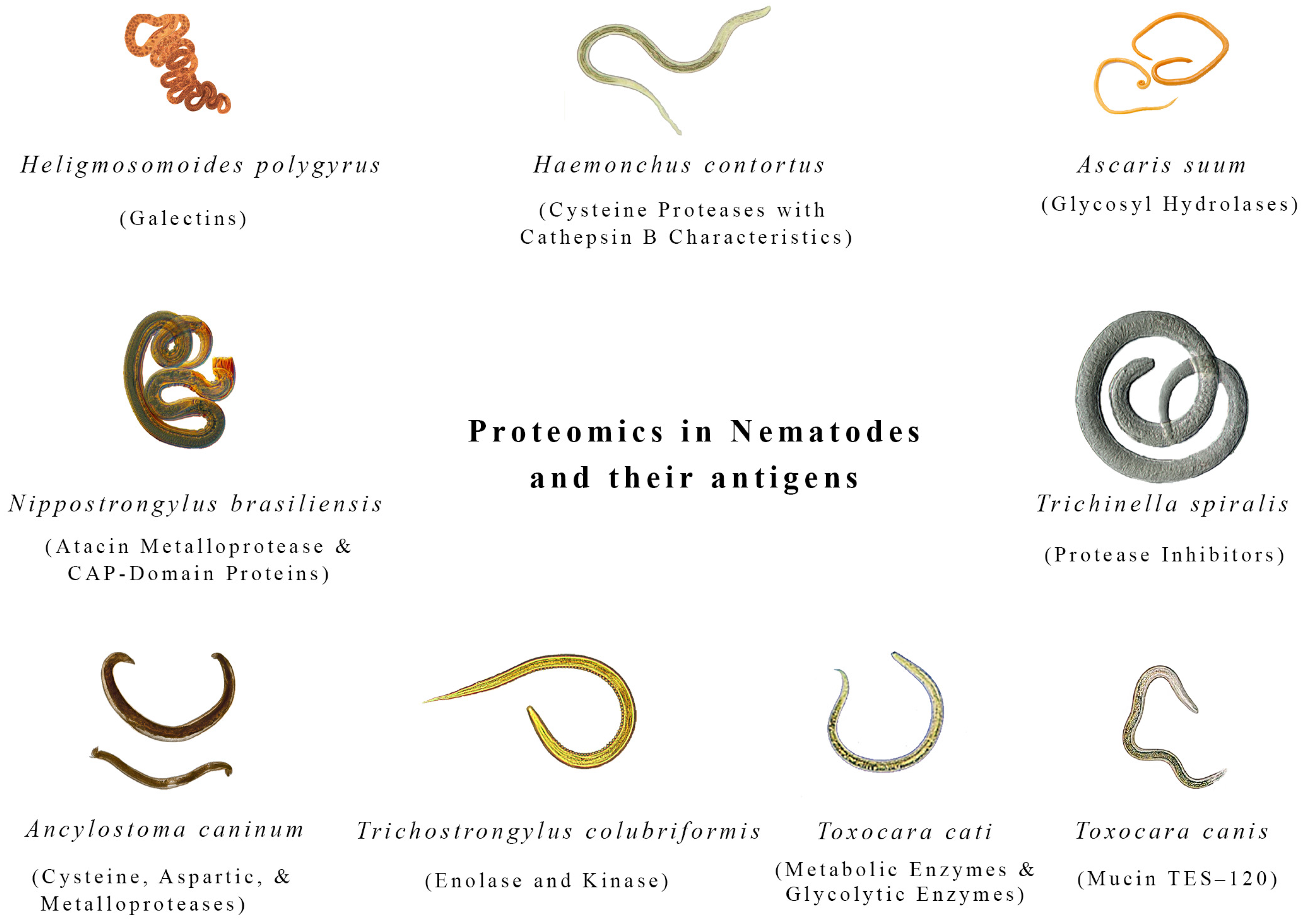
Figure 1. Most important proteomics of nematodes.
Biologics 04 00020 g001
Figure 2. Most important proteomics of trematodes and cestodes.
Biologics 04 00020 g002
Figure 3. Critical roles of proteomics in combating anthelmintic resistance. It compares resistant and non-resistant parasites, highlighting the identification of specific proteins and pathways altered in resistance. Biomarkers for early detection are discovered and represented by a diagnostic tool that identifies resistance markers and new drug targets through proteomics, with a drug molecule aimed at a key protein essential for the parasite’s survival. The comparative chart shows the enhanced efficacy of a newly developed drug over an older one, emphasizing the potential of proteomics-driven therapies in overcoming resistance.
Biologics 04 00020 g003
Figure 4. Role of proteomics in vaccine development against helminth infections through various key processes. It highlights a helminth with specific proteins, such as glucose transport protein-1 (SGTP1), tetraspanin 2 (TSP-2), and calpain, identified as potential vaccine targets on the surface of Schistosoma mansoni. Additionally, the Figure shows an immune response triggered by immunogenic proteins such as Eg14-3-3 and EgEF-1β, emphasizing their critical role in vaccine efficacy. The design of multi-epitope vaccines using proteomics and bioinformatics is also depicted, with a focus on selecting antigenic epitopes for broad immune protection against multiple helminth species. Furthermore, the Figure illustrates the surface of a helminth with proteins exposed through enzymatic shaving, highlighting their importance as targets for vaccines that enhance immune recognition and response.
Biologics 04 00020 g004
Figure 5. Navigating challenges and envisioning futures: a visual exploration of helminth proteomics. This comprehensive Figure encapsulates the challenges and prospects in helminth proteomics. The challenges, depicted through vivid illustrations, include the intricacies of sample collection symbolized by a hand attempting to collect a sample from host tissues, the complexity of helminth proteomes illustrated by a diverse array of proteins, icons representing post-translational modifications (glycosylation and phosphorylation processes), and the limited genomic resources visualized by a genome symbol with a magnifying glass. On the flip side, the prospects are artfully represented: a vaccine syringe injecting helminth proteins signifies the potential of helminth proteomics in vaccine development; a crosshair targeting helminth proteins illustrates the identification of drug targets and therapies; a handshake symbolizing interactions between host and helminth proteins signifies the insights gained into host–parasite interactions; and an advanced microscope or mass spectrometer depicts the improved analytical techniques that may revolutionize helminth proteomic analysis.
Biologics 04 00020 g005
Disclaimer/Publisher’s Note: The statements, opinions and data contained in all publications are solely those of the individual author(s) and contributor(s) and not of MDPI and/or the editor(s). MDPI and/or the editor(s) disclaim responsibility for any injury to people or property resulting from any ideas, methods, instructions or products referred to in the content.