Review

Biomechanical Behavior of Female Breast—A Review

Department of Biomedical, Industrial, and Human Factors Engineering, Wright State University, Dayton, OH 45435, USA
*
Author to whom correspondence should be addressed.
Submission received: 8 November 2024 / Revised: 19 January 2025 / Accepted: 23 January 2025 / Published: 26 January 2025

Abstract

:
Introduction: Women wear exterior breast support for most of the day. A female’s breast tissue and skin affect the comfort of the ADLs, exercise, health, and work environments. Understanding the breast tissue’s normal anatomy and mechanical and material properties is related to a woman’s daily health and quality of life outcomes. Considering the importance and impact of female breasts throughout one’s lifespan, additional research is needed to address the research gaps to provide solutions to improve daily lives and clinical interventions. Breast stability and behavior are dependent on its internal mechanical properties and applied external forces. Objective: To evaluate the current knowledge and research gaps on the adult female breast tissue’s anatomy, the factors that impact its growth and development, variations among racial populations, the internal and external mechanical properties of the tissue, and the factors employed to evaluate the pathology risk. Review sections: The review sections are as follows: 3. Anatomy of Breast, 4. Effects of Age and Stages of Breast Development, 5. Breast Skin, and 6. Breast Tissue Mechanics. Conclusions: Numerous research gaps have been identified within the field of female breasts.

1. Introduction

Women perform a multitude of physical activities while wearing poorly fitting personal protective equipment (PPE) and clothing that possesses a safety hazard and which produces physical pain [1]. Often, women have no option but to wear improperly fitting PPE and clothing in the work environment because of limited female anthropometry. A shocking 80% of women wear bras that do not have an optimal fit, of which 70% are the wrong size and 10% are too big [2,3]. The limitation on these values is that they include a narrow range of women’s ages and may lack racial diversity. The external mechanical forces exerted by clothing and wearables on the breasts may adversely impede movement, resulting in pain, discomfort, and reduced performance [1]. Continually wearing poorly fitting, unsupportive bras contribute to back pain and headaches, thus exacerbating discomfort [4]. Therefore, a greater understanding of the anatomical structure, material properties, and biomechanical behavior of female breast tissue is necessary to develop solutions to provide well-fitting bras, clothing, and equipment to support a higher quality of life [4].
The number of hours a woman wears a bra daily can vary based on lifestyle and preference. Published research has reported that 72% of women may wear a bra for more than 10 h a day, whereas Indonesian women may can wear one for more than 24 h [2,5]. A literature review has not produced research that investigated the correlation between lifestyle and cultural norm effects on the length of time a bra is worn. Despite the reduced breast movement when wearing a sports bra, which results in greater comfort during exercise, women with larger breast sizes tend to spend less time with physical activity [6,7]. Continued research is required to understand the ideal applied compression to achieve optimal comfort without sacrificing the range of motion. Understanding breast kinetics may assist manufacturers in producing better-fitting braziers and aid in reconstruction surgery and treatments [7,8]. Prior to examining the female breast kinematics, the anatomy and standard values of the mechanical properties should be established to provide a range of normal values.
A greater understanding of female breast kinetics has clinical, manufacturing, and work environment applications that may encourage retention in numerous career fields [7,9,10,11]. Variables such as age, ethnicity, body mass index (BMI), pregnancy, breastfeeding, menopause, weight fluctuation, and hormonal therapy can affect breast attributes, such as the shape and size [7,12,13]. The breast tissue undergoes mechanical changes over the female’s lifespan due to menstruation, pregnancy, lactation, menopause, and diseased tissue [14]. The lack of consensus and sparse information on breast tissue material and mechanical properties may have contributed to the variability seen in the experimental methodologies, demographics, and specimens [14,15]. Breast tissue is located internally, whereas skin is the first layer, which is located externally [7,16,17,18]. Breast stability and behavior are dependent on its internal mechanical properties and applied external forces.
This review’s objective is to explore the adult female breast tissue’s anatomy, the factors that impact its growth and development, racial variations, and internal and external effects, and characteristics that may determine pathological risk. Meeting this objective will provide insight into the research gaps and allow for the evaluation of the relationships between female breast anatomy, clinical applications, mechanical and material properties, and the variables that affect female breast tissue. Considering the importance and impact of female breasts throughout one’s lifespan, additional research is needed to address the research gaps to provide solutions to improve daily lives and clinical interventions. This will provide integral information that could be applied to the manufacturing, clinical, and future research sectors.

2. Methodology

This review was derived from examining over 135 published peer-reviewed research articles to understand the current knowledge and research gaps in the female breast. Numerous published documents were reviewed to ascertain the gaps in the research and the current knowledge on the female breast. The Firefox and Google Chrome browsers were used to access databases, such as PubMed and the National Institutes of Health (NIH), to locate and review the vast amount of information available on this expansive topic. The inclusion criteria required that 50% of the references be published peer-reviewed articles between the years of 2019 and 2024, written in English, and about adult female breast tissue. The keywords used in conjunction with adult female breast tissue were as follows: anatomy, individual female breast tissue components, growth and development, racial variations, mechanical properties, force effects, stiffness, density, and energy density because of strain. While gathering information on female breast tissue, keywords such as skin, BI-RADS, stress strain curves, and tissue expanders were explored. The images in this paper were created by utilizing information from peer-reviewed articles and purchasing a BioRender.com license, in 22 October 2023.

3. Anatomy of Breast

Understanding the anatomy, the stages of breast development, and the variables that affect female breast tissue will provide the foundation to build an understanding on female breast tissue. A multitude of anatomical factors (i.e., skin, adipose tissue, and Cooper’s ligaments) contribute to the unique shape and size of the female breast [19,20]. Breast tissue predominantly consists of fat and glandular tissue, with the role of supporting breastfeeding [10]. Since heterogenous breast tissue does not have skeletal support, ligaments attach it to the chest wall [10,21]. The breast rests on the pectoralis major, and its boundary extends from the chest wall to the mid-sternum, the axilla, and the latissimus dorsi [10,21]. Hormones such as estrogen, progesterone, and prolactin are primarily responsible for breast tissue changes, such as during the menstrual cycle, which occurs around every 28 days [21,22].
Before delving into the mechanics and material properties of the breast tissue, the anatomical structure and the variables that cause changes in it should be discussed. Figure 1 is an image of the anatomical location of the females breast subcomponents that forms the whole breast.

3.1. Rib Cage

The rib cage consists of 12 bones originating from the sternal notch to the xiphoid process [26,27]. The second to the sixth rib cage, clavicle, and sternum provide foundational support for the breast tissue [10,28]. The second to the sixth rib also have a role in the entire rib cage diameter, which increases and decreases during the respiration cycle [10,27,28]. The breath stacking method and targeted anthropometric measurements have inferred that increased age coupled with an obese BMI are correlated with a reduction in rib cage expansion when compared to those with an optimal BMI and in younger females [29]. The control group included those with a BMI within normal limits that had a total volume of the pulmonary rib cage [29]. The first group was classified as obese and did not have abdominal breathing, with a total volume of 33% [29]. The second group was also classified as obese and did not have abdominal breathing, with a total volume of 15% [29]. Research has concluded that the stiffness between each rib (ribs two, four, six, and eight) does not show variability; with an elastic modulus range of 2,000,000–14,000,000 kPa and an ultimate strength of 100 MPa [10].

3.2. Pectoralis and Intercostal Muscle

The primary muscle components that support the breast tissue are the pectoralis major, pectoralis minor, and latissimus dorsi [10]. The pectoralis major muscle is located at the breast base, which provides breast support that extends from the second to the sixth ribs [4,30]. The female pectoralis minor muscle is located approximately at the second to the fourth ribs [31]. Identifying and applying pectoralis wall variations has clinical applications, and optimizes the range of motion (ROM) and the activities of daily living (ADLs) [31,32].

3.3. Intercostal Space

The intercostal space plays an integral role in the compression and expansion of the rib cage, which is required during the respiratory cycle [27]. A prepubescent female’s nipple, located near the fourth intercostal space (between the ribs), can be palpated from the sternal notch down to the xiphoid process [26,27,28]. The palpation of this landmark can be challenging for those with a high BMI [26]. The anatomical location of the fourth intercostal space is approximately 77% of the distance between the sternal notch and the distal portion of the xiphoid process [26].

3.4. Fibroglandular Tissue

Fibroglandular tissue comprises the glands and ducts used in breastfeeding, with subcomponents of the stroma, glandular, and parenchyma [33,34]. The menstrual cycle has a follicular and luteal stage, in which the follicular stage is stiffer than the luteal stage [14]. Research has reported that fibroglandular tissue has a stiffness of 2.3 ± 0.8 kPa, and is not affected by the age of the breast tissues, the volume, or the density of the whole or a localized area of the breast [14].

3.5. Lobes, Nipple, Areola, and Ducts

The female breasts’ central role is to provide milk to a newborn through approximately 15–20 asymmetrical lobes, forming a circle which intersects the nipple, with a size of around 2–4.5 mm in diameter [10,21,28]. The lobes, also called glandular tissue, are located within the adipose tissue and play a role in the breast shape and size [20,21]. The corpus mammae contains lobes with a circular pattern that are centrally located at the nipple to deliver nourishment to newborns [7,21]. Successful breastfeeding is more likely when the nipple length is at least 7 mm [30]. The lobes have 10–100 alveoli with a 0.12 mm diameter for milk delivery to exit the nipple [7,20]. The nipple–areolar complex consists of the excretory ducts located within the lobes and drains into the lactiferous sinus [14].

3.6. Glandular Tissue

Compared to the adipose tissue, the glandular tissue stiffness has had conflicting reports. One study disclosed that glandular tissue has 5–50 times more stiffness than adipose tissue [10]. Another study reported that glandular tissue has a 1–1.67 greater stiffness than adipose tissue [7]. Two studies highlight a large discrepancy between the reported values, but both agree that glandular tissue is stiffer than adipose tissue [7,10]. The elastic modulus of glandular tissue is 2–66 kPa, but it begins to resemble fat when a low strain is applied [10,35]. Magnetic resonance elastography (MRE) estimates a stiffness of 2.45 kPa ± 0.2 kPa for glandular tissue [14]. Again, this shows variability in the reported values in glandular tissue, indicating the need for additional future research.

3.7. Adipose Tissue

Breast tissue mass consists predominantly of adipose tissue, making the palpation of its boundaries a challenge [36,37]. The distribution of adipose tissue extends from the clavicle bone to the axilla and then to the sternum [38]. The modulus of the adipose tissue does not depend on the strain rate, and its stiffness is not altered during the menstrual cycle [14,35]. An MRE determined that Young’s modulus of the adipose tissue was 0.43 kPa ± 0.07 kPa [14]. The adipose tissue weight is comprised of around 60% to 80% of the lipidic fluid, 5–30% water, and 2–3% protein [10]. An interesting behavior of the adipose tissue is that, when the strain is more than 15%, its stiffness approaches that of glandular tissue [10]. Breast tissue contains brown adipose tissue, aiding in thermoregulation and energy storage [38]. White adipose tissue prepares the breast for lactation during the second trimester of pregnancy and stores additional energy [38,39]. The only equation that was located to calculate breast firmness is in Equation (1) [10].
Breast   firmness = a d i p o s e c o n n e c t i v e   t i s s u e
Breast firmness, as shown above in Equation (1), may be a contributing factor when planning reconstruction surgery so to have a visually attractive outcome [39]. Research has revealed that firmness may significantly impact one’s body image compared to the breast size [39]. Equation (1) shows the mathematical relationship between the quantity of adipose tissue and the contribution of connective tissue to its firmness [10,39]. It is worth noting that the perspective of what constitutes attractive tissue depends on the individual’s country of origin [39].

3.8. Inframammary Fold

The inferior boundary’s landmark is the inframammary fold (IMF), also referred to as the zone of adherence, which is located at the inferior breast curvature, where the glandular tissue meets the chest wall to make the inframammary crease [2,7,40]. The IMF is located approximately between the fifth and eighth ribs and has a volume of around 27.3 cm3–205.2 cm3 [40]. Significant variability exists where the crease’s lateral and inferior location connects to a rib or the pectoralis central [40]. The skin that extends to the inframammary crease is the zone of adherence [19]. Surgical interventions should strive to preserve the original location, because of its integral role in providing definition and foundation to the inferior breast [40].

3.9. Cooper’s Ligament

The Copper suspensory ligament’s role is to anchor the breast tissue to the ribcage, influence the shape, and support movement by applying tension load support so to provide shape by shifting to the back of the skin [10,30,36,41]. An object remains stationary when the tension force is equal and pulls in opposite directions [42]. The mechanism for how ligament support is provided is through a collection of stretchable, parallel-oriented collagen fibers that extend from the fascia to the skin and to the adipose tissue [10,36]. Collagen is integral in the mechanical properties of the breast tissue, such as stiffness [14]. Prolonged stretching, seen with advanced age, will result in the decrease in the ligament’s strength, resulting in ptotic breasts [14,30,41]. The reduced ligament elasticity will cause the tissue to be displaced, leading to ptotic breasts [30,36,41]. A uniaxial test performed on Cooper’s ligaments of a cadaver resulted in a Young’s modulus of 5.8 ± 4.2 MPa with a range of 1.4 MPa–15 MPa, a rupture strain average of 8.6% ± 4.2% with a range of 1–16%, and a rupture stress average of 1.9 ± 2.5 MPa with a range of 250 kPa–13 kPa [14,25]. The breast adipose tissue is not as stiff compared to Cooper’s ligaments [25]. The mechanical properties of Cooper’s ligaments are important to evaluate and integrate into medical devices and breast remodeling [43].

4. Effects of Age and Stages of Breast Development

4.1. Tanner Staging Breast Development

The five stages of Tanner’s Stages of Breast Development (TSBD), developed by Marshall and Tanner in England between the years of 1940 and 1960, have a good agreement with the Pubertal Development Scale [44,45,46,47]. Stages 1–4 denote when significant breast tissue changes occur, and Stage 5 is when full maturation has been reached [45,47]. Although there is not full agreement on the age at which full maturation is reached, there is consensus that 4–4.5 years is required to move from Stage 2 to Stage 5 [45]. An increase in both estrogen and progesterone results in breast tissue growth and the inframammary fold, as stated in the TSBD [2,44,45].
Stage 1: Pre-adolescence occurs between the ages of 0 and 15, and there are no breast tissue changes [48,49]. Prior to the onset of puberty, the breast tissue in both genders shows similar characteristics [28].
Stage 2: Around the age of 8 to 15, the breast and papilla rise above the chest wall to form a small peak, with a minimal amount of breast tissue present [48,49]. The hormones that are primarily responsible for female breast maturation are estrogen and progesterone [28].
Stage 3: Continued breast and areola growth, with a more pronounced curve occurring approximately between the ages of 10 and 15 [48,49].
Stage 4: This stage develops greater elevation in the breast tissue, which happens approximately between the ages of 10 and 17 [48,49].
Stage 5: Full maturity of the breast tissue occurs between the ages of 12.5 and 18 [48,49].
The Nutrition Examination Survey (NHANES III) report indicates that the onset of puberty benchmarks for girls (menstrual cycle, breast development, and genital hair growth) develops earlier than for boys, particularly those that are minorities and/or multiracial [46,50]. The onset of puberty occurs earlier now than compared to the Middle Ages, during the 5th century to around the 15th century) [49,51]. The cause of the earlier onset of puberty has yet to be identified [49]. The age that TSBD stages occur is unique to the individual. Future research should examine the mechanical properties of the breast tissues within each TSBD stage and relate them to the racial composition.
Menstruation occurs around every 28 days and can be split into two halves, in which the first half has higher levels of estrogen and the second half has more progesterone [52]. The slight enlargement of the breast tissue during menstruation may cause slight breast discomfort [52]. During pregnancy, the estrogen level increases, causing breast tissue volume and body weight increases in each trimester to prepare for milk production [52]. The reduction of estrogen and progesterone levels during postpartum, initiates milk production for the newborn [2].

4.2. Effects of Hormones and Age

Females undergo various stages throughout their lifetime (i.e., the menstrual cycle, pregnancy, lactation, postpartum, and menopause), which results in changes in the hormonal levels of estrogen, progesterone, and prolactin [10,21,22]. The reduction in the body’s estrogen production is responsible for an inverse relationship between decreasing glandular tissue and increasing fatty tissue [28]. Around the age of 40, a reduction in available estrogen, less ligament support, lost skin elasticity, and the effects of gravity result in ptotic breasts [7,10]. An inverse relationship exists with age; as the glandular tissue and skin elasticity decreases, the adipose tissue increases until the glandular tissue is gone, a process that is called involution [7,10]. As the breast skin ages, the Young’s modulus increases by 30% [10]. Menopause causes there to be less available estrogen, leading to less glandular tissue [52]. Menopause occurs with advanced age, and, as a result, the estrogen level decreases, leading to breast ptosis and reduced glandular tissue [2,10,30,53].

5. Breast Skin

The skin’s heterogeneous composition provides protection from the external environment and has the ability to heal when injured [7,17,18]. Adult skin accounts for 15% of the total body weight [54]. The mechanical properties of the skin provide breast support and influence its shape [7,17,55]. Skin has a viscoelastic behavior with a heterogeneous composition material that is not uniform, and the breast tissue properties depend on the location being evaluated [55,56]. The skin’s viscoelastic behavior will be discussed in greater detail in Section 6.6, and Figure 2 is a picture of its components [57,58].
Mammography and computed tomography scans (CT) have determined that the breast tissue skin thickness is 0.5 mm–3.0 mm [59]. DermaScan determined that the proximal nipple skin is thinner than the distal portion, with the skin thickness ranging from 0.83 mm to 2.4 mm [55]. Skin thickness varies between genders, races, and the anatomical location where the skin was harvested for testing, but it is not affected by age [15,55]. A strong correlation exists between the mechanical properties of the skin thickness and the quantity of the collagen, elastin, and dermis [55].
Figure 2. The anatomy of the skin, adapted from sources [57,60]. Created in BioRender. Galbreath, S. (2025) https://BioRender.com/v81k137, accessed on 22 October 2023.
Biomed 05 00005 g002
The variables that effect skin thickness variation include age, race, the specimen location, and the testing methodology (Table 1) [7,10,15,18,58]. Although, reports state that chest skin is 2 mm thick, the Young’s modulus ranges from 0.2 to 3 MPa, the fracture is 20 MPa, and the elongation to break is 60–75%; the gender of the retrieved specimens is unknown [10].

5.1. Stratum Corneum, Epidermis, Dermis, and Hypodermis

The first layer that protects the internal environment from infection, light, and penetration is the stratum corneum [61,62]. The stratum thickness depends on its anatomical location, and has cells that are flat, highly organized, stacked, and interlocking [62].
The epidermis maintains internal homeostasis and provides additional protection [54]. The epidermis contains elastin fibers and a mesh of Type I and II collagen fibers, which have a thickness between 50 and 100 µm [7,10]. The epidermis relies on the dermis for nutrition because of its lack of blood vessels [54].
Dermal collagen and elastin fibers are responsible for the skin’s mechanical properties [55]. The thicker the dermis, the greater the presence of collagen and elastin [55]. The dermis lies under the epidermis, and is predominantly comprised of Type I and III collagen and elastin fibers, with a thickness ranging from 1 to 5 mm [54,56]. The dermis significantly affects the skin’s mechanical properties by providing the epidermis support, because it is thicker [54]. The dermis is critical in delivering nutrition to the epidermis, in waste management, and as a cushion from external forces [54]. The dermal layer’s strength originates from the presence of 70% collagen [54]. With age, the elastin and collagen will lose tautness, resulting in breast drooping; approximately 6% of the dermis is reduced every ten years [54,56]. The hypodermis lies below the dermis, which has blood vessels and contains high amounts of adipose tissue [54]. The hypodermis function provides skin support and internal protection, and absorbs external forces [54].

5.2. Breast Skin Characterization

Although there are numerous noninvasive testing modalities to evaluate the biomechanical properties of breast tissue (i.e., indention, torsion, uni-, bi-, and multi-axial, CT, etc.), their output lacks agreement [47,55,58]. Many variables can result in discrepancies in the data between the different methods [47,55]. There are a multitude of variables causing disagreements between the outputs, such as assuming the skin is uniform throughout so to simplify the calculations [35,47,58,63]. Published research has concluded that there is no significant difference between fresh specimens and specimens that have been frozen for up to 40 days [18].
Information on the correlation between breast skin thickness, modalities, specimen location, demographics, and bra size is sparse (Table 1) [7,15,18,58]. Often, the term race and ethnicity are used either interchangeably or incorrectly. Race refers to a group of people who share similar physical features (i.e., hair, skin, eyes, etc.) [64]. Ethnicity refers to the factors that are shared within a community (i.e., a shared language, religion, etc.) [64]. The National Institute of Health (NIH) established American Indian, Alaska Native, Asian, Black, African American, Hispanic, Latino, Native Hawaiian, Pacific Islander, and White to classify the different racial groups within the United States (US) [64]. The scientific community does not have an established classification for racial and ethnic groups [64]. The scientific community should establish an agreed-upon classification of monoracial and multiracial groups to allow for greater and more accurate data sharing.
The 2020 United States Census reported a 300% increase in the multiracial population in the years between 2010 and 2020, which equates to 33.8 million people [65]. Multiracial females are currently under-represented in the characterization of female breast tissue. A Korean dermis specimen harvested from breast tissue had a reported a thickness of 1.38 mm, which is close the lateral and superior breast tissue value [7,15]. The drawback to this research is that the dermis specimen did not specify the location on the breast where the tissue was harvested [15]. Size B bra had a considerable thickness of 1.76 ± 00.04 mm, then that of a 37-year-old female of African descent had a thickness of 4.4 mm, while the medial and lateral area of the breast tissue were both 1.97 ± 0.26 mm [7,15,18,58]. Ongoing research should evaluate the characterization of multiracial womens’ breast tissues to the location of the harvested specimen, the modalities, age, and breast cup size and shape. Evaluating the anatomical location of the breast tissue components and skin thickness provides a foundation when exploring the internal mechanical properties of the breast tissue components.

6. Breast Tissue Mechanics

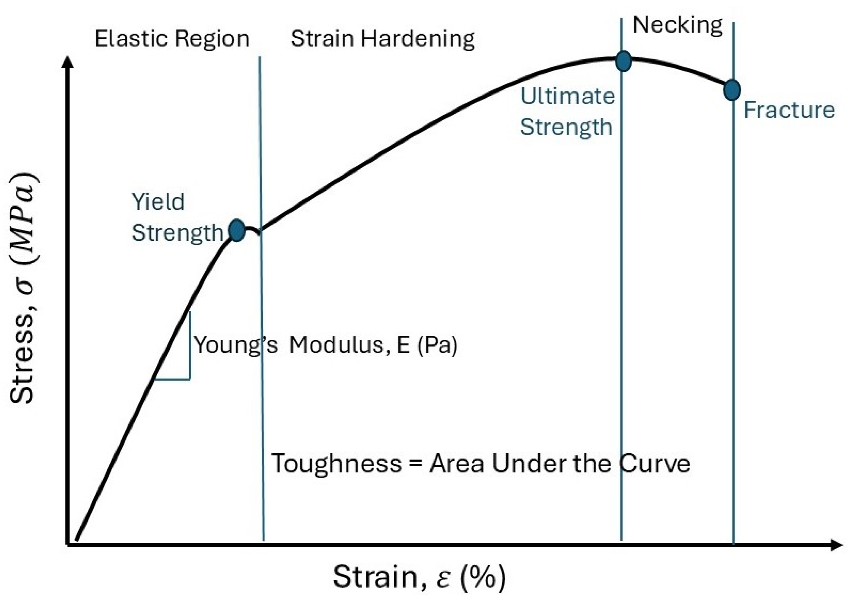
Although braziers provide breast support during the activities of daily living (ADLs), more significant research is necessary to fully understand aspects of the breasts, such as the center of mass, volume, deformation, kinetics, etc. [7]. The definition of some material properties should be defined before applying it to female breast tissue. A refresher of the stress–strain curve is presented in Figure 3. The following paragraphs provide a brief review to define the common material properties. When an external force is applied, the approximately cone-shaped female breast moves along the x, y, and z axes because it lacks skeletal support [10,36]. Beast tissue, like other tissues, are subjected to multiple forces such as tension, compression, shear, deformation, and tension–compression [43,66].
Deformation occurs when the material shape changes due to the applied stress and strain [42,67]. Stress is the strength of the forces that causes an alteration in the material’s shape [42]. The strain, a unitless dimension, describes the material’s deformation from the force applied from the stress [42,67]. When a small amount of stress is applied, a positive relationship is present between the stress and strain [42,67]. The elastic modulus or Young’s modulus are used interchangeably to quantify the tensile elastic modulus [35,42,66]. Young’s modulus quantifies the material stiffness when applying small loads [14,41]. Limited published data are available on the elastic properties of tissues without mechanical functions, such as breast tissues [30]. Young’s modulus is the slope of the elastic region of the applied stress load over the applied longitudinal strain [14,66,67]. The relationship between Young’s modulus and the stiffness expresses how each aspect affects the other [68]. Young’s modulus reflects the material stiffness and it does not depend on the material’s geometrical arrangement [14]. A large Young’s modulus represents that the material is more likely to refrain from deformation and that its value is close to the stiffness value [20,42,68]. For example, as the Young’s modulus value for either Cooper’s ligament or glandular tissue increases, then so does the stiffness, resulting in a decrease in the deformation [20]. The elastic region is when the force is removed and the material resumes its original shape and length, with the energy conserved [56,68]. The yield point is the boundary where the material’s deformation changes from elastic (resuming its initial shape) to plastic (permanent material change), with an offset between 0.1% and 0.2% on the stress–strain curve [69,70]. Elongation is when the force is removed but the material length is longer than the original length [56]. The yield strength is when the material shape does not resume its relaxed shape once the force is removed [67]. The integration of the stress–strain curve is the material toughness [67].
The mechanical properties of breast tissue go through a variety of changes because of demographics, hormonal fluctuations, and diseased tissue [14,15]. Despite the limited reports on the mechanical properties and conflicting reported data, it is necessary to continue research to prevent implant failure, to support the healthy tissue growth, and to aid in better clinical outcomes [14,43]. There are a multitude of variables that may be responsible for the conflicting data, such as the different methodologies employed, the demographic variations, and the sample source [14]. Continued research is necessary to understand breast tissue mechanics so to support solutions to alleviate the discomfort of women and to increase their quality of life [43,71]. Understanding the mechanics of breast tissue should be ongoing to prevent implant failure, support healthy tissue growth, and aid in better clinical outcomes [43].

6.1. Breast Force in Newton’s Second Law

Ideally, biological mathematical equations should be easily understood and widely applied [35]. Newton’s second law (Equation (2)) states that the total force is dependent on the mass of the object and its acceleration, (Equation (2)) [72]. Acceleration is when a force is applied to an object, resulting in fluctuating speed [72], whereas velocity is when the object’s movement is constant [71]. Acceleration is present when the net forces acting on the object do not equal zero, but velocity is when the net forces of the objects do equal zero [72]. The external force of gravity (9.8 m s 2 ) is a force that interacts with the breast tissue according to the individual’s stance (i.e., standing, prone, or supine), resulting in varying degrees of compression [12,14,72]. Gravity has forces acting on the breast regardless of the stance, but understanding its interaction provides greater insight [10,59].
F = m × a
where F = force, m = mass, a = acceleration, and the gravity acceleration = 9.8 m s 2 .
According to Newton, an object’s mass is the quantity of the matter it contains [73]. The factors that influence breast mass include the body mass index (BMI), pregnancy, lactation, hormonal and weight changes, density, and volume [7,12,13]. A study has reported the effects of force from the breast size on the spine [74]. The study concluded that a bra size of 36H exerted 52 lbs. on the spine, but a smaller size of 36D exerted only 24 lbs. [74]. The breast mass is also affected by its density and volume, as shown in Equation (3) [7]:
BM = BD × BV
where BM = breast mass, BD = breast density, and BV = breast volume.
A published experiment submerged the torso of females in water ( ρ w a t e r = 994   kg/m−3), and then again in soybean oil ( ρ o i l = 909   kg/m−3) [71]. The study measured the changes in the nipple position based on both densities [71]. The gravity-loaded results showed that the nipple position moved 25.7 mm inferiorly, 7.4 mm laterally, and 15.3 mm posteriorly, and the shape changed to a “tear drop” [71]. The submerged breast tissue changed to a “hemispherical or conical” shape [71].
A neutral buoyancy experiment was used to completely submerge breast tissue while in the prone position [59]. When the breast was submerged in water, the distance between the maximum height of the breast to the pectoral muscle was 45 mm, and when gravity was a factor, the distance was 62 mm, resulting in a difference of 17 mm in the gravity-loaded state [59]. The placement of a medical device in vivo created a more complex force body diagram, so its forces should be accounted for.

6.2. Tissue Expanders

Tissue expanders (TEs) are temporary devices utilized in breast reconstruction to establish a mound after a mastectomy, and are placed in vivo and exert forces on the chest [75,76]. Prior to exploring the device forces exerted internally, a brief description on its placement should be discussed [75,76]. After breast tissue removal, the skin is preserved and a pocket in the chest is created so to take advantage of the skin’s viscoelastic properties (see Section 6.6) [56,75,76]. The outer surface of the TE can either be smooth or textured, with an oval, round, or tear-dropped device shape [75]. A one-way port, either remote, integrated, or dual, provided access to the delivery of either saline or CO2 over the course of 2–6 weeks [75]. The function of the wings was to provide device placement and reduce the incidence of post-op device displacement [75]. The inframammary fold was the inferior landmark used to identify the placement prior to suturing the skin [75].

6.3. Breast Compression

While a force body diagram was not located on what forces TE’s exert in vivo, Equations (4)–(8) are general equations that can be applied to the breast tissue. Compression occurs when two forces of equal and opposite strength are applied to a material, in which the ending length is smaller than the starting length [42].
Compression   stress = | F A |
Compression   strain = | L L O |
Many scenarios, such as mammography, palpation, TEs, and bras, require external compression [7,14,75,76]. Bras are an external force that compresses the breast to the chest wall, thus reducing movement [7]. Palpation and other diagnostic devices compress the breast tissue to evaluate the presence of pathology within the structure [14]. The TE is placed between the chest wall and the skin, resulting in chest wall and device compression [75,76]. For the TE to remain effective in the prone position, or after receiving a hug, it must be able to withstand 35 lbs. of external compression [76]. Equations (6)–(9) is a mathematical strategy utilized to calculate the breast stiffness, with the assumption that it is a semicircle [63].
Volume = 1 2 4 3 π R volume 3
Rvolume = R1
R1 = hemisphere radius (cm).
Area = 1 2 ( π R compressed area 2 )
Area—semicircle = Rcompressed area = R2;
R2 = semicircle radius with compression.
Deformation = R1R2
Stiffness   F R 2 R 1 ( N / cm )
Compression force = F(dN)
Numerous data values can be derived from mammogram compression, such as the deformation, volume, and stiffness (Equations (5)–(8)) to calculate the compression force exerted [63]. The equations assume the following: (1) in precompression, the breast tissue shape is a hemisphere [63]; (2) the breast shape with compression is a semicircle [63]; (3) precompression of the breast tissue does not alter its volume [63]. Although the assumptions do not capture the true nature of the female breast, they do simplify the calculation.
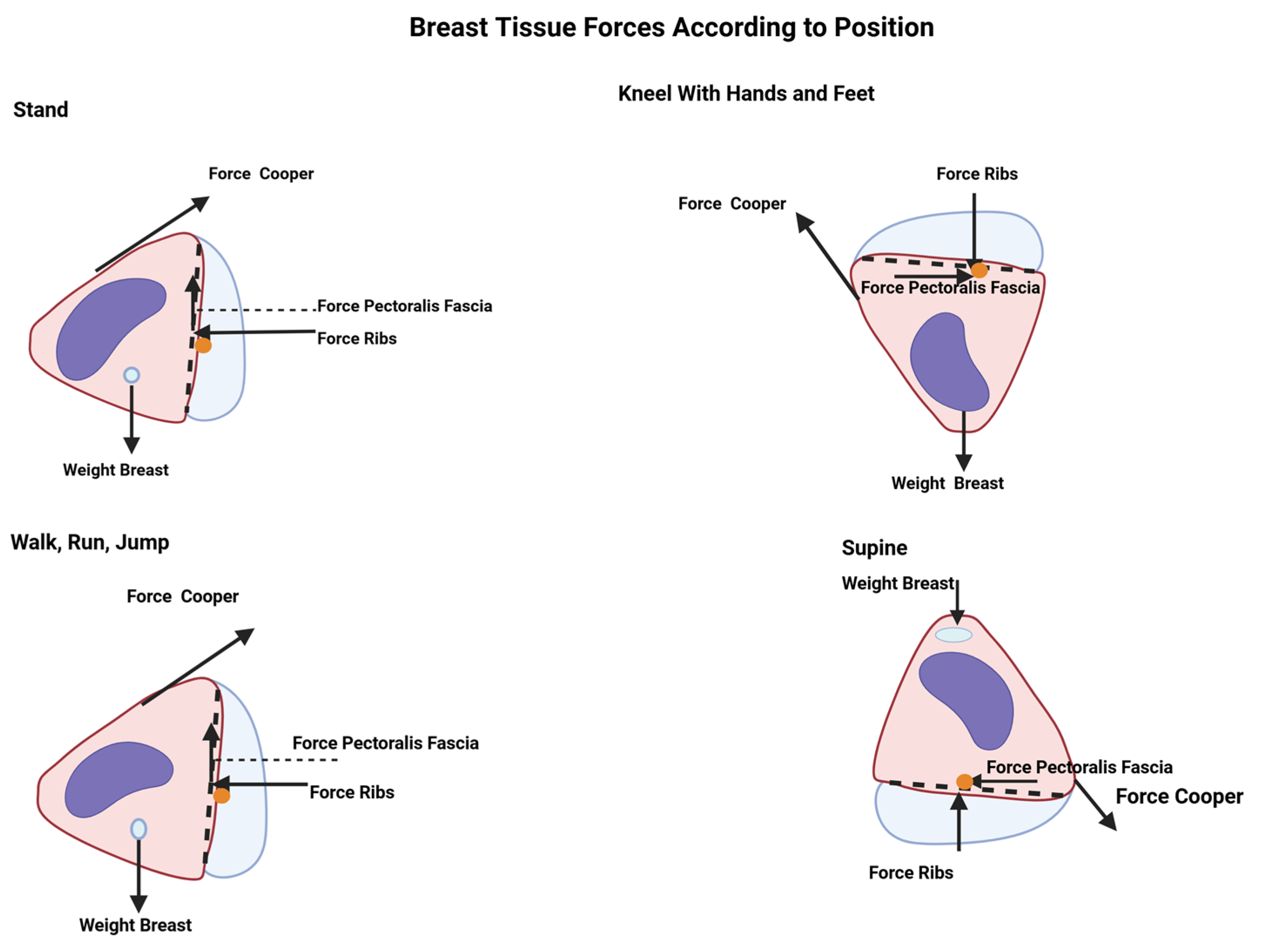
Figure 4 is a simplified force diagram showing the direction of the force from the ribs, Cooper’s ligament, gravity, pectoralis fascia, and the breast weight. The purple jelly bean shape represents a TE which also exerts in vivo according to the position [66,75,76]. For women whose breast volume is between 700 and 1000 g, they may have a mean force of 11.7 N ± 4.6 N when standing [2]. The breast tissue is in a relaxed state when in the standing stance, with gravity and internal components acting on it [10,67].

6.4. Viscoelasticity

The skin is a viscoelastic material that has the properties of both compression and sheer forces, and can be plotted on an elongation vs. force curve [56,66]. Skin is an excellent example of a viscoelastic material because, after the load removal, it returns to its original shape and stays lengthened [56]. Hysteresis is the term used to describe this behavior change based on the load quantity [14]. Hysteresis is due to varying loading and unloading pathways present in the skin’s elongation vs. force curve [14,56]. After the load is removed, the tissue will revert to its initial shape and stay lengthened [14,56].
The mechanical viscoelastic behavior is dependent on the applied load and ending shape after it is removed [14]. Stress–relaxation, creep, and dynamic testing are methods to evaluate viscoelastic tissue [69]. Testing for the viscoelastic behavior can be conducted on biological specimens and artificial materials such as elastomers [56,77,78].
Viscoelastic theory (Equation (10)) depicts the mathematical relationship between the axial strain and axial deformation to quantify the viscoelastic Poisson’s ratio [77].
Viscoelastic   Poisson s   Ratio   v ( t ) = E x ( t ) E 0
where v(t) = viscoelastic Poisson’s ratio, E x ( t ) = transversal strain over time, and E 0 = axial deformation with constant application [77].
Poisson’s ratio is the absolute value between the transverse (perpendicular to the force) and axial (direction of the force) strain to the point of elasticity of the material reached [78]. The variables that affect Poisson’s ratio are dependent on the time, temperature, load application speed (strain rates), material structure, and the interface of the applied stress and material [77,78].
There are six Poisson ratios when one material is pulled in the x, y, and z directions, originating in the middle of the applied force [78]. Stress–relaxation, creep, and dynamic testing are methods to evaluate viscoelastic tissue [69]. Poisson’s ratio is applied to soft tissue and elastomers for their viscoelastic properties [56,77]. A consensus has established that Poisson’s ratio for muscle is 0.5 due to its 75% water content [10,79]. A disadvantage of Equation (9) is its inability to accurately record minute and incremental strains when conducting stress–relaxation tests on elastomers [77]. For this reason, there are limited available data on the Poisson’s ratio of elastomers’ [77].

6.5. Creep

Creep tests are preferred over the viscoelastic Poisson’s ratio because it is easier to evaluate materials subjected to greater strains [78]. Studies have reported a good agreement between creep and relaxation testing when evaluating tissue behavior [77,78]. Stress–relaxation is a tissue phenomenon where, with a reduction in the applied stress, a constant stress is maintained [78]. When the applied stress is consistent with the tissue, creep is present because of the increased strain [78]. Viscoelastic material creep increases over time, while the relaxation decreases over time [78]. Devices can capitalize on the mechanical creep properties of the skin to support breast wound healing (see Section 6.3) [56,75,76]. Biological creep describes new tissue growth due to a force elongating the tissue over time [56]. The rate of displacement change is stated in Equation (11) [56], as follows:
Displacement   change   rate   =   F i x e d   l o a d   T i m e
Creep occurs from tension on the skin, as seen when TEs apply a constant load during treatment [56,75]. Mechanical creep is present due to the sutured pocket and stretched skin from the device placement over time [56,75]. Biological creep is when new tissue is subjected to prolonged tension (i.e., pregnancy and weight gain) [56]. Stress–relaxation, a result of creep, is when there is a reduction in the amount of force applied to the skin over time [56,75]. The material behavior of creep and stress–relaxation, originating from tension from the TEs, results in skin elongation of three to four times before the device placement [56,75]. Clinicians strive to salvage as much of the skin as possible after breast surgery so to capitalize on its mechanical behavior and to achieve better outcomes in reconstruction [75]. A greater understating of the effects of TEs and external forces will lead to desirable breast tissue shape and size outcomes [75,76]. Several devices can be utilized to exploit the viscoelastic behavior of the skin, to either pull the skin together or to elongate it [56]. Elongation of skin is when its final length is greater than its initial length [42].

6.6. Young’s Modulus of Breast Tissue

Testing has determined that the average Young’s modulus of the skin on breast tissue is 344 kPa and is not uniform, with an average thickness of 1.55 mm [55]. Studies have reported that breast tumors and healthy dense breasts have a greater stiffness than healthy, not-dense tissues [43]. Equation (5) provides a simplistic equation of Young’s modulus, but Equation (12) is a complex equation applied to breast tissue to calculate its Young’s modulus [14,30,41,66]. Since biological tissue has a high water content and is not compressible, a Poisson’s ratio of either 0.495 or 0.5 was applied [10,14,35,41,43]. Young’s modulus has received some criticism for its assessment of thickness because its assumption that the material is isotropic and uniform is not an accurate reflection of the specimen [80]. Young’s modulus has been reported to be different in horizontal and vertical measurements [80]. Although published mathematical strategies are direct and straightforward, they may not adequately reflect the actual behavior of the biological tissue [35,80].
E = 2   1 v 2 q a w
E = Young’s modulus;
v = Poisson’s ratio;
q = Load density is the force per unit area;
a = Loaded area radius;
w = Maximum displacement of the load directions.
A maximum value of 30 kPa on Cooper’s ligament was found when the applied stress increases and its length was greater [20]. A constant slope exists when Young’s modulus has an applied strain of 30%, and when the fat and glandular tissue is <10% [35]. The breast tissue stress–strain curve changes depending on the applied load applied, resulting in a larger deformation, but, if the applied strain is <50%, the skin is “linear isotropic” [10,14].

6.7. Stiffness

Imaging modalities can aid in determining the stiffness and density of the breast tissue [14,81]. The breast tissue and indention tests show that stiffness is a material property that affects the breast motion behavior [20,82]. Elastography uses soft tissue compression to provide an objective stiffness value [14,82]. Ideally, the precompression elastography setting is 10% [14]. Healthy glandular tissue is 1 to 6.7 times greater than adipose tissue [20]. Diseased tissue is 2–3 times stiffer than healthy tissue [14]. Finite element modeling (FEM) concluded that, when Cooper’s ligament or the glandular tissue is stiffer, the breast movement and deformation are reduced [20]. Establishing a baseline stiffness to the current stiffness can be used to determine breast tissue changes that may indicate the risk for the presence of masses [20,35,81]. The mechanical behavior of breast adipose tissue is constant when an applied strain is applied, whereas glandular tissues are not as constant when an applied strain is applied [35]. The average stiffness of breast tissue is 2.3 ± 0.8 m/s, but age, the breast size, and the glandular density have not been fully determined to demonstrate a correlation to stiffness [80,83].

6.7.1. Boyd’s Radial Stiffness

Boyd’s radial stiffness equations can be applied to mammograms to determine the stiffness, volume, area, and density of the breast tissue by using the breast radius [63,81]. Equations (6) and (7) are applied when the compression testing is performed on the breast tissue mammography, Equations (13)–(16) [80]. The drawback to this method is that the calculation may result in a negative value and assumes a hemispherical shape, which may not be applicable to all breast shapes (Equations (12)–(15)) [63,80].
1. Assumption: The breast is hemispherical-shaped [80]
Both directions of stiffness are calculated, as follows:
k bi   =   F 5 i   F 0 r 0 i     r 5 i
kbi = Boyd’s radial stiffness;
F 5 i = Distance at the end of the compression from the paddles;
F 0 = Preload of the breast;
roi = Radius prior to the compression, which goes with F 0 ;
r5i = After the paddle compression, which goes with F 5 i .
2. Assumption: Semicircular area [80]
The r0i is calculated for use in Equation (18), as follows:
A 01   =   1 2 π r 0 i 2   Semicircular   area
A0i = Breast cross-sectional area at the beginning.
3. Assumption: Hemispherical shape
V 5 i = 1 2 ( 4 3 π r 5 i 3 )
4. V5i = Volume after compression
V5i = A0i (h0iumax)
V5i = Final breast volume after the compression;
V o i = A 0 i ;
A 0 i = area of the cross-section of the breast;
h0i = starting distance between the compression paddles.
This study made the following assumptions: (1) the mammographic images provided the actual value of the tissue [63]; (2) the hemisphere is the shape of the breast tissue in the relaxed state [63]; (3) under mammographic conditions, the breast tissue is a semicircle [63]; (4) the area of the breast tissue area and radius is the same when the tissue is in its relaxed state and, when compressed with the screen film, the mammography is the same as before the compression [63]. The following assumptions of Boyd’s equation are captured in the following equation, in the sequence the calculation should be conducted [63]:
1. Volume of the hemispherical shape with no compression:
Volume   =   1 2 =   ( 4 3 π R volume 3 )
The first radius of the shape is measured (R1).
2. Area of the compressed area:
Area   =   1 2 ( π R compressed area 2 )
The second radius of the shape is measured (R2).
3. Breast stiffness based on the above equation
a. Compression force = F (dN)
b .   Stiffness   F ( R 2 R 1 )   ( N / cm )
Boyd’s stiffness does not account for the number of unique shapes and classifications of the female breast shapes. Manufacturers and researchers use different terminology to classify the shape of the female breast, which has not been based on objective metrics, as can be seen in the following sources [2,83,84,85,86,87,88,89]. It also does not account for the shape of ptotic breasts in relation to the inframammary fold [90].

6.7.2. Linear Stiffness

Linear stiffness lends insight into the effects of the applied force exerted from the mammography compression, but the assumption that geometry is not a factor must be applied [80]. The output of the equation will result in values representing both linear directions [80]. The linearized method to calculate the stiffness does not take into consideration the breast size, thereby resulting in no differentiation from a large cup size vs. a smaller one [80].
1. Force equation
F li = k l i μ i +   k 0 i
k l i = 6 ( p = 0 5 F i p u i p ) ( p = 0 5 F i p ) × ( p = 0 5 u i p ) 6 p = 0 5 u i p 2 ( p = 0 5 u i p ) 2
μ i = 0–5 mm displacement range;
kli = both linearized breast stiffnesses.
A limitation of Equations (20) and (21) is that, by not accounting for the different sizes, this results in a poor representation of the general population [80]. This can be inferred by the fact that, in two bras with the same cup size but different band sizes, the larger band size will have a more extensive breast tissue volume, indicating that the volume increases as the band size increases (i.e., 34B has less volume than 36B) [91]. The volume of the breast size can range from less than 150 mL to greater than 1500 mL [80].

6.7.3. Density

Research has not shown agreement on the correlation between breast tissue density and stiffness [80,92]. When the breast tissue is not dense, it has a stiffness range of 500 Pa–1000 Pa/mm, whereas dense tissue has a stiffness range of 700–2400 Pa/mm [92]. An inverse relationship exists between fibroglandular tissue and fibroadipose tissue; and their ratio determines the breast density with increasing age [7,93]. Mammograms can evaluate the amount of fibroglandular tissue related to the breast size so to determine the tissue’s density [33]. Breast tissue stiffness and density are related to collagen and fibroglandular tissue [80]. With advanced age, the quantity of fibroglandular tissue will decrease, and the quantity of fibroadipose tissue will increase, resulting in an increased density [7]. The National Cancer Institute reports that nearly 50% of women over the age of 40 have dense breasts due to the increase in fibroglandular tissue and the reduction in fibroadipose tissue [7,93]. The American College of Radiology has developed four classifications to report the breast density seen on a mammograms, titled Breast Imaging Reporting and Data System (BI-RADS) [93,94]. The mammography image is darker for fatty breasts and becomes increasingly brighter for highly dense breasts [93]. The brighter mammography image makes it a challenge to differentiate between healthy breasts, dense breasts, and masses [33,93].
Cancerous tissue is stiffer than the surrounding healthy tissue, having a positive correlation to malignant masses, density, and stiffness [63,80]. The subjective stiffness scale provided in Table 2 may be made objective by applying the directional energy formula (Equations (22)–(24)) [80]. Future research should explore if there is a correlation between the directional energy and the BI-RADS classification. Additional research should be conducted to determine if both the stiffness and density can increase the ability to evaluate the risk of breast tissue masses.

6.7.4. Increment of Strain Energy Density

The strain energy density strategy evaluates the slight increase in the strain energy by calculating the volume changes during the compression of the breast tissue [81]. Prior published research has concluded that this is a viable method to use with experienced clinical palpation, Equations (23)–(25) [81].
1. Volume of stress energy with compression
U i = E p i V i
U i = small strain energy density changes in the breast compression;
E p i = small changes in the strain energy;
V i = volume changes during compression.
2. Integration of nonlinear value of the compression force ( F i ) to the top compression paddle of the strain energy [81]
E p i = 0 u m a x F i d u i = n = 1 5 1 2 ( F i n 1 + F i n ) u i n
3. Breast volume change
V i   =   V 0 i V 5 i =   A 0 i h 0 i A 0 i h 0 i   u m a x =   A 0 i u m a x
V i = Change breast volume
V o i = A 0 i ;
A 0 i = area of cross-section of the breast;
u m a x = 5 mm.
A minimal difference was calculated between the horizontal and vertical values when using a step of 1.5 J mm−3 [81]. Additional research should evaluate if this is an appropriate step that can be applied to the stiffness scale [81].

7. Conclusions

Numerous factors related to female breast tissue need to be addressed in research to develop products that support females to engage in active and healthy lives. Various modalities can calculate the mechanical properties of breast tissue, so additional research should determine how the values differ from each of the modalities and which one represents the actual value of the tissue. Although there has been some published research on the mechanical properties of breast tissue components, future research should consider quantifying the changes in breast tissue material throughout the female’s lifespan. Determining the risk of unhealthy breast tissue can become challenging, as the tissue increases in density and stiffness. Still, research should explore the relationship between the stiffness and the density. Additional research in this area may provide excellent guidelines to evaluate the risk of unhealthy breast tissue. Published research has established that breast tissue properties vary based on age and ethnicity. Considering that there is an increase in multiracial females, this warrants additional research in the characterization of breast tissue.
This review discussed the state of the current research and has identified areas that further investigation should explore. Continued study in female breast tissue may profoundly impact the improvement in clinical outcomes, work environments, and daily lives throughout the lifespans of women. The scientific community, companies, and government departments should make this a priority in their use of resources. This research could profoundly and positively impact females’ health and daily lives due to its application in manufacturing and clinical settings.

Author Contributions

S.G. contributed to this paper by researching and reviewing published peer reviewed articles, writing and preparing the original draft. T.G. contributed to this paper by reviewing and editing the original draft, supervision of content. All authors have read and agreed to the published version of the manuscript.

Funding

This research received no external funding.

Institutional Review Board Statement

Not applicable.

Informed Consent Statement

Not applicable.

Data Availability Statement

No new data were created or analyzed in this study.

Acknowledgments

The authors would like to acknowledge Solutions Through Innovative Technologies, Inc. (STI-TEC), The University of Michigan Transportation Research Institute (UMTRI), and Anthrotech for their guidance, support, and training in the field of anthropometry. Thank you to the editors for taking the time to provide guidance in the revisions to this article.

Conflicts of Interest

The authors declare no conflicts of interest.

References

  1. Onyebeke, L.C.; Papazaharias, D.M.; Freund, A.; Dropkin, J.; McCann, M.; Sanchez, S.H.; Hashim, D.; Meyer, J.D.; Lucchini, R.G.; Zuckerman, N.C. Access to Properly Fitting Personal Protective Equipment for Female Construction Workers. Am. J. Ind. Med. 2016, 59, 1032–1040. [Google Scholar] [CrossRef] [PubMed]
  2. Hamandi, F.; Pooler, T.; Runser, A.; Asman, C.; Bailey, R.; Robinson, N.; Goswami, T. Bioenergetics Analysis of Bra-Breast Interface. Appl. Mech. 2022, 3, 70–84. [Google Scholar] [CrossRef]
  3. Wood, K.; Cameron, M.; Fitzgerald, K. Breast size, bra fit and thoracic pain in young women: A correlational study. Chiropr. Osteopat. 2008, 16, 1. [Google Scholar] [CrossRef] [PubMed]
  4. Coltman, C.E.; Steele, J.R.; McGhee, D.E. Breast volume is affected by body mass index but not age. Ergonomics 2017, 60, 1576–1585. [Google Scholar] [CrossRef] [PubMed]
  5. Pramardika, D.D.; Kasaluhe, M.D.; Barokah, S. Bra Usage Duration and Breast Cancer Risk: A Case-Control Study. J. Bidan Cerdas 2023, 5, 51–57. [Google Scholar] [CrossRef]
  6. Coltman, C.E.; Steele, J.R.; McGhee, D.E. Does breast size affect how women participate in physical activity? J. Sci. Med. Sport 2019, 22, 324–329. [Google Scholar] [CrossRef]
  7. McGhee, D.E.; Steele, J.R. Breast Biomechanics: What Do We Really Know? Physiology 2020, 35, 144–156. [Google Scholar] [CrossRef] [PubMed]
  8. Brown, T.H.; Ringrose, C.; Hyland, R.; Cole, A.; Brotherston, T. A method of assessing female breast morphometry and its clinical application. Br. J. Plast. Surg. 1999, 52, 355–359. [Google Scholar] [CrossRef] [PubMed]
  9. Mikołajczyk, M.; Kasielska-Trojan, A.; Antoszewski, B. A New Tool for Breast Anthropometric Measurements: Presentation and Validation for Women and Men. Aesthetic Plast. Surg. 2019, 43, 1160–1170. [Google Scholar] [CrossRef] [PubMed]
  10. Gefen, A.; Dilmoney, B. Mechanics of the normal woman’s breast. Technol. Health Care 2007, 15, 259–271. [Google Scholar] [CrossRef]
  11. Garamone, J. Groups Work to Eliminate, Diminish Barriers to Women’s Military Service. U.S. Department of Defense. 23 January 2022. Available online: https://www.defense.gov/News/News-Stories/Article/Article/2908233/groups-work-to-eliminate-diminish-barriers-to-womens-military-service/ (accessed on 27 April 2024).
  12. Le, Q.H.; Nguyen, H.C. Breast Anthropometry: Values and Application in Breast Surgery for Vietnamese Women. Asian Pac. J. Cancer Prev. 2022, 23, 665–671. [Google Scholar] [CrossRef]
  13. Kateina, C.; Mandalidis, D. Effects of Artificially Induced Breast Augmentation on the Electromyographic Activity of Neck and Trunk Muscles during Common Daily Movements. J. Funct. Morphol. Kinesiol. 2022, 7, 80. [Google Scholar] [CrossRef] [PubMed]
  14. Teixeira, A.M.; Martins, P. A review of bioengineering techniques applied to breast tissue: Mechanical properties, tissue engineering and finite element analysis. Front. Bioeng. Biotechnol. 2023, 11, 1161815. [Google Scholar] [CrossRef] [PubMed]
  15. Lee, Y.; Hwang, K. Skin thickness of Korean adults. Surg. Radiol. Anat. 2002, 24, 183–189. [Google Scholar] [CrossRef] [PubMed]
  16. Oskar, S.; Kehm, R.; Terry, M.B. Breast Tissue Composition—Why It Matters and How Can We Measure It More Accurately in Epidemiology Studies. Cancer Epidemiol. Biomark. Prev. 2021, 30, 590–592. [Google Scholar] [CrossRef]
  17. Montagna, W.; Parakkal, P.F. An Introduction to Skin. In Function of Skin, 3rd ed.; Academic Press, Inc.: New York, NY, USA, 1974; pp. 1–17. [Google Scholar]
  18. Diab, M.; Kumaraswamy, N.; Reece, G.P.; Hanson, S.E.; Fingeret, M.C.; Markey, M.K.; Ravi-Chandar, K. Characterization of human female breast and abdominal skin elasticity using a bulge test. J. Mech. Behav. Biomed. Mater. 2019, 103, 103604. [Google Scholar] [CrossRef] [PubMed]
  19. Matousek, S.A. Discussion: The Attachments of the Breast to the Chest Wall: A Dissection Study. Plast. Reconstr. Surg. 2020, 146, 23e–26e. [Google Scholar] [CrossRef]
  20. Chen, J.; Sun, Y.; Liu, Q.; Yip, J.; Yick, K.-L. Construction of multi-component finite element model to predict biomechanical behaviour of breasts during running and quantification of the stiffness impact of internal structure. Biomech. Model. Mechanobiol. 2024, 23, 1679–1694. [Google Scholar] [CrossRef]
  21. National Cancer Institute SEER Training Modules. Breast Anatomy. National Cancer Institute. Available online: https://training.seer.cancer.gov/breast/anatomy.html (accessed on 24 January 2025).
  22. National Library of Medicine National Center for Biotechnology Information STATPEARLS [Internet]. Physiology, Menstrual Cycle. National Library of Medicine National Center for Biotechnology Information STATPEARLS [Internet]. 24 October 2022. Available online: https://www.ncbi.nlm.nih.gov/books/NBK500020/ (accessed on 28 August 2024).
  23. Rendina, E.A.; Ciccone, A.M. The Intercostal Space. Thorac. Surg. Clin. 2007, 17, 491–501. [Google Scholar] [CrossRef]
  24. Mîra, A.; Carton, A.-K.; Muller, S.; Payan, Y. A biomechanical breast model evaluated with respect to MRI data collected in three different positions. Clin. Biomech. 2018, 60, 191–199. [Google Scholar] [CrossRef]
  25. Briot, N.; Chagnon, G.; Burlet, L.; Gil, H.; Girard, E.; Payan, Y. Experimental characterization and modelling of breast Cooper’s ligaments. Biomech. Model. Mechanobiol. 2022, 21, 1157–1168. [Google Scholar] [CrossRef]
  26. Marcus, F.; Hughes, T.; Barrios, P.; Borgstrom, M. Clinical location of the fourth and fifth intercostal spaces as a percent of the length of the sternum. J. Electrocardiol. 2018, 51, 55–59. [Google Scholar] [CrossRef]
  27. Ellis, H. The ribs and intercostal spaces. Anaesth. Intensiv. Care Med. 2008, 9, 518–519. [Google Scholar] [CrossRef]
  28. Bazira, P.J.; Ellis, H.; Mahadevan, V. Anatomy and physiology of the breast. Surgery 2022, 40, 79–83. [Google Scholar] [CrossRef]
  29. Barcelar, J.d.M.; Aliverti, A.; Rattes, C.; Ximenes, M.E.; Campos, S.L.; Brandão, D.C.; Fregonezi, G.; de Andrade, A.D. The Expansion of the Pulmonary Rib Cage during Breath Stacking Is Influenced by Age in Obese Women. PLoS ONE 2014, 9, e110959. [Google Scholar] [CrossRef] [PubMed]
  30. Rivard, A.B.; Galarza-Paez, L.; Peterson, D.C. Anatomy, Thorax, Breast. NIH National Library of Medicine National Center for Biotechnology Information. 17 July 2023. Available online: https://www.ncbi.nlm.nih.gov/books/NBK519575/ (accessed on 28 August 2024).
  31. AlHarbi, Y. Anatomical Variations in the Pectoralis Minor Muscle Origin and Insertion: A Systematic Review. Cureus 2023, 15, e46329. [Google Scholar] [CrossRef] [PubMed]
  32. Konrad, A.; Reiner, M.M.; Warneke, K.; Keiner, M.; Nakamura, M.; Tilp, M. Relationship between pectoralis major stiffness and shoulder extension range of motion. Front. Physiol. 2024, 15, 1349426. [Google Scholar] [CrossRef]
  33. Kettering Health ketteringhealth.org/Breasthealth. Breast Health Why a Mammogram; Kettering Health: Kettering, UK, 2021. [Google Scholar]
  34. Yaffe, M.J.; Byng, J.W.; Boyd, N.F. 21—Quantitative Image Analysis for Estimation of Breast Cancer Risk. In Handbook of Medical Imaging Biomedical Engineering; Academic Press: Cambridge, MA, USA, 2009; pp. 323–340. [Google Scholar] [CrossRef]
  35. Krouskop, T.A.; Wheeler, T.M.; Kallel, F.; Garra, B.S.; Hall, T. Elastic Moduli of Breast and Prostate Tissues under Compression. Ultrason. Imaging 1998, 20, 260–274. [Google Scholar] [CrossRef] [PubMed]
  36. Suh, M.; Park, J.H. Breast Geometry Characterization of Young American Females Using 3D Image Analysis. Appl. Sci. 2022, 12, 8578. [Google Scholar] [CrossRef]
  37. Veitch, D.; Burford, K.; Dench, P.; Dean, N.; Griffin, P. Measurement of breast volume using body scan technology (computer-aided anthropometry). Work 2012, 41, 4038–4045. [Google Scholar] [CrossRef] [PubMed]
  38. Kothari, C.; Diorio, C.; Durocher, F. The Importance of Breast Adipose Tissue in Breast Cancer. Int. J. Mol. Sci. 2020, 21, 57600. [Google Scholar] [CrossRef] [PubMed]
  39. Koscinski, K. Breast firmness is of greater importance for women’s attractiveness than breast size. Am. J. Hum. Biol. 2019, 31, e23287–e23299. [Google Scholar] [CrossRef]
  40. Duncan, A.M.; Al Youha, S.M.; Joukhadar, N.; Konder, R.B.; Stecco, C.; Wheelock, M.E. Anatomy of the Breast Fascial System: A Systematic Review of the Literature. Plast. Reconstr. Surg. 2021, 149, 28–40. [Google Scholar] [CrossRef] [PubMed]
  41. Ramião, N.G.; Martins, P.S.; Rynkevic, R.; Fernandes, A.A.; Barroso, M.; Santos, D.C. Biomechanical properties of breast tissue, a state-of-the-art review. Biomech. Model. Mechanobiol. 2016, 15, 1307–1323. [Google Scholar] [CrossRef] [PubMed]
  42. Openstax. University Physics Volume 1 12.3 Stress, Strain, and Elastic Modulus. Rice University. 1999–2024. ISBN 10: 1938168275. Available online: https://openstax.org/books/university-physics-volume-1/pages/12-3-stress-strain-and-elastic-modulus (accessed on 11 September 2024).
  43. Goodbrake, C.; Li, D.S.; Aghakhani, H.; Contreras, A.; Reece, G.P.; Markey, M.K.; Sacks, M.S. On the three-dimensional mechanical behavior of human breast tissue. Ann. Biomed. Eng. 2022, 50, 601–613. [Google Scholar] [CrossRef] [PubMed]
  44. Emmanuel, M.; Bokor, B.R. Tanner Stages. StatPearls [Internet]. Bookshelf ID: NBK470280. 11 December 2022. Available online: https://www.ncbi.nlm.nih.gov/books/NBK470280/ (accessed on 19 August 2023).
  45. Lteif, A.; Javed, A. Development of the Human Breast. Semin. Plast. Surg. 2013, 27, 5–12. [Google Scholar] [CrossRef]
  46. Koopman-Verhoeff, M.E.; Gredvig-Ardito, C.; Barker, D.H.; Saletin, J.M.; Carskadon, M.A. Classifying Pubertal Development Using Child and Parent Report: Comparing the Pubertal Development Scales to Tanner Staging. J. Adolesc. Health 2020, 66, 597–602. [Google Scholar] [CrossRef] [PubMed]
  47. Kaur, S.; Kumari, B.; Kalyan, G.; Kaur, B.; Devi, K.; Preeti; Saranjna; Singh, G. Anthropometric Breast Measurements and Brasserie Wearing Practices of North Indian Women. Indian J. Surg. 2022, 84, 714–721. [Google Scholar] [CrossRef]
  48. World Health Organization. Antiretroviral Therapy for HIV Infection in Infants and Children: Towards Universal Acess Recommendiations for Public Health Approach 2010 Revision; Annex H Sexual Maturity Rating (Tanner Staging) in Adolescents; World Health Organization: Geneva, Switzerland, 2010. Available online: https://www.ncbi.nlm.nih.gov/books/NBK138588/ (accessed on 11 September 2024).
  49. Krishna, K.B.; Witchel, S.F. Normal and Abnormal Puberty. Endotext. 2024. Available online: https://www.ncbi.nlm.nih.gov/books/NBK279024/ (accessed on 18 August 2024).
  50. Wang, Z.; Asokan, G.; Onnela, J.-P.; Baird, D.D.; Jukic, A.M.Z.; Wilcox, A.J.; Curry, C.L.; Fischer-Colbrie, T.; Williams, M.A.; Hauser, R.; et al. Menarche and Time to Cycle Regularity Among Individuals Born Between 1950 and 2005 in the US. JAMA Netw. Open 2024, 7, e2412854. [Google Scholar] [CrossRef]
  51. The Editors of Encylopedia. Middle Ages. Britannica. 15 September 2024. Available online: https://www.britannica.com/event/Middle-Ages (accessed on 13 October 2024).
  52. Adkins, E.N.; Anderson, S.; McKoy, T.; Maduka, N.; Goswami, T. Etiology of Breast Development and Asymmetry. Adv. Gen. Pract. Med. 2022, 4, 54–66. [Google Scholar] [CrossRef]
  53. Govrin-Yehudain, J.; Dvir, H.; Preise, D.; Govrin-Yehudain, O.; Govreen-Segal, D. Lightweight Breast Implants: A Novel Solution for Breast Augmentation and Reconstruction Mammaplasty. Aesthetic Surg. J. 2015, 35, 965–971. [Google Scholar] [CrossRef]
  54. Lawton, S. Skin 1: The structure and functions of the skin. Nurs. Times 2019, 115, 30–33. Available online: https://cdn.ps.emap.com/wp-content/uploads/sites/3/2019/11/191127-Skin-1-the-structure-and-functions-of-the-skin.pdf (accessed on 1 November 2024).
  55. Sutradhar, A.; Miller, M.J. In vivo measurement of breast skin elasticity and breast skin thickness. Ski. Res. Technol. 2012, 19, e191–e199. [Google Scholar] [CrossRef] [PubMed]
  56. Wilhelmi, B.J.; Blackwell, S.J.; Mancoll, J.S.; Phillips, L.G. Creep vs. Stretch: A review of the Viscoelastic Properties of Skin. Ann. Plast. Surg. 1998, 41, 215–219. [Google Scholar] [CrossRef]
  57. Palm Beach State Collage. Chapter 06 Lecture Outline Anatomy Physiology The Unity of Form and Function Seventh Edition. Available online: https://www.palmbeachstate.edu/slc/Documents/AandP1ch06Lecture.pdf (accessed on 15 July 2024).
  58. Huang, S.; Boone, J.M.; Yang, K.; Kwan, A.L.C.; Packard, N.J. The effect of skin thickness determined using breast CT on mammographic dosimetry. Med. Phys. 2008, 35, 1199–1206. [Google Scholar] [CrossRef]
  59. Gamage, T.P.B.; Rajagopal, V.; Nielsen, P.M.; Nash, M.P. Patient-Specific Modeling of Breast Biomechanics with Applications to Breast Cancer Detection and Treatment. Patient-Specif. Model. Tomorrow’s Med. 2011, 9, 379–412. [Google Scholar]
  60. Zherebtsov, E.; Dremin, V.; Popov, A.; Doronin, A.; Kurakina, D.; Kirillin, M.; Meglinski, I.; Bykov, A. Hyperspectral imaging of human skin aided by artificial neural networks. Biomed. Opt. Express 2019, 10, 3545–3559. [Google Scholar] [CrossRef] [PubMed]
  61. Del Rosso, J.Q.; Levin, J. The Clinical Relevance of Maintaining the Functional Integrity of the Stratum Corneum in both Healthy and Disease-affected Skin. J. Clin. Aesthetic Dermatol. 2011, 4, 22–42. [Google Scholar] [PubMed]
  62. Monteiro-Riviere, N.A.; Filon, F.L. Skin Morphology. In Adverse Effects of Engineered Nanomaterials; Academic Press: London, UK, 2017; pp. 357–380. [Google Scholar]
  63. Boyd, N.F.; Li, Q.; Melnichouk, O.; Huszti, E.; Martin, L.J.; Gunasekara, A.; Mawdsley, G.; Yaffe, M.J.; Minkin, S. Evidence that Breast Tissue Stiffness is Associated with Risk of Breast Cancer. PLoS ONE 2014, 9, e100937. [Google Scholar] [CrossRef]
  64. Lewis, C.; Cohen, P.R.; Bahl, D.; Levine, E.M.; Khaliq, W. Race and Ethnic Categories: A Brief Review of Global Terms and Nomenclature. Cureus 2023, 15, e41253. [Google Scholar] [CrossRef] [PubMed]
  65. Grilo, S.A.; Semler, M.R.; Rameau, S. The sum of all parts: A multi-level exploration of racial and ethnic identity formation during emerging adulthood. PLoS ONE 2023, 18, e0284275. [Google Scholar] [CrossRef] [PubMed]
  66. Wells, R.G. Tissue Mechanics and Fibrosis. Biochim. Biophys. Acta 2013, 1832, 884–890. [Google Scholar] [CrossRef]
  67. Alsayed, A.A. Physics of Open Fractures: Reconsidering Tissue Viability, Contamination Risk and Importance of Wound Debridement. J. Appl. Math. Phys. 2021, 9, 176–182. [Google Scholar] [CrossRef]
  68. Trębacz, H.; Barzycka, A. Mechanical Properties and Functions of Elastin: An Overview. Biomolecules 2023, 13, 574. [Google Scholar] [CrossRef] [PubMed]
  69. Abdewi, E.F. Mechanical Properties of Reinforcing Steel Rods Produced by Zliten Steel Factory. In Reference Module in Materials Science and Materials Engineering; Elsevier: Amsterdam, The Netherlands, 2017. [Google Scholar] [CrossRef]
  70. Vaidya, A.; Pathak, K. 17—Mechanical stability of dental materials. In Applications of Nanocomposite Material in Dentistry; Woodhead Publishing: Sawston, UK, 2019; pp. 285–305. ISBN 9780128137420. [Google Scholar]
  71. Mills, C.; Sanchez, A.; Scurr, J. Estimating the gravity induced three dimensional deformation of the breast. J. Biomech. 2016, 49, 4134–4137. [Google Scholar] [CrossRef] [PubMed]
  72. Openstax. University Physics Volume 1 5.3 Newton’s Second Law. Rice University. 1999–2024. ISBN 10: 1938168275. Available online: https://openstax.org/books/university-physics-volume-1/pages/5-3-newtons-second-law (accessed on 11 September 2024).
  73. Haug, E.G. Different Mass Definitions and Their Pluses and Minuses Related to Gravity. Foundations 2023, 3, 199–219. [Google Scholar] [CrossRef]
  74. Hansraj, K.J. Breast Forces on the Spine. Surg. Technol. Int. 2016, 28, 311–315. [Google Scholar] [PubMed]
  75. Cho, M.-J.; Farhadi, R.V.; Nash, D.W.; Kaleeny, J.; Povoski, S.P.; Chao, A.H. The current use of tissue expanders in breast reconstruction: Device design, features, and technical considerations. Expert Rev. Med. Devices 2023, 21, 27–35. [Google Scholar] [CrossRef]
  76. Najafali, D.; Liu, F.C.; Raman, K.; Maheta, B.; Saldana, G.M.; Heldman, L.; Cevallos, P.; Nazerali, R. The Best Under Stress: An Analysis of Breast Tissue Expander Response to External Forces. Aesthetic Surg. J. Open Forum 2023, 5, ojad018. [Google Scholar] [CrossRef]
  77. Sotomayor-Del-Moral, J.A.; Pascual-Francisco, J.B.; Susarrey-Huerta, O.; Resendiz-Calderon, C.D.; Gallardo-Hernández, E.A.; Farfan-Cabrera, L.I. Characterization of Viscoelastic Poisson’s Ratio of Engineering Elastomers via DIC-Based Creep Testing. Polymers 2022, 14, 1837. [Google Scholar] [CrossRef] [PubMed]
  78. Ashrafi, H.; Shariyat, M. A nanoindentation modeling of viscoelastic creep and relaxation behaviors of ligaments mechanical characteristics of biological tissues. In Proceedings of the 17th Iranian Conference of Biomedical Engineering (ICBME2010), Isfahan, Iran, 3–4 November 2010. [Google Scholar] [CrossRef]
  79. Tschoegl, N.; Knauss, W.G.; Emri, I. Poisson’s Ratio in Linear Viscoelasticity—A Critical Review. Mech. Time-Depend. Mater. 2002, 6, 3–51. [Google Scholar] [CrossRef]
  80. Prokop, J.; Maršálek, P.; Sengul, I.; Pelikán, A.; Janoutová, J.; Horyl, P.; Roman, J.; Sengul, D.; Junior, J.M.S. Evaluation of breast stiffness pathology based on breast compression during mammography: Proposal for novel breast stiffness scale classification. Clinics 2022, 77, 100100. [Google Scholar] [CrossRef] [PubMed]
  81. Patel, B.K.; Pepin, K.; Brandt, K.R.; Mazza, G.L.; Pockaj, B.A.; Chen, J.; Zhou, Y.; Northfelt, D.W.; Anderson, K.; Kling, J.M.; et al. Association of breast cancer risk, density, and stiffness: Global tissue stiffness on breast MR elastography (MRE). Breast Cancer Res. Treat. 2022, 194, 79–89. [Google Scholar] [CrossRef]
  82. Samani, A.; Plewes, D. A method to measure the hyperelastic parameters of ex vivo breast tissue samples. Phys. Med. Biol. 2004, 49, 4395–4405. [Google Scholar] [CrossRef] [PubMed]
  83. Chen, J.-H.; Chan, S.; Zhang, Y.; Li, S.; Chang, R.-F.; Su, M.-Y. Evaluation of breast stiffness measured by ultrasound and breast density measured by MRI using a prone-supine deformation model. Biomark. Res. 2019, 7, 20. [Google Scholar] [CrossRef] [PubMed]
  84. Zhang, B.; Sun, Y.; Zhong, Z.; Xia, Y.; Jin, S.; Li, Y.; Gu, B. Breast shape classification and discrimination driven by local features-focusing on Chinese women in their 20s. Int. J. Ind. Ergon. 2022, 90, 103304. [Google Scholar] [CrossRef]
  85. Third Love. Breast Shape Dictionary: Understanding Your Breast Shape. Third Love. 11 September 2023. Available online: https://www.thirdlove.com/blogs/learn/breast-shape-dictionary (accessed on 4 July 2024).
  86. Great Place To Work. ThirdLove Company Overview. Great Place to Work. July 2023. Available online: https://www.greatplacetowork.com/certified-company/7010800#:~:text=ThirdLove%20is%20an%20American%20lingerie,women%20in%20San%20Francisco%2C%20CA (accessed on 4 July 2024).
  87. Johnson, M. What Are the Most Common Breast Shapes? Healthline, 26 April 2019. Available online: https://www.healthline.com/health/breast-shapes (accessed on 4 July 2024).
  88. Healthline. About Us. Healthline. 2024. Available online: https://www.healthline.com/about/about-us (accessed on 4 July 2024).
  89. El-Shazly, M.; El-Oteify, M.; Megeed, H.; Ahmed, B. Assessment of the breast volume by a new simple formula. Indian J. Plast. Surg. 2006, 39, 13. [Google Scholar] [CrossRef]
  90. Pontell, M.E.; Saad, N.; Brown, A.; Rose, M.; Ashinoff, R.; Saad, A. Single Stage Nipple-Sparing Mastectomy and Reduction Mastopexy in the Ptotic Breast. Plast. Surg. Int. 2018, 2018, 1–9. [Google Scholar] [CrossRef]
  91. Choi-Rokas, H.E.; Li, P.; Mitchell, K.B. Development of US Army Tactical Brassiere (ATB) Sizing System. Digit. Hum. Model. Appl. Optim. 2023, 76, 1–10. [Google Scholar] [CrossRef]
  92. Abrahamsson, A.; Boroojeni, F.R.; Naeimipour, S.; Reustle, N.; Selegård, R.; Aili, D.; Dabrosin, C. Increased matrix stiffness enhances pro-tumorigenic traits in a physiologically relevant breast tissue-monocyte 3D model. Acta Biomater. 2024, 178, 160–169. [Google Scholar] [CrossRef]
  93. National Cancer Institute. Dense Breasts: Answers to Commonly Asked Questions. National Cancer Institute. 4 June 2024. Available online: https://www.cancer.gov/types/breast/breast-changes/dense-breasts#:~:text=The%20four%20breast%20density%20categories,be%20extremely%20dense%20(D) (accessed on 11 July 2024).
  94. Sak, M.A.; Littrup, P.J.; Duric, N.; Mullooly, M.; E Sherman, M.; Gierach, G.L. Current and Future Methods for Measuring Breast Density: A Brief Comparative Review. Breast Cancer Manag. 2015, 4, 209–221. [Google Scholar] [CrossRef]
  95. Ko, S.Y.; Kim, E.-K.; Kim, M.J.; Moon, H.J. Mammographic Density Estimation with Automated Volumetric Breast Density Measurement. Korean J. Radiol. 2014, 15, 313–321. [Google Scholar] [CrossRef]
  96. Daly, C.; Puckett, Y. New Breast Mass. National Library of Medicine National Center for Biotechnology Information. 6 October 2022. Available online: https://www.ncbi.nlm.nih.gov/books/NBK560757/ (accessed on 29 August 2024).
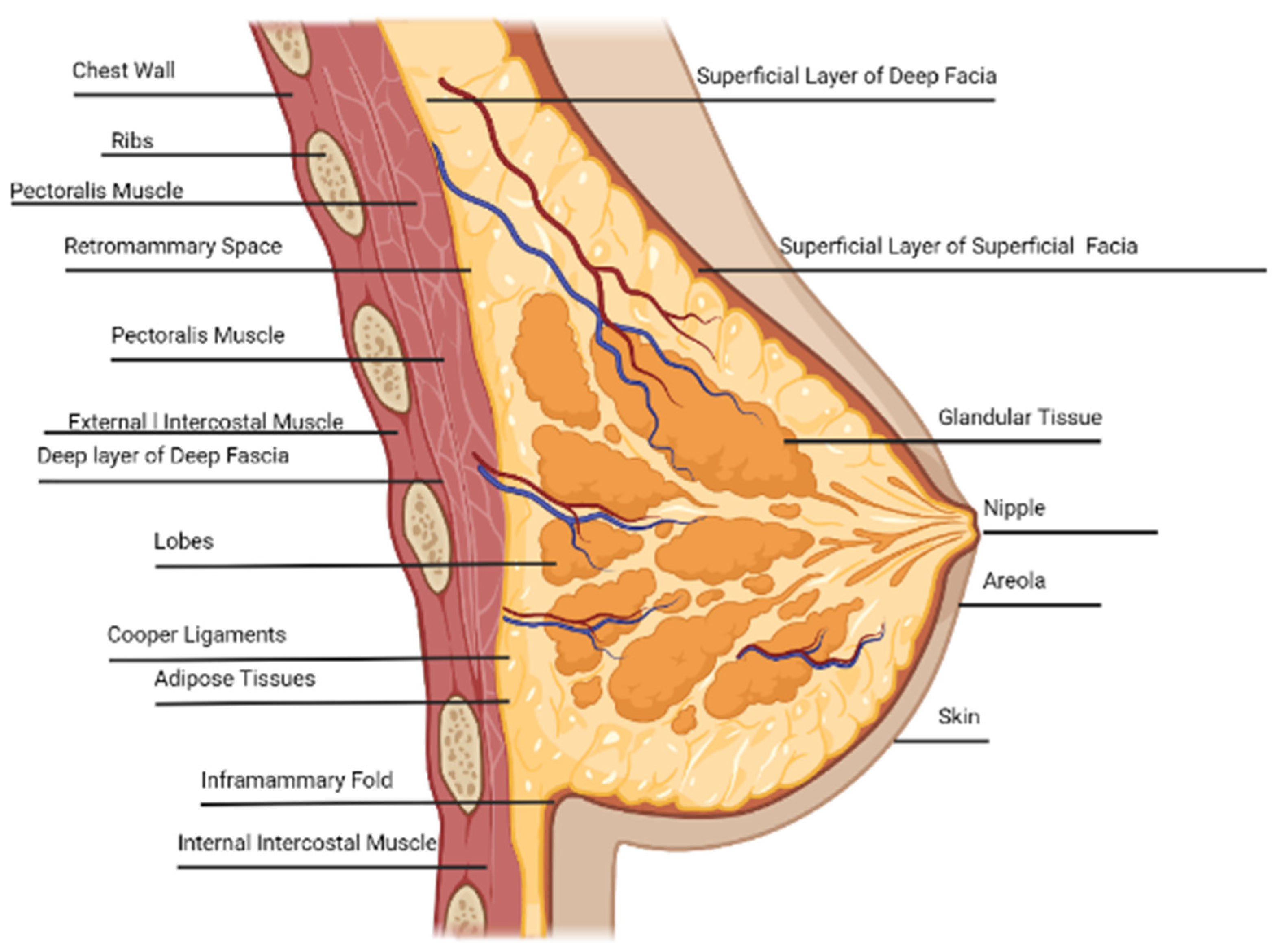
Figure 1. Lateral view of the breast anatomy, adapted by sources [23,24,25]. Created in BioRender. Galbreath, S. (2025) https://BioRender.com/p67q412, accessed on 22 October 2023.
Biomed 05 00005 g001
Figure 3. The behavior of the material as seen on the stress–strain curve [66,67]. Adapted from sources [66,67]. The stress–strain curve provides valuable information on the behavior of the material (i.e., Young’s modulus, stiffness, etc.) [68].
Biomed 05 00005 g003
Figure 4. Forces acting on the breast tissue when standing, walking, running, jumping, or kneeling on the hands and feet. The light-blue dots indicate the center of mass, and the orange dots are points of reference located at the base of the breast. The purple jellybeans represent the TEs in vivo. Figure 4 is adapted by sources [10,66,75,76]. Images were created by BioRender.com.
Biomed 05 00005 g004
Table 1. Female breast tissue skin properties based on the location and bra size [7,15,18,58].
Female Breast Tissue Skin Thickness
Anatomical LocationMethodologyAge RaceEthnicityBra Size Thickness (mm)Reference
Lateral 1.38 ± 0.24 [7]
Superior 1.38 ± 0.24 [7]
Medial 1.97 ± 0.26 [7]
Inferior 1.97 ± 0.26 [7]
Lateral Breast Fresh tissue—breast and abdomen cancer patients (mastectomy and/or reconstructive)—calipers, the Bulge test, and finite element modeling 71WhiteNon-Hispanic 2.6 [18]
Lateral Breast Fresh tissue—breast and abdomen cancer patients (mastectomy and/or reconstructive)—calipers, the Bulge test, and finite element modeling37BlackNon-Hispanic 4.4 [18]
Medial
Breast
Fresh tissue—breast and abdomen cancer patients (mastectomy and/or reconstructive)—calipers, the Bulge test, and finite element modeling37BlackNon-Hispanic 4.3 [18]
Breast—Unknown LocationFresh tissue—breast and abdomen cancer patients (mastectomy and/or reconstructive)—calipers, the Bulge test, and finite element modeling69AsianUnknown 3.55 [18]
Breast—Unknown LocationFresh tissue—breast and abdomen cancer patients (mastectomy and/or reconstructive)—calipers, the Bulge test, and finite element modeling55WhiteNon-Hispanic 3.05 [18]
Breast—Unknown LocationFresh tissue—breast and abdomen cancer patients (mastectomy and/or reconstructive)—calipers, the Bulge test, and finite element modeling41OtherHispanic 4.05 [18]
Lateral Breast Fresh tissue—breast and abdomen cancer patients (mastectomy and/or reconstructive)—calipers, the Bulge test, and finite element modeling66BlackNon-Hispanic 2.6 [18]
Lateral Breast Fresh tissue—breast and abdomen cancer patients (mastectomy and/or reconstructive)—calipers, the Bulge test, and finite element modeling66BlackNon-Hispanic 3.0 [18]
Medial BreastFresh tissue—breast and abdomen cancer patients (mastectomy and/or reconstructive)—calipers, the Bulge test, and finite element modeling33WhiteNon-Hispanic 3.15 [18]
Epidermis—Chest Fresh tissue—biopsy—measured with a microscope 16–50Korean 0.101 [15]
Dermis—Chest Fresh tissue—biopsy/skin operations—measured with a microscope16–50Korean 1.3778 [15]
Anterior–Posterior Breast CT scan prototype 1.45 ± 0.29 mean 1.0–2.2 range[58]
EpidermisCT scan prototype 0.07–1.4 range [58]
Dermis CT scan prototype 0.6 mm–3.0 range[58]
Anterior–Posterior Breast CT scan prototype A1.47 ± 0.07[58]
Anterior–Posterior Breast CT scan prototype B1.76 ± 0.04[58]
Anterior–Posterior Breast CT scan prototype C1.38 ± 0.10[58]
Anterior–Posterior Breast CT scan prototype D1.38 ± 0.10 [58]
Anterior–Posterior Breast CT scan prototype DD0.95 ±   0.07[58]
Epidermis 0.07–1.4 [58]
Dermis 0.6–3.0 [58]
Table 2. The subjective classification in BI-RADS, which has been made objective by utilizing software programs that can provide a numerical density percentage [94].
Stiffness Scale vs. BI-RADS Classification of Breast Density
Stiffness ScaleBI-RADS Classification of Breast Density
Class Defin. Palpation ResponseEnergy (x,y) Direction ∆U [J ∗ mm−3]GradeGradeClassification Description Density %Population %References
IEasily examinedBest palpation candidate. (0.0, 1.5)Grade 1 AFatty breast tissue Almost all fatty tissue <25%10[33,80,93,95]
IIWell examined <(1.5, 3.0)Grade 2 BScattered fibroglandular breast tissue Predominantly fatty tissue, with some dense glandular and fibrous connective tissue25–50%40[33,80,93,95]
IIISufficiently examined <(3.0, 4.5)Grade 3 CHeterogeneously dense breast tissueMore glandular and fibrous connective tissue compared to fatty tissue51–75%40[33,80,93,95]
IVDifficult to examineNot a candidate for palpation;
use other methods (i.e., MRI, US, etc.).
Requires exams more often.
<(4.5, 6.0)Grade 4 DExtremely dense breast tissueMostly glandular and fibrous connective tissue>75%10[33,80,93,95]
VNon-examNot a candidate for palpation;
use other methods (i.e., MRI, US, etc.).
Requires exams more often.
<(6.0, 7.5)Grade 5 >95% [80,96]
Grade 6 Malignant tissue biopsy validation[96]
Disclaimer/Publisher’s Note: The statements, opinions and data contained in all publications are solely those of the individual author(s) and contributor(s) and not of MDPI and/or the editor(s). MDPI and/or the editor(s) disclaim responsibility for any injury to people or property resulting from any ideas, methods, instructions or products referred to in the content.