Entry

Neonatal Intraventricular Hemorrhage: Current Perspectives and Management Strategies

by
Felicia H. Z. Chua
1,2,3,
Lee Ping Ng
1 and
Sharon Y. Y. Low
1,2,3,4,*
1
Neurosurgical Service, KK Women’s and Children’s Hospital, 100 Bukit Timah Road, Singapore 229899, Singapore
2
Department of Neurosurgery, National Neuroscience Institute, 11 Jalan Tan Tock Seng, Singapore 308433, Singapore
3
SingHealth Duke-NUS Neuroscience Academic Clinical Program, 11 Jalan Tan Tock Seng, Singapore 308433, Singapore
4
SingHealth Duke-NUS Pediatrics Academic Clinical Program, 100 Bukit Timah Road, Singapore 229899, Singapore
*
Author to whom correspondence should be addressed.
Encyclopedia 2024, 4(4), 1948-1961; https://doi.org/10.3390/encyclopedia4040127
Submission received: 7 October 2024 / Revised: 9 December 2024 / Accepted: 9 December 2024 / Published: 21 December 2024
(This article belongs to the Section Medicine & Pharmacology)

Definition

:
Neonatal intraventricular hemorrhage is a serious condition associated with significant acute and long-term morbidity and mortality. Neurosurgical intervention aims to relieve life-threatening raised intracranial pressure and prevent neurological deterioration. In recent years, advancements in disease understanding have paved the way for clinicians to re-evaluate conventional approaches in the management of affected patients. Examples include various neurosurgical techniques to actively reduce blood products with a view to avoid the consequences of complex hydrocephalus and intraparenchymal injury in the developing brain. In this entry paper, we aim to provide an overview of the current perspectives, pathophysiology and management strategies for this difficult condition.

1. Introduction

Neonatal intraventricular hemorrhage (nIVH) refers to the occurrence of spontaneous bleeding in the lateral and third or fourth ventricles characterized by hyper-attenuating fluid within the ventricles in imaging studies [1,2,3]. This potentially life-threatening condition is mostly associated with preterm infants, with increasing prevalence directly proportional to decreasing gestational age and birth weight. To date, the global incidence of nIVH in preterm infants has varied widely [4,5,6]. A recent systematic review by Siffel et al. summarizes the prevalence of higher grade nIVH to be in the range from 5 to 52% [6,7]. Nonetheless, most agree that the exact rates of this condition in any given population are difficult to determine owing to multiple parameters, such as antenatal factors, the availability of neonatal care, birth weight, gestational age and so forth [8,9]. To a significantly lesser extent, nIVH may occur in full-term neonates, with similar concerns of permanent neurological damage [3,10]. A feared sequelae of nIVH is ventriculomegaly causing post-hemorrhagic hydrocephalus (PHH) that results in raised intracranial pressure (ICP). If left untreated, the patient is at a high risk of impaired cognitive and functional neurodevelopment [11]. As a consequence, the pediatric neurosurgeon is often referred to intervene for cerebrospinal fluid (CSF) diversion with the aims of, firstly, reducing the raised intracranial pressure, and next, preventing further brain parenchymal injury. This entry paper attempts to discuss the current perspectives, pathophysiology, neurosurgical approaches and management challenges faced in nIVH, in corroboration with the existing literature.

2. Pathophysiology: An Overview

Broadly speaking, nIVH typically originates from the periventricular germinal matrix. Located on the head of caudate nucleus and underneath ventricular ependyma, the germinal matrix is a highly concentrated, vascular collection of glial and neuronal precursor cells. When the bleeding in the germinal matrix is substantial, the ependyma breaks, and the adjacent ventricle fills up with blood [12]. Although extensive research has been invested into its prevention, the overall incidence of nIVH has remained largely unchanged over the past few decades [13,14].

2.1. Intraventricular Hemorrhage in the Newborn: Preterm Infants Versus Full-Term Neonates

The sub-ependymal germinal matrix (GM) is located within the caudothalamic groove, which is adjacent to the fetal ventricular system. Under normal physiological circumstances, its thickness decreases after 24 weeks of gestation and almost disappears by the 36th gestational week [15]. In premature infants, up to 95% of cases of GM hemorrhage occur within the 7 days after birth [16]. The GM is a region rich in large, irregular, rapidly growing, immature capillary vessels with high blood supply harboring important neuroglial cells [17,18]. Based on this knowledge, the origin of nIVH in premature infants is exclusive to the GM. Here, bleeding primarily occurs due to the inherent fragility of the GM vasculature, cerebral blood flow disturbances and coagulation disorders [12]. Of note, we are also now aware that for these patients, intraparenchymal hemorrhage is secondary to periventricular hemorrhagic infarction as a result of impaired venous drainage of the medullary vein in the deep white matter, and not a direct extension of the original nIVH, as previously assumed [19]. In severe cases, the consequential buildup of nIVH blood products obstructs the arachnoid villi, blocking CSF reabsorption and causing obstructive hydrocephalus—often termed as ‘post-hemorrhagic hydrocephalus’ or ‘PHH’.
In contrast to well-established causative factors in preterm infants, the exact mechanism of nIVH in full-term neonates remains unelucidated. For this cohort, the GM mantle is expected to have dissipated, and hence, no longer being clinically apparent. Studies report that the source of nIVH in term infants mostly originates from the choroid plexus, residual germinal matrix tissue, the watershed area of the foramen of Monro adjacent to the caudate nucleus and thalamus [3,20,21]. The theorized risk factors include birth asphyxia [22,23], hypoxic ischemic encephalopathy [22], instrumental deliveries [22,23] and congenital heart disease causing altered hemodynamics and use of anticoagulant pharmacotherapy [24]. The other reasons cited in the limited literature include genetic factors, thrombocytopenia and undiagnosed coagulation factor deficiencies [25,26,27,28,29,30].

2.2. Neuroimaging in Neonatal Intraventricular Hemorrhage

As a rule of thumb, most neonatologists recommend brain imaging within 72 h after birth in preterm infants. This is essential to exclude GM bleeds and nIVH in this group of high-risk patients [31,32]. In contrast, the indication for neuroimaging in full-term infants is often prompted by clinical signs of seizures, neurological deterioration and apnea [33]. Here, cranial ultrasound (CUS) is the preferred brain imaging modality for neonates due to its portability and avoidance of ionizing radiation. In recent years, the utility of fetal magnetic resonance imaging (MRI) scans in evaluating detailed central nervous system (CNS) anatomy has increased, especially for selected cases, where CUS findings are equivocal [16,34]. This is because MRI is superior to CUS in identifying intracranial hemorrhage and white matter injury [35]. Based on the presence of germinal matrix bleeding and the amount of blood in the lateral ventricles, the radiological severity of nIVH is historically classified into four categories, according to the Papile system [36,37]. The original Papile system was conceived from computed tomographic (CT) brain imaging and subsequently adapted by Volpe et al. on the basis of CUS findings [36,37]. Although primarily used in preterm infants, this classification is sometimes referred to for cases of nIVH in full-term neonates for ease of description. (See Table 1).
To date, MRI modalities provide the best anatomical and structural details of lesions in the neuroaxis [38]. This is especially so for small parenchymal bleeds that may be difficult to visualize with CUS. Adjunct MRI techniques can quantitatively measure the biophysical properties of brain tissue in vivo, allowing better characterization of regional changes in the tissue microstructural environment [38]. Specifically for assessing blood products, these include susceptibility-weighted imaging (SWI) and T2*-weighted gradient echo imaging (GRE) sequences [16]. Furthermore, MRI has the added advantage for detection of temporal, occipital or cerebellar hemorrhages, which are not usually detected on CUS unless they are substantially large [16]. Although MRI scans are not routinely used as part of the imaging protocol in nIVH patients, they may be indicated for cases where there is suspicion of other underlying causes of the bleed, such as neoplasm, vascular anomalies or infection. At our institution, early MRI brain scans may be initiated for term infants presenting with nIVH.

2.3. The Consequences of Neonatal Intraventricular Hemorrhage and Post-Hemorrhagic Hydrocephalus

Regardless of the age of the newborn (i.e., premature versus full-term), the presence of nIVH has significant impacts in both early and late phases of life. Broadly speaking, higher radiological grades of nIVH are associated with increased rates of deficit in motor function, neurodevelopment and intellectual ability, especially in preterm infants [16,39,40,41,42]. Even the lower grades of nIVH (that is, I and II) have been implicated as risk factors for cerebral palsy and other neurocognitive impairments [16,43,44,45].
Insights from translational research on this topic highlight the following at the parenchymal level in the acute setting. Firstly, nIVH triggers significant periventricular inflammation and damages axons, thereby inducing apoptosis and maturational arrest of oligodendrocyte progenitors, leading to reduced myelination of white matter [46]. Concurrently, blockage of arachnoid villi prompts TGF-ß1 to be released into the CSF, leading to the immediate accumulation of extracellular matrix and glial fibrillary acidic proteins [14,15]. Next, a constellation of blood-induced reactions, including oxidative stress, glutamate excitotoxicity, inflammation, deranged signaling pathways and alteration of the extracellular matrix, contributes to irreversible white matter injury [46]. Additional studies evaluating nIVH breakdown products in the CSF of preterm neonates report elevated hemoglobin and ferritin levels corresponding to increasing ventricular size [16]. Put together, the current theory is that PHH is a consequence of endogenous iron and excess brain-injury-related protein clearance mechanisms being overwhelmed [14,15,16]. Furthermore, PHH also contributes to direct brain injury via enlargement of the ventricles and shearing of surrounding white matter [47]. Building on these, we are now aware that the severity of nIVH and associated PHH corresponds to the extent of brain tissue damage, with proportional degrees of neurodevelopmental delay, motor impairment and even death. Another theorized reason for their poor outcomes is hypothesized to be due to an early onset of ventriculomegaly, resulting in a protracted course of irreversible cellular damage [14,48].
To address this very challenging condition, various trials have been initiated with the objective of decreasing the morbidity and mortality associated with nIVH [49,50,51]. One example is drainage, irrigation and fibrinolytic therapy (DRIFT)—a randomized, controlled trial that attempted to remove nIVH blood, inflammatory cytokines and ferritin, which are associated with the development of hydrocephalus [50]. However, the results of the DRIFT study showed that, although it did not significantly lower the need for permanent shunt surgery, severe cognitive disability at 2 years was significantly reduced [49]. Separately, a study by Bassan et al. reports that early EVD insertion to facilitate continuous drainage of blood-burdened CSF in preterm infants with PHH is associated with lower rates of neurodevelopmental disabilities in comparison with those whose EVDs are inserted later [52]. Overall, these findings do concur that the active reduction of nIVH and its by-products has a positive correlation with long-term neurological outcomes [53].
From a neurosurgical perspective, we are aware that ventriculoperitoneal shunt (VPS) dependency is reported in more than 80% of these patients, with its innate risks of long-term implant complications [54]. Following that, a notable concern related to nIVH is the risk of interval multiloculated hydrocephalus—a condition that arises when intraventricular and parenchymal septations form in the aftermath of tissue scarring, creating discrete fluid-filled compartments [55,56]. This condition is extremely difficult to treat and may require several CSF diversion interventions in the patient’s lifetime; and thus, experience even more negative effects on their neurodevelopment in comparison to patients with non-complex hydrocephalus [14,57].

3. Review of Conventional Management Approaches

The established bedside techniques to temporize the effects of nIVH and PHH include serial tapping of CSF either via lumbar puncture (LP) or anterior fontanelle ventricular puncture, removing a minimum of 10 mg/kg of CSF with each tap [58]. Both procedures are relatively straightforward and can be performed safely by experienced neonatologists [59]. The rationale for active removal of old blood and protein from the CSF is to reduce the risk of hydrocephalus and the interval needed for a permanent VPS [60]. An LP removes CSF from the lower back via direct needle puncture of the lumbar cistern. (See Figure 1a). However, only low volumes can be removed each time, and repeated procedures may be required to effectively lower ICP. In contrast, ventricular tapping can remove more CSF via direct puncture of lateral ventricle by a small needle [61]. (See Figure 1b). Specific to this latter technique, the complications include cortical intracranial hemorrhage, and infection or porencephalic cyst formation may occur after multiple punctures [61,62,63,64,65]. Also, there are studies that show no significant difference in the risks of disability, death or permanent shunting between serial tapping and conservative treatment [62,63].

4. The Role of the Pediatric Neurosurgeon

To date, there are no established international guidelines for the treatment of nIVH and PHH. The existing literature demonstrates a heterogeneity of approaches, leading to a concurrent paucity of data to confirm the efficacy of the various treatment options [66]. At the time of writing, no single neurosurgical approach has shown definitive superiority over another. For most patients, the final choice of intervention is dependent on the operating neurosurgeon and patient-related factors. In recent years, there has been a paradigm shift toward a more proactive approach to remove nIVH blood products as prevention against biochemical parenchymal injury from the by-products of clot lysis. Under such circumstances, intervention is justified because, in theory, there is prolonged, ongoing damage to the developing brain tissue in cases of severe nIVH due to higher hematoma volumes.

4.1. Neurosurgical Temporizing Measures: A Summary of Options

In cases where a trial period of LP or ventricular tapping fails to control ICP, the patient may be referred to the neurosurgeon for consideration of a longer term temporizing option. Often, these are patients who may still have significant nIVH blood burden, other ongoing medical issues or extremely low birth weight and are hence unsuitable for an immediate permanent CSF shunt procedure. Examples of temporizing measures include external ventricular drain (EVD), ventricular access device (VAD) or ventriculo-subgaleal shunt (VSGS). A systemic review published in 2015 showed that VSGS and VAD could be effective temporary procedures, as well as EVD [54]. A prospective cohort study published in 2017 found no evidence of differences in the conversion rate to a permanent VP shunt among these temporary methods [67]. Overall, there is no definitive evidence to support one procedure being superior to the other at this juncture. In addition, patient factors tend to vary between individuals, including the interim resorption and clearance of nIVH. Put together, it may be difficult to predict the duration required for the temporizing measure. Ultimately, the choice of intervention is dependent on the infant, nursing experience, quality of CSF components and parental consent. At our institution, the emphasis is on open lines of communication among the multi-disciplinary clinical team, nursing staff and the parents for a cohesive approach to decision making.

4.1.1. External Ventricular Drain (EVD)

Under asepsis, a catheter is typically inserted into the selected anterior horn of a dilated lateral ventricle via Kocher’s point. Next, the end of the proximal catheter is subcutaneously tunneled to a site on the scalp (or body) distant from the initial incision and connected to an adjustable external drainage system [60]. Here, continuous drainage of old blood and protein material and CSF reduces ventricle size and ICP [52,60]. With an EVD in situ, repeated skin punctures to remove CSF are not necessary [61]. Next, the EVD provides clinicians with an opportunity to titrate the amount of CSF drained in order to prevent underdrainage [65]. The disadvantages of this procedure include its lack of longevity, infection (as it is not a closed system), the need for repeated CSF examinations, obstruction and dislodgement of the catheter and its attachments and CSF leakage from the catheter exit site on the scalp [65]. Overall, the complication rates in several studies vary widely, which may be due to small sample sizes and differences in institutional practice, including the methods of infection control, theatre conditions and the threshold for diagnosis of complications [8]. At our institution, EVD is often the choice of intervention for patients who have ongoing sepsis or are suspected of having an underlying infection.

4.1.2. Ventricular Access Device (VAD)

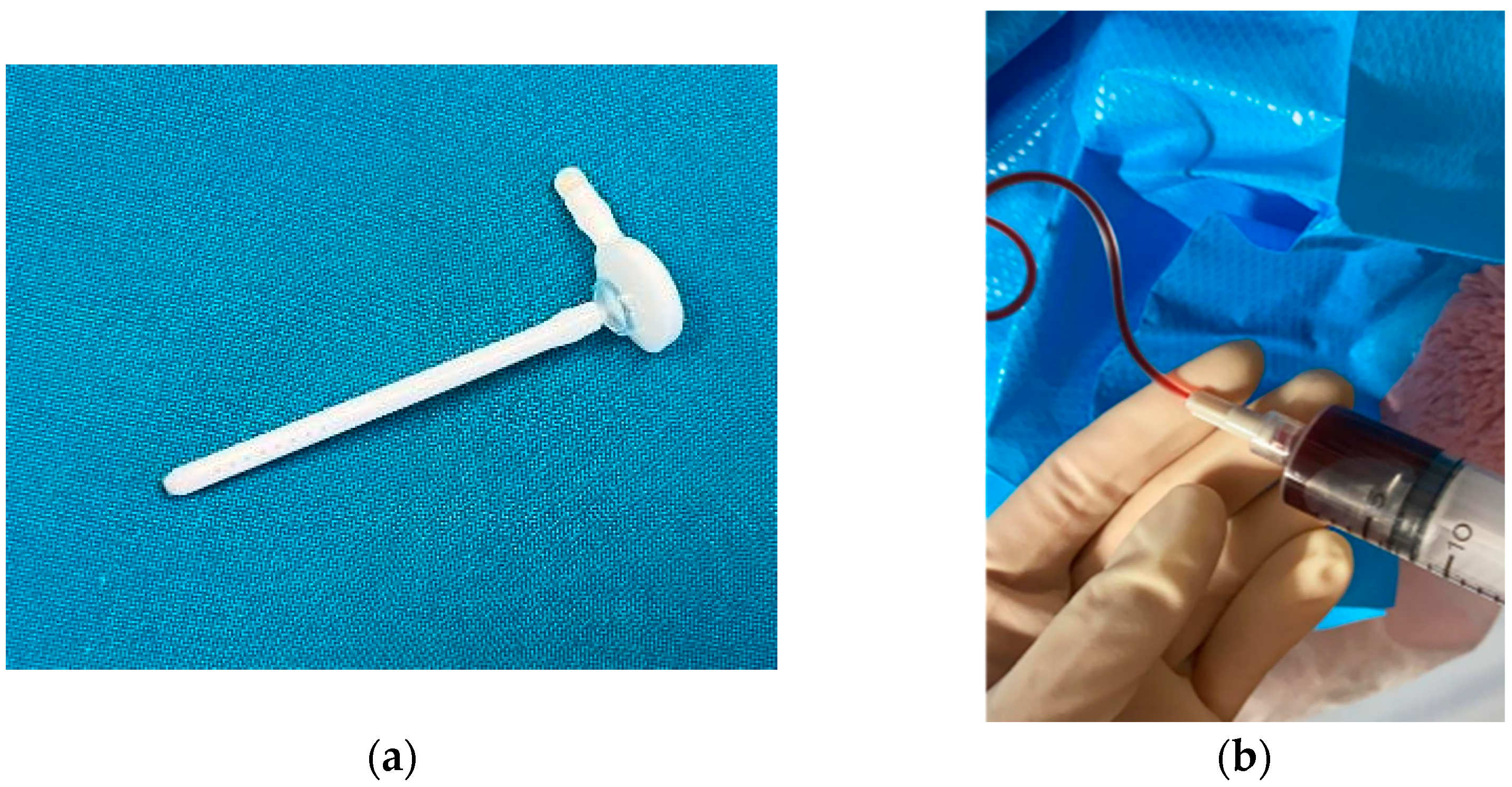
The VAD is a subcutaneous reservoir that has a catheter positioned in the lateral ventricle and is attached to a reservoir implanted under the scalp [61,63]. (See Figure 2a). This device helps to avoid repeated direct needle tracks through the brain and potential injury. The advantages are that it is a closed system that is easily accessible, and the amount of CSF removed can be adjusted to each patient’s opening pressure. (See Figure 2b). However, an important drawback of VADs is that the removal of CSF is intermittent. Under such circumstances, the fluid buildup and resulting ICP increase between taps may be detrimental. Other reported complications of VADs include CSF infection, skin necrosis, CSF fistula or subdural hygroma [60]. Although it has not been proven that VADs are superior to other temporization methods, the prevalent consensus is that they are still beneficial compared to early VP shunt insertion [8].

4.1.3. Ventriculo-Subgaleal Shunt (VSGS)

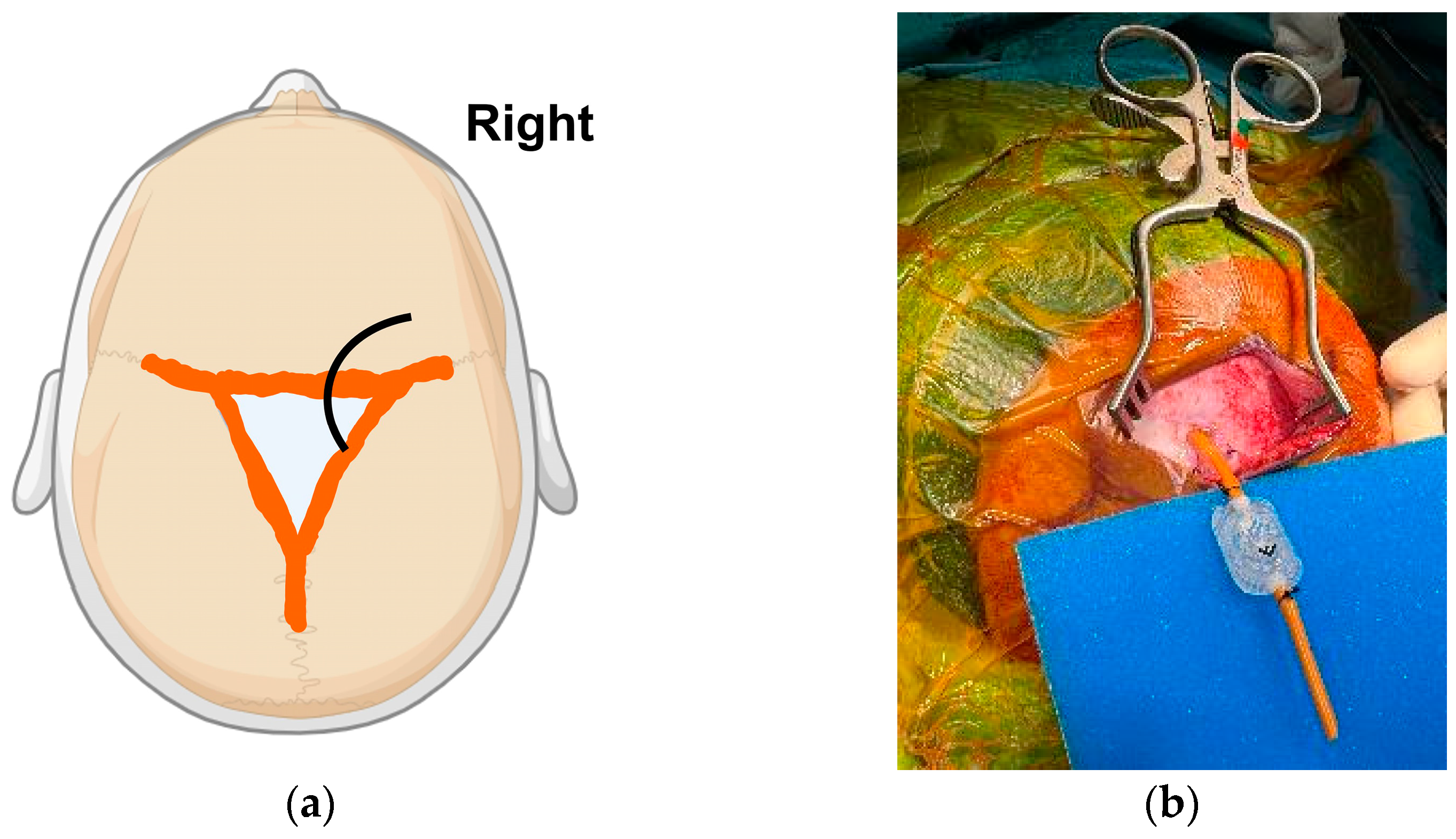
The scalp’s subgaleal space is a fibroareolar layer with elastic and absorptive properties that is found between the galea aponeurotica and the scalp periosteum [68]. Building on this, the VSGS capitalizes this advantage by directly connecting the ventricles to the patient’s own subgaleal space to allow continuous CSF diversion, providing sustained relief [8,69]. This approach has emerged as a suitable approach for neonates to control PVH until the patient reaches an adequate body weight and the CSF clarifies [68,70]. The surgical technique for VSGS has been well described in the literature [70,71]. Briefly, the patient is positioned in the supine position, with the coronal sutures, anterior fontanelle and midline identified. (See Figure 3a). Next, a ventricular catheter is placed into the frontal horn. Once smooth CSF flow is established, the catheter is connected to a right-angled extension to either a shortened piece of close-end tubing or a low-pressure valve to establish one-way flow from the ventricle into the subgaleal pocket [65,71]. (See Figure 3b). The creation of a generous subgaleal pocket is associated with increased longevity of the VSGS [71]. In essence, CSF is resorbed through a surgically dissected subgaleal scalp pocket, reducing the need for intermittent tapping required with a VAD [72,73,74]. This technique is also associated with lower infection rates than EVD due to the closed system of CSF drainage and lack of external tubes [65]. Following that, the VSGS establishes permanent decompression without causing electrolyte and nutritional losses [74,75]. Overall, VSGS is cited as a more physiological and less invasive means of achieving CSF diversion until a permanent shunt can be placed [60]. Notable complications of VSGS include catheter migration or malfunction, scarring of the subgaleal pocket [65,73,76] and/or CSF leak [76,77,78]. Some series also observe notable infection and failure rates [77,79]. Very rarely, a subgaleal encephalocele may develop, whereby patients present with seizures, meningitis, abscess formation and infarction of herniated brain parenchyma [65]. Another drawback unique to VSGS is the cosmetically unappealing swelling following the collection of egressed CSF in the subgaleal space. To mitigate this concern, caregivers are usually informed of the expected postoperative scalp swelling before the VSGS is inserted [77].

4.2. Neuroendoscopic Lavage (NEL): Background and Current Applications

In this era of modern medicine, neuroendoscopy has become a well-established neurosurgical technique in resection of intraventricular tumors, CSF diversion techniques, washout of ventriculitis and hematoma evacuation. Neuroendoscopy in pediatrics was first formally evaluated in 2014 in Part 2 of the Congress of Neurological Surgeons Pediatric Hydrocephalus Guidelines, where its use in endoscopic third ventriculostomy (ETV) in premature infants with PHH failed to show sufficient evidence to be recommended (Level III) [80]. In 2020, this recommendation was updated, stating that NEL was deemed a feasible and safe option for the removal of intraventricular clots, along with the benefit of reducing the rates of VP shunt placement [81]. The main objectives of NEL are, firstly, to clear the intraventricular blood products, and next, to reduce the protein burden [82]. Concurrently, the aligned publications on the use of NEL in neonatal IVH have reported motor and cognitive benefits in these patients, accompanied by low perioperative complication rates and shorter hospital stays [53,64,70,82,83,84]. Truth be said, most of these studies are retrospective in nature, with small patient cohorts [53,64,70,82,83,84,85]. At the time of writing, definitive results from prospective, multi-centered randomized controlled trials are still lacking. Nevertheless, there is an increasing consensus among pediatric neurosurgeons that NEL has superior benefits in the long-term neurodevelopmental outcomes of nIVH infants. To objectively address the benefits of NEL versus conventional treatment strategies, there are ongoing efforts by some international groups to collect multi-centered, prospective data [66,85,86].
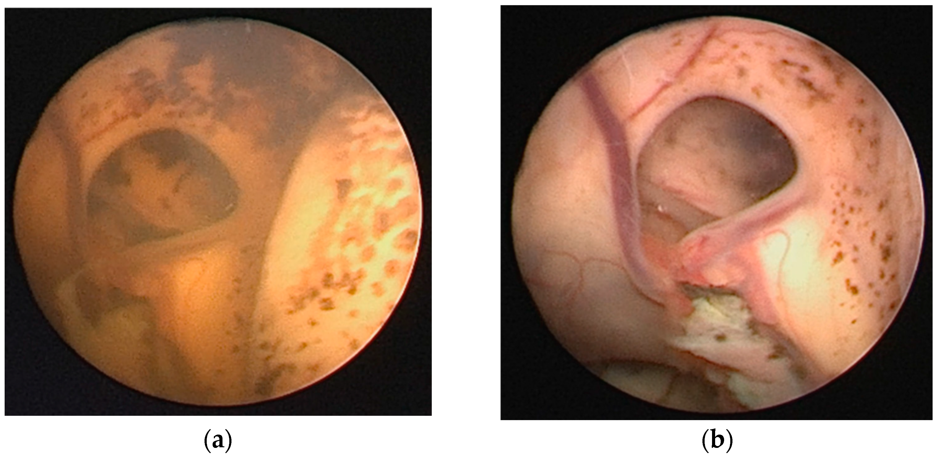
The technique for NEL has been described in the literature by several publications [64,82,83,84,87]. Briefly, the neuroendoscope is used to continuously irrigate, remove clots and perform a septostomy to communicate both hemispheres during the procedure. At our institution, the rigid, 0-degree Lotta® [Storz, Tuttlingen, Germany] is the neuroendoscope of choice, and irrigation of the ventricular system is facilitated by warm Ringer’s solution. (See Figure 4a,b).

4.3. The Ventriculoperitoneal Shunt (VPS) Is Imperfect but Still Relevant

Globally, the VPS is one of the most frequent life-saving procedures performed by pediatric neurosurgeons [88,89]. Most nIVH infants who undergo temporary CSF diversion will eventually still require permanent VPS placement. Interventions to decrease the likelihood of needing VPS have been trialed, but none have been proven effective [90]. Several studies cite a range between 2 and 3 kg as the minimum weight suitable for a VPS insertion, if there are no other comorbidities [91,92,93,94]. Surgical techniques and their nuances, morbidity rates and failure metrics are extensively discussed in the literature [89,95,96,97]. Despite its purported benefits, the VPS is associated with higher failure rates and implant-related complications in the pediatric population [65]. Particularly in premature neonates, young age and very low birth weight are identified as contributing factors for VPS failure. This is largely due to their higher risks of shunt infection because of an immature immune system, which makes them especially prone to pathogens and poor wound healing [14,90]. In addition, these patients are at risk of inadequate peritoneal CSF absorption, and a notable proportion have concurrent abdominal complications of prematurity [90]. Some studies also report that the high protein load in the ventricles due to blood degradation products may predispose VPS valves to blockage or dysfunction [98,99]. For the pediatric neurosurgeon, the following assumptions are certain: firstly, the existing VPS options are flawed, and many patients inevitably require multiple surgeries throughout their lifetime for shunt-related complications [100]. In congruency with other parts of the world, our neurosurgical unit faces similar issues from traditional fixed pressure shunt valves. These include CSF overdrainage due to the siphoning effect or, conversely, underdrainage causing subdural bleeding, the development of slit-ventricle syndrome, loss of cerebral compliance and cranio-cephalic disproportion [101]. To mitigate these potential problems, we may consider the choice of a programmable shunt valve for selected patients in this cohort [101].

5. Future Directions

As demonstrated in the above discussion, we acknowledge that most contemporary studies are still centered on the undertaking of acute and long-term effects of this catastrophic condition. Although extensive research has looked into the prevention of nIVH, its overall incidence has been largely unchanged over the past few decades [13,14]. We advocate for continued research on elucidating the causative factors of nIVH. This is because there are studies that observe extreme preterm infants with lower grades of nIVH (i.e., Papile grades I and II) that may not require neurosurgical intervention but are also associated with adverse neurodevelopmental outcomes [16,43,44,45]. Hence, emphasis on nIVH prevention is paramount for these vulnerable patients. Examples of the clinical prenatal factors to investigate may include chorioamnionitis, cord blood erythropoietin levels and so forth [90]. Following that, there is a role for concerted efforts by managing clinicians to consider NEL for selected cases of nIVH with PHH. This is especially so in the face of the growing body of evidence that patients who undergo preemptive removal of blood products via this approach have lower rates of shunt dependence and good long-term neurodevelopmental outcomes [82,87].

6. Conclusions

Neonatal intraventricular hemorrhage and its downstream sequelae continue to be challenging entities. This entry article highlights the current disease understanding and various treatment options for management. In this era of modern medicine, we advocate for active consensus efforts from like-minded clinicians to optimize the management of these vulnerable patients.

Author Contributions

Conceptualization, F.H.Z.C. and S.Y.Y.L.; methodology, S.Y.Y.L.; software, S.Y.Y.L.; validation, F.H.Z.C., L.P.N. and S.Y.Y.L.; formal analysis, F.H.Z.C. and S.Y.Y.L.; resources, F.H.Z.C., L.P.N. and S.Y.Y.L.; data curation, L.P.N. and S.Y.Y.L.; writing—original draft preparation, F.H.Z.C. and S.Y.Y.L.; writing—review and editing, F.H.Z.C. and S.Y.Y.L.; project administration, L.P.N. and S.Y.Y.L. All authors have read and agreed to the published version of the manuscript.

Funding

This research received no external funding.

Institutional Review Board Statement

Ethical review and approval were waived for this study since it is an Entry paper. It is meant for education purposes and does not contain any of our research data analysis on the topic of Neonatal Intraventricular Haemorrhage. Figure 2, Figure 3 and Figure 4 were taken as part of a broader study on ‘pediatric hydrocephalus’ that is approved by the Institutional Review Board (or Ethics Committee) of SingHealth Centralized Institutional Review Board (protocol code Singhealth CIRB Ref: 2020/2416 and 7 August 2023).

Informed Consent Statement

Written informed consent was obtained from the subjects’ parents/ legal guardians for the relevant intraoperative images used in the manuscript. The consents are taken as part of a broader study on ‘pediatric hydrocephalus’ that is approved by the Institutional Review Board (or Ethics Committee) of SingHealth Centralized Institutional Review Board (protocol code Singhealth CIRB Ref: 2020/2416 and 7 August 2023).

Data Availability Statement

Not applicable.

Conflicts of Interest

The authors declare no conflicts of interest.

References

  1. Nanba, E.; Eda, I.; Takashima, S.; Ohta, S.; Ohtani, K.; Takeshita, K. Intracranial hemorrhage in the full-term neonate and young infant: Correlation of the location and outcome. Brain Dev. 1984, 6, 435–443. [Google Scholar] [CrossRef] [PubMed]
  2. Trounce, J.Q.; Fagan, D.; Levene, M.I. Intraventricular haemorrhage and periventricular leucomalacia: Ultrasound and autopsy correlation. Arch. Dis. Child. 1986, 61, 1203–1207. [Google Scholar] [CrossRef] [PubMed]
  3. Afsharkhas, L.; Khalessi, N.; Karimi Panah, M. Intraventricular Hemorrhage in Term Neonates: Sources, Severity and Outcome. Iran. J. Child Neurol. 2015, 9, 34–39. [Google Scholar] [PubMed]
  4. Pande, G.S.; Vagha, J.D. A Review of the Occurrence of Intraventricular Hemorrhage in Preterm Newborns and its Future Neurodevelopmental Consequences. Cureus 2023, 15, e48968. [Google Scholar] [CrossRef]
  5. Maduray, T.; Mamdoo, F.; Masekela, R. A retrospective study on the prevalence, severity and outcomes of intraventricular haemorrhage in infants with a low birth weight in a quarternary hospital in a low- to middle-income country. S. Afr. J. Child Health 2019, 13, 56–62. [Google Scholar] [CrossRef]
  6. Siffel, C.; Kistler, K.D.; Sarda, S.P. Global incidence of intraventricular hemorrhage among extremely preterm infants: A systematic literature review. J. Perinat. Med. 2021, 49, 1017–1026. [Google Scholar] [CrossRef]
  7. Lee, J.; Lee, C.Y.M.; Naiduvaje, K.; Wong, Y.; Bhatia, A.; Ereno, I.L.; Ho, S.K.Y.; Yeo, C.L.; Rajadurai, V.S. Trends in neonatal mortality and morbidity in very-low-birth-weight (VLBW) infants over a decade: Singapore national cohort study. Pediatr. Neonatol. 2023, 64, 585–595. [Google Scholar] [CrossRef]
  8. Pettorini, B.; Keh, R.; Elllenbogen, J.; Williams, D.; Zebian, B. Intraventricular haemorrhage in prematurity. Infant 2014, 10, 186–190. [Google Scholar]
  9. du Plessis, A.J. The role of systemic hemodynamic disturbances in prematurity-related brain injury. J. Child Neurol. 2009, 24, 1127–1140. [Google Scholar] [CrossRef]
  10. Mitchell, W.; O’Tuama, L. Cerebral intraventricular hemorrhages in infants: A widening age spectrum. Pediatrics 1980, 65, 35–39. [Google Scholar] [CrossRef]
  11. Brouwer, A.J.; van Stam, C.; Uniken Venema, M.; Koopman, C.; Groenendaal, F.; de Vries, L.S. Cognitive and Neurological Outcome at the Age of 5–8 Years of Preterm Infants with Post-Hemorrhagic Ventricular Dilatation Requiring Neurosurgical Intervention. Neonatology 2011, 101, 210–216. [Google Scholar] [CrossRef] [PubMed]
  12. Ballabh, P. Intraventricular hemorrhage in premature infants: Mechanism of disease. Pediatr. Res. 2010, 67, 1–8. [Google Scholar] [CrossRef] [PubMed]
  13. Ballabh, P. Pathogenesis and prevention of intraventricular hemorrhage. Clin. Perinatol. 2014, 41, 47–67. [Google Scholar] [CrossRef] [PubMed]
  14. Low, S.Y.Y.; Kestle, J.R.W.; Walker, M.L.; Seow, W.T. Cerebrospinal fluid shunt malfunctions: A reflective review. Child’s Nerv. Syst. 2023, 39, 2719–2728. [Google Scholar] [CrossRef]
  15. Egesa, W.I.; Odoch, S.; Odong, R.J.; Nakalema, G.; Asiimwe, D.; Ekuk, E.; Twesigemukama, S.; Turyasiima, M.; Lokengama, R.K.; Waibi, W.M.; et al. Germinal Matrix-Intraventricular Hemorrhage: A Tale of Preterm Infants. Int. J. Pediatr. 2021, 2021, 6622598. [Google Scholar] [CrossRef]
  16. You, S.K. Neuroimaging of Germinal Matrix and Intraventricular Hemorrhage in Premature Infants. J. Korean Neurosurg. Soc. 2023, 66, 239–246. [Google Scholar] [CrossRef]
  17. Özek, E.; Kersin, S.G. Intraventricular hemorrhage in preterm babies. Turk. Arch. Pediatr. 2020, 55, 215–221. [Google Scholar] [CrossRef]
  18. McMillan, N.; Sharma, H.; Manganas, L.N.; Kirschen, G.W. Chapter 23—Development and pathology of the germinal matrix. In Factors Affecting Neurodevelopment; Martin, C.R., Preedy, V.R., Rajendram, R., Eds.; Academic Press: Cambridge, MA, USA, 2021; pp. 259–271. [Google Scholar]
  19. Takashima, S.; Mito, T.; Ando, Y. Pathogenesis of periventricular white matter hemorrhages in preterm infants. Brain Dev. 1986, 8, 25–30. [Google Scholar] [CrossRef]
  20. Fink, S. Intraventricular hemorrhage in the term infant. Neonatal Netw. 2000, 19, 13–18. [Google Scholar] [CrossRef]
  21. Berger, R.; Bender, S.; Sefkow, S.; Klingmüller, V.; Künzel, W.; Jensen, A. Peri/intraventricular haemorrhage: A cranial ultrasound study on 5286 neonates. Eur. J. Obstet. Gynecol. Reprod. Biol. 1997, 75, 191–203. [Google Scholar] [CrossRef]
  22. Brouwer, A.J.; Groenendaal, F.; Koopman, C.; Nievelstein, R.-J.A.; Han, S.K.; de Vries, L.S. Intracranial hemorrhage in full-term newborns: A hospital-based cohort study. Neuroradiology 2010, 52, 567–576. [Google Scholar] [CrossRef] [PubMed]
  23. Looney, C.B.; Smith, J.K.; Merck, L.H.; Wolfe, H.M.; Chescheir, N.C.; Hamer, R.M.; Gilmore, J.H. Intracranial hemorrhage in asymptomatic neonates: Prevalence on MR images and relationship to obstetric and neonatal risk factors. Radiology 2007, 242, 535–541. [Google Scholar] [CrossRef] [PubMed]
  24. Bruno, C.J.; Beslow, L.A.; Witmer, C.M.; Vossough, A.; Jordan, L.C.; Zelonis, S.; Licht, D.J.; Ichord, R.N.; Smith, S.E. Haemorrhagic stroke in term and late preterm neonates. Arch. Dis. Child. Fetal Neonatal Ed. 2014, 99, F48–F53. [Google Scholar] [CrossRef] [PubMed]
  25. Jhawar, B.S.; Ranger, A.; Steven, D.; Del Maestro, R.F. Risk Factors for Intracranial Hemorrhage among Full-term Infants: A Case-Control Study. Neurosurgery 2003, 52, 581–590. [Google Scholar] [CrossRef]
  26. Sahriarian, S.; Akbari, P.; Amini, E.; Dalili, H.; Esmaeilnia Shrivany, T.; Niknafs, N.; Shariat, M.; Ghorban Sabagh, V. Intraventricular Hemorrhage in a Term Neonate: Manifestation of Protein S Deficiency—A Case Report. Iran. J. Public Health 2016, 45, 531–534. [Google Scholar]
  27. Abraham, B.M.; Zaazoue, M.A.; Xu, G.; Ducis, K.A. Intraventricular hemorrhage in term infants: A single institutional experience between 2016 and 2020. Childs Nerv. Syst. 2023, 39, 2123–2129. [Google Scholar] [CrossRef]
  28. Tarantino, M.D.; Gupta, S.L.; Brusky, R.M. The incidence and outcome of intracranial haemorrhage in newborns with haemophilia: Analysis of the Nationwide Inpatient Sample database. Haemophilia 2007, 13, 380–382. [Google Scholar] [CrossRef]
  29. Mochida, G.H.; Ganesh, V.S.; Felie, J.M.; Gleason, D.; Hill, R.S.; Clapham, K.R.; Rakiec, D.; Tan, W.H.; Akawi, N.; Al-Saffar, M.; et al. A homozygous mutation in the tight-junction protein JAM3 causes hemorrhagic destruction of the brain, subependymal calcification, and congenital cataracts. Am. J. Hum. Genet. 2010, 87, 882–889. [Google Scholar] [CrossRef]
  30. Meuwissen, M.E.; Halley, D.J.; Smit, L.S.; Lequin, M.H.; Cobben, J.M.; de Coo, R.; van Harssel, J.; Sallevelt, S.; Woldringh, G.; van der Knaap, M.S.; et al. The expanding phenotype of COL4A1 and COL4A2 mutations: Clinical data on 13 newly identified families and a review of the literature. Genet. Med. 2015, 17, 843–853. [Google Scholar] [CrossRef]
  31. Guillot, M.; Chau, V.; Lemyre, B. Routine imaging of the preterm neonatal brain. Paediatr. Child Health 2020, 25, 249–262. [Google Scholar] [CrossRef]
  32. Caro-Domínguez, P.; Lecacheux, C.; Hernandez-Herrera, C.; Llorens-Salvador, R. Cranial ultrasound for beginners. Transl. Pediatr. 2021, 10, 1117–1137. [Google Scholar] [CrossRef] [PubMed]
  33. Szpecht, D.; Frydryszak, D.; Miszczyk, N.; Szymankiewicz, M.; Gadzinowski, J. The incidence of severe intraventricular hemorrhage based on retrospective analysis of 35939 full-term newborns-report of two cases and review of literature. Childs Nerv. Syst. 2016, 32, 2447–2451. [Google Scholar] [CrossRef] [PubMed]
  34. Radoš, M.; Judaš, M.; Kostović, I. In vitro MRI of brain development. Eur. J. Radiol. 2006, 57, 187–198. [Google Scholar] [CrossRef] [PubMed]
  35. Guillot, M.; Sebastianski, M.; Lemyre, B. Comparative performance of head ultrasound and MRI in detecting preterm brain injury and predicting outcomes: A systematic review. Acta Paediatr. 2021, 110, 1425–1432. [Google Scholar] [CrossRef]
  36. Papile, L.A.; Burstein, J.; Burstein, R.; Koffler, H. Incidence and evolution of subependymal and intraventricular hemorrhage: A study of infants with birth weights less than 1500 gm. J. Pediatr. 1978, 92, 529–534. [Google Scholar] [CrossRef]
  37. Volpe, J.J. Intraventricular hemorrhage in the premature infant--current concepts. Part II. Ann. Neurol. 1989, 25, 109–116. [Google Scholar] [CrossRef]
  38. Sawlani, V.; Patel, M.D.; Davies, N.; Flintham, R.; Wesolowski, R.; Ughratdar, I.; Pohl, U.; Nagaraju, S.; Petrik, V.; Kay, A.; et al. Multiparametric MRI: Practical approach and pictorial review of a useful tool in the evaluation of brain tumours and tumour-like lesions. Insights Imaging 2020, 11, 84. [Google Scholar] [CrossRef]
  39. Hollebrandse, N.L.; Spittle, A.J.; Burnett, A.C.; Anderson, P.J.; Roberts, G.; Doyle, L.W.; Cheong, J.L.Y. School-age outcomes following intraventricular haemorrhage in infants born extremely preterm. Arch. Dis. Child. Fetal Neonatal Ed. 2021, 106, 4–8. [Google Scholar] [CrossRef]
  40. Pinto, C.; Malik, P.; Desai, R.; Shelar, V.; Bekina-Sreenivasan, D.; Satnarine, T.A.; Lavado, L.K.; Singla, R.; Chavda, D.; Kaul, S.; et al. Post-Hemorrhagic Hydrocephalus and Outcomes Amongst Neonates With Intraventricular Hemorrhage: A Systematic Review and Pooled Analysis. Cureus 2021, 13, e18877. [Google Scholar] [CrossRef]
  41. Dorner, R.A.; Burton, V.J.; Allen, M.C.; Robinson, S.; Soares, B.P. Preterm neuroimaging and neurodevelopmental outcome: A focus on intraventricular hemorrhage, post-hemorrhagic hydrocephalus, and associated brain injury. J. Perinatol. 2018, 38, 1431–1443. [Google Scholar] [CrossRef]
  42. Nongena, P.; Ederies, A.; Azzopardi, D.V.; Edwards, A.D. Confidence in the prediction of neurodevelopmental outcome by cranial ultrasound and MRI in preterm infants. Arch. Dis. Child. Fetal Neonatal Ed. 2010, 95, F388–F390. [Google Scholar] [CrossRef] [PubMed]
  43. Rees, P.; Callan, C.; Chadda, K.R.; Vaal, M.; Diviney, J.; Sabti, S.; Harnden, F.; Gardiner, J.; Battersby, C.; Gale, C.; et al. Preterm Brain Injury and Neurodevelopmental Outcomes: A Meta-analysis. Pediatrics 2022, 150, e2022057442. [Google Scholar] [CrossRef] [PubMed]
  44. Patra, K.; Wilson-Costello, D.; Taylor, H.G.; Mercuri-Minich, N.; Hack, M. Grades I-II intraventricular hemorrhage in extremely low birth weight infants: Effects on neurodevelopment. J. Pediatr. 2006, 149, 169–173. [Google Scholar] [CrossRef] [PubMed]
  45. Bolisetty, S.; Dhawan, A.; Abdel-Latif, M.; Bajuk, B.; Stack, J.; Lui, K. Intraventricular hemorrhage and neurodevelopmental outcomes in extreme preterm infants. Pediatrics 2014, 133, 55–62. [Google Scholar] [CrossRef]
  46. Ballabh, P.; de Vries, L.S. White matter injury in infants with intraventricular haemorrhage: Mechanisms and therapies. Nat. Rev. Neurol. 2021, 17, 199–214. [Google Scholar] [CrossRef]
  47. Cherian, S.; Whitelaw, A.; Thoresen, M.; Love, S. The pathogenesis of neonatal post-hemorrhagic hydrocephalus. Brain Pathol. 2004, 14, 305–311. [Google Scholar] [CrossRef]
  48. McAllister, J.P., 2nd. Pathophysiology of congenital and neonatal hydrocephalus. Semin. Fetal Neonatal Med. 2012, 17, 285–294. [Google Scholar] [CrossRef]
  49. Whitelaw, A.; Jary, S.; Kmita, G.; Wroblewska, J.; Musialik-Swietlinska, E.; Mandera, M.; Hunt, L.; Carter, M.; Pople, I. Randomized trial of drainage, irrigation and fibrinolytic therapy for premature infants with posthemorrhagic ventricular dilatation: Developmental outcome at 2 years. Pediatrics 2010, 125, e852–e858. [Google Scholar] [CrossRef]
  50. Whitelaw, A.; Evans, D.; Carter, M.; Thoresen, M.; Wroblewska, J.; Mandera, M.; Swietlinski, J.; Simpson, J.; Hajivassiliou, C.; Hunt, L.P.; et al. Randomized clinical trial of prevention of hydrocephalus after intraventricular hemorrhage in preterm infants: Brain-washing versus tapping fluid. Pediatrics 2007, 119, e1071–e1078. [Google Scholar] [CrossRef]
  51. Kennedy, C.R.; Ayers, S.; Campbell, M.J.; Elbourne, D.; Hope, P.; Johnson, A. Randomized, controlled trial of acetazolamide and furosemide in posthemorrhagic ventricular dilation in infancy: Follow-up at 1 year. Pediatrics 2001, 108, 597–607. [Google Scholar] [CrossRef]
  52. Bassan, H.; Eshel, R.; Golan, I.; Kohelet, D.; Ben Sira, L.; Mandel, D.; Levi, L.; Constantini, S.; Beni-Adani, L. Timing of external ventricular drainage and neurodevelopmental outcome in preterm infants with posthemorrhagic hydrocephalus. Eur. J. Paediatr. Neurol. 2012, 16, 662–670. [Google Scholar] [CrossRef] [PubMed]
  53. Behrens, P.; Tietze, A.; Walch, E.; Bittigau, P.; Bührer, C.; Schulz, M.; Aigner, A.; Thomale, U.W. Neurodevelopmental outcome at 2 years after neuroendoscopic lavage in neonates with posthemorrhagic hydrocephalus. J. Neurosurg. Pediatr. 2020, 26, 495–503. [Google Scholar] [CrossRef] [PubMed]
  54. Badhiwala, J.H.; Hong, C.J.; Nassiri, F.; Hong, B.Y.; Riva-Cambrin, J.; Kulkarni, A.V. Treatment of posthemorrhagic ventricular dilation in preterm infants: A systematic review and meta-analysis of outcomes and complications. J. Neurosurg. Pediatr. 2015, 16, 545–555. [Google Scholar] [CrossRef] [PubMed]
  55. Sandberg, D.I.; McComb, J.G.; Krieger, M.D. Craniotomy for fenestration of multiloculated hydrocephalus in pediatric patients. Oper. Neurosurg. 2005, 57, 100–106. [Google Scholar] [CrossRef]
  56. Vinnakota, A.; Wright, J.M.; Tomei, K.L. A Precursor to Multiloculated Hydrocephalus: Case Report and Review of Literature. World Neurosurg. 2019, 130, 216–221. [Google Scholar] [CrossRef]
  57. Vinchon, M.; Rekate, H.; Kulkarni, A.V. Pediatric hydrocephalus outcomes: A review. Fluids Barriers CNS 2012, 9, 18. [Google Scholar] [CrossRef]
  58. Hislop, J.E.; Dubowitz, L.M.; Kaiser, A.M.; Singh, M.P.; Whitelaw, A.G. Outcome of infants shunted for post-haemorrhagic ventricular dilatation. Dev. Med. Child Neurol. 1988, 30, 451–456. [Google Scholar] [CrossRef]
  59. Bedetti, L.; Lugli, L.; Marrozzini, L.; Baraldi, A.; Leone, F.; Baroni, L.; Lucaccioni, L.; Rossi, C.; Roversi, M.F.; D’Amico, R.; et al. Safety and Success of Lumbar Puncture in Young Infants: A Prospective Observational Study. Front. Pediatr. 2021, 9, 692652. [Google Scholar] [CrossRef]
  60. Shooman, D.; Portess, H.; Sparrow, O. A review of the current treatment methods for posthaemorrhagic hydrocephalus of infants. Cerebrospinal Fluid. Res. 2009, 6, 1. [Google Scholar] [CrossRef]
  61. Park, E.K.; Kim, J.Y.; Kim, D.S.; Shim, K.W. Temporary Surgical Management of Intraventricular Hemorrhage in Premature Infants. J. Korean Neurosurg. Soc. 2023, 66, 274–280. [Google Scholar] [CrossRef]
  62. Kreusser, K.L.; Tarby, T.J.; Kovnar, E.; Taylor, D.A.; Hill, A.; Volpe, J.J. Serial lumbar punctures for at least temporary amelioration of neonatal posthemorrhagic hydrocephalus. Pediatrics 1985, 75, 719–724. [Google Scholar] [CrossRef] [PubMed]
  63. Whitelaw, A.; Lee-Kelland, R. Repeated lumbar or ventricular punctures in newborns with intraventricular haemorrhage. Cochrane Database Syst. Rev. 2017, 4, Cd000216. [Google Scholar] [CrossRef] [PubMed]
  64. Dvalishvili, A.; Khinikadze, M.; Gegia, G.; Khutsishvili, L. Neuroendoscopic lavage versus traditional surgical methods for the early management of posthemorrhagic hydrocephalus in neonates. Childs Nerv. Syst. 2022, 38, 1897–1902. [Google Scholar] [CrossRef] [PubMed]
  65. Kuo, M.-F. Surgical management of intraventricular hemorrhage and posthemorrhagic hydrocephalus in premature infants. Biomed. J. 2020, 43, 268–276. [Google Scholar] [CrossRef]
  66. Thomale, U.W.; Cinalli, G.; Kulkarni, A.V.; Al-Hakim, S.; Roth, J.; Schaumann, A.; Bührer, C.; Cavalheiro, S.; Sgouros, S.; Constantini, S.; et al. TROPHY registry study design: A prospective, international multicenter study for the surgical treatment of posthemorrhagic hydrocephalus in neonates. Childs Nerv. Syst. 2019, 35, 613–619. [Google Scholar] [CrossRef]
  67. Wellons, J.C., 3rd; Shannon, C.N.; Holubkov, R.; Riva-Cambrin, J.; Kulkarni, A.V.; Limbrick, D.D., Jr.; Whitehead, W.; Browd, S.; Rozzelle, C.; Simon, T.D.; et al. Shunting outcomes in posthemorrhagic hydrocephalus: Results of a Hydrocephalus Clinical Research Network prospective cohort study. J. Neurosurg. Pediatr. 2017, 20, 19–29. [Google Scholar] [CrossRef]
  68. Eid, S.; Iwanaga, J.; Oskouian, R.J.; Loukas, M.; Jerry Oakes, W.; Shane Tubbs, R. Ventriculosubgaleal shunting—A comprehensive review and over two-decade surgical experience. Child’s Nerv. Syst. 2018, 34, 1639–1642. [Google Scholar] [CrossRef]
  69. Perret, G.E.; Graf, C.J. Subgaleal shunt for temporary ventricle decompression and subdural drainage. J. Neurosurg. 1977, 47, 590–595. [Google Scholar] [CrossRef]
  70. Frassanito, P.; Serrao, F.; Gallini, F.; Bianchi, F.; Massimi, L.; Vento, G.; Tamburrini, G. Ventriculosubgaleal shunt and neuroendoscopic lavage: Refining the treatment algorithm of neonatal post-hemorrhagic hydrocephalus. Childs Nerv. Syst. 2021, 37, 3531–3540. [Google Scholar] [CrossRef]
  71. Hansasuta, A.; Boongird, A. Ventriculo-subgaleal shunt: Step-by-step technical note. J. Med. Assoc. Thai 2007, 90, 473–478. [Google Scholar]
  72. McComb, J.G.; Ramos, A.D.; Platzker, A.C.; Henderson, D.J.; Segall, H.D. Management of hydrocephalus secondary to intraventricular hemorrhage in the preterm infant with a subcutaneous ventricular catheter reservoir. Neurosurgery 1983, 13, 295–300. [Google Scholar] [CrossRef] [PubMed]
  73. Fulmer, B.B.; Grabb, P.A.; Oakes, W.J.; Mapstone, T.B. Neonatal ventriculosubgaleal shunts. Neurosurgery 2000, 47, 80–83, discussion 83–84. [Google Scholar] [CrossRef] [PubMed]
  74. Fountain, D.M.; Chari, A.; Allen, D.; James, G. Comparison of the use of ventricular access devices and ventriculosubgaleal shunts in posthaemorrhagic hydrocephalus: Systematic review and meta-analysis. Childs Nerv. Syst. 2016, 32, 259–267. [Google Scholar] [CrossRef] [PubMed]
  75. Köksal, V.; Öktem, S. Ventriculosubgaleal shunt procedure and its long-term outcomes in premature infants with post-hemorrhagic hydrocephalus. Childs Nerv. Syst. 2010, 26, 1505–1515. [Google Scholar] [CrossRef] [PubMed]
  76. Tubbs, R.S.; Smyth, M.D.; Wellons, J.C., 3rd; Blount, J.; Grabb, P.A.; Oakes, W.J. Life expectancy of ventriculosubgaleal shunt revisions. Pediatr. Neurosurg. 2003, 38, 244–246. [Google Scholar] [CrossRef]
  77. Willis, B.K.; Kumar, C.R.; Wylen, E.L.; Nanda, A. Ventriculosubgaleal shunts for posthemorrhagic hydrocephalus in premature infants. Pediatr. Neurosurg. 2005, 41, 178–185. [Google Scholar] [CrossRef]
  78. Sklar, F.; Adegbite, A.; Shapiro, K.; Miller, K. Ventriculosubgaleal shunts: Management of posthemorrhagic hydrocephalus in premature infants. Pediatr. Neurosurg. 1992, 18, 263–265. [Google Scholar] [CrossRef]
  79. Kadri, H.; Mawla, A.A.; Kazah, J. The incidence, timing, and predisposing factors of germinal matrix and intraventricular hemorrhage (GMH/IVH) in preterm neonates. Childs Nerv. Syst. 2006, 22, 1086–1090. [Google Scholar] [CrossRef]
  80. Mazzola, C.A.; Choudhri, A.F.; Auguste, K.I.; Limbrick, D.D.; Rogido, M.; Mitchell, L.; Flannery, A.M. Pediatric hydrocephalus: Systematic literature review and evidence-based guidelines. Part 2: Management of posthemorrhagic hydrocephalus in premature infants. J. Neurosurg. Pediatr. PED 2014, 14, 8–23. [Google Scholar] [CrossRef]
  81. Bauer, D.F.; Baird, L.C.; Klimo, P.; Mazzola, C.A.; Nikas, D.C.; Tamber, M.S.; Flannery, A.M. Congress of Neurological Surgeons Systematic Review and Evidence-Based Guidelines on the Treatment of Pediatric Hydrocephalus: Update of the 2014 Guidelines. Neurosurgery 2020, 87, 1071–1075. [Google Scholar] [CrossRef]
  82. Honeyman, S.I.; Boukas, A.; Jayamohan, J.; Magdum, S. Neuroendoscopic lavage for the management of neonatal post-haemorrhagic hydrocephalus: A retrospective series. Child’s Nerv. Syst. ChNS Off. J. Int. Soc. Pediatr. Neurosurg. 2022, 38, 115–121. [Google Scholar] [CrossRef] [PubMed]
  83. Etus, V.; Kahilogullari, G.; Karabagli, H.; Unlu, A. Early Endoscopic Ventricular Irrigation for the Treatment of Neonatal Posthemorrhagic Hydrocephalus: A Feasible Treatment Option or Not? A Multicenter Study. Turk. Neurosurg. 2018, 28, 137–141. [Google Scholar] [CrossRef] [PubMed]
  84. d’Arcangues, C.; Schulz, M.; Bührer, C.; Thome, U.; Krause, M.; Thomale, U.W. Extended Experience with Neuroendoscopic Lavage for Posthemorrhagic Hydrocephalus in Neonates. World Neurosurg. 2018, 116, e217–e224. [Google Scholar] [CrossRef] [PubMed]
  85. Schulz, M.; Bührer, C.; Pohl-Schickinger, A.; Haberl, H.; Thomale, U.W. Neuroendoscopic lavage for the treatment of intraventricular hemorrhage and hydrocephalus in neonates. J. Neurosurg. Pediatr. 2014, 13, 626–635. [Google Scholar] [CrossRef] [PubMed]
  86. A standardised protocol for neuro-endoscopic lavage for post-haemorrhagic ventricular dilatation: A Delphi consensus approach. Childs Nerv. Syst. 2022, 38, 2181–2187. [CrossRef]
  87. Tirado-Caballero, J.; Rivero-Garvia, M.; Arteaga-Romero, F.; Herreria-Franco, J.; Lozano-Gonzalez, Á.; Marquez-Rivas, J. Neuroendoscopic lavage for the management of posthemorrhagic hydrocephalus in preterm infants: Safety, effectivity, and lessons learned. J. Neurosurg. Pediatr. 2020, 26, 237–246. [Google Scholar] [CrossRef]
  88. Lee, R.P.; Ajmera, S.; Thomas, F.; Dave, P.; Lillard, J.C.; Wallace, D.; Broussard, A.; Motiwala, M.; Norrdahl, S.P.; Venable, G.T.; et al. Shunt Failure-The First 30 Days. Neurosurgery 2020, 87, 123–129. [Google Scholar] [CrossRef]
  89. Lim, J.X.; Han, H.P.; Foo, Y.W.; Chan, Y.H.; Ng, L.P.; Low, D.C.Y.; Seow, W.T.; Low, S.Y.Y. Paediatric ventriculoperitoneal shunt failures: 12-year experience from a Singapore children’s hospital. Childs Nerv. Syst. 2023, 39, 3445–3455. [Google Scholar] [CrossRef]
  90. Robinson, S. Neonatal posthemorrhagic hydrocephalus from prematurity: Pathophysiology and current treatment concepts. J. Neurosurg. Pediatr. 2012, 9, 242–258. [Google Scholar] [CrossRef]
  91. Harada, A. Permanent Surgical Treatment for Posthemorrhagic Hydrocephalus in Preterm Infants. J. Korean Neurosurg. Soc. 2023, 66, 281–288. [Google Scholar] [CrossRef]
  92. Fulkerson, D.H.; Vachhrajani, S.; Bohnstedt, B.N.; Patel, N.B.; Patel, A.J.; Fox, B.D.; Jea, A.; Boaz, J.C. Analysis of the risk of shunt failure or infection related to cerebrospinal fluid cell count, protein level, and glucose levels in low-birth-weight premature infants with posthemorrhagic hydrocephalus. J. Neurosurg. Pediatr. 2011, 7, 147–151. [Google Scholar] [CrossRef] [PubMed]
  93. Whitelaw, A.; Aquilina, K. Management of posthaemorrhagic ventricular dilatation. Arch. Dis. Child. Fetal Neonatal Ed. 2012, 97, F229–F233. [Google Scholar] [CrossRef] [PubMed]
  94. Valdez Sandoval, P.; Hernández Rosales, P.; Quiñones Hernández, D.G.; Chavana Naranjo, E.A.; García Navarro, V. Intraventricular hemorrhage and posthemorrhagic hydrocephalus in preterm infants: Diagnosis, classification, and treatment options. Childs Nerv. Syst. 2019, 35, 917–927. [Google Scholar] [CrossRef] [PubMed]
  95. Elmesallamy, W.A.E.A.; Abofaid, A.M.A.; Mohamed, M.S.; Taha, M.M. Pediatric ventriculoperitoneal shunt: A comparative study between anterior fontanel ultrasound-guided versus conventional cranial end insertion. Child’s Nerv. Syst. 2023, 39, 921–928. [Google Scholar] [CrossRef]
  96. Gluski, J.; Zajciw, P.; Hariharan, P.; Morgan, A.; Morales, D.M.; Jea, A.; Whitehead, W.; Marupudi, N.; Ham, S.; Sood, S.; et al. Characterization of a multicenter pediatric-hydrocephalus shunt biobank. Fluids Barriers CNS 2020, 17, 45. [Google Scholar] [CrossRef]
  97. Limwattananon, P.; Kitkhuandee, A. Ventriculoperitoneal shunt failure in pediatric patients: An analysis of a national hospitalization database in Thailand. J. Neurosurg. Pediatr. 2021, 28, 128–138. [Google Scholar] [CrossRef]
  98. Brydon, H.L.; Hayward, R.; Harkness, W.; Bayston, R. Physical properties of cerebrospinal fluid of relevance to shunt function. 2: The effect of protein upon CSF surface tension and contact angle. Br. J. Neurosurg. 1995, 9, 645–651. [Google Scholar] [CrossRef]
  99. Kaestner, S.; Sani, R.; Graf, K.; Uhl, E.; Godau, J.; Deinsberger, W. CSF shunt valve occlusion-does CSF protein and cell count matter? Acta Neurochir. 2021, 163, 1991–1996. [Google Scholar] [CrossRef]
  100. Tomei, K.L. The Evolution of Cerebrospinal Fluid Shunts: Advances in Technology and Technique. Pediatr. Neurosurg. 2017, 52, 369–380. [Google Scholar] [CrossRef]
  101. Tey, M.L.; Ng, L.P.; Low, D.C.Y.; Seow, W.T.; Low, S.Y.Y. Programmable Shunt Valves for Pediatric Hydrocephalus: 22-Year Experience from a Singapore Children’s Hospital. Brain Sci. 2021, 11, 1548. [Google Scholar] [CrossRef]
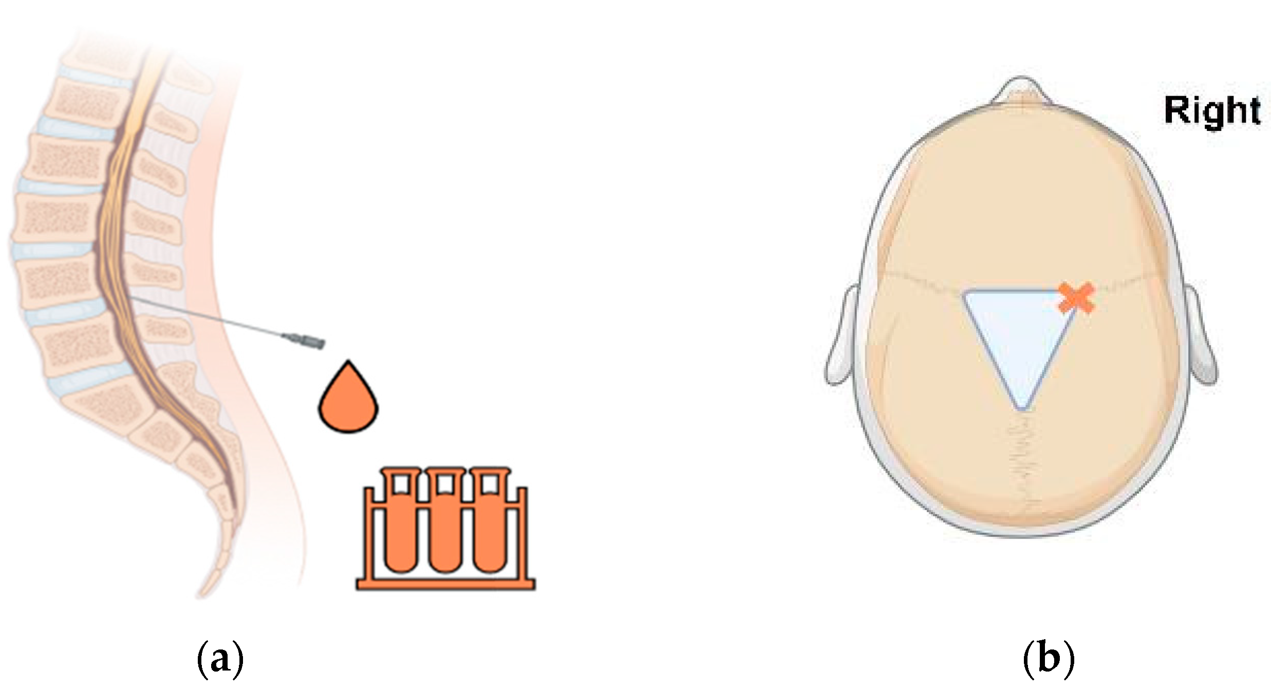
Figure 1. (a) Representative image of the lumbar puncture procedure. Under asepsis, a spinal tap needle is inserted in the lumbar subarachnoid space below the conus. Here, drips of blood-stained or xanthochromic CSF are collected. (b) Representative illustration of the location for inserting the needle for an anterior fontanelle tap. Under asepsis, a needle is inserted as far laterally as possible at the border of the anterior fontanelle or along the coronal suture to avoid injuring the superior sagittal sinus (marked by ‘X’). All images were created with the use of BioRender (BioRender.com; accessed on 1 September 2024) and Microsoft® Powerpoint® for Microsoft 365 (Microsoft Corporation, Redmond, WA, USA).
Encyclopedia 04 00127 g001
Figure 2. (a) Photographic example of a VAD used in our institution (MEDTRONIC™ CSF-Ventriculostomy Reservoir Kit, product reference 44201, Minneapolis, MN, USA). Our choice of implant is selected based on its small size, and hence, lower risk of wound tension and subsequent scalp necrosis in neonates. (b) Photograph depicting blood-stained CSF aspirated via a VAD using a small-gauge butterfly needle under asepsis. Extra care is taken to ensure the fluid is removed slowly at a rate of 1 mL/min (10 mL/kg) while monitoring the patient’s vital signs.
Encyclopedia 04 00127 g002
Figure 3. (a) Pictorial illustration of preoperative view of neonate’s head. The edges of the anterior fontanelle, coronal sutures and midline are marked out (orange outline). Next, a short curvilinear incision is made over the right frontal area (black line). This image was created with the use of BioRender (BioRender.com; accessed on 1 September 2024) and Microsoft® Powerpoint® for Microsoft 365 (Microsoft Corporation, Redmond, WA, USA). (b) Intraoperative photo of the VSGS implant used in our institution. Briefly, upon confirmation of continuous CSF flow during ventriculostomy, the ventricular catheter is connected to a low-pressure shunt valve and subsequently placed into a pre-dissected subgaleal pocket. Of note, the CUS is routinely used to confirm the depth and placement of the ventricular catheter. Our preference for the VSGS construct is an antibiotic-impregnated catheter (MEDTRONIC™ Ares™ ventricular catheter, Minneapolis, MN, USA) connected to an ultra-small low-pressure valve (MEDTRONIC™ CSF-Flow Control Valve, Ultra Small, Low Pressure, product reference 42410, Minneapolis, MN, USA).
Encyclopedia 04 00127 g003
Figure 4. Sequential neuroendoscopic images from a case of NEL for nIVH with PHH performed in our institution. (a) Neuroendoscopic view of the right foramen of Monro. Of note, the CSF is xanthochromic and turbid. Hemosiderin products are seen lining the ventricle walls and within the third ventricle. (b) After a period of continuous lavage with Ringer’s solution, the CSF is significantly clearer, with less hemosiderin products in view.
Encyclopedia 04 00127 g004
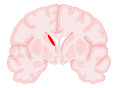
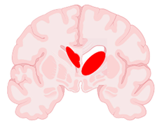
Table 1. Classification of nIVH severity according to Papile and Volpe [36,37]. Illustrations created with the use of BioRender (BioRender.com; accessed on 1 September 2024) and Microsoft® Powerpoint® for Microsoft 365 (Microsoft Corporation, Redmond, WA, USA). (Abbreviations: CT = computed tomography; CUS = cranial ultrasound).
GradePapile Criteria (Based on CT Scans)Volpe Criteria (Based on CUS Imaging)Representative Illustration (Brain Section in Coronal View)
IHemorrhage limited to the
sub-ependymal matrix
Hemorrhage without intraventricular extension or with hemorrhage occupying <10% of ventricular areaEncyclopedia 04 00127 i001
IIHemorrhage extending into
the ventricular system (<50%),
without acute ventriculomegaly
Hemorrhage occupying 10–50% of ventricular areaEncyclopedia 04 00127 i002
IIIHemorrhage extending into
the ventricular system ≥50% or ≥1 lateral ventricles
OR
Hemorrhage extending into a dilated ventricle
Hemorrhage occupying >50% of ventricular areaEncyclopedia 04 00127 i003
IVHemorrhage grade I, II or III
with extension into brain tissue
Periventricular echodensity (periventricular venous hemorrhagic infarction)Encyclopedia 04 00127 i004
Disclaimer/Publisher’s Note: The statements, opinions and data contained in all publications are solely those of the individual author(s) and contributor(s) and not of MDPI and/or the editor(s). MDPI and/or the editor(s) disclaim responsibility for any injury to people or property resulting from any ideas, methods, instructions or products referred to in the content.