Case Report

High Serum Levels of Anti-Proteinase 3, Low Clues: Detecting Occult Granulomatosis with Polyangiitis Activity Beyond Clinical Remission


U.O.C. Nefrologia e Dialisi, Dipartimento delle Medicine Specialistiche, Ospedale “Guglielmo da Saliceto”, 29121 Piacenza, Italy
*
Author to whom correspondence should be addressed.
These authors contributed equally to this work.
Kidney Dial. 2026, 6(2), 37; https://doi.org/10.3390/kidneydial6020037
Submission received: 13 March 2026 / Revised: 15 May 2026 / Accepted: 20 May 2026 / Published: 1 June 2026

Abstract

Granulomatosis with polyangiitis (GPA) is a systemic vasculitis frequently associated with serum anti-neutrophil cytoplasmic antibodies (ANCAs), particularly anti-proteinase 3 (PR3). Multisystem involvement is typical, although disease onset with simultaneous manifestations affecting both the ocular and otorhinolaryngologic systems is rare and poorly reported in the literature. We describe a 65-year-old woman with renal impairment with microscopic hematuria and proteinuria and PR3-positive who developed bilateral otitis leading to sensorineural hearing loss and severe anterior scleritis, with a high suspicion of ANCA-associated vasculitis. Despite immunosuppression therapy, persistently elevated serum anti-PR3 levels in the absence of overt clinical activity prompted further diagnostic evaluation, which revealed previously unrecognized subglottic stenosis (SGS), confirmed by MRI, and fully resolved after anti-CD20 therapy. Concurrent involvement of the upper airways and ocular district as the initial presentation of GPA is unusual and may precede renal involvement by several months. Persistent elevation of PR3 levels in an apparent state of clinical remission may indicate ongoing active disease in other anatomical sites. The integration of laboratory tests, imaging studies, and multidisciplinary assessment is essential for early diagnosis and effective therapeutic management.
Keywords:
vasculitis; ANCA; kidney

1. Introduction

Granulomatosis with polyangiitis (GPA) is a rare, systemic vasculitis characterized by tissue inflammation and involvement of small- to medium-sized blood vessels. It is strongly associated with serum anti-neutrophil cytoplasmic antibodies (ANCAs), especially those directed against proteinase 3 (PR3) [1]. The clinical presentations of GPA is highly variable, ranging from localized disease of the upper respiratory tract to severe multisystem disease that impacts the lungs, kidneys, and other organs. Kidney involvement is a key and potentially severe feature of GPA, and is observed in most patients with ANCA-associated vasculitis (AAV) [2,3].
The typical renal histopathological finding is pauci-immune, focal, and segmental necrotizing and crescentic glomerulonephritis [4,5]. Clinically, this presents as microscopic hematuria with dysmorphic red blood cells, red cell casts, and moderate proteinuria. The disease can progress rapidly, leading to a sharp decline in renal function, although a more indolent course can also occur [6,7]. The presence of PR3-ANCA positivity alongside kidney and multisystem involvement strongly supports the diagnosis of ANCA-associated vasculitis, especially GPA [8,9]. Kidney involvement is associated with increased morbidity and mortality, and the chance of progression to end-stage kidney disease closely depends on kidney function upon presentation. Early recognition and prompt initiation of immunosuppressive therapy are crucial to improving outcomes and reducing the risk of permanent renal damage [7,10].
Ear, nose and throat (ENT) symptoms are among the most common early signs of GPA. Conditions such as chronic rhinosinusitis, nasal ulcers, otitis media, and subglottic stenosis can appear months or even years before more serious systemic symptoms [7,11,12]. Eye involvement, such as scleritis, episcleritis, orbital inflammation, and dacryocystitis, is also frequent, affecting up to one-third of patients and potentially leading to significant complications if not treated promptly [12,13,14].
In contrast, the simultaneous onset of ocular and ENT manifestations is relatively uncommon and can make diagnosis more difficult. This presentation can mimic more common infections, inflammatory diseases, or malignancies [13,14,15]. Recent studies show that sinonasal disease increases the risk of ocular involvement. When these symptoms coexist at diagnosis, they are linked to a higher chance of ongoing or recurring disease and more widespread organ damage [16,17].
Recognizing this clinical pattern early is essential. Organ complications, such as vision loss, hearing impairment, and progressive glomerulonephritis, can develop rapidly. This requires immediate immunosuppressive treatment to improve outcomes [10,14,18]. This case report highlights the importance of maintaining a high index of suspicion and adopting a multidisciplinary approach in atypical presentations of AAV, particularly when renal involvement occurs alongside concurrent ocular and ENT manifestations.

2. Case Report

A 65-year-old woman with a past medical history notable for Raynaud’s phenomenon, arterial hypertension, and seronegative rheumatoid arthritis, was hospitalized in 2021 for worsening of kidney function and proteinuria during follow-up evaluation. Laboratory tests revealed serum creatinine of 2.0 mg/dL, microhematuria, proteinuria (1.7 g/day), anemia, and elevated C-reactive protein. Serological studies demonstrated positivity for anti-neutrophil cytoplasmic antibodies (C-ANCA).
Renal biopsy did not show histological features diagnostic of vasculitis due to the presence of only one glomerulus that did not show crescents. Given the high clinical suspicion and supportive laboratory findings, the patient was treated with high-dose glucocorticoids (methylprednisolone 750 mg/day for three consecutive days intravenously, followed by oral prednisone 0.8 mg/kg/day), achieving progressive improvement in renal function (serum creatinine 1.0 mg/dL). She continued corticosteroid therapy with a gradual tapering of the dose. Since the beginning of steroid therapy, proteinuria levels have remained stable and consistently below 300 mg/day, with no trace of blood in the urine and consistently negative PR3-ANCA levels.
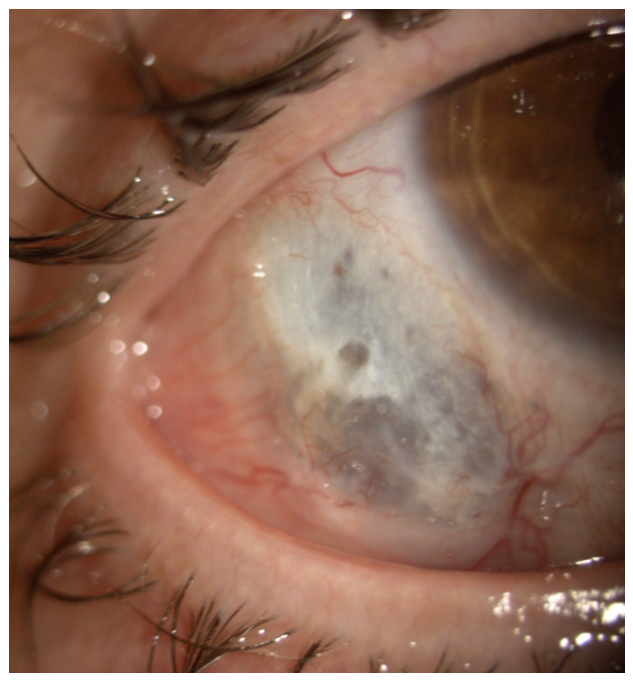
In June 2023, the patient developed marked right-sided hearing loss, attributed to suspected serous otitis media, which was unresponsive to corticosteroid therapy. One month later, she developed ocular involvement characterized by redness, pain, and decreased visual acuity in the right eye. Ophthalmologic evaluation was consistent with severe anterior scleritis associated with a conjunctival lesion (Figure 1).
After three months of follow-up, persistent scleral exposure at the site of prior attempted suturing was observed, in the absence of active inflammation, vitreitis, or retinal vasculitic signs.
The combination of otolaryngologic and ocular manifestations was highly suggestive of a relapse of ANCA-associated vasculitis.
Accordingly, in October 2023, the patient experienced further deterioration in renal function (serum creatinine 1.7 mg/dL, proteinuria 0.53 g/day, microhematuria), accompanied by a significant rise in PR3-ANCA levels (PR3 = 66 U/L, normal values < 2 U/mL). Therefore, she was started on high-dose corticosteroid therapy (methylprednisolone 750 mg/day for three days, followed by prednisone 0.8 mg/kg/day) in combination with oral cyclophosphamide (50–100 mg/day) due to organ-threatening multisystem involvement.
A nasal cavity biopsy was subsequently performed due to frequent epistaxis, revealing, on histological examination, nonspecific chronic inflammation with granulocytes located peripherally to capillary with focal phenomena of wall permeation.
After one month of immunosuppressive therapy, renal function progressively improved, with decreasing proteinuria (creatinine 1.3 mg/dL, proteinuria 0.2 g/day) and resolution of microhematuria. A parallel decline in PR3-ANCA levels (PR3 = 17 U/L, normal values < 2 U/mL) was also obtained.
After six months of treatment, in February 2024, cyclophosphamide was discontinued and azathioprine was initiated; however, it was rapidly withdrawn due to leukopenia. Maintenance therapy with mycophenolate mofetil (1 g/day) was subsequently started, selected as a less intensive maintenance approach.
In February 2025, due to the detection of wheezing on chest examination and an increase in PR3-ANCA levels (PR3 = 91 U/L) in the absence of renal, ocular or otologic symptoms, computed tomography of the chest and neck was subsequently performed. Imaging revealed a subglottic stenosis at the level of the cricoid cartilage, with irregular thickening of the mucosal surface extending into the first tracheal rings in the cranio-caudal direction, measuring approximately 17 mm, causing tracheal narrowing and consistent with vasculitic involvement.
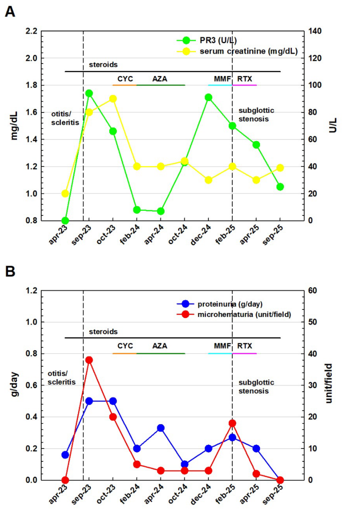
Due to clear clinical disease reactivation, mycophenolate mofetil was discontinued and rituximab was started (375 mg/m2 weekly for four weeks), in line with guideline recommendation for relapsing, multi-organ PR3-positive disease, resulting in complete resolution of the subglottic stenosis, as confirmed by magnetic resonance imaging and laryngoscopy. Renal function remained stable (latest serum creatinine 1.19 mg/dL, with no microhematuria or proteinuria as of September 2025) with reduction in PR3 (Figure 2A,B).

3. Discussion

Diagnosing AAV can be challenging due to its heterogeneous clinical manifestations and overlapping features with other autoimmune diseases [19].
A negative renal biopsy does not exclude the diagnosis of ANCA-associated vasculitis, particularly in early or focal forms in which characteristic histological lesions, such as crescents, may not yet be fully developed. In addition, renal biopsy is subject to inherent limitations, including sampling variability, especially in focal and segmental disease where affected glomeruli may be missed. In this context, integrating histopathological frameworks such as the Berden classification [20] may help better contextualize non-crescentic or pauci-lesional presentations. Therefore, in a compatible clinical and serological setting, histological negativity should be interpreted with caution and should not delay appropriate diagnosis and treatment. Moreover, the differential diagnosis of subglottic stenosis and associated ENT, ocular, and systemic manifestations should be carefully considered, especially in the absence of unequivocal histological confirmation of vasculitis. Relapsing polychondritis may present with airway involvement and ocular inflammation but typically manifests with auricular or nasal chondritis and lacks ANCA positivity. IgG4-related disease can involve the orbit and upper airways; however, it is usually characterized by tumefactive lesions, elevated serum IgG4 levels, and a distinct histopathological pattern consisting of storiform fibrosis and IgG4-positive plasma cell infiltration, none of which were present in this case. Infectious etiologies were also considered but excluded based on the clinical course, laboratory findings, and response to immunosuppressive therapy. Idiopathic subglottic stenosis, while a potential mimic, is typically isolated, occurs predominantly in middle-aged women, and does not explain the multisystemic and serologically active disease observed in our patient.
Taken together, although histological confirmation was limited by sampling constraints, the combination of compatible clinical features, PR3-ANCA positivity, multisystem involvement, and response to immunosuppressive therapy strongly supports a diagnosis within the spectrum of GPA. A major challenge is the early identification of patients at higher risk of relapse, death, or kidney failure. Benichou et al., in a post hoc analysis of 571 patients from five European randomized clinical trials, demonstrated that persistent hematuria after induction therapy independently predicts relapse [21], whereas persistent proteinuria is associated with death and kidney failure.
Several studies and meta-analyses have also focused on monitoring serum ANCA levels, showing that persistent ANCA positivity during clinical remission, as well as reappearance of ANCAs, is associated with an increased risk of clinical relapse [22]. Data from the Mayo Clinic [23] confirmed these findings, demonstrating a reduced risk of clinical relapse in patients with persistent ANCA negativity. Several studies have quantified the differential relapse risk between PR3- and MPO-ANCA-associated vasculitis, consistently demonstrating a higher relapse rate in PR3-positive disease. Long-term cohort analyses report relapse rates exceeding 50–60% in PR3-ANCA-positive patients compared with approximately 10–20% in MPO-ANCA-associated vasculitis, supporting the concept that PR3 positivity represents a major prognostic factor for recurrent disease activity. In particular, data from large European cohorts have shown that PR3-ANCA positivity is independently associated with an increased hazard of relapse even after remission has been achieved, whereas MPO-ANCA-associated disease tends to follow a more monophasic course [24,25]. These observations are consistent with findings from the EUVAS long-term follow-up cohort, which confirmed significantly higher relapse rates in PR3-ANCA disease over time [25]. Similarly, a systematic review by Walsh et al. identified PR3-ANCA positivity as one of the strongest predictors of relapse in ANCA-associated vasculitis [24]. This pattern is also reflected in granulomatosis with polyangiitis (GPA) cohorts with predominant ENT and ocular involvement, in which disease relapse is particularly frequent and often manifests with sequential or compartmentalized organ involvement rather than simultaneous systemic flares. In such cases, ENT and orbital disease may precede systemic relapse or occur in isolation, as described in classic GPA cohorts [26]. An increase in ANCA titer often precedes disease relapse within six months and, to a lesser extent, within 12 months, although elevated serum ANCA levels alone do not indicate active relapse [27]. This finding may appear controversial. Al-Soudi et al. speculated that the autoimmune cascade induced by ANCA may be counterbalanced by mechanisms that maintain immune homeostasis. For example, serum anti-inflammatory cytokines such as IL-10 are increased in patients with persistent serum ANCA positivity [28] and may inhibit inflammation caused by neutrophil activation [29]. In such cases, fluctuations in serum ANCA levels do not necessarily lead to clinical relapse. Conversely, if these compensatory mechanisms become overwhelmed, local tissue damage may increase, ultimately leading to relapse. For these reasons, increases in ANCA titers and clinical relapse do not necessarily occur simultaneously, although they are clearly associated. In a large retrospective study [26], 50% of patients with rising serum ANCA titers experienced relapse within 18 months of the increase; therefore, decisions regarding immunosuppressive therapy should not be based solely on changes in serum ANCA titers. On the other hand, some patients exhibit an increase in serum ANCA levels before each clinical relapse. In these selected cases, prophylactic therapy may be considered. Our case exemplifies the asynchronous progression of organ involvement in ANCA-associated vasculitis, with discordant timing between renal, otorhinolaryngological, ocular, and airway manifestations. Despite initial renal remission, persistent and subsequently rising PR3-ANCA levels preceded the sequential development of ENT, ocular, and later subglottic disease, suggesting that serological activity may reflect ongoing subclinical immune dysregulation rather than organ-specific inflammation at a given time. Although ANCA kinetics have been widely studied, their clinical interpretation remains challenging, as fluctuations in ANCA levels have limited specificity for active disease and should not be considered reliable standalone markers of relapse. Indeed, increases in ANCA titers may occur in the absence of systemic manifestations, highlighting the need to distinguish between serological activity and clinically overt disease. The apparent discordance observed in this case may be explained by heterogeneous tissue-specific inflammatory pathways, in which local susceptibility, microenvironmental factors, and differential neutrophil activation drive site-specific disease expression. Notably, persistent PR3-ANCA positivity in our patient was not initially associated with renal relapse but instead paralleled the emergence of anatomically localized involvement, suggesting that sustained serological activity may reflect ongoing site-specific inflammation rather than a generalized systemic flare [30].
Overall, this clinical trajectory likely represents a continuum of systemic immune activation with temporally and anatomically distinct manifestations, underscoring the importance of comprehensive longitudinal assessment beyond single-organ monitoring and careful evaluation for localized, potentially subclinical disease in the presence of persistent ANCA positivity. In particular, in GPA, ocular and ENT manifestations should be considered warning signs of disease activity.

4. Conclusions

In conclusion, the diagnosis and monitoring of ANCA-associated vasculitis (AAV) remain challenging due to the heterogeneity of clinical manifestations and the imperfect correlation between serological markers and disease activity. Serological changes alone are not sufficient to predict disease reactivation or guide therapeutic decisions. Therefore, careful clinical assessment remains essential. Our case highlights the importance of maintaining a high index of suspicion for extrarenal disease activity in patients with persistent ANCA positivity. In granulomatosis with polyangiitis, ocular and ENT manifestations should be considered potential indicators of disease activity even in the absence of renal relapse. Overall, this case underscores the importance of recognizing extrarenal manifestations as early indicators of disease activity in patients with persistent ANCA positivity.

Author Contributions

M.G., V.B.: writing; M.D.: figures and resources; C.R., T.V.: data curation; R.S.: supervision. All authors have read and agreed to the published version of the manuscript.

Funding

This research received no external funding.

Institutional Review Board Statement

Not applicable, because for case reports, informed consent is enough; since it is not a clinical trial, an ethics committee is not required.

Informed Consent Statement

Written informed consent has been obtained from the patient to publish this paper.

Data Availability Statement

The data presented in this study are available on request from the corresponding author due to privacy and ethical restrictions.

Conflicts of Interest

The authors declare no conflicts of interest.

References

  1. Falde, S.D.; Fussner, L.A.; Tazelaar, H.D.; O’Brien, E.K.; Lamprecht, P.; Konig, M.F.; Specks, U. Proteinase 3-specific antineutrophil cytoplasmic antibody-associated vasculitis. Lancet Rheumatol. 2024, 6, e314–e327. [Google Scholar] [CrossRef]
  2. Kitching, A.R.; Anders, H.-J.; Basu, N.; Brouwer, E.; Gordon, J.; Jayne, D.R.; Kullman, J.; Lyons, P.A.; Merkel, P.A.; Savage, C.O.S.; et al. ANCA-associated vasculitis. Nat. Rev. Dis. Prim. 2020, 6, 71. [Google Scholar] [CrossRef]
  3. Kronbichler, A.; Bajema, I.M.; Bruchfeld, A.; Kirsztajn, G.M.; Stone, J.H. Diagnosis and management of ANCA-associated vasculitis. Lancet 2024, 403, 683–698. [Google Scholar] [CrossRef]
  4. Floege, J.; Jayne, D.R.W.; Sanders, J.-S.F.; Tesar, V.; Rovin, B.H. KDIGO 2024 Clinical Practice Guideline for the Management of Antineutrophil Cytoplasmic Antibody (ANCA)–Associated Vasculitis. Kidney Int. 2024, 105, S71–S116, Corrigendum in Kidney Int. 2024, 106, 160–163. Corrigendum in Kidney Int. 2025, 107, 367. [Google Scholar] [CrossRef]
  5. Binda, V.; Moroni, G.; Messa, P. ANCA-associated vasculitis with renal involvement. J. Nephrol. 2018, 31, 197–208. [Google Scholar] [CrossRef] [PubMed]
  6. Sethi, S.; De Vriese, A.S.; Fervenza, F.C. Acute glomerulonephritis. Lancet 2022, 399, 1646–1663. [Google Scholar] [CrossRef] [PubMed]
  7. Comarmond, C.; Cacoub, P. Granulomatosis with polyangiitis (Wegener): Clinical aspects and treatment. Autoimmun. Rev. 2014, 13, 1121–1125. [Google Scholar] [CrossRef] [PubMed]
  8. Knopf, A.; Chaker, A.; Stark, T.; Hofauer, B.; Lahmer, T.; Thürmel, K.; Bas, M. Clinical aspects of granulomatosis with polyangiitis affecting the head and neck. Eur. Arch. Oto-Rhino-Laryngol. 2015, 272, 185–193. [Google Scholar] [CrossRef]
  9. Chung, S.A.; Langford, C.A.; Maz, M.; Abril, A.; Gorelik, M.; Guyatt, G.; Archer, A.M.; Conn, D.L.; Full, K.A.; Grayson, P.C.; et al. 2021 American College of Rheumatology/Vasculitis Foundation Guideline for the Management of Antineutrophil Cytoplasmic Antibody–Associated Vasculitis. Arthritis Care Res. 2021, 73, 1088–1105. [Google Scholar] [CrossRef]
  10. Taylor, S.C.; Clayburgh, D.R.; Rosenbaum, J.T.; Schindler, J.S. Progression and management of Wegener’s granulomatosis in the head and neck. Laryngoscope 2012, 122, 1695–1700. [Google Scholar] [CrossRef]
  11. Trimarchi, M.; Sinico, R.A.; Teggi, R.; Bussi, M.; Specks, U.; Meroni, P.L. Otorhinolaryngological manifestations in granulomatosis with polyangiitis (Wegener’s). Autoimmun. Rev. 2013, 12, 501–505, Erratum in Autoimmun. Rev. 2015, 14, 80. [Google Scholar] [CrossRef]
  12. Mei, L.; Wang, L.; Yan, H. Updates of ocular involvement in granulomatosis with polyangiitis. Graefe’s Arch. Clin. Exp. Ophthalmol. 2023, 261, 1515–1523. [Google Scholar] [CrossRef] [PubMed]
  13. Junek, M.L.; Zhao, L.; Garner, S.; Cuthbertson, D.; Pagnoux, C.; Koening, C.L.; Langford, C.A.; McAlear, C.A.; Monach, P.A.; Moreland, L.W.; et al. Ocular manifestations of ANCA-associated vasculitis. Rheumatology 2023, 62, 2517–2524. [Google Scholar] [CrossRef]
  14. Hinojosa-Azaola, A.; García-Castro, A.; Juárez-Flores, A.; Recillas-Gispert, C. Clinical significance of ocular manifestations in granulomatosis with polyangiitis: Association with sinonasal involvement and damage. Rheumatol. Int. 2019, 39, 489–495. [Google Scholar] [CrossRef]
  15. Kühn, D.; Hospowsky, C.; Both, M.; Hey, M.; Laudien, M. Manifestation of granulomatosis with polyangiitis in head and neck. Clin. Exp. Rheumatol. 2018, 36, 78–84. [Google Scholar] [PubMed]
  16. Sahyouni, R.; Moshtaghi, O.; Abouzari, M.; Le, P.; Birkenbeuel, J.; Cheung, D.; Lin, H.W.; Djalilian, H.R. A Case Series of Granulomatosis with Polyangiitis Primarily Diagnosed by Otological Manifestations. Ann. Otol. Rhinol. Laryngol. 2019, 128, 263–266. [Google Scholar] [CrossRef] [PubMed]
  17. Asín, M.A.P.-J.; Charles, P.; Rothschild, P.-R.; Terrier, B.; Brézin, A.; Mouthon, L.; Guillevin, L.; Puéchal, X. Ocular involvement in granulomatosis with polyangiitis: A single-center cohort study on 63 patients. Autoimmun. Rev. 2019, 18, 493–500. [Google Scholar] [CrossRef]
  18. Sattui, S.E.; Lally, L. Localized Granulomatous with Polyangiitis (GPA): Varied Clinical Presentations and Update on Treatment. Curr. Allergy Asthma Rep. 2020, 20, 56. [Google Scholar] [CrossRef]
  19. Tan, M.H.; Jayne, D. Top ten tips in managing ANCA vasculitis. Clin. Kidney J. 2025, 18, sfae389. [Google Scholar] [CrossRef]
  20. Berden, A.E.; Ferrario, F.; Hagen, E.C.; Jayne, D.R.; Jennette, J.C.; Joh, K.; Neumann, I.; Noël, L.-H.; Pusey, C.D.; Waldherr, R.; et al. Histopathologic classification of ANCA-associated glomerulonephritis. J. Am. Soc. Nephrol. 2010, 21, 1628–1636. [Google Scholar] [CrossRef]
  21. Benichou, N.; Charles, P.; Terrier, B.; Jones, R.B.; Hiemstra, T.; Mouthon, L.; Bajema, I.; Berden, A.; Thervet, E.; Guillevin, L.; et al. Proteinuria and hematuria after remission induction are associated with outcome in ANCA-associated vasculitis. Kidney Int. 2023, 103, 1144–1155. [Google Scholar] [CrossRef]
  22. McClure, M.E.M.; Wason, J.; Gopaluni, S.M.; Tieu, J.F.; Smith, R.M.M.; Jayne, D.R.F.; Jones, R.B.F. Evaluation of PR3-ANCA Status After Rituximab for ANCA-Associated Vasculitis. J. Clin. Rheumatol. 2019, 25, 217–223. [Google Scholar] [CrossRef] [PubMed]
  23. Moura, M.C.; Specks, U.; Tehranian, S.; Sethi, S.; Zubidat, D.; Nardelli, L.; dos Santos, F.G.; Sousa, C.; León-Róman, J.; Bobart, S.A.; et al. Maintenance of Remission and Risk of Relapse in Myeloperoxidase-Positive ANCA-Associated Vasculitis with Kidney Involvement. Clin. J. Am. Soc. Nephrol. 2023, 18, 47–59. [Google Scholar] [CrossRef]
  24. Walsh, M.; Flossmann, O.; Berden, A.; Westman, K.; Höglund, P.; Stegeman, C.; Jayne, D. European Vasculitis Study Group. Risk factors for relapse of antineutrophil cytoplasmic antibody-associated vasculitis. Arthritis Rheum. 2012, 64, 542–548. [Google Scholar] [CrossRef] [PubMed]
  25. Flossmann, O.; Berden, A.; de Groot, K.; Hagen, C.; Harper, L.; Heijl, C.; Höglund, P.; Jayne, D.; Luqmani, R.; Mahr, A.; et al. Long-term patient survival in ANCA-associated vasculitis. Ann. Rheum. Dis. 2011, 70, 488–494. [Google Scholar] [CrossRef] [PubMed]
  26. Hoffman, G.S.; Kerr, G.S.; Leavitt, R.Y.; Hallahan, C.W.; Lebovics, R.S.; Travis, W.D.; Rottem, M.; Fauci, A.S. Wegener granulomatosis: An analysis of 158 patients. Ann. Intern. Med. 1992, 116, 488–498. [Google Scholar] [CrossRef]
  27. Al-Soudi, A.; Vegting, Y.; Klarenbeek, P.L.; Hilhorst, M.L. Do Relapses Follow ANCA Rises? A Systematic Review and Meta-Analysis on the Value of Serial ANCA Level Evaluation. Front. Med. 2022, 9, 844112. [Google Scholar] [CrossRef]
  28. Hoffmann, J.C.; Patschan, D.; Dihazi, H.; Müller, C.; Schwarze, K.; Henze, E.; Ritter, O.; Müller, G.A.; Patschan, S. Cytokine profiling in anti neutrophil cytoplasmic antibody-associated vasculitis: A cross-sectional cohort study. Rheumatol. Int. 2019, 39, 1907–1917. [Google Scholar] [CrossRef]
  29. Saitoh, T.; Komano, J.; Saitoh, Y.; Misawa, T.; Takahama, M.; Kozaki, T.; Uehata, T.; Iwasaki, H.; Omori, H.; Yamaoka, S.; et al. Neutrophil Extracellular Traps Mediate a Host Defense Response to Human Immunodeficiency Virus-1. Cell Host Microbe 2012, 12, 109–116. [Google Scholar] [CrossRef]
  30. Kemna, M.J.; Damoiseaux, J.; Austen, J.; Winkens, B.; Peters, J.; van Paassen, P.; Tervaert, J.W.C. ANCA as a predictor of relapse: Useful in patients with renal involvement but not in patients with nonrenal disease. J. Am. Soc. Nephrol. 2015, 26, 537–542. [Google Scholar] [CrossRef]
Figure 1. Severe anterior scleritis associated with a conjunctival lesion in the patient’s right eye, in August 2023.
Kidneydial 06 00037 g001
Figure 2. (A) Trend of PR3 (in green) and serum creatinine (in yellow) throughout the follow up, from the diagnosis to the last visit. (B) Trend of proteinuria (in blue) and microhematuria (in red) throughout the follow up, from the diagnosis to the last visit. In both panel (A,B), the dashed lines represent clinical manifestation onset of otitis/scleritis (the first line) and subglottic stenosis (the second line). In both panel (A,B), the black horizontal line represents the length of steroids therapy; the orange line represents treatment with cyclophosphamide (CYC), the dark green line treatment with azathioprine (AZA), the light blue with mycophenolate mofetil (MMF) and the pink line treatment with rituximab (RTX).
Kidneydial 06 00037 g002
Disclaimer/Publisher’s Note: The statements, opinions and data contained in all publications are solely those of the individual author(s) and contributor(s) and not of MDPI and/or the editor(s). MDPI and/or the editor(s) disclaim responsibility for any injury to people or property resulting from any ideas, methods, instructions or products referred to in the content.