Article

Cystatin C-Based eGFR Predicts Post-Treatment Kidney Prognosis in Patients with Severe Obstructive Nephropathy

1
Department of Nephrology, Graduate School of Medical Science, Kyoto Prefectural University of Medicine, 465 Kajii-cho, Kamigyo-ku, Kyoto 602-8566, Japan
2
Department of Nephrology, The Omihachiman Community Medical Center, 1379 Tsuchida-cho, Omihachiman 523-0082, Japan
3
Division of Medical Engineering, Faculty of Medical Care Sciences, Himeji Dokkyo University, 7-2-1 Kami-ohno, Himeji 670-8524, Japan
4
Department of Mathematics and Statistics in Medical Sciences, Kyoto Prefectural University of Medicine, 465 Kajii-cho, Kamigyo-ku, Kyoto 602-8566, Japan
5
Department of Cardiovascular Medicine, Graduate School of Medical Science, Kyoto Prefectural University of Medicine, 465 Kajii-cho, Kamigyo-ku, Kyoto 602-8566, Japan
*
Author to whom correspondence should be addressed.
Kidney Dial. 2022, 2(3), 474-481; https://doi.org/10.3390/kidneydial2030043
Submission received: 7 July 2022 / Revised: 30 August 2022 / Accepted: 5 September 2022 / Published: 9 September 2022

Abstract

:
A discrepancy between serum concentrations of cystatin C (CysC) and creatinine (sCr) has been reported in patients with acute obstructive nephropathy. However, the usefulness of CysC for predicting the recovery of kidney function in patients with severe obstructive nephropathy remains unclear. We examined the predictability of the estimated glomerular filtration rate calculated with CysC or sCr (eGFRcys or eGFRcreat) for the post-treatment recovery of kidney function. We retrospectively collected patients with severe obstructive nephropathy (eGFRcreat < 30 mL/min/1.73 m2) whose baseline sCr and CysC were measured between 48 h before and 24 h after the release of urinary tract obstruction (UTO). The primary outcome was recovery from severe eGFRcreat depression (i.e., eGFRcreat ≥ 30 mL/min/1.73 m2) 7 days after the release of UTO. We calculated the area under the curve (AUC) of the receiver operating characteristic (ROC) curve for the relationship between eGFRcys or eGFRcreat and recovery. Thirty-four patients (20 males) with a median age of 76 years were eligible. We identified 20 recovery cases. The AUCs of the ROC curves (95% confidence interval) for eGFRcys and eGFRcreat were 0.81 (0.66–0.96) and 0.53 (0.32–0.73), respectively. These results imply cystatin C-based eGFR may help predict kidney prognosis in patients with severe obstructive nephropathy.

1. Introduction

Kidney dysfunction characterized by urinary tract obstruction (UTO) is called obstructive nephropathy or obstructive uropathy. UTO increases pressure in the proximal tubules and decreases the hydraulic pressure gradient across glomerular capillaries, which reduces the glomerular filtration rate (GFR) in the kidneys [1,2].
In evaluations of GFR, serum creatinine concentrations (sCr) are generally used as a biomarker to estimate GFR. In obstructive nephropathy, the cessation of urinary flow in the bilateral urinary tracts or solitary urinary tract increases sCr. However, serum cystatin C concentrations (CysC), another biomarker for assessing kidney function, do not increase with sCr in patients with acute obstructive nephropathy, which is also known as postrenal acute kidney injury (AKI) [3,4,5,6]. Due to this discrepancy, a high sCr-to-CysC ratio is useful for diagnosing obstructive nephropathy [3,5]. In terms of the relationship between CysC and kidney prognosis, Matsuki et al. reported a relationship between this discrepancy (sCr–CysC) and reductions in sCr after the treatment of obstructive nephropathy [6]. Okuda et al. also reported that CysC in obstructive nephropathy was correlated with sCr after the release of UTO [3]. Accordingly, CysC may be a marker predicting the post-treatment kidney function in obstructive nephropathy patients. Previous reports have demonstrated the superiority of CysC as a marker of kidney function and as a prognostic marker in the field of chronic kidney disease [7,8,9]; however, the usefulness of CysC for predicting the post-treatment recovery of kidney function in patients with severe obstructive nephropathy has not yet been demonstrated. Patients with severe obstructive nephropathy sometimes require renal replacement therapy, and the reversibility of kidney function is a matter of great concern for clinicians. If it is possible for clinicians to predict after-treatment kidney function, they may be able to estimate the need for maintenance dialysis or supportive therapy for chronic kidney disease. This estimation is also useful for explaining estimated clinical courses to patients.
We hypothesized that CysC may predict post-treatment recovery from severe estimated GFR (eGFR) depression among patients with obstructive nephropathy. The aim of this study was to assess the predictability of CysC compared to sCr for post-treatment kidney function in obstructive nephropathy.

2. Materials and Methods

2.1. Patients and Measurements

This was a retrospective single-center study to assess the usefulness of kidney biomarkers. Clinical data were collected from the medical records of patients with severe obstructive nephropathy who were admitted to the Omihachiman Community Medical Center in Japan between January 2010 and December 2021. Patients with obstructive nephropathy were initially screened with the insurance disease names of “postrenal failure” or “acute renal failure”. Then, the patients whose CysC had been measured were reviewed further. Each patient’s clinical course was checked in their medical records, and those appropriate candidates for the present study were identified if they met all of the following criteria: (i) diagnosed with obstructive nephropathy by ultrasonography or abdominal CT based on the findings of bilateral hydronephrosis or hydronephrosis in a solitary kidney; (ii) underwent the release of UTO to recover kidney function; (iii) baseline CysC and sCr measured with the same sample between 48 h before and 24 h after the release of UTO; (iv) severe estimated GFR (eGFR) depression defined as eGFR calculated with sCr (eGFRcreat) below 30 mL/min/1.73 m2 at baseline; (v) admitted to the hospital with a clinical course that was followed. We collected patient data regarding sex, age, etiology of obstructive nephropathy, laboratory data, and clinical course. CysC was measured using colloidal gold immunoassay; sCr was assessed with an enzymatic method.
We calculated eGFRcys and eGFRcreat using the following equations: eGFRcys (mL/min/1.73 m2) = (104 × CysC (mg/L)−1.019 × 0.996age × 0.929 (if female)) − 8 [10], eGFRcreat (mL/min/1.73 m2) = 194 × sCr (mg/dL)−1.094 × age−0.287 × 0.739 (if female) [11], where eGFR—estimated glomerular filtration rate; eGFRcys—eGFR calculated with serum cystatin C; eGFRcreat—eGFR calculated with serum creatinine; CysC—serum cystatin C; sCr—serum creatinine. The normal range of eGFRcys and eGFRcreat is ≥60 mL/min/1.73 m2.

2.2. Outcomes

The primary outcome was recovery from severe eGFRcreat depression within 7 days of the release of UTO. The patients were considered recovering if they were not under dialysis and their eGFRcreat exceeded 30 mL/min/1.73 m2 as described in a previous study [12]. For example, a patient with an eGFR of 45 mL/min/1.73 m2 on the fifth day was considered recovering, and another with an eGFR of 15 mL/min/1.73 m2 on the seventh day was not considered recovering.

2.3. Statistical Analysis

Initially, patient characteristics were described. Then, the baseline clinical findings were described and compared between the recovery and non-recovery cases. The Wilcoxon rank-sum test was used for continuous variables, Fisher’s exact test—for categorical variables. Logistic regression analysis was performed to examine whether eGFRcys or eGFRcreat was associated with recovery from severe eGFRcreat depression. Then, ROC curves were used to examine the relationship between the baseline eGFRcys or eGFRcreat and the recovery. The AUCs of the ROC curves were calculated. Then, a tentative cutoff point of eGFRcys for distinguishing recovery cases from non-recovery cases was explored.
Statistical analyses were performed using JMP version 14.3.0 (SAS Institute Inc., Cary, NC, USA) and R version 3.4.3 (R Foundation for Statistical Computing, Vienna, Austria).

3. Results

A total of 1078 patients with suspected AKI, including obstructive nephropathy, were screened based on their medical records, 462 of whom had their CysC measured. Thirty-four patients met the criteria and were considered to be appropriate candidates for the present study (Figure 1).

3.1. Patient Characteristics

Table 1 shows patient characteristics. Twenty out of 34 patients were male. The median age of the patients was 76 years. The primary cause of postrenal failure was neurogenic bladder (n = 12), followed by benign prostatic hyperplasia (n = 11).

3.2. Baseline eGFR and Recovery from Severe eGFRcreat Depression

Table 2 shows the baseline clinical findings of recovery and non-recovery cases. We identified 20 recovery cases. Among the 14 non-recovery cases, three needed maintenance hemodialysis. Significant differences were observed in eGFRcys (p = 0.002) and hemoglobin (p = 0.004) between the recovery and non-recovery cases, whereas eGFRcreat did not differ significantly (p = 0.803).
Individual kidney function at baseline and follow-up, timing of sampling, etiology, and interventions are shown in Table S1.
Table 3 shows the association between eGFRcys or eGFRcreat and recovery from severe eGFRcreat depression using logistic regression analysis. The baseline eGFRcys was significantly associated with recovery, whereas eGFRcreat was not. Figure 2 shows the receiver operating characteristic (ROC) curves of the relationship between eGFRcys or eGFRcreat and recovery. The areas under the ROC curves (AUCs) for eGFRcys and eGFRcreat were 0.81 (95% confidence interval (CI): 0.66–0.96) and 0.53 (95% CI: 0.32–0.73), respectively.
Table 4 shows a crosstabulation table between eGFRcys ≥ 25 and recovery from severe eGFRcreat depression; eGFRcys ≥ 25 identified recovery cases with a sensitivity of 70% (14/20) and specificity of 100% (14/14).

4. Discussion

In this hospital-based retrospective study, we examined the relationship between eGFRcys or eGFRcreat and post-treatment recovery from severe eGFRcreat depression in patients with obstructive nephropathy. The main result obtained was that the baseline eGFRcys was associated with recovery from severe eGFRcreat depression, whereas the baseline eGFRcreat was not. It is important to note that the reversibility of kidney function in obstructive nephropathy was predicted by eGFRcys, not by eGFRcreat. This is the first study to examine the usefulness of CysC for predicting kidney prognosis in severe obstructive nephropathy patients. Our findings imply that CysC may help predict kidney prognosis in addition to diagnosing obstructive nephropathy. Although further investigation by means of prospective studies is needed, CysC may play an important role in the treatment of obstructive nephropathy.
The mechanism underlying the discrepancy between eGFRcys and eGFRcreat in patients with obstructive nephropathy has been a matter of debate [3,4,6,13]. This discrepancy was recently explained by the degradation of low-molecular-weight proteins in proximal tubules. Cystatin C is reabsorbed and broken down by proximal tubular cells, whereas creatinine cannot be reabsorbed and accumulates in the urinary space [13,14]. In the present study, some patients with obstructive nephropathy had elevated CysC at baseline, and their kidney prognosis was worse than in those without CysC elevations. The reason for this phenomenon may be explained by the residual functions of proximal tubules. Complete UTO increases the intratubular pressure and reduces renal blood flow and GFR in the acute phase [1,2,15,16]. Sustained UTO then leads to interstitial inflammatory infiltration, tubular epithelial cell death, and subsequent tubular atrophy and interstitial fibrosis [2,16,17,18]. CysC may be absorbed by proximal tubules in the acute phase; however, this absorption may not occur in prolonged obstructive nephropathy because of irreversible structural changes in proximal tubular cells. Further studies on the relationship between tubular changes and CysC are needed to verify this hypothesis.
Based on these results, eGFRcys of 25 mL/min/1.73 m2 was suggested as a provisional cutoff point. In the present study, eGFRcys ≥ 25 identified recovery cases with a sensitivity of 70% (14/20) and specificity of 100% (14/14) (Table 4); eGFRcys ≥ 25 was a favorable sign for obstructive nephropathy with severe eGFRcreat depression. However, six out of the 20 patients with eGFRcys < 25 also recovered. This phenomenon may be partly explained by a complication of another curable AKI. These patients received antibiotic therapy other than the release of UTO (patient Nos. 6, 12, 25, 27, 29, and 30 in Table S1). CysC is elevated in patients with prerenal or intrinsic AKI, including septic AKI [19,20,21]. Therefore, CysC in those patients may have been elevated because of another curable AKI complicated by obstructive nephropathy. The recovery of kidney function is expected in patients with obstructive nephropathy with elevated CysC if it is complicated by a curable AKI. Interpreted with the existing evidence, the discrepancy between Cre and CysC seems to be useful as a marker of favorable kidney prognosis as well as for the diagnosis of obstructive nephropathy. This finding may encourage clinicians to release UTO for severe obstructive nephropathy. The present results cannot be generalized because this was a retrospective study with a small sample size. Further prospective multicenter studies with a large sample size are needed to validate our explanations.
The present study has several limitations. As mentioned before, this was a single-center retrospective study with a small sample size. In addition to the issue of generalizability, the present results may have been affected by selection bias. CysC was often, but not systematically, measured in obstructive nephropathy patients with severe eGFRcreat depression. Therefore, some patients whose CysC was not measured for some reason may have been excluded. Furthermore, we were unable to obtain the patients’ sCr levels before the development of obstructive nephropathy. Some patients with chronic kidney disease (CKD) complications might have been included among the patients with elevated CysC (acute-on-chronic kidney disease). The predictability of eGFRcys for kidney prognosis in these cases may differ from that in obstructive nephropathy without CKD. In addition, our follow-up period was no longer than 7 days. Since many of our patients were discharged after approximately 7 days, we were unable to follow up for longer outcomes. However, a 7-day follow-up may be reasonable because the recovery of kidney function generally occurs within 7–10 days of the release of the obstruction [22,23].

5. Conclusions

The present study showed that eGFRcys was associated with the post-treatment recovery of kidney function from severe eGFRcreat depression in patients with obstructive nephropathy. Cystatin C-based eGFR may help predict kidney prognosis in patients with severe obstructive nephropathy.

Supplementary Materials

The following supporting information can be downloaded at: https://www.mdpi.com/article/10.3390/kidneydial2030043/s1. Table S1. Individual kidney function at baseline and follow-up, timing of sampling, etiology, and interventions for severe obstructive nephropathy patients (n = 34, Omihachiman Community Medical Center, 2010–2021).

Author Contributions

Conceptualization, K.N., H.S. and M.Y.; Methodology, K.N., H.S., M.Y., T.K., S.M., K.T., T.H. and H.K.; Formal analysis, H.S. and K.Y.; Investigation, K.N. and H.S.; Resources, K.N., H.S., T.H. and H.K.; Data curation, K.N.; Writing—original draft preparation, K.N. and H.S.; Writing—review and editing, M.Y., K.Y., T.K., S.M., K.T., T.H. and H.K.; Visualization, K.N., H.S. and K.Y.; Supervision, M.Y.; Project administration, H.S. All authors have read and agreed to the published version of the manuscript.

Funding

No funding was received to conduct this study.

Institutional Review Board Statement

The present study was conducted according to the guidelines of the Declaration of Helsinki and approved by the Institutional Ethics Board of the Omihachiman Community Medical Center (No. R4-1, 2022).

Informed Consent Statement

Written informed consent for participation in this study was replaced by an opt-out disclosure on the website of the Omihachiman Community Medical Center (https://www.kenkou1.com/medical-list/dep37 (accessed on 1 July 2022) because this study was performed on the basis of secondhand use of previous medical records.

Data Availability Statement

Not applicable.

Acknowledgments

The authors wish to express their appreciation to Hideyuki Motonaka, Mayuka Yokoe (medical technologists), and Mio Tsurubou (the information management staff at the Omihachiman Community Medical Center) for their contribution to data collection from medical records.

Conflicts of Interest

The authors declare no conflict of interest.

References

  1. Basile, D.P.; Anderson, M.D.; Sutton, T.A. Pathophysiology of acute kidney injury. Compr. Physiol. 2012, 2, 1303–1353. [Google Scholar] [CrossRef] [PubMed]
  2. Klahr, S. Obstructive nephropathy. Intern. Med. 2000, 39, 355–361. [Google Scholar] [CrossRef] [PubMed]
  3. Okuda, Y.; Namba, S.; Nagata, M.; Hara, H.; Morita, T. Plasma creatinine and cystatin C ratio is useful for discriminate diagnosis of postrenal renal failure. Rinsho Byori. Jpn. J. Clin. Pathol. 2008, 56, 101–107. [Google Scholar]
  4. Fujisawa, N.; Yashiro, M.; Segawa, H.; Kadoya, Y.; Kamata, T. Discrepancy between cystatin C and creatinine points leading to a diagnosis of postrenal acute kidney injury and its reversibility: Three case reports. Clin. Exp. Nephrol. 2010, 14, 608–613. [Google Scholar] [CrossRef]
  5. İnal, S.; Altuntaş, A.; Kidir, V.; Özorak, A.; İlgin, Y.; Sezer, M.T. Utility of serum creatinine/cystatin C ratio in diagnosis of postrenal acute kidney injury. J. Res. Med. Sci. Off. J. Isfahan Univ. Med. Sci. 2014, 19, 1086–1089. [Google Scholar]
  6. Matsuki, M.; Tanaka, T.; Maehana, T.; Kyoda, Y.; Ichihara, K.; Hashimoto, K.; Yanase, M.; Matsukawa, M.; Adachi, H.; Takahashi, S.; et al. The discrepancy between serum creatinine and cystatin C can predict renal function after treatment for postrenal acute kidney injury: Multicenter study and pooled data analysis. Clin. Exp. Nephrol. 2017, 21, 852–857. [Google Scholar] [CrossRef] [PubMed]
  7. Dharnidharka, V.R.; Kwon, C.; Stevens, G. Serum cystatin C is superior to serum creatinine as a marker of kidney function: A meta-analysis. Am. J. Kidney Dis. 2002, 40, 221–226. [Google Scholar] [CrossRef] [PubMed]
  8. Perkins, B.A.; Nelson, R.G.; Ostrander, B.E.; Blouch, K.L.; Krolewski, A.S.; Myers, B.D.; Warram, J.H. Detection of renal function decline in patients with diabetes and normal or elevated GFR by serial measurements of serum cystatin C concentration: Results of a 4-year follow-up study. J. Am. Soc. Nephrol. JASN 2005, 16, 1404–1412. [Google Scholar] [CrossRef]
  9. Peralta, C.A.; Katz, R.; Sarnak, M.J.; Ix, J.; Fried, L.F.; De Boer, I.; Palmas, W.; Siscovick, D.; Levey, A.S.; Shlipak, M.G. Cystatin C identifies chronic kidney disease patients at higher risk for complications. J. Am. Soc. Nephrol. JASN 2011, 22, 147–155. [Google Scholar] [CrossRef]
  10. Horio, M.; Imai, E.; Yasuda, Y.; Watanabe, T.; Matsuo, S. GFR estimation using standardized serum cystatin C in Japan. Am. J. Kidney Dis 2013, 61, 197–203. [Google Scholar] [CrossRef]
  11. Matsuo, S.; Imai, E.; Horio, M.; Yasuda, Y.; Tomita, K.; Nitta, K.; Yamagata, K.; Tomino, Y.; Yokoyama, H.; Hishida, A. Revised equations for estimated GFR from serum creatinine in Japan. Am. J. Kidney Dis 2009, 53, 982–992. [Google Scholar] [CrossRef] [PubMed]
  12. Hamdi, A.; Hajage, D.; Van Glabeke, E.; Belenfant, X.; Vincent, F.; Gonzalez, F.; Ciroldi, M.; Obadia, E.; Chelha, R.; Pallot, J.L.; et al. Severe post-renal acute kidney injury, post-obstructive diuresis and renal recovery. BJU Int. 2012, 110, E1027–E1034. [Google Scholar] [CrossRef] [PubMed]
  13. Tsuda, H.; Isaka, Y.; Takahara, S.; Horio, M. Discrepancy between serum levels of low molecular weight proteins in acute kidney injury model rats with bilateral ureteral obstruction and bilateral nephrectomy. Clin. Exp. Nephrol. 2009, 13, 567–570. [Google Scholar] [CrossRef] [PubMed]
  14. Tenstad, O.; Roald, A.B.; Grubb, A.; Aukland, K. Renal handling of radiolabelled human cystatin C in the rat. Scand. J. Clin. Lab. Investig. 1996, 56, 409–414. [Google Scholar] [CrossRef] [PubMed]
  15. Vaughan, E.D., Jr.; Marion, D.; Poppas, D.P.; Felsen, D. Pathophysiology of unilateral ureteral obstruction: Studies from Charlottesville to New York. J. Urol. 2004, 172, 2563–2569. [Google Scholar] [CrossRef]
  16. Chevalier, R.L. Specific molecular targeting of renal injury in obstructive nephropathy. Kidney Int. 2006, 70, 1200–1201. [Google Scholar] [CrossRef]
  17. Klahr, S.; Pukerson, M.L. The pathophysiology of obstructive nephropathy: The role of vasoactive compounds in the hemodynamic and structural abnormalities of the obstructed kidney. Am. J. Kidney Dis 1994, 23, 219–223. [Google Scholar] [CrossRef]
  18. Chevalier, R.L.; Forbes, M.S.; Thornhill, B.A. Ureteral obstruction as a model of renal interstitial fibrosis and obstructive nephropathy. Kidney Int. 2009, 75, 1145–1152. [Google Scholar] [CrossRef]
  19. Nejat, M.; Pickering, J.W.; Devarajan, P.; Bonventre, J.V.; Edelstein, C.L.; Walker, R.J.; Endre, Z.H. Some biomarkers of acute kidney injury are increased in pre-renal acute injury. Kidney Int. 2012, 81, 1254–1262. [Google Scholar] [CrossRef]
  20. Teo, S.H.; Endre, Z.H. Biomarkers in acute kidney injury (AKI). Best Pract. Res. Clin. Anaesthesiol. 2017, 31, 331–344. [Google Scholar] [CrossRef]
  21. Leem, A.Y.; Park, M.S.; Park, B.H.; Jung, W.J.; Chung, K.S.; Kim, S.Y.; Kim, E.Y.; Jung, J.Y.; Kang, Y.A.; Kim, Y.S.; et al. Value of Serum Cystatin C Measurement in the Diagnosis of Sepsis-Induced Kidney Injury and Prediction of Renal Function Recovery. Yonsei Med. J. 2017, 58, 604–612. [Google Scholar] [CrossRef] [PubMed]
  22. Better, O.S.; Arieff, A.I.; Massry, S.G.; Kleeman, C.R.; Maxwell, M.H. Studies on renal function after relief of complete unilateral ureteral obstruction of three months’ duration in man. Am. J. Med. 1973, 54, 234–240. [Google Scholar] [CrossRef]
  23. Green, J.; Vardy, Y.; Munichor, M.; Better, O.S. Extreme unilateral hydronephrosis with normal glomerular filtration rate: Physiological studies in a case of obstructive uropathy. J. Urol. 1986, 136, 361–365. [Google Scholar] [CrossRef]
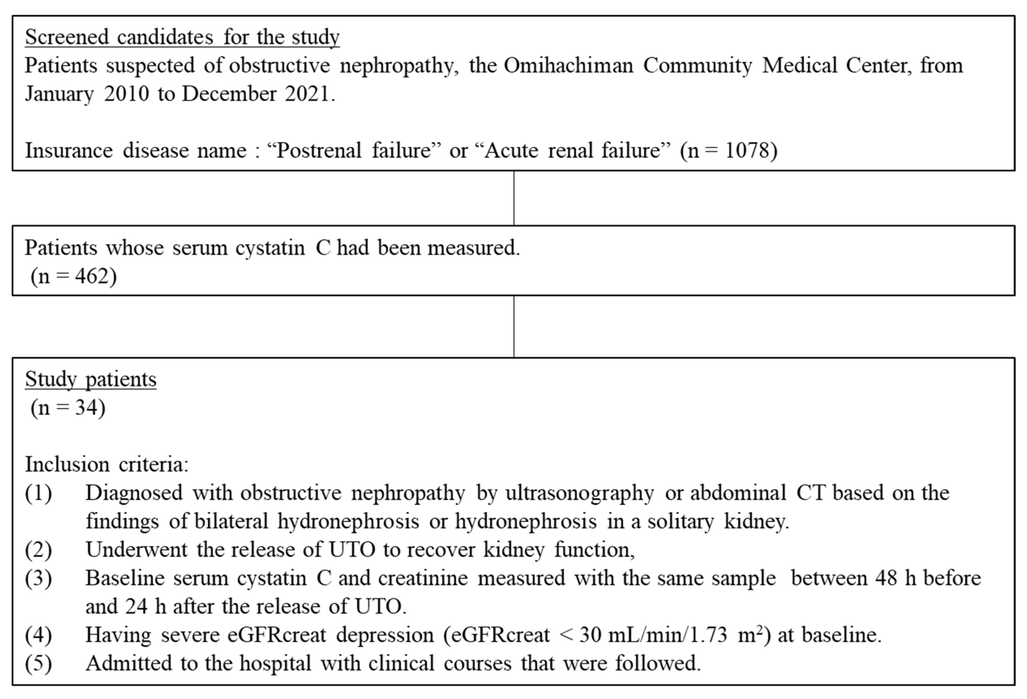
Figure 1. Flow diagram for the inclusion of study patients. Patients with severe obstructive nephropathy admitted to the Omihachiman Community Medical Center in Japan between January 2010 and December 2021 were enrolled. Overall, 34 patients, 20 of whom were male, were eligible for inclusion in this study. UTO, urinary tract obstruction; eGFRcreat, estimated glomerular filtration rate calculated with serum creatinine.
Kidneydial 02 00043 g001
Figure 2. ROC curves of the relationship between eGFRcys (solid line) or eGFRcreat (dashed line) and recovery from severe eGFRcreat depression defined as eGFRcreat < 30 mL/min/1.73 m2 in patients with obstructive nephropathy (n = 34). The baseline eGFRcys was associated with recovery from severe eGFRcreat depression, whereas the baseline eGFRcreat was not. Abbreviations: eGFR, estimated glomerular filtration rate; eGFRcys, eGFR calculated with serum cystatin C; eGFRcreat, eGFR calculated with serum creatinine; AUC, area under the curve.
Kidneydial 02 00043 g002
Table 1. Patient characteristics (n = 34).
Sex (male)20 (59%)
Age (years)76 (67, 83)
Weight (kg)58.7 (48.1, 66.4)
Etiology of obstructive nephropathy
Benign prostatic hyperplasia11 (32%)
Malignant tumor7 (21%)
Neurogenic bladder12 (35%)
Retroperitoneal fibrosis3 (9%)
Ureteral stone (solitary functional kidney)1 (3%)
Continuous variables are described as medians (interquartile range); categorical variables are described as n (%).
Table 2. Baseline clinical findings of the recovery and non-recovery cases.
Recovery * (n = 20)Non-recovery (n = 14)p
Sex (male)12 (60%)8 (57%)1.000
Age (years)80 (73, 85)69 (64, 78)0.069
Weight (kg)61.0 (49.8, 68.7)52.3 (44.5, 61.1)0.336
Etiology of obstructive nephropathy 0.521
Benign prostatic hyperplasia5 (25%)6 (43%)
Malignant tumor3 (15%)4 (29%)
Neurogenic bladder9 (45%)3 (21%)
Retroperitoneal fibrosis2 (10%)1 (7%)
Ureteral stone (solitary functional kidney)1 (5%)0 (0%)
Maintenance hemodialysis3 (21%)
Laboratory data
Serum creatinine (mg/dL)5.89 (4.74, 8.14)6.09 (4.53, 10.97)1.000
Serum cystatin C (mg/L)1.91 (1.45, 3.05)3.83 (3.19, 4.17)<0.001
eGFRcreat (mL/min/1.73 m2)7 (5, 10)6 (4, 9)0.803
eGFRcys (mL/min/1.73 m2)31 (14, 40)13 (9, 14)0.002
Blood urea nitrogen (mg/dL)110.1 (53.0, 138.5)78.8 (55.4, 120.1)0.877
White blood cells (103/μL)9.1 (6.6, 10.6)7.6 (5.3, 9.3)0.345
Hemoglobin (g/L)11.5 (9.4, 13.0)9.3 (8.3, 9.9)0.004
Platelets (103/μL)221 (175, 341)167 (134, 215)0.083
Serum albumin (g/dL)3.0 (2.8, 3.8)3.2 (2.8, 4.0)0.726
Serum sodium (mmol/L)139 (136, 147)141 (135, 143)0.672
Serum potassium (mmol/L)5.0 (4.4, 6.3)4.8 (3.4, 5.6)0.201
Bicarbonate (mmol/L)17.2 (13.8, 18.4)17.9 (13.5, 22.3)0.551
Serum calcium (mg/dL)8.7 (8.5, 9.0)8.2 (7.6, 9.0)0.072
Serum phosphate (mg/dL)5.5 (3.9, 8.0)5.1 (3.9, 6.4)0.619
Serum uric acid (mg/dL)9.8 (7.2, 14.1)8.7 (7.6, 9.6)0.241
C-reactive protein (mg/dL)8.4 (3.4, 15.2)4.9 (1.6, 10.6)0.138
Continuous variables are described as medians (interquartile range); categorical variables are described as n (%). * Recovery from severe eGFRcreat depression (i.e., eGFRcreat ≥ 30 mL/min/1.73 m2) 7 days after the release of UTO. Abbreviations: eGFR, estimated glomerular filtration rate; eGFRcreat, eGFR calculated with serum creatinine; eGFRcys, eGFR calculated with serum cystatin C. Note: p-values were calculated using Fisher’s exact test for the categorical variables and the Wilcoxon rank-sum test for the continuous variables.
Table 3. Association between the baseline eGFRcys or eGFRcreat and recovery from severe eGFRcreat depression a (n = 34).
OR (95% CI)p
eGFRcys (mL/min/1.73 m2)1.17 (1.04–1.31)0.010
eGFRcreat (mL/min/1.73 m2)1.00 (0.87–1.15)0.987
Abbreviations: eGFR, estimated glomerular filtration rate; eGFRcys, eGFR calculated with serum cystatin C; eGFRcreat, eGFR calculated with serum creatinine; OR, odds ratio; CI, confidence interval. a Severe eGFRcreat depression was defined as eGFRcreat < 30 mL/min/1.73 m2.
Table 4. Crosstabulation table between eGFRcys ≥ 25 and recovery from severe eGFRcreat depression a.
RecoveryTotal
YesNo
eGFRcys ≥ 25Yes14014
No61420
Total201434
Abbreviations: eGFR, estimated glomerular filtration rate; eGFRcreat, eGFR calculated with serum creatinine; eGFRcys, eGFR calculated with serum cystatin C. a Severe eGFRcreat depression was defined as eGFR < 30 mL/min/1.73 m2.
Publisher’s Note: MDPI stays neutral with regard to jurisdictional claims in published maps and institutional affiliations.