Brief Report

Development of a Nanogold-Based Lateral Flow Immunoassay for Point-of-Care Detection of SARS-CoV-2 Nucleocapsid Proteins and Antibodies

1
Department of Medical, Laboratory Science and Biotechnology, China Medical University, Taichung 404328, Taiwan
2
PhD Program for Health Science and Industry, College of Health Care, China Medical University, Taichung 404328, Taiwan
3
Graduate Institute of Biomedical Sciences, College of Medicine, China Medical University, Taichung 404328, Taiwan
4
Department of Food Science and Biotechnology, National Chung Hsing University, Taichung 40227, Taiwan
5
Department of Pediatric Infectious Diseases, China Medical University Children’s Hospital, Taichung 404327, Taiwan
6
Division of Infectious Diseases, Department of Internal Medicine, China Medical University Hospital, China Medical University, Taichung 404327, Taiwan
7
Department of Laboratory Medicine, China Medical University Hospital, China Medical University, Taichung 404327, Taiwan
8
School of Medicine, China Medical University, Taichung 404328, Taiwan
9
Office of Research and Development, Asia University, Taichung 413305, Taiwan
*
Author to whom correspondence should be addressed.
COVID 2025, 5(9), 158; https://doi.org/10.3390/covid5090158
Submission received: 19 August 2025 / Revised: 12 September 2025 / Accepted: 16 September 2025 / Published: 18 September 2025
(This article belongs to the Section COVID Clinical Manifestations and Management)

Abstract

The ongoing COVID-19 pandemic has underscored the urgent need for rapid, sensitive, and versatile diagnostic tools. In this study, we developed a nanogold-based lateral flow immunoassay (LFIA) capable of detecting both SARS-CoV-2 nucleocapsid (N) protein antigens and anti-N IgG antibodies at the point of care. Following optimization of colloidal gold nanoparticle size, pH, and protein conjugation parameters, LFIA strips were assembled in two formats: a competitive assay for antigen detection and a sandwich assay for antibody detection. In the competitive format, gold nanoparticles (AuNPs)-conjugated N protein were used to detect varying concentrations of free N protein. The test line signal inversely correlated with antigen concentration, confirming the assay’s specificity and effectiveness. For antibody detection, the sandwich LFIA format employed immobilized anti-human IgG to capture anti-N antibodies in serum samples from COVID-19 patients. Strong test line signals were observed in samples collected ≥11 days post-symptom onset, indicating a time-dependent increase in IgG detectability. These results demonstrate that the AuNP-based LFIA platform provides a flexible, rapid, and low-cost diagnostic solution, suitable for both early antigen detection and serological monitoring during SARS-CoV-2 infection and recovery.

1. Introduction

The global outbreak of coronavirus disease 2019 (COVID-19), caused by severe acute respiratory syndrome coronavirus 2 (SARS-CoV-2), has presented major challenges to public health systems worldwide [1]. Although nucleic acid amplification tests (NAATs) remain the gold standard for early detection due to their high sensitivity and specificity, their widespread use is often limited by cost, technical complexity, and reliance on centralized laboratories [2,3]—particularly in point-of-care (POC) and resource-limited settings.
Lateral flow immunoassays (LFIAs) have emerged as a practical alternative for serological testing, particularly for detecting SARS-CoV-2 antibodies [4]. Offering simplicity, rapid results, and cost-effectiveness, LFIAs are well-suited for individual antibody monitoring and large-scale seroprevalence studies [5]. Most LFIAs employ gold nanoparticles (AuNPs) as labels due to their favorable optical properties, biocompatibility, and ease of large-scale synthesis [6,7]. These nanoparticles are typically conjugated with specific antigens or antibodies and incorporated into the conjugate pad of the test strip. When a sample is applied, capillary action drives the fluid along the strip, allowing analytes to interact with immobilized reagents and generating visible colored lines. In the sandwich format, higher analyte concentrations correspond to stronger test line signals. Conversely, in the competitive format—commonly used for antigen detection—the analyte competes with immobilized antigens on the test line for binding to nanoparticle–antibody conjugates, leading to weaker test line signals at higher analyte concentrations. Thus, the capillary-driven flow ensures analyte–reagent interactions that produce clear visual outputs, enabling straightforward interpretation of results.
SARS-CoV-2 nucleocapsid (N) protein-based antigen tests are useful for identifying active infections, while anti-N IgG assays indicate past exposure [8,9]. In this study, we developed a gold nanoparticle-based LFIA using both competitive and sandwich formats for the detection of SARS-CoV-2 N protein and anti-N IgG, respectively. Recombinant His-tagged N protein was expressed in E. coli BL21 (DE3) using the pET-32a(+) vector (excluding the thioredoxin tag), induced with IPTG, purified via Ni-NTA chromatography, and subsequently conjugated to gold nanoparticles. In the competitive assay, anti-N and mouse IgG antibodies were immobilized at the test (T) and control (C) lines, respectively. Increasing concentrations of free N protein led to decreased T line intensity. In the sandwich assay, anti-human IgG and anti-N IgG were stripped at the T and C lines; positive serum samples produced red lines at both positions, while negative samples showed only the C line. These LFIA formats provide a rapid and reliable platform for detecting both active and past SARS-CoV-2 infections.

2. Materials and Methods

2.1. Expression and Purification of Recombinant SARS-CoV-2 N Protein

The recombinant SARS-CoV-2 nucleocapsid (N) protein was expressed in E. coli DH5α transformed with the pET-32a(+)-nCoV-2-N plasmid. Cultures were grown overnight at 37 °C in LB broth containing 100 µg/mL ampicillin. Protein expression was induced with 0.2 µM IPTG for 3 h at 37 °C. Cells were harvested by centrifugation at 6000 rpm for 15 min at 4 °C and resuspended in 20 mM imidazole buffer. Lysis was performed via sonication on ice for 20 min using a Misonix SONICATOR 3000. After centrifugation, supernatant and pellet fractions were purified using Ni-NTA affinity chromatography. Elution was carried out using increasing concentrations of imidazole (10, 100, 400, and 500 µM). Eluted proteins were dialyzed against 50 µM Tris and 150 µM NaCl to remove residual imidazole. To concentrate the protein, dialysis tubing was coated externally with PEG20000 and incubated at 4 °C. Additional PEG20000 was added as needed until the concentration reached saturation. Protein concentration was measured using a bicinchoninic acid (BCA) assay (Thermo Fisher Scientific, Waltham, MA, USA). Samples from supernatant, pellet, and concentrated eluate were mixed with 2× SDS-PAGE loading buffer, denatured at 100 °C for 5 min, and analyzed by 8–10% SDS-PAGE. Gels were stained with Coomassie Brilliant Blue or transferred to nitrocellulose membranes for Western blotting. Membranes were blocked with 5% skim milk in TBST (Tris-buffered saline with 0.1% Tween-20) for 1 h, followed by overnight incubation at 4 °C with primary antibodies: anti-6× His tag (1:2000) or human anti-SARS-CoV-2 N protein IgG (#7 and #33; 1:2000). After TBST washes, membranes were incubated with HRP-conjugated secondary antibodies (anti-mouse IgG-HRP or anti-human IgG-HRP; 1:5000) for 1 h. Detection was performed using Trident Plus Western HRP substrate and visualized with the MultiGel-21 imaging system (Gentaur, Voortstraat, Belgium).

2.2. Immunoreagents and Chemicals

All chemicals were analytical grade. Tris, NaCl, and PEG20000 were obtained from KINGFEX (Taichung, Taiwan) and Merck (Darmstadt, Germany). SDS-PAGE loading buffer was from Abbexa (Cambridge, UK). SARS-CoV-2 N protein human IgG antibodies (#7 and #33) were purchased from PEPTIDE CHAM BIOTECH (Kaohsiung, Taiwan); anti-6× His, anti-mouse IgG-HRP, and anti-human IgG-HRP were from GeneTex (Hsinchu, Taiwan). Additional reagents included potassium carbonate (K2CO3), sucrose, sodium azide (NaN3; Sigma-Aldrich, Darmstadt, Germany), and Tween-20 (EMPEROR CHEMICAL, Taipei, Taiwan). Gold(III) chloride (CAS No. 16903-35-8, SKU: 484385-10g) was used for colloidal gold synthesis. LFIA components included: nitrocellulose membrane (FF120HP, Whatman/GE, Pittsburgh, PA, USA), sample pad (Ahlstrom Grade 8964, Stockholm, Sweden), absorbent pad (Ahlstrom Grade 222, Stockholm, Sweden), conjugate pad (Millipore GFDX, Darmstadt, Germany), and adhesive backing card (KJNbi, Shanghai, China). Human serum samples were obtained from Boca Biolistics (Pompano Beach, FL, USA), collected 6–17 days post-symptom onset from individuals recovering from COVID-19. Samples were stored at −80 °C and thawed immediately before testing to evaluate the performance of the developed LFIA for anti-N IgG detection.

2.3. Preparation of Colloidal Gold Nanoparticles

Gold nanoparticles (AuNPs) were synthesized by reducing gold(III) chloride with sodium citrate. Particle size was controlled by citrate concentration: 0.4 µL of 1% citrate yielded ~40 nm particles; 0.6 µL of 4% citrate produced ~20 nm particles. In brief, 19 µL of deionized water was brought to a boil with stirring, followed by the addition of 1 µL gold(III) chloride. After 5 min, sodium citrate was added, and the color change from yellow to fuchsia confirmed AuNP formation. The mixture was stirred for 10 min off-heat, cooled, and stored at 4 °C. UV–visible spectroscopy (400–800 nm) (Merck, Darmstadt, Germany) was used to assess optical properties. The effect of pH on absorbance was evaluated by adjusting AuNP solutions with K2CO3. pH ranges tested were: 10.0–11.5 for 20 nm particles, and 9.5–11.0 for 40 nm particles [10].

2.4. Optimization of Colloidal Gold–N Antigen Conjugation

Conjugation efficiency was optimized by adjusting pH and N antigen concentration, with stability assessed via a salt-induced aggregation assay. A stable conjugate maintained its red color and exhibited a peak absorbance around 520–530 nm without aggregation after NaCl addition. For pH optimization, 500 µL of AuNPs was adjusted with K2CO3 to pH 10.0 or 10.5 (20 nm particles) and pH 9.5, 10.0, or 10.5 (40 nm particles). N antigen (1 mg/mL final concentration) was incubated with AuNPs at room temperature for 30 min, followed by UV–Vis analysis after adding 10% NaCl to evaluate stability [11,12]. For conjugation, 10 mL of colloidal gold was adjusted to pH 10.5 with 0.2 M K2CO3. Recombinant N protein, diluted in 20 mM borax buffer (pH 9.3), was added to a final concentration of 0.05 mg/mL and gently mixed at room temperature for 40 min. To block nonspecific binding sites, 1 mL of 10% BSA in borax buffer was added and incubated for 15 min. The mixture was centrifuged at 15,000× g for 30 min at 4 °C, and the pellet was resuspended in storage buffer for further use.

2.5. Preparation of LFIA Strips

LFIA strips were assembled on backing cards with nitrocellulose membranes, conjugate pads, sample pads, and absorbent pads, with ~2 mm overlaps. Conjugate pads (30 cm × 1 cm) were pre-treated with buffer (0.2% Tween-20, 0.1% NaN3, 6% sucrose in PBS, pH 7.5), dried at 37 °C overnight, then coated with 1 mL of gold-conjugated N protein mixed with 1 mL of Solution I (0.15% Tween-20, 6% sucrose, 0.075% NaN3 in PBS), and dried again. Sample pads (30 cm × 2.2 cm) were treated with 5% skim milk, 0.1% NaN3, and 0.2% Tween-20 in PBS, agitated for 1 h, and dried overnight. For N protein detection (competitive format), the NC membrane was striped with SARS-CoV-2 N-specific IgG antibody (#7; 0.5 mg/mL) as the test (T) line and mouse IgG (0.5 mg/mL) as the control (C) line, 6 mm apart. After drying, membranes were blocked with 5% skim milk in PBST for 40 min, rinsed, soaked, blotted, and dried. Assembled strips were cut into 4 mm widths and stored in cassettes (Figure 1). For testing, 100 µL of AuNP-conjugated N protein (0.5 mg/mL) mixed with various concentrations of free N protein (0, 0.005, 0.05, 0.5 mg/mL) was applied. The absence of free N protein resulted in a visible T line; higher antigen concentrations decreased T line intensity. For anti-N IgG detection (sandwich format), the NC membrane was striped with anti-human IgG (0.5 mg/mL) at the T line and anti-N IgG (#7; 0.5 mg/mL) at the C line. After drying, blocking and washing procedures were repeated as described above. Strips were assembled and cut as before. To perform the assay, 100 µL of serum was applied, and results were interpreted after 10–15 min. A positive result displayed red lines at both T and C; a negative result showed only the C line.

3. Results

3.1. Expression and Purification of Recombinant SARS-CoV-2 N Protein

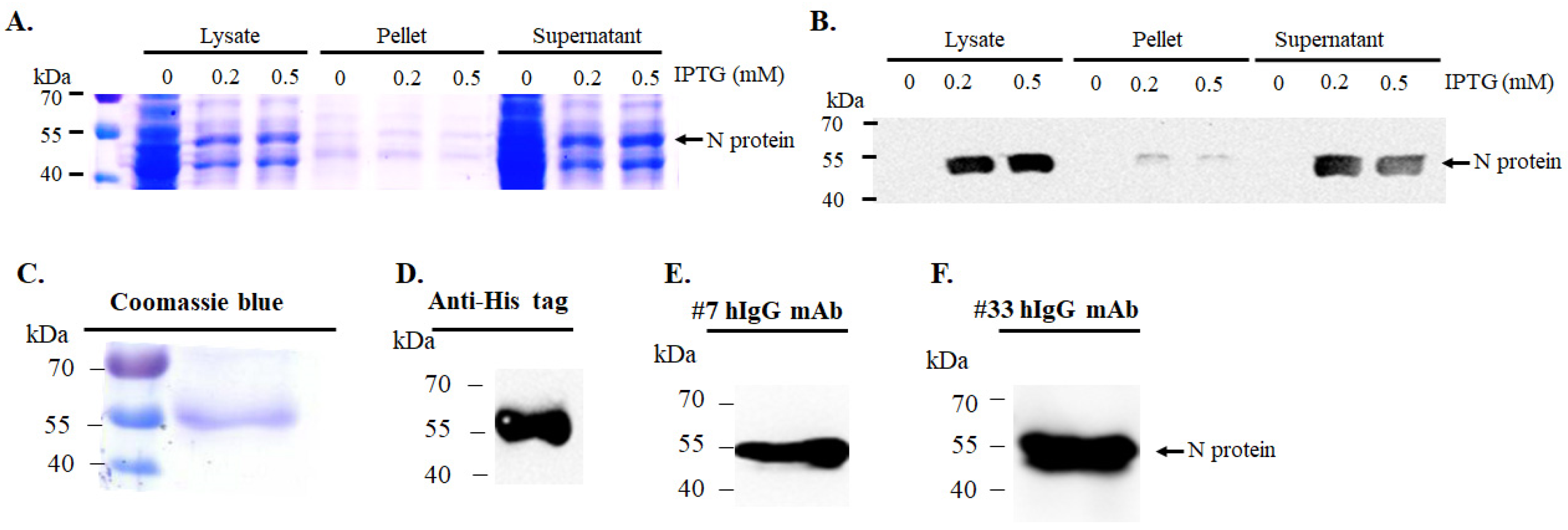
The recombinant plasmid pET-32a(+)-nCoV-2-N-protein was initially propagated in E. coli 10B and transformed into E. coli BL21 (DE3) for protein expression. Induction with IPTG at 0.2 mM and 0.5 mM at 37 °C triggered expression of the His-tagged SARS-CoV-2 nucleocapsid (N) protein. Following sonication, lysates were separated into total, soluble (supernatant), and insoluble (pellet) fractions and analyzed by SDS-PAGE (Figure 2A). A distinct ~55 kDa band, corresponding to the N protein, appeared predominantly in the total and supernatant fractions, with stronger expression at 0.2 mM IPTG. Western blotting with anti-His-tag antibodies confirmed the identity of the protein (Figure 2B), showing a strong ~55 kDa signal, particularly in the soluble fraction from the 0.2 mM IPTG condition, which was selected for purification.
Purification was carried out via nickel affinity chromatography. After cell lysis and clarification, the supernatant was loaded onto a Ni2⁺-charged column and eluted with increasing imidazole concentrations (10–400 mM). SDS-PAGE showed efficient enrichment of the N protein in the 400 mM elution fraction, with minimal background (Figure 2C). This fraction, along with the flow-through, was dialyzed against Tris-HCl buffer (50 mM, pH 8.0) containing 150 mM NaCl to remove imidazole. Protein concentration was enhanced by PEG 20000-mediated precipitation in three 1-hour incubations at 4 °C. Western blot analysis confirmed protein identity in the dialyzed and concentrated samples using anti-His-tag antibodies (Figure 2D).
To verify antigenicity, Western blotting was performed using two monoclonal antibodies specific to the SARS-CoV-2 N protein (Antibodies #33 and #7). Both detected immunoreactive bands at ~55 kDa (Figure 2E,F), consistent with the expected molecular weight, indicating the purified protein retained its immunological properties. These findings confirm successful expression, purification, and preservation of antigenicity, supporting the protein’s suitability for downstream LFIA development.

3.2. Stability of Gold Nanoparticles and Optimization of SARS-CoV-2 N Protein Conjugation

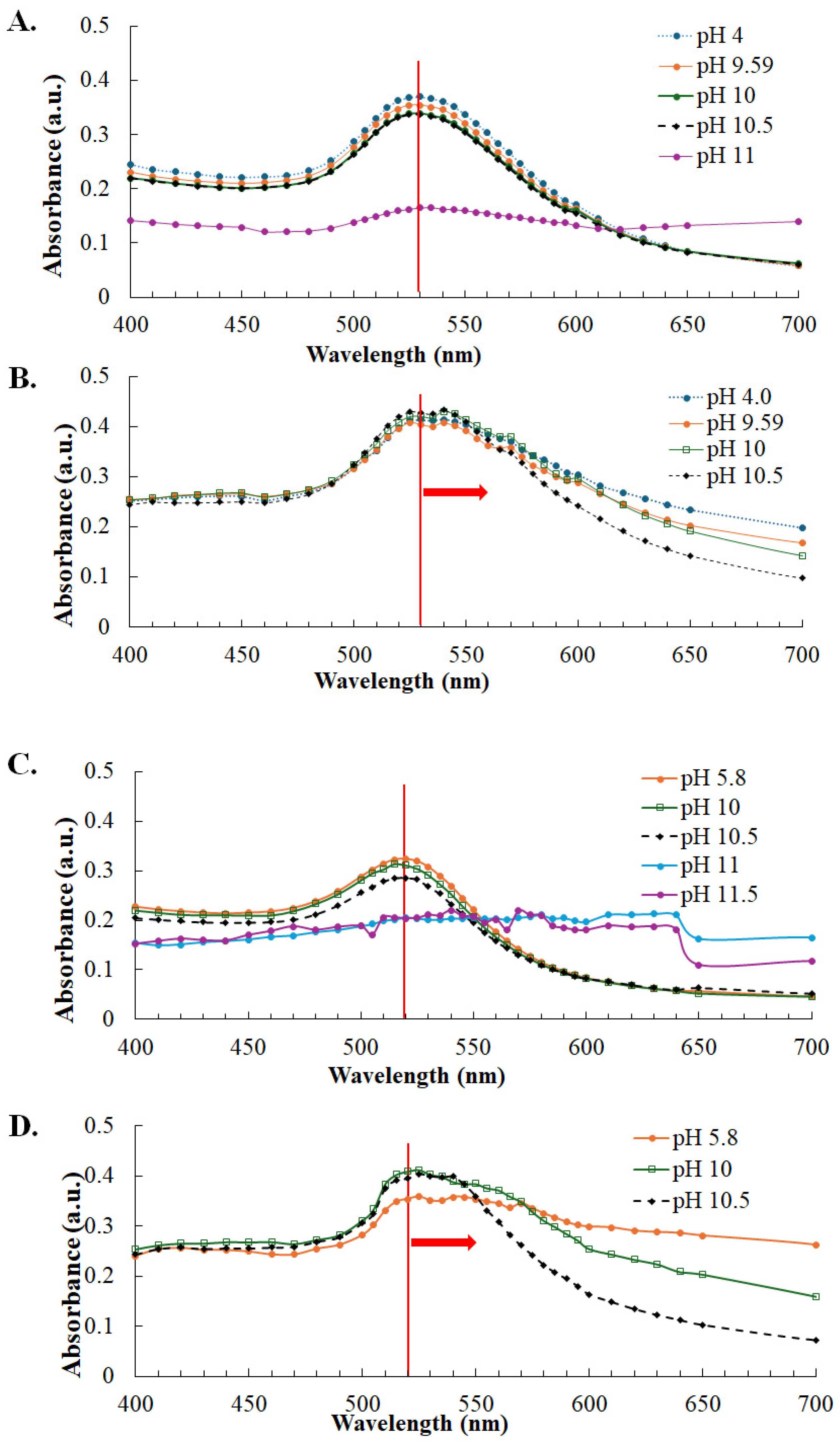
To optimize the conjugation of recombinant N protein with colloidal gold, two types of gold nanoparticles were synthesized using 1% and 4% sodium citrate. These nanoparticles were adjusted to pH values ranging from 4.0 to 11.5, and their optical properties were analyzed via UV-Vis spectroscopy (400–800 nm) (Figure 3). Both nanoparticle types remained stable from pH 4.0 to 10.5, exhibiting characteristic plasmon resonance peaks at 530 nm (1% citrate) and 520 nm (4% citrate). At pH levels above 11.0, these peaks disappeared, indicating nanoparticle instability (Figure 3A,C).
To evaluate protein conjugation, recombinant N protein (1 mg/mL) was mixed with pH-adjusted colloidal gold and incubated at 4 °C for 30 min. Following this, NaCl was added, and the mixtures were incubated at 37 °C for 10 min to assess aggregation. UV-Vis analysis showed spectral shifts in all N protein-containing samples, confirming successful conjugation (Figure 3B,D). The intrinsic pH of the synthesized AuNPs was approximately 4.0 (1% citrate) or 5.8 (4% citrate), which served as the unconjugated controls (Figure 3A,C). The choice of conjugation pH was based on the calculated isoelectric point of the recombinant N protein (pI ≈ 9.0), highlighting the importance of using conditions at or above the pI to achieve stable colloidal gold-protein interactions. Among them, the complex prepared at pH 10.5 displayed a sharp, well-defined peak, indicating reduced aggregation and improved stability compared with those prepared at lower pH values.

3.3. Assembly of Lateral Flow Immunoassay (LFIA) Strips for SARS-CoV-2 N Protein and Antibody Detection

Following optimization of gold nanoparticle size, pH, and protein conjugation conditions, lateral flow immunoassay (LFIA) strips were assembled. Immunogold conjugates were dispensed onto the conjugate pad and dried at 37 °C. All components were cut to a uniform width of ~0.4 cm before assembly, as illustrated in Figure 4A and Figure 5A.
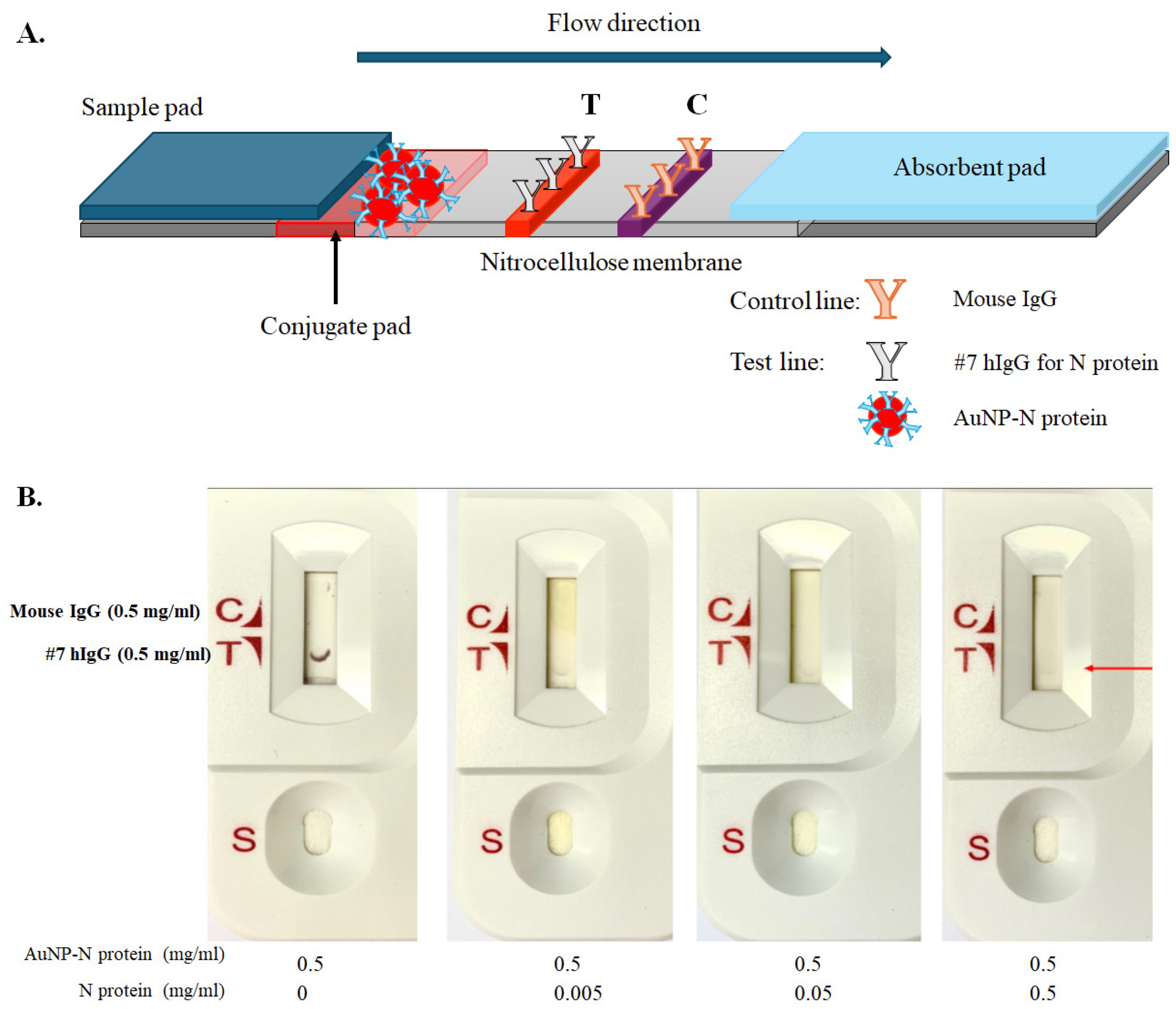
For SARS-CoV-2 nucleocapsid (N) protein detection, a competitive LFIA format was constructed (Figure 4). The nitrocellulose membrane was mounted on a backing card with monoclonal anti-N antibody #7 at the test (T) line and mouse IgG at the control (C) line. An absorbent pad was placed at the top and a sample pad with conjugate pad at the bottom, each overlapping the membrane by ~2 mm to ensure consistent capillary flow (Figure 4A). AuNP-conjugated N protein (0.5 mg/mL) was mixed with varying concentrations of free N protein (0–0.5 mg/mL) and applied to the sample pad. In the absence of free antigen, a strong T line appeared. Increasing concentrations of unconjugated N protein competed with the conjugated form, leading to reduced T line intensity (Figure 4B). This confirms the functionality of the competitive format, where higher antigen levels suppress the signal at the test line.
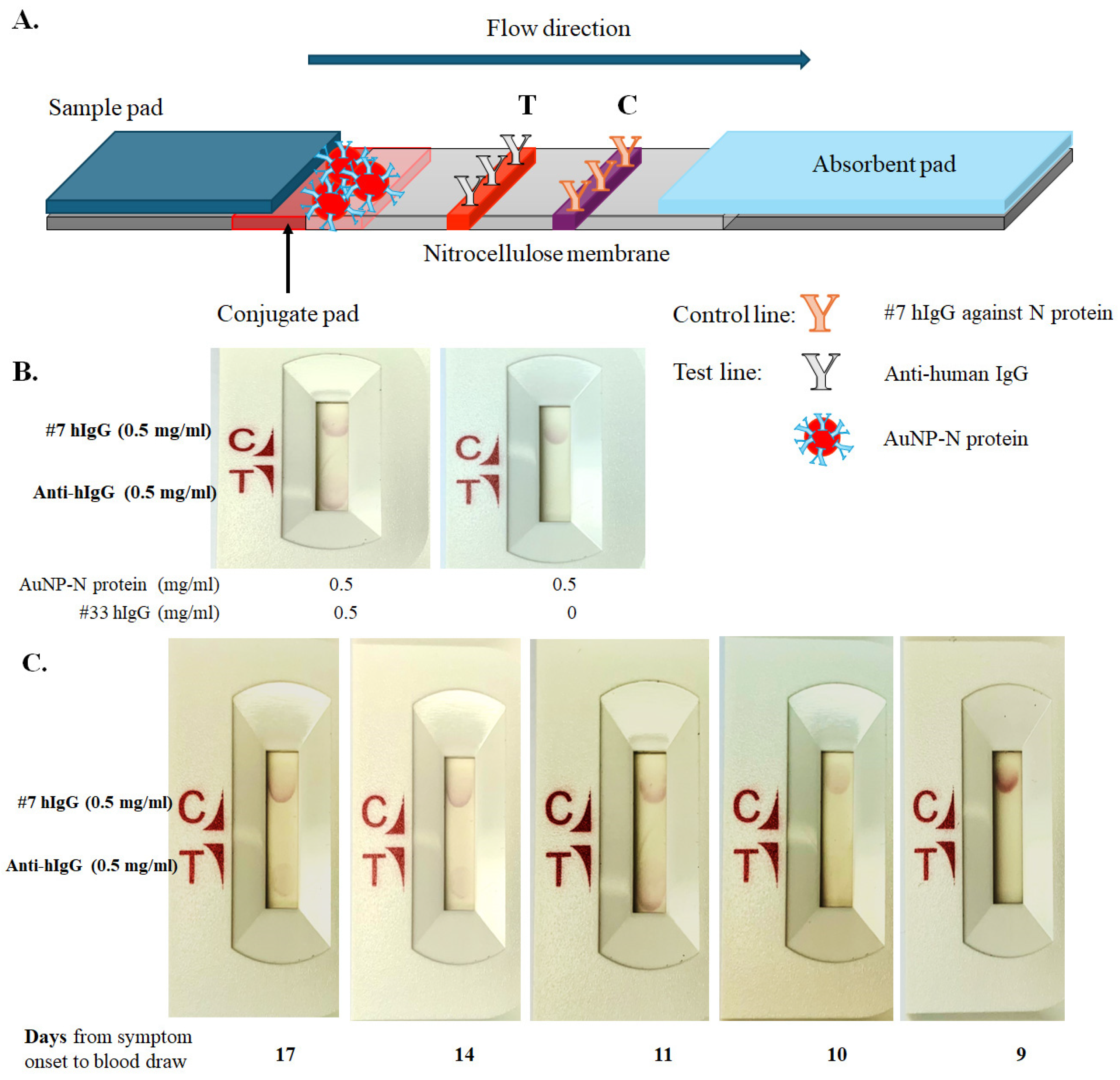
To detect anti-SARS-CoV-2 N IgG, a sandwich-format LFIA strip was developed (Figure 5). Anti-human IgG was applied to the T line and monoclonal antibody #7 hIgG to the C line. The strip layout mirrored the competitive format, with appropriate overlap to maintain flow direction (Figure 5A). Validation with monoclonal antibody #33 hIgG (0.5 mg/mL) produced visible red lines at both T and C positions (Figure 5B). Human serum samples collected 6–17 days after symptom onset (Boca Biolistics, USA) were also tested. Strong T line signals were observed in samples from Days 17, 14, and 11, while earlier samples produced weaker or no signals (Figure 5C), indicating that anti-N IgG becomes reliably detectable around 10 days post-symptom onset.
Overall, these results demonstrate the versatility of the AuNP-based LFIA platform, which can be configured for either competitive antigen detection or sandwich-format antibody detection.

4. Discussion

We developed a cost-effective lateral flow immunoassay (LFIA) for detecting SARS-CoV-2 N antigen and anti-N IgG antibodies using gold nanoparticles synthesized in-house by citrate reduction and conjugated with recombinant N protein (Figure 1, Figure 2, Figure 3, Figure 4 and Figure 5). The stability and conjugation efficiency of N protein–gold nanoparticle complexes were assessed at various pH levels and with NaCl. UV-Vis spectra showed characteristic shifts (400–800 nm), with complexes at pH 10.5 exhibiting narrow peaks and strong resistance to salt-induced aggregation (Figure 3), matching the N protein’s isoelectric point (~10.2) and indicating pH 10.5 as optimal.
We demonstrated that the competitive LFIA format was capable of detecting the N antigen at concentrations of 5, 50, and 500 μg/mL in a 50 μL sample volume (Figure 4). In a previous study, several antigen point-of-care tests (AgPOCTs)—including the Abbott Panbio COVID-19 Ag Rapid Test, RapiGEN BIOCREDIT COVID-19 Ag, Healgen Coronavirus Ag Rapid Test Cassette (Swab), Coris BioConcept COVID-19 Ag Respi-Strip, R-Biopharm RIDA QUICK SARS-CoV-2 Antigen, nal von minden NADAL COVID-19 Ag Test, and Roche-SD Biosensor SARS-CoV Rapid Antigen Test—were evaluated using recombinant SARS-CoV-2 nucleoprotein. These commercial assays were able to detect recombinant N protein at concentrations as low as 5–25 ng/mL per 50 μL sample [13]. Therefore, based on the results shown in Figure 4, our competitive LFIA format appears to be significantly less sensitive than these AgPOCTs. Further optimization will be necessary to enhance its performance, particularly to enable detection of recombinant N protein at lower concentrations (e.g., 5 μg/mL) and to ensure reliable detection of antigen in cell culture supernatants from SARS-CoV-2-infected cells. In addition, this prototype exhibited faint control line visibility when coated with mouse IgG (control antibodies) and lacked a reaction-based positive control, both of which limit the reliability of the assay and highlight the need for further refinement.
We also demonstrated that the sandwich LFIA format effectively detected monoclonal antibody #33 hIgG against the SARS-CoV-2 N protein at a concentration of 500 μg/mL in a 100 μL sample volume, as well as anti-N antibodies in commercial human serum samples collected between 6 and 17 days post-symptom onset (Figure 5). Strong positive signals were consistently observed in samples collected on days 11, 14, and 17 (Figure 5), aligning with established IgG seroconversion timelines during SARS-CoV-2 infection [14,15,16]. The assay exhibited high specificity and minimized false positives, achieved without reliance on costly commercial reagents [17,18]. While numerous antibody-based LFIAs have been developed throughout the COVID-19 pandemic for serological surveillance [19,20], many depend on commercially sourced recombinant antigens [21], which increase production costs and limit accessibility in low-resource settings. In contrast, our platform utilizes in-house recombinant N protein production, offering a more sustainable and scalable solution suitable for decentralized deployment.
This study aimed to demonstrate feasibility at the prototype level, showing that recombinant SARS-CoV-2 N protein can be successfully conjugated to AuNPs and applied in both competitive and sandwich LFIA formats. Future work will focus on systematic evaluation of key parameters, including specificity, cross-reactivity, long-term stability, and batch-to-batch reproducibility, to support product-oriented development of LFIA strips. In this prototype study, the faintness of the test lines limited our assessment to qualitative interpretation; future studies will incorporate quantitative signal analysis to enhance measurement accuracy and enable more rigorous comparisons across samples and conditions. Moreover, in-house production of recombinant N protein offers potential advantages in terms of cost reduction and scalability, which could facilitate broader application of the developed LFIA platform.

5. Conclusions

Recombinant SARS-CoV-2 N protein conjugated to gold nanoparticles was successfully applied in both competitive and sandwich LFIA formats, providing a versatile platform for viral antigen detection and humoral immune monitoring. Although the prototype exhibited lower sensitivity compared with commercial tests, it demonstrates feasibility, simplicity, and adaptability, particularly for resource-limited settings. Future work will focus on optimizing sensitivity, assessing specificity and reproducibility, expanding clinical validation with patient samples, and enhancing nanoparticle characterization to advance the platform toward practical diagnostic applications.

Author Contributions

Conceptualization, methodology, W.-J.T., Y.C., J.-L.H., H.-C.L., P.-R.H. and C.-W.L.; validation, formal analysis, data curation, W.-J.T. and C.-W.L.; writing—original draft preparation, writing—review and editing, W.-J.T., Y.C., J.-L.H. and C.-W.L.; funding acquisition, H.-C.L., P.-R.H. and C.-W.L. All authors have read and agreed to the published version of the manuscript.

Funding

This work was supported by the National Science and Technology Council, Taiwan (NSTC112-2320-B-039-054-MY3), and China Medical University (CMU113-ASIA-11, CMU114-ASIA-04, and CMU113-MF-15), including funding from the Higher Education Sprout Project, Ministry of Education, Taiwan.

Institutional Review Board Statement

Not applicable.

Informed Consent Statement

Not applicable.

Data Availability Statement

Data are contained within the articles.

Acknowledgments

Experiments were conducted at the Department of Medical Laboratory Science and Biotechnology and the Medical Research Core Facilities Center, China Medical University. We also thank PEPTIDE CHAM BIOTECH CO., LTD. (Kaohsiung, Taiwan) for providing SARS-CoV-2 nucleocapsid antibodies and technical guidance, which greatly supported assay development.

Conflicts of Interest

The authors declare no conflicts of interest.

References

  1. World Health Organization (WHO). Novel Coronavirus 2019—Situation Report; WHO: Geneva, Switzerland, 2020; Available online: https://www.who.int/emergencies/diseases/novel-coronavirus-2019/situation-reports/ (accessed on 1 August 2020).
  2. Jalandra, R.; Yadav, A.K.; Verma, D.; Dalal, N.; Sharma, M.; Singh, R.; Kumar, A.; Solanki, P.R. Strategies and perspectives to develop SARS-CoV-2 detection methods and diagnostics. Biomed. Pharmacother. 2020, 129, 110446. [Google Scholar] [CrossRef] [PubMed]
  3. Keshav, V.; Scott, L.; David, A.; Noble, L.; Mayne, E.; Stevens, W. Antigen-based point of care testing (POCT) for diagnosing SARS-CoV-2: Assessing performance. In SARS-CoV-2; Chu, J.J.H., Ahidjo, B.A., Mok, C.K., Eds.; Methods in Molecular Biology; Humana: New York, NY, USA, 2022; Volume 2452, pp. 45–62. [Google Scholar]
  4. Kakkar, S.; Gupta, P.; Yadav, S.P.S.; Raj, D.; Singh, G.; Chauhan, S.; Mishra, M.K.; Martín-Ortega, E.; Chiussi, S.; Kant, K. Lateral flow assays: Progress and evolution of recent trends in point-of-care applications. Mater Today Bio. 2024, 25, 100817. [Google Scholar] [CrossRef]
  5. Ang, G.Y.; Chan, K.G.; Yean, C.Y.; Yu, C.Y. Lateral flow immunoassays for SARS-CoV-2. Diagnostics 2022, 12, 2854. [Google Scholar] [CrossRef] [PubMed]
  6. Mirica, A.C.; Stan, D.; Chelcea, I.C.; Mihailescu, C.M.; Ofiteru, A.; Bocancia-Mateescu, L.A. Latest trends in lateral flow immunoassay (LFIA) detection labels and conjugation process. Front. Bioeng. Biotechnol. 2022, 10, 922772. [Google Scholar] [CrossRef] [PubMed]
  7. Posthuma-Trumpie, G.A.; Korf, J.; van Amerongen, A. Lateral flow (immuno)assay: Its strengths, weaknesses, opportunities and threats. Anal. Bioanal. Chem. 2009, 393, 569–582. [Google Scholar] [CrossRef] [PubMed]
  8. He, J.; Zhu, S.; Zhou, J.; Jiang, W.; Yin, L.; Su, L.; Zhang, X.; Chen, Q.; Li, X. Rapid detection of SARS-CoV-2: The gradual boom of lateral flow immunoassay. Front. Bioeng. Biotechnol. 2023, 10, 1090281. [Google Scholar] [CrossRef] [PubMed]
  9. Ince, B.; Sezgintürk, M.K. Lateral flow assays for viruses diagnosis: Up-to-date technology and future prospects. TrAC Trends Anal. Chem. 2022, 157, 116725. [Google Scholar] [CrossRef] [PubMed]
  10. Fagúndez, P.; Botasini, S.; Tosar, J.P.; Méndez, E. Systematic process evaluation of the conjugation of proteins to gold nanoparticles. Heliyon 2021, 7, e07392. [Google Scholar] [CrossRef] [PubMed]
  11. Shahjahan, T.; Javed, B.; Sharma, V.; Tian, F. pH and NaCl optimisation to improve the stability of gold and silver nanoparticles’ anti-zearalenone antibody conjugates for immunochromatographic assay. Methods Protoc. 2023, 6, 93. [Google Scholar] [CrossRef] [PubMed]
  12. Zhou, Y.; Wang, H.; Lin, W.; Lin, L.; Gao, Y.; Yang, F.; Du, M.; Fang, W.; Huang, J.; Sun, D.; et al. Quantitative nucleation and growth kinetics of gold nanoparticles via model-assisted dynamic spectroscopic approach. J. Colloid Interface Sci. 2013, 407, 8–16. [Google Scholar] [CrossRef] [PubMed]
  13. Corman, V.M.; Haage, V.C.; Bleicker, T.; Schmidt, M.L.; Mühlemann, B.; Zuchowski, M.; Jo, W.K.; Tscheak, P.; Möncke-Buchner, E.; Müller, M.A.; et al. Comparison of seven commercial SARS-CoV-2 rapid point-of-care antigen tests: A single-centre laboratory evaluation study. Lancet Microbe 2021, 2, e311–e319. [Google Scholar] [CrossRef] [PubMed]
  14. Kumar, P.K.; Banerjee, M.; Bajpayee, A.; Mandal, S.; Mitra, P.; Sharma, P.; Misra, S.; Bhardwaj, P. SARS-CoV-2 IgG Antibody and its Clinical Correlates in Convalescent Plasma Donors: An Indian Experience. Indian J. Clin. Biochem. 2022, 37, 423–431. [Google Scholar] [CrossRef] [PubMed]
  15. Long, Q.X.; Tang, X.J.; Shi, Q.L.; Li, Q.; Deng, H.J.; Yuan, J.; Hu, J.L.; Xu, W.; Zhang, Y.; Lv, F.J.; et al. Clinical and immunological assessment of asympto-matic SARS-CoV-2 infections. Nat. Med. 2020, 26, 1200–1204. [Google Scholar] [CrossRef]
  16. Liu, C.; Yu, X.; Gao, C.; Zhang, L.; Zhai, H.; Hu, Y.; Liu, E.; Wang, Q.; Gao, Y.; Wei, D.; et al. Characterization of antibody responses to SARS-CoV-2 in convalescent COVID-19 patients. J. Med. Virol. 2020, 93, 2227–2233. [Google Scholar] [CrossRef]
  17. Alhammadi, M.; Yoo, J.; Sonwal, S.; Park, S.Y.; Umapathi, R.; Oh, M.H.; Huh, Y.S. A highly sensitive lateral flow immuno-assay for the rapid and on-site detection of enrofloxacin in milk. Front Nutr. 2022, 9, 1036826. [Google Scholar] [CrossRef] [PubMed]
  18. Natarajan, S.; Su, F.; Jayaraj, J.; Shah, M.I.; Huang, Y. Paper microfluidics-based fluorescent lateral flow immunoassay for point-of-care diagnostics of noncommunicable diseases. Analyst 2019, 144, 7026–7034. [Google Scholar] [CrossRef] [PubMed]
  19. Castrejón-Jiménez, N.S.; García-Pérez, B.E.; Reyes-Rodríguez, N.E.; Vega-Sánchez, V.; Martínez-Juárez, V.M.; Hernández-González, J.C. Challenges in the detection of SARS-CoV-2: Evolution of the lateral flow immunoassay as a valuable tool for viral diagnosis. Biosensors 2022, 12, 728. [Google Scholar] [CrossRef] [PubMed]
  20. Ong, D.S.Y.; Fragkou, P.C.; Schweitzer, V.A.; Chemaly, R.F.; Moschopoulos, C.D.; Skevaki, C. How to interpret and use COVID-19 serology and immunology tests. Clin. Microbiol. Infect. 2021, 27, 981–986. [Google Scholar] [CrossRef] [PubMed]
  21. Swadźba, J.; Bednarczyk, M.; Anyszek, T.; Martin, E. A comparison of 7 commercial anti-SARS-CoV-2 antibody immunoassays. Arch. Med. Sci. 2020, 19, 1281–1288. [Google Scholar] [CrossRef] [PubMed]
Figure 1. Photograph of the assembled strip, showing the configuration of the sample pad, conjugate pad, nitrocellulose membrane with test and control lines, and absorbent pad.
Covid 05 00158 g001
Figure 2. Expression, purification, and verification of His-tagged SARS-CoV-2 N protein in E. coli BL21 (DE3). (A) SDS-PAGE analysis showing total, soluble (supernatant), and insoluble (pellet) protein fractions from cultures induced with 0, 0.2, or 0.5 mM IPTG. A distinct ~55 kDa band was most prominent at 0.2 mM IPTG. (B) Western blot using anti-His-tag antibodies confirmed the identity of the ~55 kDa protein as recombinant N protein. (C) SDS-PAGE analysis of the purified protein eluted with 400 mM imidazole from Ni2⁺ affinity chromatography. (DF) Western blot analyses using anti-His-tag and commercial anti-N antibodies (#33 and #7) verified the identity and antigenicity of the purified protein.
Covid 05 00158 g002
Figure 3. Stability and SARS-CoV-2 N protein conjugation of gold nanoparticles synthesized with 1% or 4% sodium citrate. (A) UV-Vis spectra of gold nanoparticles synthesized with 1% sodium citrate measured across pH 4.0–11.5. (B) Spectral shifts observed after conjugation of these nanoparticles with SARS-CoV-2 N protein and subsequent NaCl treatment. (C) UV-Vis spectra of gold nanoparticles synthesized with 4% sodium citrate showed stable plasmon resonance around 520 nm across the same pH range. (D) After conjugation with N protein and NaCl treatment, spectral shifts were detected at all pH levels for the 4% sodium citrate nanoparticles. Red arrow, spectral shift; red vertical line, plasmon resonance peak.
Covid 05 00158 g003
Figure 4. Assembly and performance of a competitive lateral flow immunoassay (LFIA) strip for SARS-CoV-2 N antigen detection. (A) The LFIA strip consisted of a sample pad, a conjugate pad containing gold-labeled N protein, a nitrocellulose membrane with monoclonal anti-N antibody #7 on the test (T) line and mouse IgG on the control (C) line, and an absorbent pad to maintain capillary flow. (B) In the competitive assay, varying concentrations of unlabeled N protein were applied to the sample pad. Representative results showing the T line in red are presented.
Covid 05 00158 g004
Figure 5. Assembly and performance of a sandwich-format lateral flow immunoassay (LFIA) strip for SARS-CoV-2 IgG detection. (A) The LFIA strip was assembled with a sample pad, a conjugate pad containing gold-labeled N protein, a nitrocellulose membrane with anti-human IgG on the test (T) line and monoclonal anti-N antibody #7 hIgG on the control (C) line, and an absorbent pad to support capillary flow. (B) Anti-N protein human IgG (#33 hIgG) was applied to the sample pad, and representative T line results (in red) were shown. (C) Human serum samples collected 6–17 days after symptom onset were tested, with representative results displayed.
Covid 05 00158 g005
Disclaimer/Publisher’s Note: The statements, opinions and data contained in all publications are solely those of the individual author(s) and contributor(s) and not of MDPI and/or the editor(s). MDPI and/or the editor(s) disclaim responsibility for any injury to people or property resulting from any ideas, methods, instructions or products referred to in the content.