Article

Breaking the Oxygen Dogma: How High FiO2 May Disrupt Pulmonary Physiology in COVID-19

by
Francisco Javier González Ruiz
*,
Blanca Estela Broca-García
,
Daniel Manzur-Sandoval
,
Luis Efrén Santos-Martínez
,
Uriel Encarnación-Martínez
,
Emmanuel Adrián Lazcano-Díaz
and
Angel Ramos-Enriquez
Department of Cardiovascular Critical Care, National Institute of Cardiology “Dr. Ignacio Chávez”, Mexico City 14080, Mexico
*
Author to whom correspondence should be addressed.
COVID 2025, 5(8), 139; https://doi.org/10.3390/covid5080139
Submission received: 13 July 2025 / Revised: 6 August 2025 / Accepted: 18 August 2025 / Published: 20 August 2025
(This article belongs to the Section COVID Clinical Manifestations and Management)

Abstract

Background: High concentrations of supplemental oxygen (FiO2 > 0.6) are commonly used to treat acute hypoxemia in critically ill patients. However, the effects of High FiO2 in patients with COVID-19 remain unclear, particularly regarding its impact on hypoxic pulmonary vasoconstriction (HPV) and ventilation–perfusion (V/Q) mismatch. Objective: This study aims to evaluate whether administering lower concentrations of inspired oxygen (FiO2 < 0.6) is associated with improved outcomes—namely reduced need for mechanical ventilation and mortality—in patients with COVID-19 and severe pulmonary involvement. Methods: This retrospective observational cohort included 201 patients with confirmed COVID-19. Patients were grouped by mean FiO2 during the first 24–48 h: High FiO2 (≥0.60) or Low FiO2 (<0.60). The primary outcome was the requirement for mechanical ventilation; the secondary outcome was in-hospital mortality. A composite endpoint (mechanical ventilation and in-hospital death) was also evaluated. Analyses included logistic regression and Kaplan–Meier survival with log-rank testing. Results: High FiO2 (≥0.60) was associated with higher odds of the composite outcome (mechanical ventilation and in-hospital death). In multivariable analysis, Low FiO2 remained associated with lower odds (adjusted OR 0.18; 95% CI 0.08–0.39; p < 0.001). Unadjusted rates were 43.1% vs. 16.1% for mechanical ventilation and 34.3% vs. 8.1% for in-hospital death (High vs. Low FiO2; both p < 0.001). Event-free survival favored the Low FiO2 group (log-rank p < 0.001). The model showed excellent discrimination (AUC 0.96; 95% CI 0.92–0.99). Conclusions: Higher early FiO2 exposure was associated with worse clinical outcomes in severe COVID-19. These findings are consistent with physiological models in which excess oxygen may attenuate hypoxic pulmonary vasoconstriction and increase shunt/dead space. Prospective studies are warranted to assess causality and refine oxygen targets.

1. Introduction

Hypoxic pulmonary vasoconstriction (HPV), also known as the von Euler–Liljestrand mechanism, plays a critical role in optimizing pulmonary gas exchange by redirecting blood flow away from poorly ventilated alveolar units toward better-ventilated regions, thereby improving arterial oxygenation [1,2,3,4]. This physiological response is crucial in several pathological states characterized by ventilation–perfusion (V/Q) mismatch, such as pulmonary embolism, acute respiratory distress syndrome (ARDS), and hepatopulmonary syndrome [5].
Although first described decades ago [6,7], the molecular mechanisms underlying oxygen sensing and signal transduction in HPV remain incompletely understood [8]. In the setting of COVID-19, mounting evidence indicates a unique alteration in pulmonary vascular physiology. Autopsy studies have revealed widespread microvascular thrombosis, fibrin deposition, and diffuse alveolar damage, often in the proliferative exudative phase [9,10,11,12]. These changes are frequently accompanied by features of organized pneumonia and early interstitial fibrosis, suggesting a rapidly evolving lung injury pattern distinct from classic ARDS [13].
A hallmark of severe COVID-19 is profound arterial hypoxemia, often disproportionate to the degree of respiratory distress—a phenomenon attributed to impaired HPV, increased dead space, and extensive V/Q mismatch [14,15,16]. In this context, the administration of high inspired oxygen concentrations (FiO2 > 0.6), while intended to correct hypoxemia, may paradoxically worsen gas exchange by inhibiting HPV and promoting resorption atelectasis.
Historically, trials in ARDS have recommended targeting oxygen saturation (SpO2) levels between 88 and 94%, with FiO2 titration guided by this range [17,18]. In contrast, recent guidelines for COVID-19 have proposed more liberal targets (SpO2 92–96%) [19], potentially leading to widespread use of High FiO2 in clinical practice. However, the safety and efficacy of this approach remain uncertain. A meta-analysis by Chu et al. [13] and findings by Ohshimo et al. [20] have suggested increased mortality associated with liberal oxygen therapy, though these studies focused primarily on non-COVID-19 ARDS populations.
Given the distinct pathophysiology of COVID-19-related lung injury, including altered vascular responses and early fibrotic changes, it is critical to reassess oxygenation strategies in this patient population. We hypothesize that conservative oxygen therapy—defined as maintaining FiO2 < 0.6—may preserve pulmonary autoregulation via HPV and reduce the need for mechanical ventilation and associated mortality.
This study aimed to compare clinical outcomes in patients with severe COVID-19 receiving high versus low concentrations of inspired oxygen during the first 24–48 h of hospitalization. We also explore the potential role of HPV preservation in mitigating disease progression.

2. Methods

2.1. Study Design and Setting

This study was a retrospective observational cohort study conducted at the National Institute of Cardiology “Dr. Ignacio Chávez” in Mexico City between 11 April and 8 August 2020. This study was approved by the Institutional Review Board and conducted in accordance with the principles outlined in the Declaration of Helsinki. Informed consent was waived due to the retrospective nature of this study, which involved minimal risk to participants and was approved by the Institutional Review Board.
The primary objective was to evaluate the association between the fraction of inspired oxygen (FiO2) administered during the first 24–48 h of hospitalization and two major outcomes: (1) the requirement for mechanical ventilation and (2) in-hospital mortality. Patients were categorized into two groups according to the FiO2 strategy followed:
High FiO2 group: FiO2 ≥ 0.6;
Low FiO2 group: FiO2 < 0.6.
The allocation to either group was based on the clinical judgment of the treating physicians. Although there was no formal institutional protocol dictating FiO2 thresholds, oxygen therapy was administered via high-flow nasal cannula (HFNC) in all cases, with settings tailored to patient oxygenation status, respiratory rate, and overall severity.

2.2. Oxygen Titration in Clinical Practice

Oxygen supplementation was titrated by the treating physicians to achieve a target SpO2 of 92–96%, avoiding both hypoxemia and hyperoxia, consistent with international guidance [20,21]. Initial FiO2 and subsequent adjustments were based on oxygen saturation, work of breathing, and gas-exchange indices (PaO2/FiO2, A–a). Patients were categorized as High FiO2 (≥0.60) or Low FiO2 (<0.60) according to the mean FiO2 delivered during the first 24–48 h.

2.2.1. Eligibility Criteria

We screened all adult patients (aged 18 to 80 years old) admitted to the intensive care unit (ICU) with confirmed COVID-19 infection by reverse transcription polymerase chain reaction (RT-PCR) assay.

2.2.2. Inclusion Criteria

The following inclusion criteria were applied:
Confirmed diagnosis of SARS-CoV-2 infection;
Acute hypoxemic respiratory failure requiring supplemental oxygen;
Thoracic computed tomography (CT) categorized as CO-RADS 4 or 5, indicating high or very high suspicion of COVID-19 pneumonia.

2.2.3. Exclusion Criteria

The following exclusion criteria were applied:
Pre-existing chronic cardiopulmonary conditions (e.g., coronary artery disease, congestive heart failure, chronic kidney disease, chronic obstructive pulmonary disease);
CT evidence of acute pulmonary thromboembolism;
Incomplete arterial blood gas (ABG) data within the first 48 h.

2.3. Data Collection

Clinical and laboratory data were collected retrospectively from medical records and electronic monitoring systems. Baseline demographics, comorbidities, vital signs, and laboratory markers (e.g., D-dimer, ferritin, fibrinogen, C-reactive protein) were recorded upon ICU admission. Chest CT scans were evaluated by a board-certified radiologist who was blinded to the FiO2 status and patient outcomes. CO-RADS classification was used to quantify the severity of pulmonary involvement (Table 1).
Oxygen therapy was delivered via high-flow nasal cannula with FiO2 ranging from 0.3 to 1.0 and flow rates of 10–40 L/min. Arterial blood gas analysis was performed at admission (before oxygen administration) and repeatedly during the first 48 h. Clinical decisions regarding medication use (e.g., dexamethasone, NSAIDs, anticoagulation) were made in accordance with institutional standards but left to the discretion of the treating physicians.
Patients were followed until ICU discharge, death, or intubation for mechanical ventilation.

2.4. Oxygenation Metrics

The alveolar–arterial oxygen gradient (A–a gradient) was calculated as follows:
PAO 2 = FiO 2 × ( Patm PH 2 O ) ( PaCO 2 / 0.8 )
where Patm was assumed to be 585 mmHg, corresponding to the average barometric pressure at 2240 m above sea level (Mexico City altitude), and PH2O = 47 mmHg. The age-adjusted A–a gradient was calculated as
Expected A–a = ( Age / 4 ) + 4
The P/F ratio (PaO2/FiO2) and other ABG parameters were used to assess gas exchange.

2.5. Statistical Analysis

All statistical analyses were performed using Stata Statistical Software: Release 14 (StataCorp LP, College Station, TX, USA) and IBM SPSS Statistics for Windows, Version 26.0 (IBM Corp., Armonk, NY, USA).
Continuous variables were first tested for normality by the Shapiro–Wilk test. Variables with approximately normal distributions are reported as mean ± standard deviation (SD) and compared by Student’s t-test; non-normal variables are reported as medians (interquartile range [IQR]) and compared by the Mann–Whitney U test (Table 2 and Table 3). In addition to p-values, effect sizes for continuous comparisons were summarized as follows: for approximately normally distributed variables, we reported standardized mean differences with 95% confidence intervals; for non-normally distributed variables compared with the Mann–Whitney U test, and emphasized distribution-free summaries (median [IQR]) p-value (Table 3). Categorical variables are presented as frequencies and percentages and compared by the chi-square test or Fisher’s exact test for expected counts < 5 (Table 2 and Table 4).
A multivariable logistic regression model was constructed to identify independent predictors of the composite outcome (mechanical ventilation and in-hospital death). Covariates were chosen a priori based on clinical relevance and a bivariate screening threshold of p < 0.20, and they included the following:
  • FiO2 group (High ≥ 0.60 vs. Low < 0.60);
  • P/F ratio (per 10 mmHg increment);
  • A–a gradient (per 5 mmHg increment);
  • C-reactive protein (per 10 mg/L increment);
  • APACHE II score (per point).
Given the marked imbalance in sex distribution between groups and its known prognostic relevance in COVID-19, we conducted a prespecified sensitivity analysis by refitting the logistic model and additionally adjusting for sex (male vs. female). Estimates and inferences were qualitatively unchanged.
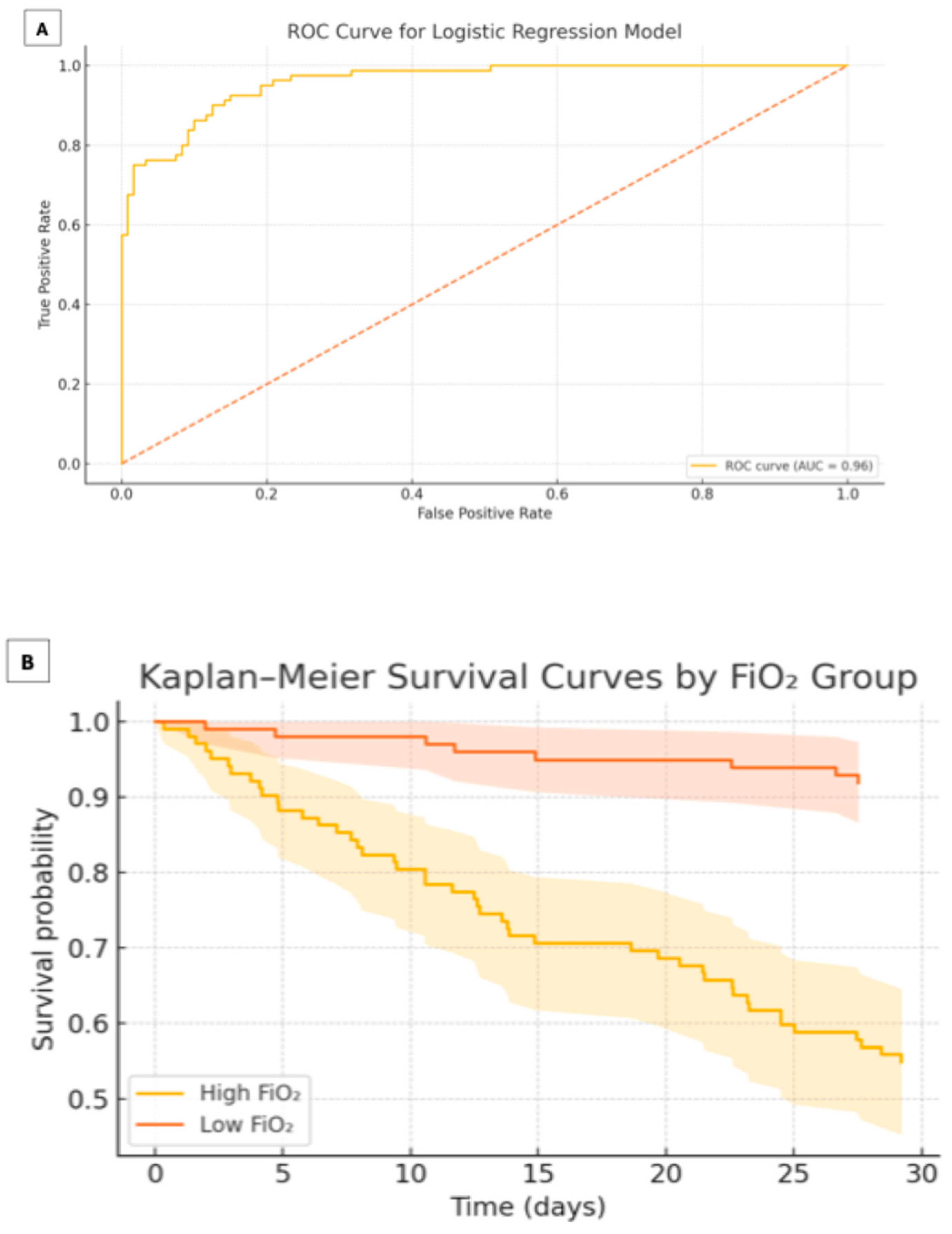
Results are reported as odds ratios (OR) with 95% CI and two-tailed p-values (Table 5). Model fit was assessed by the Hosmer–Lemeshow goodness-of-fit test (p = 0.47) and discrimination by the area under the receiver-operating characteristic (ROC) curve (AUC = 0.96; 95% CI 0.92–0.99) (Figure 1). Multicollinearity was ruled out by variance inflation factors (VIF < 2 for all covariates).
Time-to-event analyses for the composite outcome were performed by Kaplan–Meier survival estimation, with differences between FiO2 groups assessed by the log-rank test (Figure 1). Follow-up was censored at 30 days. A Cox proportional-hazards model was considered but not included in the final analysis due to the violation of the proportional-hazards assumption.
All statistical tests were two-tailed, and a p-value < 0.05 was considered statistically significant.

3. Results

3.1. Baseline Characteristics

A total of 201 patients with confirmed COVID-19 were included. The median age was 55.3 years (IQR 45–66), and 62.6% were male; however, sex distribution differed significantly between groups (High FiO2 = 84/102 [82.4%] vs. Low FiO2 = 42/99 [42.4%]; p < 0.001) (Table 2). Hypertension was more frequent in the High FiO2 group (39/102, 38.2%) than in the Low FiO2 group (22/99, 22.2%; p = 0.01), whereas smoking prevalence was similar between groups. Vital signs and inflammatory markers (D-dimer, ferritin, fibrinogen, CRP) did not differ meaningfully at ICU admission. The median CO-RADS score was 5 (IQR 5–5) in both cohorts, reflecting a high degree of pulmonary involvement (Table 2).
Table 2. Baseline patient characteristics stratified by FiO2 group.
High FiO2
(n = 102)
Low FiO2
(n = 99)
p-Value
Sex, n (%) <0.001
Male84 (82.4%)42 (42.4%)
Female18 (17.6%)57 (57.6%)
Hypertension, n (%)39 (38.2%)22 (22.2%)0.01
Smoking, n (%)51 (50.0%)36 (36.4%)0.24
Age (years), mean ± SD55.3 ± 13.255.2 ± 15.30.98
Height (cm), mean ± SD164 ± 7.9166 ± 7.30.42
Weight (kg), median (IQR)75 (68–85)78 (68–80)0.95
BMI (kg/m2), median (IQR)27.3 (25.0–29.4)27.6 (24.3–29.5)0.91
Heart rate (bpm), median (IQR)95 (82–109)95 (84–105)0.93
Respiratory rate (rpm), median (IQR)25 (20–28)26 (24–28)0.10
CO-RADS, median (IQR)5 (5–5)5 (5–5)0.55
D-dimer (µg/mL), median (IQR)0.36 (0.23–0.56)0.36 (0.24–0.51)0.86
Fibrinogen (g/L), median (IQR)5.5 (4.7–6.2)5.6 (4.2–6.1)0.59
Ferritin (ng/mL), median (IQR)594 (268–1082)581 (355–1232)0.88
CRP (mg/L), median (IQR)159 (47–259)139 (55–196)0.45
Data are presented as n (%) for categorical variables, mean ± SD for normally distributed continuous variables, and median (IQR) for non-normally distributed variables. High FiO2 ≥ 0.60; Low FiO2 < 0.60. Categorical comparisons were performed by the chi-square test, t-test for normal data and the Mann–Whitney U test for non-normal data. Abbreviations: BMI, body mass index; CRP, C-reactive protein; CO-RADS, COVID-19 Reporting and Data System.

3.2. Arterial Blood Gas Parameters

Baseline oxygen saturation at ICU admission was comparable between groups (Table 3). At admission, ABG parameters did not differ meaningfully between groups for pH, PaO2, or PaCO2 (all p > 0.05). The A–a gradient was slightly higher in the High FiO2 group than in the Low FiO2 group (p < 0.001), and the P/F ratio was lower in the High FiO2 group (242 [209–280] vs. 276 [261–290]; p = 0.01) (Table 3).
By 24–48 h, the High FiO2 group—despite receiving a greater FiO2 (70% vs. 40%; p < 0.001)—showed significantly lower PaO2 (67 [59–75] vs. 77 [72–85] mmHg; p < 0.001), lower SaO2 (93.5% [87–95] vs. 96% [94–97]; p < 0.001), and a persistently reduced P/F ratio (95 [83–147] vs. 219 [140–282]; p < 0.001), indicating ongoing V/Q mismatch (Table 3).
Table 3. Arterial blood gas levels at admission and 24–48 h by FiO2 group.
High FiO2 (n = 102), Median (IQR)Low FiO2 (n = 99), Median (IQR)p-Value
Admission
pH7.44 (7.39–7.48)7.45 (7.44–7.47)0.18
PaO2, mmHg55 (50–64)58 (55–61)0.29
PaCO2, mmHg32 (27–35)30 (26–34)0.56
SaO2, %85.6 (79.5–92)87 (84–88)0.79
FiO2, %21 (21–21)21 (21–21)1.00
A–a gradient, mmHg32 (25–48)34 (27–53)<0.001
P/F ratio242 (209–280)276 (261–290)0.01
24–48 h
pH7.41 (7.30–7.45)7.45 (7.42–7.47)0.01
PaO2, mmHg67 (59–75)77 (72–85)<0.001
PaCO2, mmHg36 (31–43)34 (31–37)0.44
SaO2, %93.5 (87–95)96 (94–97)<0.001
FiO2, %70 (60–90)40 (32–59)<0.001
A–a gradient, mmHg257 (170–369)198 (143–306)0.10
P/F ratio95 (83–147)219 (140–282)<0.001
Data are median (IQR). Between-group comparisons were performed by the Mann–Whitney U test. High FiO2 ≥ 0.60; Low FiO2 < 0.60. Abbreviations: FiO2, fraction of inspired oxygen; PaO2, arterial oxygen tension; PaCO2, arterial carbon dioxide tension; SaO2, arterial oxygen saturation; P/F, PaO2/FiO2 ratio; A–a, alveolar–arterial oxygen gradient.

3.3. Clinical Outcomes

Mechanical ventilation was required in 44/102 (43.1%) of the High FiO2 group versus 16/99 (16.1%) of the Low FiO2 group (p < 0.001). The median time to intubation did not differ (0 [0–1] days vs. 0 [0–0] days; p = 0.27). In-hospital mortality was also higher with High FiO2 (35/102, 34.3% vs. 8/99, 8.1%; p < 0.001). The composite endpoint (mechanical ventilation and death) occurred in 39/102 (38.2%) vs. 10/99 (10.1%) of cases, p < 0.001 (Table 4).
Table 4. Clinical outcomes by FiO2 group.
OutcomeHigh FiO2
(n = 102)
Low FiO2
(n = 99)
p-Value
Mechanical ventilation, n (%)44 (43.1%)16 (16.1%)<0.001
Days to mechanical ventilation, median (IQR)0 (0–1)0 (0–0)0.27
In-hospital death, n (%)35 (34.3%)8 (8.1%)<0.001
Mechanical ventilation and in-hospital death, n (%)39 (38.2%)10 (10.1%)<0.001
Data are n (%) for categorical outcomes and median (IQR) for time-to-event outcomes. Categorical comparisons were performed by the chi-square test and continuous variables by Mann–Whitney U test. The composite endpoint was defined as the concurrent occurrence of mechanical ventilation and in-hospital death during index hospitalization. High FiO2 ≥ 0.60; Low FiO2 < 0.60. Abbreviations: FiO2, fraction of inspired oxygen.

3.4. Multivariable Analysis

A logistic regression model adjusting for FiO2 group (High ≥ 0.60 vs. Low < 0.60), P/F ratio (per 10 mmHg), A–a gradient (per 5 mmHg), CRP (per 10 mg/L), and APACHE II score (per point) identified Low FiO2 as independently protective (OR 0.18; 95% CI 0.08–0.39; p < 0.001). Model calibration was good (Hosmer–Lemeshow p = 0.47) and discrimination was excellent (AUC = 0.96; 95% CI: 0.92–0.99) (Table 5). Variance inflation factors were all <2, excluding multicollinearity.
Table 5. Multivariable logistic regression predicting composite outcome of mechanical ventilation or in-hospital death.
VariableOR (95% CI)p-Value
Low FiO2 (<0.60 vs. ≥0.60)0.18 (0.08–0.39)<0.001
P/F ratio (per 10 mmHg increase)0.95 (0.92–0.98)0.002
A–a gradient (per 5 mmHg increase)1.03 (1.01–1.05)0.010
CRP (per 10 mg/L increase)1.01 (1.00–1.02)0.030
APACHE II score (per point)1.12 (1.04–1.21)0.004
Multivariable logistic regression model predicting the composite outcome of mechanical ventilation or in-hospital death. Odds ratios (ORs) with 95% confidence intervals (CIs) and p-values are shown for each covariate. The model was adjusted for P/F ratio (per 10 mmHg increase), alveolar–arterial (A–a) gradient (per 5 mmHg increase), C-reactive protein (CRP; per 10 mg/L increase), and APACHE II score (per point). Goodness-of-fit was assessed by the Hosmer–Lemeshow test (p = 0.47). Discrimination is reported as the area under the receiver-operating characteristic (ROC) curve (AUC = 0.96; 95% CI: 0.92–0.99). Variance inflation factors (VIFs) for all covariates were <2, indicating no significant multicollinearity. Abbreviations: FiO2, fraction of inspired oxygen; OR, odds ratio; CI, confidence interval; P/F, PaO2/FiO2 ratio; A–a, alveolar–arterial gradient; CRP, C-reactive protein; APACHE, Acute Physiology And Chronic Health Evaluation.

3.5. Kaplan–Meier Analysis

Kaplan–Meier curves used for the time to composite outcome showed a significant early separation favoring the Low FiO2 group (log-rank p < 0.001) (Figure 1—A: ROC; B: Kaplan–Meier). Follow-up was censored at 30 days.
Figure 1. ROC curve (A) and Kaplan–Meier survival analysis (B) for the composite outcome. (A) Receiver-operating characteristic (ROC) curve for the multivariable logistic regression model predicting the composite outcome of mechanical ventilation or in-hospital death. The model demonstrated excellent discrimination with an area under the curve (AUC) of 0.96. (B) Kaplan–Meier survival curves comparing time to composite outcome between the High FiO2 (≥0.6) and Low FiO2 (<0.6) groups. A significant early divergence in event-free survival was observed in favor of the Low FiO2 group (log-rank p < 0.001), indicating a protective association of conservative oxygen therapy during the first 48 h of hospitalization.
Covid 05 00139 g001

4. Discussion

This study provides novel insights into the effects of oxygen therapy strategies in patients with severe COVID-19. Our results demonstrate that the use of high inspired oxygen concentrations (FiO2 ≥ 0.6) is associated with a significantly higher rate of mechanical ventilation (43.1% vs. 16.1%; p < 0.001) and a significantly higher in-hospital mortality (34.3% vs. 8.1%; p < 0.001) compared to a more conservative oxygen approach (FiO2 < 0.6). Importantly, these differences persisted after multivariable adjustment (Low FiO2 protective: OR 0.18; 95% CI 0.08–0.39; p < 0.001) and were supported by excellent model discrimination (AUC = 0.96; 95% CI: 0.92–0.99), indicating the strong predictive capacity of the logistic model. Furthermore, the Kaplan–Meier survival analysis demonstrated a significant early divergence between groups (log-rank p < 0.001) consistent with a potential benefit of early conservative oxygen titration and warranting prospective evaluation of optimal oxygen targets. Given that our cohort was managed at high altitude (~2240 m), we also considered how altitude might influence oxygenation metrics and the generalizability of these findings.
At this elevation, barometric pressure and inspired oxygen tension are lower; accordingly, we calculated the A–a gradient using the locally appropriate barometric pressure and reported ABG values as medians (IQR). Although absolute PaO2/SaO2 at admission may differ from sea-level settings, all patients in this study shared the same altitude, preserving internal comparisons. Importantly, exposure was defined by the mean FiO2 delivered during the first 24–48 h, capturing early clinical evolution rather than a single time point. The association between higher early FiO2 and worse outcomes is biologically plausible across altitudes because the relevant mechanisms—shunt, ventilation–perfusion mismatch, and attenuation of hypoxic pulmonary vasoconstriction—do not depend on elevation, providing the physiological context for the patterns observed.
Within this context, these patterns are consistent with greater ventilation–perfusion mismatch in the High FiO2 group. They align with physiological models indicating that oxygen supplementation has limited efficacy in shunt physiology and that excess FiO2 may attenuate hypoxic pulmonary vasoconstriction, thereby worsening gas exchange [22,23,24].
Our findings support the hypothesis that excessive oxygen administration may disrupt hypoxic pulmonary vasoconstriction (HPV), a fundamental adaptive mechanism that optimizes V/Q matching by diverting blood flow away from poorly ventilated alveoli [1,2,3,4]. In COVID-19, this mechanism appears particularly vulnerable due to extensive microvascular thrombosis, interstitial edema, and alveolar injury, as shown in postmortem histopathologic studies [9,10,11,12]. The suppression of HPV by High FiO2 may exacerbate shunting and dead space ventilation, impairing oxygenation and promoting further lung injury.
These pathophysiological considerations are consistent with contemporary evidence. Direct randomized evidence on oxygen targets in COVID-19 remains limited, and trials across mixed ICU populations have yielded mixed results when comparing conservative versus liberal strategies. A large meta-analysis has associated liberal oxygen with increased mortality, whereas ICU randomized trials have alternately shown benefit with conservative protocols, neutral effects, or no difference in mortality despite lower oxygen exposure. Together with the biological rationale that excess FiO2 can worsen V/Q mismatch in the presence of shunt, these observations align with our findings and support prospective, protocolized evaluations of oxygen targets in COVID-19 [18,25,26,27].
Despite receiving higher FiO2 during the first 48 h, patients in the High FiO2 group did not exhibit improved PaO2 or P/F ratios. At ICU admission, the A–a gradient was slightly higher in the High FiO2 group than in the Low FiO2 group (34 vs. 32 mmHg; p < 0.001). By 24–48 h, the between-group difference in A–a was not significant (257 vs. 198 mmHg; p = 0.10). In contrast, oxygenation indices remained worse in the High FiO2 group at 24–48 h, with a lower P/F ratio and lower SaO2 (both p < 0.001).
Moreover, the risk of resorption atelectasis—a known consequence of High FiO2 due to rapid diffusion of oxygen and nitrogen washout—may further impair lung function [26]. This phenomenon likely contributes to alveolar collapse, especially in inflamed and edematous lung regions, compounding V/Q mismatch.
Recent evidence from non-COVID-19 ARDS trials and meta-analyses has raised concerns about liberal oxygen strategies, showing associations with increased mortality [13,20,23]. However, COVID-19 is distinct from typical ARDS in both its pathophysiology and radiologic presentation. In this context, our study is among the first to demonstrate that tailoring oxygen targets in the early phase of severe COVID-19 may impact the need for mechanical ventilation, a significant determinant of prognosis.
A critical physiological consideration is that SpO2 does not always reflect PaO2 in COVID-19 due to alterations in the oxyhemoglobin dissociation curve. This can occur secondary to respiratory alkalosis, interstitial edema, microthrombosis, and altered hemoglobin affinity [28,29]. Thus, reliance on pulse oximetry alone to titrate FiO2 may be misleading and promote over-oxygenation.
Individual variability in the HPV response, affected by factors such as age, diabetes, and chronic inflammation, may further influence susceptibility to oxygen-induced impairment [30,31,32,33,34]. In older adults and those with metabolic disorders, the hypoxic response is often blunted, making them more vulnerable to the adverse effects of high oxygen concentrations.
From a mechanistic perspective, High FiO2 administration likely contributes to the loss of HPV, increased pulmonary vascular permeability, and ultimately pulmonary decompensation. By maintaining FiO2 levels below 0.6, the compensatory HPV mechanism may remain intact for a longer period, thereby preserving V/Q matching and delaying the progression to mechanical ventilation. This is further illustrated by the early divergence in Kaplan–Meier curves for time to mechanical ventilation or death (log-rank p < 0.001) (Figure 2).

5. Implications for Practice and Future Research

Our findings support a reassessment of oxygen therapy targets in patients with COVID-19-related respiratory failure. Rather than aiming for SpO2 > 96%, a strategy targeting SpO2 between 88% and 92%, as suggested in classic ARDS protocols [17,18], may offer physiologic and clinical advantages in selected patients. Prospective studies are needed to confirm these observations and to define optimal oxygenation thresholds in COVID-19. The role of individualized oxygen titration, combined with markers of V/Q mismatch and pulmonary vascular function, represents a promising area of research.

6. Strengths and Limitations

This study contributes to the growing body of evidence evaluating oxygen therapy strategies in patients with COVID-19, highlighting that higher early FiO2 exposure is associated with impaired gas exchange and worse clinical outcomes. By integrating early oxygenation parameters with clinically meaningful endpoints—mechanical ventilation, in-hospital death, and a composite of both—our findings offer practice-relevant insights into the consequences of liberal oxygen supplementation. Methodological strengths include objective physiological measurements (arterial blood gases), standardized radiologic classification (CO-RADS; Table 1) with blinded interpretation, and prespecified exposure categories (High FiO2 ≥ 0.60 vs. Low FiO2 < 0.60) based on the mean FiO2 during the first 24–48 h. Multivariable modeling adjusted for baseline severity (P/F ratio, A–a gradient, APACHE II) showed excellent discrimination (AUC 0.96), and results were robust in a sensitivity analysis additionally adjusting for sex, a baseline characteristic that differed between groups.
Nonetheless, several limitations must be acknowledged. First, the retrospective, single-center design precludes causal inference and may limit generalizability. Second, FiO2 group assignment reflected clinician-driven titration rather than randomization, introducing potential confounding by indication; reverse causality cannot be fully excluded (i.e., sicker patients may have received more oxygen). Although we adjusted for key severity markers and conducted a sex-adjusted sensitivity analysis, residual confounding may persist. Third, while baseline oxygenation metrics were systematically recorded, unmeasured factors influencing oxygen titration (e.g., provider preference or subtle clinical cues) could still bias estimates. Finally, the sample size, though adequate for the primary analyses, may have limited power for some secondary endpoints and subgroup assessments. Prospective, protocolized studies are warranted to confirm these associations and refine target oxygen ranges in severe COVID-19.

7. Conclusions

Our findings suggest that the administration of high concentrations of supplemental oxygen (FiO2 ≥ 0.6) in patients with severe COVID-19 is independently associated with worse clinical outcomes, including a higher likelihood of requiring mechanical ventilation and increased mortality, likely mediated by the disruption of hypoxic pulmonary vasoconstriction, impaired ventilation–perfusion matching, and the development of resorption atelectasis. These findings were further validated by robust multivariable modeling (AUC = 0.96; 95% CI: 0.92–0.99) and a significant early separation in Kaplan–Meier survival curves, underscoring the clinical importance of early FiO2 titration strategies in the management of severe COVID-19.

Author Contributions

All authors contributed substantially to the conception, design, and execution of this study. F.J.G.R. and B.E.B.-G. contributed to study design and manuscript drafting. D.M.-S., U.E.-M., E.A.L.-D. and L.E.S.-M. participated in data collection and interpretation. A.R.-E. supervised the overall project and provided critical revisions. All authors have read and agreed to the published version of the manuscript.

Funding

This research received no specific grant from any funding agency in the public, commercial, or not-for-profit sectors.

Acknowledgments

We thank the medical and nursing staff of the Cardiovascular Intensive Care Unit for their dedication and support during the COVID-19 pandemic. Their clinical efforts were essential to the care and documentation of the cases included in this study.

Conflicts of Interest

The authors declare no conflicts of interest related to this study.

References

  1. Dunham-Snary, K.J.; Wu, D.; Sykes, E.A.; Thakrar, A.; Parlow, L.R.G.; Mewburn, J.D.; Parlow, J.L.; Archer, S.L. Hypoxic Pulmonary Vasoconstrictiction. Chest 2017, 1511, 181–192. [Google Scholar] [CrossRef] [PubMed]
  2. Tarry, D.; Powell, M. Hypoxic pulmonary vasoconstriction. BJA Educ. 2017, 17, 208–213. [Google Scholar] [CrossRef]
  3. Sommer, N.; Strielkov, I.; Pak, O.; Weissmann, N. Oxygen sensing and signal transduction in hypoxic pulmonary vasoconstriction. Eur. Respir. J. 2016, 47, 288–303. [Google Scholar] [CrossRef] [PubMed]
  4. Marshall, B.E.; Hanson, C.W.; Frasch, F.; Marshall, C. Role of hypoxic pulmonary vasoconstriction in pulmonary gas exchange and blood flow distribution. Intensive Care Med. 1994, 20, 379–389. [Google Scholar] [CrossRef]
  5. Lumb, A.B.; Slinger, P. Hypoxic pulmonary vasoconstriction: Physiology and anesthetic implications. Anesthesiology 2015, 122, 932–946. [Google Scholar] [CrossRef]
  6. Bradford, J.R.; Dean, H.P. Pulmonary circulation. J. Physiol. 1894, 16, 34–110. [Google Scholar] [CrossRef]
  7. Euler, U.S.V.; Liljestrand, G. Observations on the pulmonary arterial blood pressure in the cat. Acta Physiol. Scand. 1946, 12, 301–320. [Google Scholar] [CrossRef]
  8. Carsana, L.; Sonzogni, A.; Nasr, A.; Rossi, R.S.; Pellegrinelli, A.; Zerbi, P.; Rech, R.; Colombo, R.; Antinori, S.; Corbellino, M.; et al. Pulmonary post-mortem findings in a series of COVID-19 cases from northern Italy: A two-center descriptive study. Lancet 2020, 20, 1135–1140. [Google Scholar] [CrossRef]
  9. Magro, C.; Mulvey, J.J.; Berlin, D.; Nuovo, G.; Salvatore, S.; Harp, J.; Baxter-Stoltzfus, A.; Laurence, J. Complement associated microvascular injury and thrombosis in the pathogenesis of severe COVID-19 infection: A report of five cases. Transl. Res. 2020, 220, 1–3. [Google Scholar] [CrossRef]
  10. Lang, M.; Som, A.; Mendoza, D.P.; Flores, E.J.; Reid, N.; Carey, D.; Li, M.D.; Witkin, A.; Rodriguez-Lopez, J.M.; Shepard, J.-A.O.; et al. Hypoxaemia related to COVID-19: Vascular and perfusion abnormalities on dual-energy CT. Lancet Infect. Dis. 2020, 20, 1365–1366. [Google Scholar] [CrossRef]
  11. Santamarina, M.G.; Boiser, D.; Contreras, R.; Baque, M.; Volpacchio, M.; Beddings, I. COVID-19: A hypothesis regarding the ventilation–perfusion mismatch. Crit. Care 2020, 24, 395. [Google Scholar] [CrossRef]
  12. Dhont, S.; Derom, E.; Van Braeckel, E.; Depuydt, P.; Lambrecht, B.N. The pathophysiology of “happy” hypoxemia in COVID-19. Respir. Res. 2020, 21, 198. [Google Scholar] [CrossRef]
  13. Prokop, M.; Van Everdingen, W.; van Rees Vellinga, T.; Quarles van Ufford, H.; Stöger, L.; Beenen, L.; Geurts, B.; Gietema, H.; Krdzalic, J.; Schaefer-Prokop, C.; et al. CO-RADS: A categorical CT assessment scheme for patients suspected of having COVID-19—Definition and evaluation. Radiology 2020, 296, E97–E104. [Google Scholar] [CrossRef] [PubMed]
  14. Abdo, W.F.; Heunks, L.M. Oxygen-induced hypercapnia in COPD: Myths and facts. Crit. Care 2012, 16, 323. [Google Scholar] [CrossRef] [PubMed]
  15. Papazian, L.; Forel, J.-M.; Gacouin, A.; Penot-Ragon, C.; Perrin, G.; Loundou, A.; Jaber, S.; Arnal, J.-M.; Perez, D.; Seghboyan, J.-M.; et al. Neuromuscular blockers in early acute respiratory distress syndrome. N. Engl. J. Med. 2010, 363, 1107–1116. [Google Scholar] [CrossRef] [PubMed]
  16. Combes, A.; Hajage, D.; Capellier, G.; Demoule, A.; Lavoué, S.; Guervilly, C.; Da Silva, D.; Zafrani, L.; Tirot, P.; Veber, B.; et al. Extracorporeal membrane oxygenation for severe acute respiratory syndrome. N. Engl. J. Med. 2018, 378, 1965–1975. [Google Scholar] [CrossRef]
  17. Alhazzani, W.; Møller, M.H.; Arabi, Y.M.; Loeb, M.; Gong, M.N.; Fan, E.; Oczkowski, S.; Levy, M.M.; Derde, L.; Dzierba, A.; et al. Surviving Sepsis Campaign: Guidelines on the management of critically ill adults with Coronavirus Disease 2019 (COVID-19). Intensive Care Med. 2020, 46, 854–887. [Google Scholar] [CrossRef]
  18. Chu, D.K.; Kim, L.H.-Y.; Young, P.J.; Zamiri, N.; Almenawer, S.A.; Jaeschke, R.; Szczeklik, W.; Schünemann, H.J.; Neary, J.D.; Alhazzani, W. Mortality and morbidity in acutely ill adults treated with liberal versus conservative oxygen therapy (IOTA): A systematic review and meta-analysis. Lancet 2018, 391, 1693–1705. [Google Scholar] [CrossRef]
  19. Ohshimo, S. Oxygen administration for patients with ARDS. J. Intensive Care 2021, 9, 17. [Google Scholar] [CrossRef]
  20. O’Driscoll, B.R.; Howard, L.S.; Earis, J.; Mak, V. British Thoracic Society Guideline for oxygen use in adults in healthcare and emergency settings. BMJ Open Respir. Res. 2017, 4, e000170. [Google Scholar] [CrossRef] [PubMed] [PubMed Central]
  21. Alhazzani, W.; Evans, L.; Alshamsi, F.; Møller, M.H.; Ostermann, M.; Prescott, H.C.; Arabi, Y.M.; Loeb, M.; Ng Gong, M.; Fan, E.; et al. Surviving Sepsis Campaign Guidelines on the Management of Adults With Coronavirus Disease 2019 (COVID-19) in the ICU: First Update. Crit. Care Med. 2021, 49, e219–e234. [Google Scholar] [CrossRef] [PubMed]
  22. Peterson, J.W.; Glenny, R. Gas exchange and ventilation–perfusion relationships in the lung. Eur. Respir. J. 2014, 44, 1023–1041. [Google Scholar] [CrossRef]
  23. Busana, M.; Giosa, L.; Cressoni, M.; Gasperetti, A.; Di Girolamo, L.; Martinelli, A.; Sonzogni, A.; Lorini, L.; Palumbo, M.M.; Romitti, F.; et al. The impact of ventilation–perfusion inequality in COVID-19: A computational model. J. Appl. Physiol. 2021, 130, 865–876. [Google Scholar] [CrossRef]
  24. Dantzker, D.R.; Brook, C.J.; Dehart, P.; Lynch, J.P.; Weg, J.G. Ventilation–perfusion distributions in the adult respiratory distress syndrome. Am. Rev. Respir. Dis. 1979, 120, 1039–1052. [Google Scholar] [CrossRef] [PubMed]
  25. Girardis, M.; Busani, S.; Damiani, E.; Donati, A.; Rinaldi, L.; Marudi, A.; Morelli, A.; Antonelli, M.; Singer, M. Effect of conservative vs conventional oxygen therapy on mortality among patients in an intensive care unit: The Oxygen-ICU randomized clinical trial. JAMA 2016, 316, 1583–1589. [Google Scholar] [CrossRef]
  26. Mackle, D.; Bellomo, R.; Bailey, M.; Beasley, R.; Deane, A.; Eastwood, G.; Finfer, S.; Freebairn, R.; King, V.; Linke, N.; et al. Conservative oxygen therapy during mechanical ventilation in the ICU. N. Engl. J. Med. 2019, 381, 989–998. [Google Scholar] [CrossRef]
  27. Schjørring, O.L.; Klitgaard, T.L.; Perner, A.; Wetterslev, J.; Lange, T.; Siegemund, M.; Bäcklund, M.; Keus, F.; Laake, J.H.; Morgan, M.; et al. Lower or higher oxygenation targets for acute hypoxemic respiratory failure. N. Engl. J. Med. 2021, 384, 139–150. [Google Scholar] [CrossRef] [PubMed]
  28. Hamilton, C.; Steinlechner, B.; Gruber, E.; Simon, P.; Wollenek, G. The oxygen dissociation curve: Quantifying the shift. Perfusion 2004, 19, 141–144. [Google Scholar] [CrossRef]
  29. Woyke, S.; Rauch, S.; Ströhle, M.; Gatterer, H. Modulation of Hb-O2 affinity to improve hypoxemia in COVID-19 patients. Clin. Nutr. 2021, 40, 38–39. [Google Scholar] [CrossRef] [PubMed]
  30. Pfeiffer, B.; Hachenberg, T.; Feyerherd, F.; Wendt, M. Ventilation–perfusion distribution with volume-reduced, pressure-limited ventilation with permissive hypercapnia. Anasthesiol. Intensivmed. Notfallmedizin Schmerzther. 1998, 33, 367–372. [Google Scholar] [CrossRef]
  31. Swenson, E.R. Hypoxic pulmonary vasoconstriction. High. Alt. Med. Biol. 2013, 14, 101–110. [Google Scholar] [CrossRef] [PubMed]
  32. Moosavi, S.H.; Golestanian, E.; Binks, A.P.; Lansing, R.W.; Brown, R.; Banzett, R.B. Hypoxic and hypercapnic drives to breathe generate equivalent levels of air hunger in humans. J. Appl. Physiol. 2003, 94, 141–154. [Google Scholar] [CrossRef] [PubMed]
  33. Weisbrod, C.J.; Eastwood, P.R.; O’Driscoll, G.; Green, D.J. Abnormal ventilatory responses to hypoxia in Type 2 diabetes. Diabet. Med. 2005, 22, 563–568. [Google Scholar] [CrossRef]
  34. Nishimura, M.; Miyamoto, K.; Suzuki, A.; Yamamoto, H.; Tsuji, M.; Kishi, F.; Kawakami, Y. Ventilatory and heart rate responses to hypoxia and hypercapnia in patients with diabetes mellitus. Thorax 1989, 44, 251–257. [Google Scholar] [CrossRef] [PubMed]
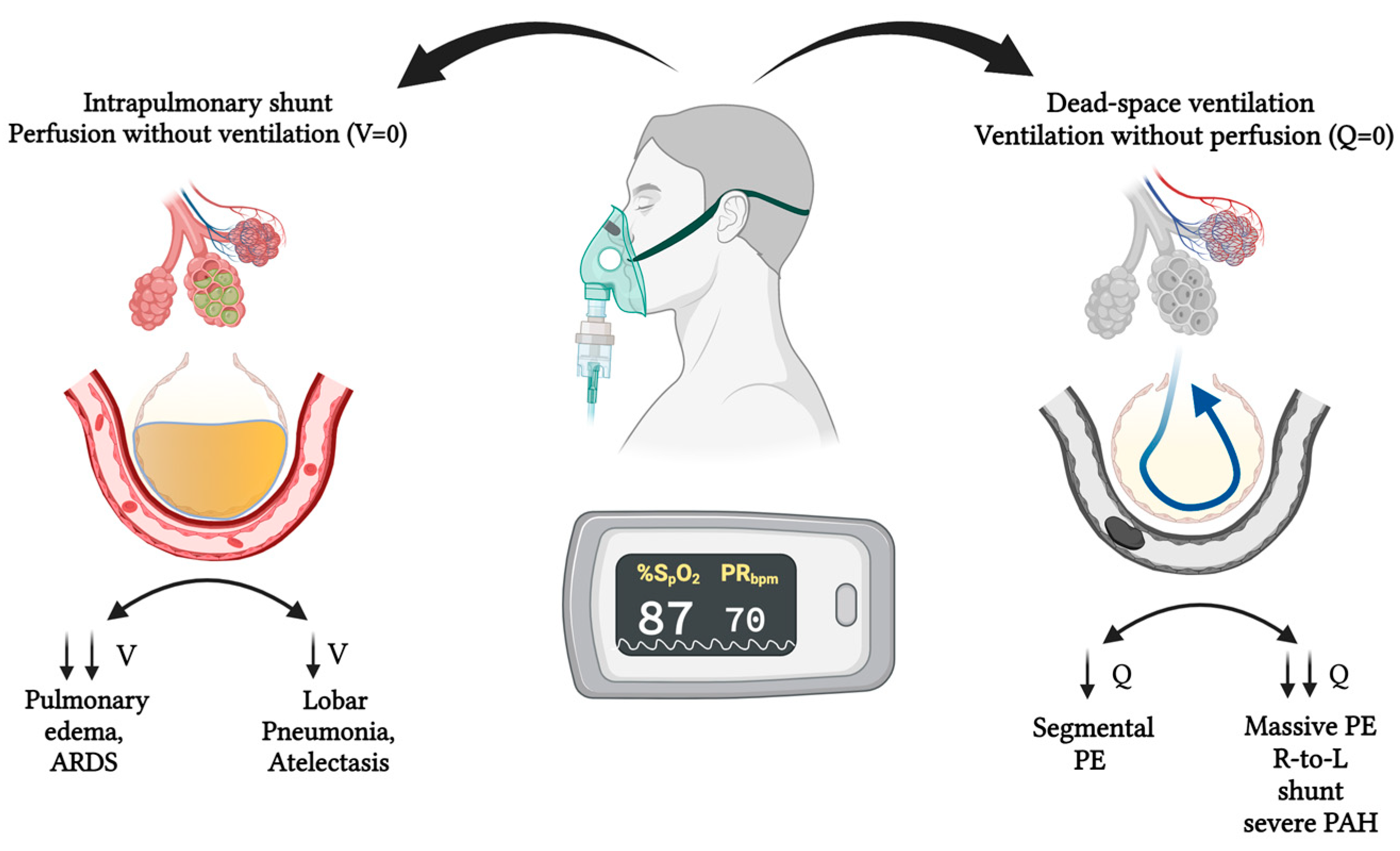
Figure 2. The pathophysiological spectrum of ventilation–perfusion (V/Q) mismatch in COVID-19. In COVID-19, multiple overlapping mechanisms contribute to V/Q mismatch. Direct viral injury and inflammatory cytokine release increase alveolar–capillary permeability, leading to interstitial and alveolar edema. Concurrent prothrombotic effects impair regional perfusion through microvascular obstruction, exacerbating hypoxemia. In both shunt-like and dead space scenarios, supplemental oxygen fails to significantly increase arterial PaO2. Moreover, high inspired oxygen concentrations accelerate nitrogen washout, enhancing alveolar collapse via resorption atelectasis and further disrupting gas exchange.
Covid 05 00139 g002
Table 1. CO-RADS classification for pulmonary CT findings.
CO-RADSDescription
0Incomplete or technically insufficient CT scan preventing evaluation.
1Very low level of suspicion: normal findings or findings clearly unrelated to infection (e.g., emphysema, fibrosis).
2Low level of suspicion: findings typical of other infectious etiologies (e.g., “tree-in-bud” pattern, lobar consolidation).
3Indeterminate level of suspicion: equivocal findings compatible with viral pneumonia or non-infectious pathology (e.g., peripheral ground-glass).
4High level of suspicion: imaging features typical of COVID-19 but with atypical distribution or overlap with other diseases (e.g., unilateral).
5Very high level of suspicion: multifocal bilateral peripheral ground-glass opacities with or without consolidation, predominantly subpleural.
CO-RADS, COVID-19 Reporting and Data System, is a standardized classification for grading the level of suspicion for pulmonary involvement by COVID-19 on chest CT. Categories range from 0 (non-diagnostic) to 5 (very high suspicion). Adapted from Prokop et al., 2020 [13].
Disclaimer/Publisher’s Note: The statements, opinions and data contained in all publications are solely those of the individual author(s) and contributor(s) and not of MDPI and/or the editor(s). MDPI and/or the editor(s) disclaim responsibility for any injury to people or property resulting from any ideas, methods, instructions or products referred to in the content.