Article

Impact of COVID-19 Pandemic on the Diagnosis and Management of Infective Endocarditis

by
Andrej Preveden
1,2,
Marina Bandulaja
1,
Vanja Drljevic Todic
1,2,
Ranko Zdravkovic
1,2,*,
Miodrag Golubovic
1,2,
Teodora Pantic
2,
Branislav Crnomarkovic
2,
Nikola Mladenovic
1,2,
Srdjan Maletin
1,2,
Milana Jarakovic
1,2,
Dragana Dabovic
1,2,
Dragica Andric
1,2,
Aleksandar Milosavljevic
1,2,
Aleksandra Mladenovic
2,
Sanja Maletin
2,
Stefan Andric
2 and
Mihaela Preveden
1,2
1
Faculty of Medicine, University of Novi Sad, 21000 Novi Sad, Serbia
2
Institute of Cardiovascular Diseases of Vojvodina, 21208 Sremska Kamenica, Serbia
*
Author to whom correspondence should be addressed.
COVID 2025, 5(8), 138; https://doi.org/10.3390/covid5080138
Submission received: 22 July 2025 / Revised: 8 August 2025 / Accepted: 18 August 2025 / Published: 20 August 2025
(This article belongs to the Section COVID Clinical Manifestations and Management)

Abstract

Background: The lockdown and other measures for coronavirus disease 2019 (COVID-19) prevention have presented an enormous burden on healthcare systems, causing reorganization in work and lowering the number of elective hospitalizations, along with a consequent reduction in hospital-acquired infections and bacteremia. The aim of this study was to examine the influence of the COVID-19 pandemic on the diagnosis and management of infective endocarditis (IE). Methods: The study included 111 patients admitted for IE from 1 January 2017 to 31 December 2021. They were divided in two groups chronologically according to the COVID-19 pandemic: the first group consisted of patients before the pandemic (n = 85), while the second group consisted of patients during the pandemic (n = 26). Results: Before the pandemic there were 2.26 ± 1.41 cases/month with diagnosis of IE, while during the pandemic there were only 1.14 ± 1.15 cases/month (p = 0.004). IE patients during the pandemic were younger (53 [41–65] vs. 68 [52–74] years; p < 0.05). A similar number of patients underwent surgical treatment before and during COVID-19 (53% vs. 67%; p = 0.210) and 30-day mortality was comparable (28% vs. 22%; p = 0.539). Conclusion: The COVID-19 pandemic and lockdown measures have led to a reduction in the number of patients with IE, although this change did not influence management strategies and mortality.

1. Introduction

Since the end of 2019, the coronavirus disease 2019 (COVID-19), first discovered in China, has spread across the world and become a global concern. It is estimated that over 200 million people got infected, 5 million of which have died [1]. The pandemic caused not only a health crisis, but also an economic, social, and demographic issue [2]. Consequences of the infection and infection-related diseases have presented an enormous burden on healthcare systems across the globe [3]. Not long after the first registered case of COVID-19 in Serbia, a state of emergency with a national lockdown was declared on 15 March 2020 [4].
Infective endocarditis (IE) represents an inflammation of the endocardium caused most commonly by bacteria (particularly Gram-positive cocci). It is a serious condition with high morbidity and mortality if left untreated, with an estimated incidence of 10 cases per 100,000 population in Western countries [5]. The main symptoms of IE include fever and dyspnea [6], although a more dramatic clinical presentation due to systemic embolism can also sometimes be seen [7]. The symptoms are frequently unspecific and can be misinterpreted as COVID-19, which has taken the spotlight for healthcare providers since the pandemic outbreak.
The lockdown and measures for COVID-19 prevention have presented an enormous burden on healthcare systems, causing reorganization in work and lowering the number of elective hospitalizations, along with a consequent reduction in hospital-acquired infections and bacteremia [8]. Social distancing and preventive measures have limited the transmission rate of bacterial infections [9]. Finally, a decline in outpatient physician visits has led to under-diagnosing of IE symptoms [10]. In contrast, COVID-19 is a systemic disease, causing cardiovascular injury and endothelial dysfunction, which can be risk factors for developing IE [11].
Given all these multiple factors associated with COVID-19 and IE epidemiology and pathophysiology, we tried to explore and enlighten their complex relationship. The aim of this study was to investigate the impact of the COVID-19 pandemic on the diagnosis, management, and outcomes of patients with IE in the territory of the Autonomous Province of Vojvodina, Serbia.

2. Materials and Methods

This was a retrospective, single center, observational study that included all consecutive adult patients (≥18 years old) who were admitted with a diagnosis of IE between 1 January 2017 and 31 December 2021. The study was conducted at the Institute of Cardiovascular Diseases Vojvodina, a tertiary care hospital serving approximately 2 million people in the Autonomous Province of Vojvodina, Serbia. Only patients with definite diagnosis of IE according to the modified Duke criteria [12] were included in the study. The patients were divided into two groups according to the date of diagnosis: (1) first group, consisting of IE patients from 1 January 2017 to 15 March 2020 (i.e., before the COVID-19 pandemic); or (2) second group, consisting of IE patients from 16 March 2020 to 31 December 2021 (i.e., during the COVID-19 pandemic).
The study was approved by the institutional ethical committee (permission no. 2537-1/6) and conducted within the principles of Good Clinical Practice and following the Declaration of Helsinki. Patient consent was waived due to retrospective study design.
The observed patient-related characteristics included demographic data, comorbidities, and risk factors for IE, such as intravenous drug use and presence of prosthetic heart valves or cardiac implantable electronic devices (pacemaker/implantable cardioverter defibrillator (ICD)/cardiac resynchronization therapy (CRT)). The data on frequency of performed transthoracic and transesophageal echocardiography studies and cardiac computed tomography (CT) were also collected and compared between the two periods. Regarding the IE presentation, the studied data included laboratory findings, localization and number of vegetations, as well as complications, including abscess, pseudoaneurysm, and prosthetic valve dehiscence. Finally, we analyzed treatment modalities (conservative or surgical), length of hospital stay, and all-cause 30-day in-hospital mortality.

Statistical Analysis

Continuous variables are expressed as mean values ± standard deviation for normally distributed data or median with interquartile range (IQR) [25th to 75th percentile] for non-normally distributed data, whereas categorical variables are presented as absolute numbers and percentages. The Kolmogorov–Smirnov test was used for the determination of quantitative data distribution. Mean values of continuous variables were compared using the independent samples t-test or Mann–Whitney U test, while categorical variables were compared using the chi-square test. Determination of the influence of the examined variables on the mortality of IE patients was performed using univariate and multivariate binary logistic regression. Statistical significance for all tests was set at the p-value of <0.05.

3. Results

The study included a total of 111 patients with confirmed diagnosis of IE. According to the two study periods, there were 85 patients before the COVID-19 pandemic (39 months in total) and 26 patients during the COVID-19 pandemic (21 months in total) (Figure 1). A significant decline in the monthly number of IE cases was observed, from 2.26 ± 1.41 cases/month before COVID-19 to 1.14 ± 1.15 cases/month during COVID-19, which represented a 50% decrease in the monthly number of cases (p = 0.004).
Baseline characteristics of patients in the two observed periods are displayed in Table 1. IE patients before COVID-19 were older than during the pandemic (68 [IQR 52–74] vs. 53 [IQR 41–65] years, p = 0.002) and there were more females (41.2% vs. 18.5%, p = 0.032), while no major differences in body mass index and comorbidities were observed.
On admission, fever was present in a similar number of cases, and there were no differences in blood laboratory results (Table 2). Notably, during the pandemic period, none of the IE patients were diagnosed with COVID-19, although each one was tested upon admission in accordance with hospital protocol.
The main imaging modality for the diagnosis of IE was transthoracic echocardiography, which was performed in every patient during both periods. The use of transesophageal echocardiography decreased by 50% during the pandemic period, from 23.3% to 12.0% of patients, although this difference was not statistically significant (p = 0.222). Cardiac CT was performed in 3.5% and 8.0% of patients (p = 0.338) before and during the pandemic, respectively.
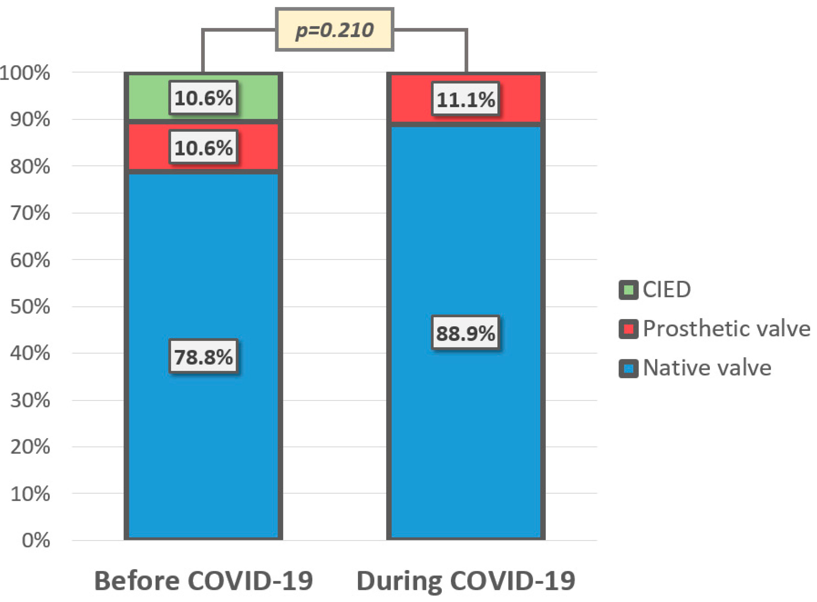
The majority of patients in both groups had endocarditis of native heart valves, with no significant difference in the affection of prosthetic valves and implantable electronic device leads (p = 0.210) (Figure 2). Interestingly, during the COVID-19 pandemic there were no IEs related to implantable electronic device leads. During the pandemic, a significantly higher proportion of patients had infection located on the aortic valve (36.0% vs. 60.0%, p = 0.032), while involvement of other heart valves remained similar, with the mitral valve being the most commonly affected in both periods (45.3% vs. 40.0%, p = 0.635). Multiple valve involvement was more frequent during the pandemic (8.1% vs. 16.0%, p = 0.247), although the difference was not statistically significant.
In the vast majority of patients in both observed periods, IE was characterized by vegetations. Other more complicated forms of IE, including abscess, pseudoaneurysm, and prosthetic valve dehiscence, were present in a similar number of cases before and during the COVID-19 pandemic (Table 2).
All IE patients were treated with antibiotic therapy, while surgical treatment was performed in 52.9% and 66.7% of patients (p = 0.210) before and during the COVID-19 pandemic, respectively (Figure 3). In patients who underwent surgery, the procedure was performed after a median of 5 [2–8] days of hospitalization before the pandemic, and 7 [4–13] days during the pandemic (p = 0.202). While hospital stay was 5 days shorter before than during the COVID-19 pandemic (p = 0.095), both study periods expressed similar 30-day in-hospital mortality (28.2% vs. 22.2%, p = 0.539).
For the whole cohort of IE patients, univariate and multivariate binary logistic regression were performed to identify predictors of 30-day in-hospital mortality (Table 3). At univariate analysis, stroke, fever, C-reactive protein (CRP) on admission, treatment modality, and length of hospital stay were significantly associated with 30-day in-hospital mortality. Multivariate analysis identified length of hospital stay and CRP on admission as predictors of 30-day in-hospital mortality in IE patients. Reducing the length of hospital stay by one day reduces the risk of mortality by 10%. Each increase of CRP at admission by one point increases the risk of mortality by 2.5%.

4. Discussion

The present study investigated the impact of the COVID-19 pandemic on the diagnosis, management, and outcomes of IE in a tertiary referral center providing care to approximately 2 million people. Our results showed that there was a significant 50% decrease in the number of newly diagnosed IE cases during the COVID-19 pandemic, while no major differences in the clinical presentation, treatment modalities, and mortality were observed.
Lockdown measures, including home isolation and social distancing, have proven to be effective in reducing the spread of the COVID-19 infection and its mortality [13], while on the other hand, the lockdown has arguably produced negative effects on various economic and social aspects on global scales [14]. Elective open-heart surgeries have also been largely reduced during the pandemic [15,16]. In contrast, IE patients usually present to the emergency department with severe symptoms, requiring surgical treatment on an emergency or urgent basis. Hence, the lockdown expectedly would not have influenced the volume of surgically treated IE patients in the same manner as other elective open-heart surgical procedures. Nonetheless, the COVID-19 pandemic has been shown to reduce the number of patients presenting with most common cardiology emergencies, including acute coronary syndrome, decompensated heart failure, hypertensive emergencies, and aortic dissection [17,18,19,20,21].
Our study demonstrated a decline in the number of IE patients during the COVID-19 pandemic. During the pandemic, our hospital canceled all elective admissions and focused exclusively on emergency cardiovascular cases during the lockdown period. Most of our staff were temporarily reassigned to COVID-19 facilities to assist with the management of COVID-19 cases at the peak of the pandemic, which meant the hospital was operating under unusual and challenging conditions. As a result, we are unable to compare the number and character of IE cases with the overall number of elective or emergency admissions between the two time periods.
Although there are conflicting data across the literature regarding the volume of IE patients, the majority of studies show lower numbers of IE cases during the pandemic [22,23,24]. According to the literature, this reduction ranged between 33–55%, which is consistent with our results. This decline in the number of cases could be either a real drop in IE incidence due to various measures for COVID-19 prevention, or a false drop because of the reduced physician consultations and restricted access to healthcare facilities for patients with mild and unspecific symptoms. Most probably, it was a more or less balanced combination of the two aforementioned aspects. Despite a decline in IE cases during the lockdown period, the most recent data indicate that the incidence of IE has risen excessively after the end of the pandemic, surpassing the levels both before and during the lockdown [25]. On the other hand, some studies reported an increase in the number of IE cases during the pandemic period [26,27], which can be explained by special attention given to patients presenting with fever or signs of infection.
There are multiple reasons that could be responsible for the drop in IE incidence during the COVID-19 pandemic, mainly by affecting the spread of pathogens due to lockdown and social distancing. During the COVID-19 pandemic, the majority of dental procedures were canceled or postponed due to lockdown measures. Given that dental procedures carry a well-recognized risk for the development of IE [12], this might have had a contribution to the decline in the incidence of IE during the pandemic. Other procedures (respiratory, gastrointestinal, urogenital), including diagnostic and elective surgical procedures, were also suspended during the pandemic. Although these are not necessarily considered to be at-risk for IE development, their reduction resulted in fewer contacts with healthcare institutions and hospitalizations, thus reducing the risk of contact with multi-resistant pathogens associated with intrahospital infections.
During the pandemic period, fear of infections was substantially increased in the general population [28], leading to massive antibiotics prescription without any additional laboratory tests and diagnostics. This wide antibiotics overuse, although undisputedly unjustified from the microbiological point of view, could have had a positive effect on bacteria suppression in population at risk for and susceptible to IE development, or even in individuals with initial and mild clinical forms of IE.
The examination with highest sensitivity for IE is transesophageal echocardiography, which is most often used for definite diagnosis, as well as management choice and monitoring of these patients [29]. Since the start of the COVID-19 pandemic, a strict restriction in the volume of transesophageal echocardiography examinations has been advocated, mainly due to the high risk of contamination with droplets and aerosol containing virus particles [30]. Consequently, a majority of patients with suspected IE during the pandemic have been examined only by transthoracic echocardiography, which is less sensitive and can potentially miss smaller and indistinct vegetations, which could be another factor contributing to the drop in the number of IE cases. In our cohort, all IE patients underwent transthoracic echocardiography, while there was a 50% drop in the use of transesophageal echocardiography during the pandemic period, which is consistent with the trends across the literature [31]. As a result, some cases of subtle or non-specific IE may have gone undiagnosed due to the reduced utilization of this important diagnostic tool.
The most common locations for endocarditis are the aortic and mitral valves, accounting for 50% and 40% of cases, respectively, according to the literature [6], which is consistent with our findings. Unexpectedly, we observed a significant increase in the number of patients with aortic valve endocarditis during the COVID-19 pandemic, rising from 36% to 60% (p = 0.032). Also, there was an increase in the number of patients with multiple valve involvement, which, along with the higher proportion of patients requiring surgical treatment, supports the hypothesis of more complex IE patients during the pandemic. Fortunately, it did not have a significant impact on the observed short-term mortality, which is consistent with the findings from other centers [32]. Notably, we did not record any cases of cardiac implantable electronic device-related endocarditis during the pandemic. This is most likely attributed to a significant reduction in the number of pacemaker and ICD implantations during the lockdown [33].
The study has a few limitations that should be acknowledged. Its retrospective design limits the ability to establish causal relationships and may be subject to selection and information biases. The study was conducted at a single center, which limits its generalizability to broader populations and different healthcare systems, particularly in the context of varying lockdown measures during the COVID-19 pandemic. Finaly, the relatively small sample size may reduce the statistical power to detect potential additional statistical differences. Future multicenter studies and meta-analyses with larger cohorts are warranted to validate and expand upon these findings.

5. Conclusions

In conclusion, the COVID-19 pandemic and associated lockdown measures led to a reduction in the number of patients with IE, although this change did not impact management strategies and short-term mortality.

Author Contributions

Conceptualization, A.P.; methodology, A.P. and M.B.; formal analysis, A.P., M.B. and R.Z.; investigation, M.B., M.G., T.P., B.C., N.M., S.M. (Srdjan Maletin), M.J., D.D., D.A., A.M. (Aleksandar Milosavljevic), A.M. (Aleksandra Mladenovic), S.M. (Sanja Maletin) and S.A.; writing—original draft preparation, A.P., M.B., R.Z., V.D.T., T.P. and M.P.; writing—review and editing, A.P.; visualization, A.P. and M.P. All authors have read and agreed to the published version of the manuscript.

Funding

This research received no external funding.

Institutional Review Board Statement

The study was conducted in accordance with the Declaration of Helsinki and approved by the Ethics Committee of the Institute of Cardiovascular Diseases Vojvodina (protocol code 2537-1/6 on 16 December 2021).

Informed Consent Statement

Patient consent was waived due to the retrospective design of the study.

Data Availability Statement

The raw data supporting the conclusions of this article will be made available by the authors on request.

Conflicts of Interest

The authors declare no conflicts of interest.

Abbreviations

The following abbreviations are used in this manuscript:
COVID-19Coronavirus disease 2019
IEInfective endocarditis
CTComputed tomography
ICDImplantable cardioverter defibrillator
CRTCardiac resynchronization therapy
CRPC-reactive protein
NT-proBNPN-terminal pro-brain natriuretic peptide
PCTProcalcitonin

References

  1. Chang, D.; Chang, X.; He, Y.; Tan, K.J.K. The determinants of COVID-19 morbidity and mortality across countries. Sci. Rep. 2022, 12, 5888. [Google Scholar] [CrossRef]
  2. Kalabikhina, I.E. Demographic and social issues of the pandemic. Popul. Econ. 2020, 4, 103–122. [Google Scholar] [CrossRef]
  3. Menges, D.; Ballouz, T.; Anagnostopoulos, A.; Aschmann, H.E.; Domenghino, A.; Fehr, J.S.; Puhan, M.A. Burden of post-COVID-19 syndrome and implications for healthcare service planning: A population-based cohort study. PLoS ONE 2021, 16, e0254523. [Google Scholar] [CrossRef]
  4. The Government of the Republic of Serbia. State of Emergency Declared Throughout Serbia. Published 15 March 2020. Available online: https://www.srbija.gov.rs/vest/en/151398/state-of-emergency-declared-throughout-serbia.php (accessed on 26 March 2023).
  5. Williams, M.L.; Doyle, M.P.; McNamara, N.; Tardo, D.; Mathew, M.; Robinson, B. Epidemiology of infective endocarditis before versus after change of international guidelines: A systematic review. Ther. Adv. Cardiovasc. Dis. 2021, 15, 17539447211002688. [Google Scholar] [CrossRef]
  6. Habib, G.; Erba, P.A.; Iung, B.; Donal, E.; Cosyns, B.; Laroche, C.; Popescu, B.A.; Prendergast, B.; Tornos, P.; Sadeghpour, A.; et al. Clinical presentation, aetiology and outcome of infective endocarditis. Results of the ESC-EORP EURO-ENDO (European infective endocarditis) registry: A prospective cohort study. Eur. Heart J. 2019, 40, 3222–3232. [Google Scholar] [CrossRef]
  7. Redzek, A.; Preveden, A.; Kaloci, S.R.; Samardzija, G.; Preveden, M.; Golubovic, M.; Velicki, L. Unusual non-valvular left ventricular endocarditis presenting as multiple brain embolism. Acta Clin. Belg. 2022, 77, 142–146. [Google Scholar] [CrossRef]
  8. Havers-Borgersen, E.; Fosbøl, E.L.; Butt, J.H.; Petersen, J.K.; Dalsgaard, A.; Kyhl, F.; Schou, M.; Phelps, M.; Kragholm, K.; Gislason, G.H.; et al. Incidence of infective endocarditis during the coronavirus disease 2019 pandemic: A nationwide study. IJC Heart Vasc. 2020, 31, 100675. [Google Scholar] [CrossRef] [PubMed]
  9. Feldman, I.; Natsheh, A.; Nesher, G.; Breuer, G.S. Social distancing and bacteraemia in the time of COVID-19. Intern. Med. J. 2022, 52, 223–227. [Google Scholar] [CrossRef] [PubMed]
  10. Kumagai, N. The Impact of the COVID-19 Pandemic on Physician Visits in Japan. Front. Public Health 2021, 9, 743371. [Google Scholar] [CrossRef] [PubMed]
  11. Arbune, M.; Iancu, A.V.; Lupasteanu, G.; Vasile, M.C.; Stefanescu, V. A Challenge of COVID-19: Associated Infective Endocarditis with Streptococcus gordonii in a Young Immunocompetent Patient. Medicine 2021, 57, 1298. [Google Scholar] [CrossRef]
  12. Habib, G.; Lancellotti, P.; Antunes, M.J.; Bongiorni, M.G.; Casalta, J.P.; Del Zotti, F.; Dulgheru, R.; El Khoury, G.; Erba, P.A.; Iung, B.; et al. 2015 ESC Guidelines for the management of infective endocarditis: The Task Force for the Management of Infective Endocarditis of the European Society of Cardiology (ESC)Endorsed by: European Association for Cardio-Thoracic Surgery (EACTS), the European Association of Nuclear Medicine (EANM). Eur. Heart J. 2015, 36, 3075–3128. [Google Scholar]
  13. Ghosal, S.; Bhattacharyya, R.; Majumder, M. Impact of complete lockdown on total infection and death rates: A hierarchical cluster analysis. Diabetes Metab. Syndr. 2020, 14, 707–711. [Google Scholar] [CrossRef] [PubMed]
  14. Nicola, M.; Alsafi, Z.; Sohrabi, C.; Kerwan, A.; Al-Jabir, A.; Iosifidis, C.; Agha, M.; Agha, R. The socio-economic implications of the coronavirus pandemic (COVID-19): A review. Int. J. Surg. 2020, 78, 185–193. [Google Scholar] [CrossRef] [PubMed]
  15. Gopal, K.; Krishna, N.; Jose, R.; Biju, S.S.C.; Pichandi, J.S.; Varma, P.K. Effects of the COVID-19 Pandemic on Cardiac Surgery Practice and Outcomes. J. Chest Surg. 2022, 55, 61–68. [Google Scholar] [CrossRef]
  16. COVIDSurg Collaborative. Elective surgery cancellations due to the COVID-19 pandemic: Global predictive modelling to inform surgical recovery plans. Br. J. Surg. 2020, 107, 1440–1449. [Google Scholar] [CrossRef]
  17. El–Hamamsy, I.; Brinster, D.R.; DeRose, J.J.; Girardi, L.N.; Hisamoto, K.; Imam, M.N.; Itagaki, S.; Kurlansky, P.A.; Lau, C.; Nemeth, S.; et al. The COVID-19 Pandemic and Acute Aortic Dissections in New York. J. Am. Coll. Cardiol. 2020, 76, 227–229. [Google Scholar] [CrossRef] [PubMed]
  18. Mesnier, J.; Cottin, Y.; Coste, P.; Ferrari, E.; Schiele, F.; Lemesle, G.; Thuaire, C.; Angoulvant, D.; Cayla, G.; Bouleti, C.; et al. Hospital admissions for acute myocardial infarction before and after lockdown according to regional prevalence of COVID-19 and patient profile in France: A registry study. Lancet Public Health 2020, 5, e536–e542. [Google Scholar] [CrossRef]
  19. Milovančev, A.; Petrović, M.; Popadić, V.; Miljković, T.; Klašnja, S.; Djuran, P.; Ilić, A.; Kovačević, M.; Stojšić Milosavljević, A.; Brajković, M.; et al. Characteristics and Outcomes of Patients with Acute Coronary Syndrome and COVID-19. J. Clin. Med. 2022, 11, 1791. [Google Scholar] [CrossRef]
  20. Hall, M.E.; Vaduganathan, M.; Khan, M.S.; Papadimitriou, L.; Long, R.C.; Hernandez, G.A.; Moore, C.K.; Lennep, B.W.; Mcmullan, M.R.; Butler, J. Reductions in Heart Failure Hospitalizations During the COVID-19 Pandemic. J. Card. Fail. 2020, 26, 462–463. [Google Scholar] [CrossRef]
  21. Hartnett, K.P.; Kite-Powell, A.; DeVies, J.; Coletta, M.A.; Boehmer, T.K.; Adjemian, J.; Gundlapalli, A.V.; National Syndromic Surveillance Program Community of Practice. Impact of the COVID-19 Pandemic on Emergency Department Visits—United States, January 1, 2019–May 30, 2020. MMWR Morb. Mortal. Wkly. Rep. 2020, 69, 699–704. [Google Scholar] [CrossRef]
  22. Escolà-Vergé, L.; Cuervo, G.; de Alarcón, A.; Sousa, D.; Barca, L.V.; Fernández-Hidalgo, N. Impact of the COVID-19 pandemic on the diagnosis, management and prognosis of infective endocarditis. Clin. Microbiol. Infect. 2021, 27, 660–664. [Google Scholar] [CrossRef]
  23. Cosyns, B.; Motoc, A.; Arregle, F.; Habib, G. A Plea Not to Forget Infective Endocarditis in COVID-19 Era. JACC Cardiovasc. Imaging 2020, 13, 2470–2471. [Google Scholar] [CrossRef]
  24. Van Camp, G.; De Beenhouwer, H.; Beles, M.; Collet, C.; Nasser, R.; Schelfaut, D.; Penicka, M. Disturbing effect of lockdown for COVID-19 on the incidence of infective endocarditis: A word of caution. Clin. Res. Cardiol. 2020, 109, 1573–1576. [Google Scholar] [CrossRef]
  25. Pommier, T.; Benzenine, E.; Bernard, C.; Mariet, A.S.; Béjot, Y.; Giroud, M.; Morgant, M.C.; Steinmetz, E.; Guenancia, C.; Bouchot, O.; et al. Trends of Myocarditis and Endocarditis Cases Before, During, and After the First Complete COVID-19-Related Lockdown in 2020 in France. Biomedicines 2022, 10, 1231. [Google Scholar] [CrossRef] [PubMed]
  26. Morawiec, R.; Matuszewska-Brycht, O.; Maeser, P.; Kośny, M.; Krejca, M.; Drożdż, J. Decreasing numbers of valve-related infective endocarditis cases. An urgent call to action to improve diagnostic pathways: A retrospective tertiary center perspective (2015–2022). Kardiol. Pol. 2024, 82, 79–81. [Google Scholar] [CrossRef] [PubMed]
  27. Cabral, M.; Fernandes, S.; Santos, L.G.; Carvalho, R.; Sa, F.M.; Martins, H.; Pernencar, S.; Ruivo, C.; Santos, B.; Morais, J. An outbreak of infective endocarditis during the COVID-19 pandemic?—An observational retrospective single centre study. Eur. Heart J. Acute Cardiovasc. Care 2021, 10 (Suppl. 1), zuab020.184. [Google Scholar] [CrossRef]
  28. Spatafora, F.; Matos Fialho, P.M.; Busse, H.; Helmer, S.M.; Zeeb, H.; Stock, C.; Wendt, C.; Pischke, C.R. Fear of Infection and Depressive Symptoms among German University Students during the COVID-19 Pandemic: Results of COVID-19 International Student Well-Being Study. Int. J. Environ. Res. Public Health 2022, 19, 1659. [Google Scholar] [CrossRef]
  29. Habib, G.; Badano, L.; Tribouilloy, C.; Vilacosta, I.; Zamorano, J.L.; Galderisi, M. Recommendations for the practice of echocardiography in infective endocarditis. Eur. J. Echocardiogr. 2010, 11, 202–219. [Google Scholar] [CrossRef]
  30. Skulstad, H.; Cosyns, B.; Popescu, B.A.; Galderisi, M.; Salvo, G.D.; Donal, E.; Petersen, S.; Gimelli, A.; Haugaa, K.H.; Muraru, D.; et al. COVID-19 pandemic and cardiac imaging: EACVI recommendations on precautions, indications, prioritization, and protection for patients and healthcare personnel. Eur. Heart J. Cardiovasc. Imaging 2020, 21, 592–598. [Google Scholar] [CrossRef]
  31. Pang, L.; Stahl, E.P.; Fujikura, K.; Chen, M.; Li, W.; Zhang, M.; Levsky, J.M.; Travin, M.I.; Ho, E.C.; Goldberg, Y.; et al. Echocardiography Abnormal Findings and Laboratory Operations during the COVID-19 Pandemic at a High Volume Center in New York City. Healthcare 2020, 8, 534. [Google Scholar] [CrossRef]
  32. Mikus, E.; Fiorentino, M.; Sangiorgi, D.; Fiaschini, C.; Tenti, E.; Tremoli, E.; Calvi, S.; Costantino, A.; Tripodi, A.; Zucchetta, F.; et al. Surgical Treatment of Active Endocarditis Pre- and Post-COVID-19 Pandemic Onset. Biomedicines 2024, 12, 233. [Google Scholar] [CrossRef] [PubMed]
  33. Zecchin, M.; Ciminello, E.; Mari, V.; Proclemer, A.; D’Onofrio, A.; Zanotto, G.; De Ponti, R.; Capovilla, T.M.; Laricchiuta, P.; Biondi, A.; et al. A global analysis of implants and replacements of pacemakers and cardioverter-defibrillators before, during, and after the COVID-19 pandemic in Italy. Intern. Emerg. Med. 2024, 19, 107–114. [Google Scholar] [CrossRef] [PubMed]
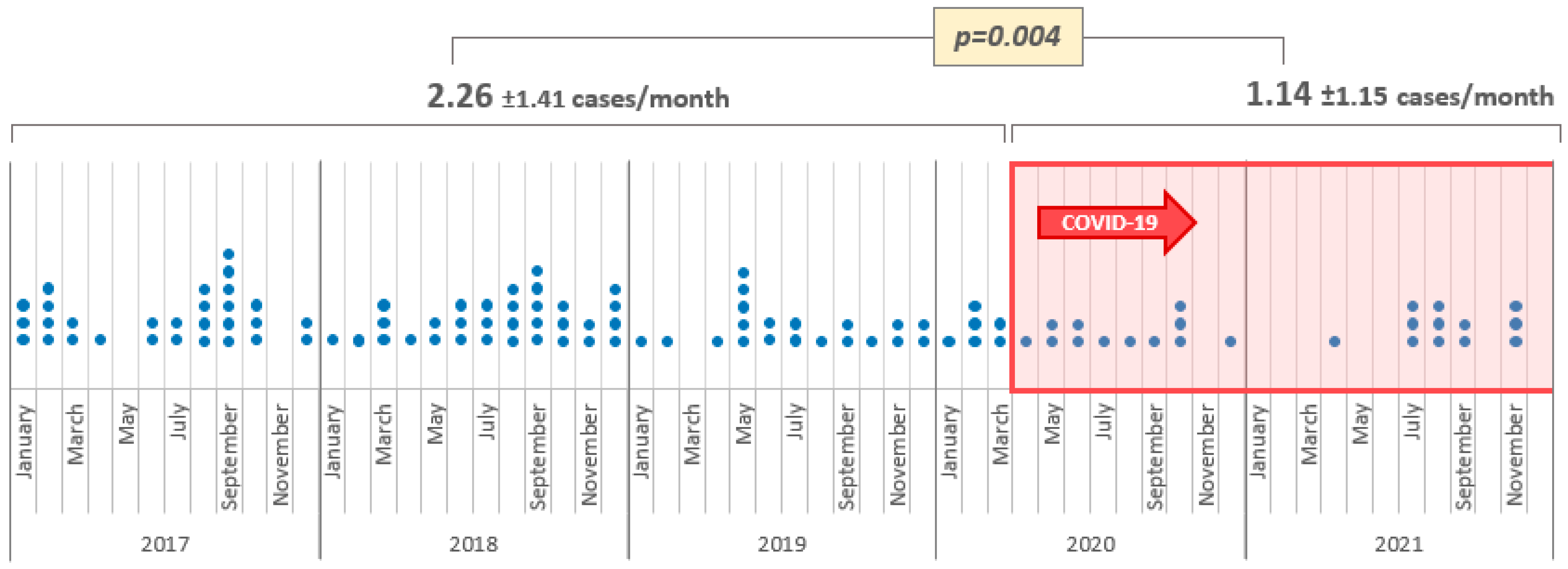
Figure 1. Monthly number of IE cases before and during the COVID-19 pandemic.
Covid 05 00138 g001
Figure 2. Type of endocarditis before and during the COVID-19 pandemic. (Abbreviations: CIED—cardiac implantable electronic devices.).
Covid 05 00138 g002
Figure 3. Treatment modalities of IE patients before and during the COVID-19 pandemic.
Covid 05 00138 g003
Table 1. General characteristics of patients diagnosed with IE before and during the COVID-19 pandemic.
Overall
n = 111
Before COVID-19
n = 85
During COVID-19
n = 26
p-Value
Females, n (%)40 (35.7%)35 (41.2%)5 (18.5%)0.032
Age (years)65 [46–65]68 [52–74]53 [41–65]0.002
Body mass index (kg/m2)25.3 [23.7–27.8]24.9 [23.4–27.9]25.7 [24.2–27.7]0.373
Comorbidities
    Hypertension, n (%)65 (58.0%)53 (62.4%)12 (44.4%)0.100
    Prior MI, n (%)9 (8.0%)8 (9.4%)1 (3.7%)0.342
    Heart failure, n (%)24 (21.4%)15 (17.6%)9 (33.3%)0.084
    Stroke, n (%)16 (14.3%)13 (15.3%)3 (11.1%)0.588
    COPD, n (%)7 (6.2%)5 (5.9%)2 (7.4%)0.775
    Diabetes mellitus, n (%)28 (25.0%)23 (27.1%)5 (18.5%)0.372
    Renal failure, n (%)25 (22.3%)18 (21.2%)7 (25.9%)0.606
Intravenous drug use, n (%)6 (5.4%)3 (3.5%)3 (11.1%)0.127
Prosthetic heart valve, n (%)12 (10.7%)9 (10.6%)3 (11.1%)0.591
Pacemaker/ICD/CRT, n (%)9 (8.0%)9 (10.6%)00.075
Abbreviations: COPD—chronic obstructive pulmonary disease; CRT—cardiac resynchronization therapy; ICD—implantable cardioverter defibrillator; MI—myocardial infarction.
Table 2. Clinical findings of patients diagnosed with IE before and during the COVID-19 pandemic.
Overall
n = 111
Before COVID-19
n = 85
During COVID-19
n = 26
p-Value
Fever, n (%)21 (19.1%)18 (21.7%)3 (11.1%)0.225
Laboratory
    CRP (mg/L)59.1 [25.5–98.5]59.9 [27.3–95.9]53.5 [19.6–101.4]0.568
    PCT (µg/L)0.28 [0.09–0.79]0.24 [0.09–0.96]0.30 [0.20–0.51]0.671
    NT-proBNP (ng/L)13,141 [2315–25,000]9023 [2219–25,000]17,664 [3695–25,000]0.096
Performed imaging studies
    TTE, n (%)111 (100%)85 (100%)26 (100%)1.000
    TEE, n (%)23 (20.7%)20 (23.3%)3 (12.0%)0.222
    Cardiac CT, n (%)5 (4.5%)3 (3.5%)2 (8.0%)0.338
Endocarditis finding
    Vegetation, n (%)107 (97.3%)83 (97.6%)24 (96.0%)0.657
    Abscess, n (%)12 (10.8%)9 (10.5%)3 (12.0%)0.828
    Pseudoaneurysm, n (%)1 (0.9%)0 (0.0%)1 (4.0%)0.062
    Prosthetic valve dehiscence, n (%)4 (3.6%)3 (3.5%)1 (4.0%)0.904
Affected valve
    Aortic, n (%)46 (41.4%)31 (36.0%)15 (60.0%)0.032
    Mitral, n (%)49 (44.1%)39 (45.3%)10 (40.0%)0.635
    Tricuspid, n (%)14 (12.6%)10 (11.6%)4 (16.0%)0.562
    Pulmonary, n (%)3 (2.7%)3 (3.5%)0 (0.0%)0.344
    Multiple valves, n (%)11 (9.9%)7 (8.1%)4 (16.0%)0.247
Treatment modality
    Conservative treatment, n (%)49 (43.8%)40 (47.1%)9 (33.3%)0.210
    Surgery, n (%)63 (56.2%)45 (52.9%)18 (66.7%)
Time until surgery (days)5 [3–8]5 [2–8]7 [4–13]0.202
Hospital stay (days)18 [12–24]16 [12–24]21 [15–27]0.095
30-day all-cause mortality, n (%)30 (26.8%)24 (28.2%)6 (22.2%)0.539
Abbreviations: CRP—C-reactive protein; CT—computed tomography; NT-proBNP—N-terminal pro-brain natriuretic peptide; PCT—procalcitonin; TEE—transesophageal echocardiography; TTE—transthoracic echocardiography.
Table 3. Univariate and multivariate analysis of predictors of in-hospital mortality.
UnivariateMultivariate
OR (95% CI)p ValueOR (95% CI)p Value
Stroke3.364 (1.132–9.999)0.029-ns
Fever3.136 (1.165–8.445)0.024-ns
Treatment modality3.467 (1.435–8.376)0.006-ns
Hospital stay0.911 (0.862–0.963)0.0010.907 (0.852–0.965)0.002
CRP1.021 (1.011–1.031)<0.00051.025 (1.013–1.036)<0.0005
Abbreviations: CI—confidence interval; CRP—C-reactive protein; ns—non significant; OR—odds ratio.
Disclaimer/Publisher’s Note: The statements, opinions and data contained in all publications are solely those of the individual author(s) and contributor(s) and not of MDPI and/or the editor(s). MDPI and/or the editor(s) disclaim responsibility for any injury to people or property resulting from any ideas, methods, instructions or products referred to in the content.