Case Report

SARS-CoV-2 and HCoV IgG Antibodies in the Breast Milk of a Postpartum SARS-CoV-2 Patient Following Bamlanivimab Administration: A Case Report

1
Pathology and Laboratory Medicine, University of British Columbia, Vancouver, BC V6T 2B5, Canada
2
Surrey Memorial Hospital Clinical Research Unit, Surrey, BC V3V 1Z2, Canada
3
Fraser Health, Surrey, BC V3T 0H1, Canada
4
Public Health Laboratories, British Columbia Centre for Disease Control, Vancouver, BC V5Z 4R4, Canada
5
Department of Medicine, University of British Columbia, Vancouver, BC V5Z 1M9, Canada
6
Department of Obstetrics and Gynecology, University of British Columbia, Vancouver, BC V6T 1Z3, Canada
7
Women’s Health Research Institute, Vancouver, BC V6H 3V4, Canada
*
Author to whom correspondence should be addressed.
COVID 2025, 5(8), 123; https://doi.org/10.3390/covid5080123
Submission received: 1 July 2025 / Revised: 26 July 2025 / Accepted: 30 July 2025 / Published: 1 August 2025
(This article belongs to the Section COVID Clinical Manifestations and Management)

Abstract

Breast milk can provide passive immunity to infants, serving as a valuable source of maternal antibodies while remaining a non-invasive sample for investigating maternal immune responses. To date, no studies have evaluated SARS-CoV-2 and potentially cross-reactive HCoV antibodies in breast milk following bamlanivimab administration. A 36-year-old postpartum female was PCR-positive for SARS-CoV-2 four days post-delivery. Bamlanivimab was administered intravenously two days later. Breast milk was collected before bamlanivimab infusion, daily for two weeks post-infusion, then weekly until 102 days post-infusion. Mother and infant sera were collected only at 102 days post-infusion. All milk and serum samples were tested for IgG antibodies against SARS-CoV-2 and HCoV. We observed two distinct SARS-CoV-2 antibody peaks at days 3 and 29 post-infusion, likely representing bamlanivimab transfer and the post-infection antibody response. Beta-HCoV antibodies showed two peaks at days 6 and 29, potentially representing backboosted beta-HCoV responses and/or antibody cross-reactivity with SARS-CoV-2. Infant seropositivity for SARS-CoV-2 102 days post-infusion may represent antibodies from passive transfer via breastfeeding or a subclinical infection. This case highlights the value of breast milk as a non-invasive and repeatable sample to help understand maternal immune responses post-infection, exogenous antibody infusion, and passive antibody transfer during breastfeeding, which can provide insights into maternal–infant health research.

1. Introduction

In addition to providing infant nutrients, breast milk serves as a source of passive immunity for infants during a time when the infant’s immune system remains immature in early life [1]. Beyond passive immunity, breast milk is a promising non-invasive sample type that can provide a window into the maternal immune history [2]; which can be used for surveillance and immune profiling studies.
Studies have demonstrated severe acute respiratory syndrome coronavirus 2 (SARS-CoV-2) antibodies in breast milk following SARS-CoV-2 infection and vaccination [3,4,5,6], along with a variety of immune cells and cytokines [7,8], highlighting the complex immunological profiles within human milk following exposure to SARS-CoV-2. However, assessment of SARS-CoV-2 antibodies in breast milk in the context of monoclonal antibody (mAb) therapy has rarely been described [9]. Bamlanivimab was a commercially available neutralizing immunoglobulin G (IgG) mAb that binds to the receptor-binding domain (RBD) within the spike protein of SARS-CoV-2, preventing viral attachment to the human angiotensin converting enzyme 2 receptor [10]. Bamlanivimab does not elicit a response against nucleocapsid; thus, SARS-CoV-2 nucleocapsid antibodies are expected to follow the normal course of an immune response to infection. As such, detection of antibodies against RBD and spike in breast milk, but not nucleocapsid, may indicate mAb derived from bamlanivimab therapy. Bamlanivimab was previously authorized for use in severe COVID-19, but while no longer authorized as of July 2021 due to its lack of efficacy against COVID-19 variants [11], understanding whether these mAbs can be identified in breast milk can provide valuable insights into the use of breast milk samples in understanding maternal–infant immunity in the context of mAb therapy.
Beyond SARS-CoV-2, endemic human coronaviruses (HCoVs) of alpha (HCoV-229E and HCoV-NL63) and beta (HCoV-HKU1 and HCoV-OC43) lineages circulate widely [12,13], with population antibody levels in serum remaining high throughout a lifetime due to repeated boosting from seasonal exposures [10,12]. Several studies have reported antibody cross-reactivity between HCoV and SARS-CoV-2 in the blood [14,15,16,17,18], though evidence of such cross-reactivity in breast milk remains scarce [19,20]. Our case is the first to evaluate HCoV responses in breast milk following administration of a SARS-CoV-2 mAb, highlighting its value as a non-invasive and easy to obtain sample for investigating maternal immune profiles and interactions beyond recent exposures for maternal–infant health research and surveillance.
Here, we report a novel case featuring the longitudinal detection of SARS-CoV-2 and HCoV IgG antibodies in the breast milk of a SARS-CoV-2-infected postpartum patient following clinical bamlanivimab treatment. Furthermore, we report detection of SARS-CoV-2 antibodies in infant serum after 102 days post-infusion.

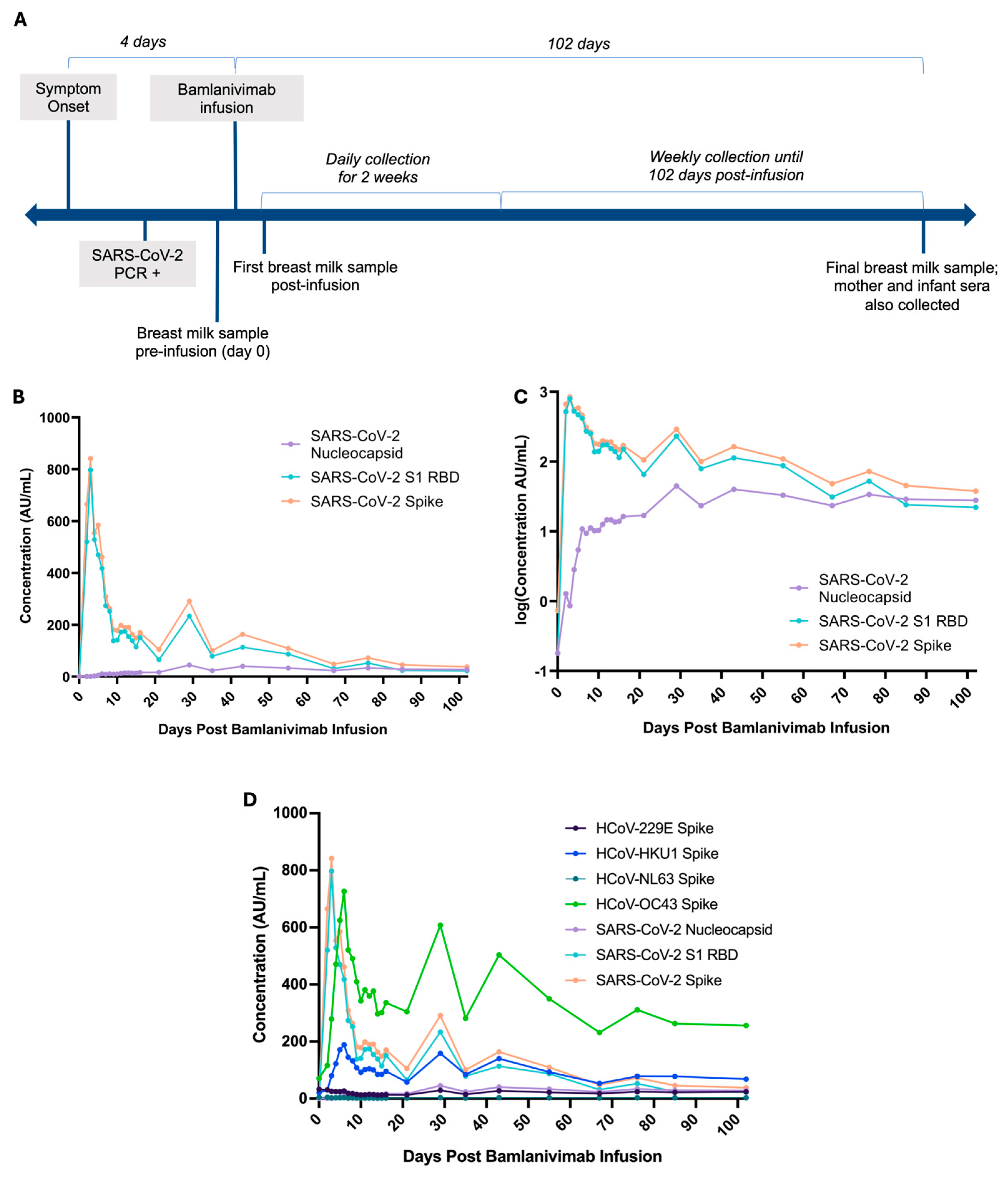
2. Detailed Case Description

A 36-year-old female, early postpartum and unvaccinated for SARS-CoV-2, presented with a moderate cough, shortness of breath, mild loss of appetite, severe loss of taste and smell, and fatigue. In May 2021, she tested positive by PCR for SARS-CoV-2 infection shortly after two days following symptom onset, when her infant was 1-week-old. Two days following PCR-confirmation of a SARS-CoV-2 infection, bamlanivimab was administered intravenously (700 mg/20 mL) as a single infusion (Figure 1A). Written consent was provided by the patient for the collection of breast milk samples. A breast milk sample was collected immediately prior to bamlanivimab infusion (day 0). Breast milk samples were then collected daily until 15 days post-infusion, when the patient was switched to a weekly collection schedule until 102 days post-infusion (Figure 1A). A total of 25 breast milk samples were collected. Both mother and infant sera were collected only at 102 days post-infusion. No sera were collected prior to bamlanivimab infusion.
All breast milk samples were self-collected and kept at −20 °C until testing. No specific instructions for self-collection were provided to the patient, and approximately 50 mL was provided at each time point. Testing was carried out in the fall of 2021, shortly after the collection of all samples. Breast milk samples were thawed at 37 °C, and 1 mL was centrifuged at 1300 RCF for 20 min to separate the opaque cream (top) and aqueous (bottom) layers. The top layer was subsequently discarded, and the remaining aqueous layer was used for antibody testing. Processing methods yielded approximately 50% of IgG recovery, which had been demonstrated in validation studies testing breast milk spiked with known amounts of IgG.
Processed breast milk and serum IgG antibodies against the spike, RBD, and nucleocapsid regions of SARS-CoV-2, along with the spike protein of four HCoVs (HCoV-229E, HCoV-NL63, HCoV-HKU1, and HCoV-OC43), were quantified using the Meso Scale Discovery (MSD) V-PLEX Coronavirus Panel 2 (Rockville, MD, USA), an electrochemiluminescent multiplex immunoassay that has been previously validated and demonstrated for research use on milk and serum samples [21,22]. Briefly, antigens were printed on the bottom of a multi-spot 96 well plate from MSD, and the assay was performed according to the manufacturer’s instructions. Following plate blocking, processed milk (diluted 1:100) and serum (diluted at 1:5000) specimens were added and incubated for two hours. Bound antibodies were then labelled with SULFO-TAG™ Anti-human IgG for an hour. MSD Gold Read buffer was added after a final wash, and plates were immediately read using the MSD QuickPlex SQ120. All incubation steps were carried out shaking at room temperature (20–26 °C), and all washes were performed using the MSD Wash Buffer. Raw data was imported into the MSD Discovery Workbench software for data processing. Figures were prepared on GraphPad Prism 10.
Given the mother’s infection pattern, we expected to see the natural antibody response for SARS-CoV-2 peak at around 21–49 days post symptom onset, as previous studies have described [23,24]. Here, we observed two distinct peaks in SARS-CoV-2 anti-spike and anti-RBD IgG levels were observed at days 3 and 29 post-infusion. The first peak in anti-spike and anti-RBD IgG, but not anti-nucleocapsid, occurred within 3 days post-infusion of bamlanivimab (7 days post-symptom onset) followed by a rapid decline (Figure 1B), suggesting the transfer of bamlanivimab-derived IgG to breast milk. The second peak in anti-spike and anti-RBD IgG occurred 29 days post-infusion (33 days post-symptom onset), which coincided with the single peak in SARS-CoV-2 anti-nucleocapsid IgG antibody levels (Figure 1B,C). Anti-nucleocapsid IgG exhibited a gradual increase (Figure 1C), reached peak levels at 29 days post-infusion, and remained stable thereafter. IgG antibodies to SARS-CoV-2 spike, nucleocapsid, and RBD all persisted in breast milk by day 102 post-infusion and were higher than baseline pre-infusion antibody levels as expected (Table 1). Additionally, both mother and infant sera at 102 days post-infusion were seropositive for IgG against all three SARS-CoV-2 proteins tested (Table 1); however, we did not have pre-infusion sera for either mother or infant to determine seroconversion prior to bamlanivimab infusion. Furthermore, the infant’s infection status remains unknown, as the infant was never symptomatic and had not been tested for SARS-CoV-2 by PCR; thus, we are unable to rule out a possible subclinical infection in the infant.
Breast milk IgG antibodies for beta-HCoV (HCoV-HKU1 and HCoV-OC43) were both 3X higher at day 102 post-infusion in comparison to day 0 pre-infusion levels (Table 1, Figure 1D). In contrast, alpha-lineage (HCoV-229E and HCoV-NL63) IgG levels in breast milk remained similar between day 0 and day 102 post-infusion (Table 1, Figure 1D). Beta-HCoV IgG levels also exhibited two significant peaks: the first peak 6 days post-infusion (10 days post-symptom onset), then a second peak at 29 days post-infusion (33 days post-symptom onset) (Figure 1D). The first beta-HCoV IgG peak at day 6 post-infusion did not overlap with any of the SARS-CoV-2 antibody peaks, while the latter beta-HCoV IgG peak at day 29 post-infusion coincided with the singular peak in SARS-CoV-2 nucleocapsid IgG levels and the second peak in SARS-CoV-2 anti-spike and anti-RBD. Both beta-HCoV IgG in breast milk followed a stable and gradual decline thereafter until 102 days post-infusion, exhibiting a pattern similar to the SARS-CoV-2 antibodies.

3. Discussion

In this case report of a postpartum patient with SARS-CoV-2 following bamlanvimab administration, we detected two distinct peaks in anti-spike and anti-RBD IgG against SARS-CoV-2 in breast milk, with the earlier peak likely due to bamlanivimab administration, and the later peak likely representing the natural immune response in an unvaccinated individual. We also found elevated beta-HCoV antibodies in breast milk over 102 days post-infection, likely representing a backboost of memory beta-HCoV responses and/or cross-reactive antibody responses to SARS-CoV-2. Finally, we detected antibodies against SARS-CoV-2 in infant serum at 102 days post-infection.
The two distinct peaks in anti-spike and anti-RBD IgG against SARS-CoV-2 at days 3 and 29 post-infusion (days 7 and 33 post-symptom onset) likely represented both the impact of the mAb infusion and the mother’s immune response, respectively. Since bamlanivimab is an IgG mAb that targets the RBD within the spike region, but not nucleocapsid, the first peak we see at day 3 post-infusion for anti-spike and anti-RBD IgG antibodies, but not anti-nucleocapsid, likely represents the transfer of bamlanivimab to breast milk. This also aligns with the rapid action and short-lived nature of IgG mAb, such as bamlanivimab, which was previously reported to possess a half-life of 20.9 days [24]. In contrast to the first peak in anti-spike and anti-RBD IgG against SARS-CoV-2, the second peaks for both antibodies overlapped with the single peak in anti-nucleocapsid IgG levels at day 29 post-infusion (day 33 post-infection), likely representing the natural antibody response to SARS-CoV-2 infection, in addition to any residual mAb responses. This is consistent with the expected timeline for SARS-CoV-2 IgG response during a primary immune response, which typically arises around days 7–14 post-infection, reaching peak levels between 21 and 49 days post symptom onset [23,25]. While our methods do not conclusively differentiate between endogenous IgG from the mother’s natural immune response or exogenously administered bamlanivinab, we observed a sharp decline following the initial spike in antibodies after day 3, likely represented by declining bamlanivimab. As such, antibodies following this likely represents both endogenous and exogenous antibodies, though the relative level of bamlanivimab is likely much lower compared to that of the natural antibody response by the second peak. To date, few studies have described breast milk IgG to mAb therapy in SARS-CoV-2, and while one other case reported on breast milk anti-RBD IgG following administration of casirivimab–imdevimab [7], there is currently no published data on breast milk antibody responses in the context of bamlanivimab. Our findings demonstrate the significance of using breast milk as a non-invasive and easy to collect sample to monitor mAb and other antibody responses in mothers and passive transfer to infants.
We could not rule out the possibility that bamlanivimab administration can modulate the natural antibody response in breast milk. Previous studies have described that treatment with SARS-CoV-2 anti-spike IgG mAb can influence the endogenous serological response, following administration of casirivimab-imdevimab, bamlanivimab, and bamlanivmab–estevimab in human and mice models [26,27]. Specifically, neutralizing and binding antibody profiles post-infusion differed based on an individual’s serostatus at the time of mAb administration, where mAb infusion of SARS-CoV-2 seronegative individuals demonstrated a significantly blunted endogenous antibody response to spike and RBD, with trends of lower nucleocapsid antibody levels, compared to mAb infusion of seropositive individuals. This blunting phenomenon after passive mAb administration has been attributed to a weaker induction of the natural immune response due to faster viral clearance and less viral replication [26]. Currently, most studies investigating mAb-associated blunting of the endogenous antibody response have been limited to human serum and mice studies, thus, the influence of mAb treatment on breast milk antibody responses is not well-studied. However, breast milk mAb have been previously shown to reflect a 1:20 ratio relative to serum [28]; therefore, future studies can explore how mAb treatments can affect the natural antibody responses in breast milk to further understand how these can influence protection in breastfeeding infants.
We also observed two distinct peaks in IgG levels against the spike protein of beta-HCoV (HCoV-HKU1 and HCoV-OC43) at days 6 and 29 post-infusion, with breast milk IgG levels at day 102 post-infusion 3X higher than pre-infusion IgG levels. The first peak in beta-HCoV IgG at day 6 exhibited distinct patterns of antibody elevation and decline from the SARS-CoV-2 specific peak at day 3, thus, this lack of an overlap suggests that the observed response was not due to cross-reactivity from bamlanivimab, but rather, from a rapid and transient back-boost of HCoV-HKU1 and HCoV-OC43 spike antibodies from pre-existing immune memory. The rapid and transient peaks we observed for HCoV-HKU1 and HCoV-OC43 IgG at day 6 post-infusion were also consistent with previous findings of IgG back-boosting for HKU1, specifically against the highly conserved S2 region within the Spike protein [14]. On the other hand, the second beta-HCoV peak at day 29 post-infusion overlapped with the peak in SARS-CoV-2 antibodies, suggesting potential beta-HCoV antibody cross-reactivity with SARS-CoV-2. While we cannot clearly differentiate between either back-boosted response and/or cross-reactivity, our findings of two distinct peaks suggest that both back-boosting and cross-reactive mechanisms are not mutually exclusive and both may possibly be at play. Additionally, the serological response is also dependent on the timing of sample collection post-mAb infusion and post-infection. To date, the function of these back-boosted and/or cross-reactive HCoV responses in SARS-CoV-2 infections remains unknown, with studies describing both contributions to protection and exacerbation of SARS-CoV-2 [29,30,31]. A better understanding of broad coronavirus antibody responses in breast milk could potentially inform how passive immunity from breast milk in infants correlates with immune protection from infection and/or severe disease. Additionally, our findings of HCoV antibodies in breast milk further emphasize the potential utility of breast milk to understand broader maternal immune history beyond recent exposures and infections, supporting its use in further immunologic studies regarding maternal and infant immune protection.
We detected SARS-CoV-2 antibodies in the infant serum at 102 days post-infusion, but it remains unknown whether these infant SARS-CoV-2 antibodies were acquired through passive immunization during breastfeeding or through the development of a natural immune response secondary to horizontal transmission or a subclinical infection in the infant.
The infant was breastfed and remained close to the mother throughout the 102 days; however, the infant was never symptomatic nor tested for SARS-CoV-2 by PCR. Notably, infant serum antibodies at 102 days post-infusion were 3X lower than maternal serum antibodies, but remarkably higher compared to the mother’s breast milk. While breast milk transfer of IgG antibodies is possible, most breast milk-derived IgG are often digested in the infant’s gut, with only minimal absorption into the bloodstream facilitated by the neonatal Fc receptors in the gut. Additionally, breast milk IgG passively transferred to infants rapidly wane and are often only detectable in infant circulation during the breastfeeding period [32]. The infant was still breastfed at the time of our serum collection of 102 days post-infusion; therefore, any absorbed antibodies via breast milk would likely still be detectable at this time. While transplacental transfer of antibodies would also have been possible, this was unlikely due to the timing of symptom onset (4 days post-delivery) and SARS-CoV-2 PCR positivity shortly after, suggesting unlikely development of a maternal immune response prior to delivery [25]. However, we cannot rule out the possibility of a prior occult infection in the mother, since we did not have serum available to determine seronegativity prior to PCR. Another plausible explanation would be the development of a natural immune response following horizontal transmission or the transfer of low amounts of microbial antigens via breast milk, allowing the infant to mount an immune response [32], though the presence of SARS-CoV-2 antigens in breast milk is not well-studied and infant immune systems have yet to fully mature and develop at this stage [33]. Nonetheless, our findings reinforce the importance of investigating antibodies in breast milk to better understand the potential influence of maternal immunity on breastfeeding infants.
Case report limitations include that only one mother–infant pair was studied and therefore the results are not generalizable to other individuals or for other mAb. Future studies could evaluate breast milk antibodies in larger study populations to improve generalizability. Also, we only evaluated binding antibody quantities and not antibody function in breast milk, as such, our findings cannot infer protection. Lastly, we only have a single serum sample from both the mother and the infant at 102 days post-infusion, with no serum collected during baseline for both. Thus, we are unable to evaluate the evolution of serum antibody levels in the infant and determine whether these SARS-CoV-2 antibodies are present due to breastfeeding, the horizontal transmission of SARS-CoV-2, or a transplacental transfer of antibodies.
Despite these limitations, our study possesses several strengths. First, we demonstrated detailed longitudinal sampling of breast milk over 102 days, allowing us to discern between monoclonal and naturally derived origins of antibodies. Additionally, we simultaneously tested antibodies against several SARS-CoV-2 and HCoV targets to illustrate the longitudinal evolution of antibody levels simultaneously, highlighting the value of using breast milk as a non-invasive sample type for other immune studies.

4. Conclusions

In conclusion, this case demonstrates that antibodies from both the mAb therapy and the natural immune response can be detected in breast milk. Our findings highlight the value of breast milk as a sample type to better understand maternal immune responses, as well as the potential passive transfer of antibodies and immune sero-protection in breastfed infants, to better understand maternal and infant serological immune responses.

Author Contributions

Conceptualization, G.T., C.C., A.W., D.M., M.K., M.M., A.N.J., G.H., and I.S.; methodology, G.T., C.C., A.C.M., S.L., N.B., M.G., and B.G.; formal analysis, G.T.; investigation, G.T., C.C., A.W., D.M., M.K., M.M., A.N.J., and I.S.; resources, D.M.; data curation, G.T., C.C., A.C.M., S.L., N.B., M.G., and B.G.; writing—original draft preparation, G.T.; writing—review and editing, all authors; visualization, G.T., I.S., and M.K.; supervision, D.M., M.K., M.M., A.N.J., and I.S.; Funding acquisition, D.M. All authors have read and agreed to the published version of the manuscript.

Funding

This research was supported by the Public Health Agency of Canada through the COVID-19 Immune Task Force (GR020544).

Institutional Review Board Statement

Written informed consent was obtained from the patient for sample collection, testing, and storage as part of the CanCOVID-Preg Biorepository project and to support the attending clinical team’s management of the patient and her infant.

Informed Consent Statement

Consent for testing was obtained as above. Methods used for testing were validated within our laboratory within our quality assurance framework.

Data Availability Statement

Dataset is available on request from the authors.

Acknowledgments

We would like to express our gratitude to the patient and her enthusiastic participation on this case report, without whom this work would not have been possible.

Conflicts of Interest

The authors declare no conflicts of interest.

Abbreviations

The following abbreviations are used in this manuscript:
SARS-CoV-2Severe acute respiratory syndrome coronavirus 2
HCoVHuman coronaviruses
IgGImmunoglobulin G
PCRPolymerase chain reaction
mAbMonoclonal antibody

References

  1. Vassilopoulou, E.; Feketea, G.; Koumbi, L.; Mesiari, C.; Berghea, E.C.; Konstantinou, G.N. Breastfeeding and COVID-19: From Nutrition to Immunity. Front. Immunol 2021, 12, 661806. [Google Scholar] [CrossRef]
  2. Scrimin, F.; Campisciano, G.; Comar, M.; Ragazzon, C.; Davanzo, R.; Quadrifoglio, M.; Giangreco, M.; Stabile, G.; Ricci, G. IgG and IgA Antibodies Post SARS-CoV-2 Vaccine in the Breast Milk and Sera of Breastfeeding Women. Vaccines 2022, 10, 125. [Google Scholar] [CrossRef]
  3. Olearo, F.; Radmanesh, L.S.; Felber, N.; von Possel, R.; Emmerich, P.; Pekarek, N.; Pfefferle, S.; Nörz, D.; Hansen, G.; Diemert, A.; et al. Anti-SARS-CoV-2 antibodies in breast milk during lactation after infection or vaccination: A cohort study. J. Reprod. Immunol. 2022, 153, 103685. [Google Scholar] [CrossRef] [PubMed]
  4. Narayanaswamy, V.; Pentecost, B.T.; Schoen, C.N.; Alfandari, D.; Schneider, S.S.; Baker, R.; Arcaro, K.F. Neutralizing Antibodies and Cytokines in Breast Milk After Coronavirus Disease 2019 (COVID-19) mRNA Vaccination. Obstet. Gynecol. 2022, 139, 181–191. [Google Scholar] [CrossRef] [PubMed]
  5. Perl, S.H.; Uzan-Yulzari, A.; Klainer, H.; Asiskovich, L.; Youngster, M.; Rinott, E.; Youngster, I. SARS-CoV-2–Specific Antibodies in Breast Milk After COVID-19 Vaccination of Breastfeeding Women. JAMA 2021, 325, 2013–2014. [Google Scholar] [CrossRef]
  6. Perez, S.E.; Luna Centeno, L.D.; Cheng, W.A.; Marentes Ruiz, C.J.; Lee, Y.; Congrave-Wilson, Z.; Powell, R.L.; Stellwagen, L.; Pannaraj, P.S. Human Milk SARS-CoV-2 Antibodies up to 6 Months After Vaccination. Pediatrics 2022, 149, e2021054260. [Google Scholar] [CrossRef]
  7. Graciliano, N.G.; Goulart, M.O.F.; de Oliveira, A.C.M. Impact of Maternal Exposure to SARS-CoV-2 on Immunological Components of Breast Milk. Int. J. Mol. Sci. 2025, 26, 2600. [Google Scholar] [CrossRef]
  8. Armistead, B.; Jiang, Y.; Carlson, M.; Ford, E.S.; Jani, S.; Houck, J.; Wu, X.; Jing, L.; Pecor, T.; Kachikis, A.; et al. Spike-specific T cells are enriched in breastmilk following SARS-CoV-2 mRNA vaccination. Mucosal Immunol. 2023, 16, 39–49. [Google Scholar] [CrossRef] [PubMed]
  9. Marshall, N.E.; Blanton, M.B.; Doratt, B.M.; Malherbe, D.C.; Rincon, M.; Messaoudi, I. Monoclonal Antibody Therapy of Breastfeeding Patient Infected with SARS-CoV-2: A Case Report. Breastfeed. Med. 2023, 18, 626–630. [Google Scholar] [CrossRef]
  10. Eli Lilly Canada, Inc. Bamlanivimab Product Monograph; Eli Lilly Canada, Inc.: Toronto, ON, Canada, 2021; pp. 1–19. [Google Scholar]
  11. Regulatory Decision Summary—Bamlanivimab—Health Canada [Internet]. Available online: https://covid-vaccine.canada.ca/info/regulatory-decision-summary-detailTwo.html?linkID=RDS00719& (accessed on 30 May 2025).
  12. Huang, A.T.; Garcia-Carreras, B.; Hitchings, M.D.T.; Yang, B.; Katzelnick, L.C.; Rattigan, S.M.; Borgert, B.A.; Moreno, C.A.; Solomon, B.D.; Trimmer-Smith, L.; et al. A systematic review of antibody mediated immunity to coronaviruses: Kinetics, correlates of protection, and association with severity. Nat. Commun. 2020, 11, 4704. [Google Scholar] [CrossRef]
  13. Tanunliong, G.; Liu, A.C.; Kaweski, S.; Irvine, M.; Reyes, R.C.; Purych, D.; Krajden, M.; Morshed, M.; Sekirov, I.; Gantt, S.; et al. Age-Associated Seroprevalence of Coronavirus Antibodies: Population-Based Serosurveys in 2013 and 2020, British Columbia, Canada. Front. Immunol. 2022, 13, 836449. [Google Scholar] [CrossRef]
  14. Aydillo, T.; Rombauts, A.; Stadlbauer, D.; Aslam, S.; Abelenda-Alonso, G.; Escalera, A.; Amanat, F.; Jiang, K.; Krammer, F.; Carratala, J.; et al. Immunological imprinting of the antibody response in COVID-19 patients. Nat. Commun. 2021, 12, 3781. [Google Scholar] [CrossRef]
  15. Tso, F.Y.; Lidenge, S.J.; Peña, P.B.; Clegg, A.A.; Ngowi, J.R.; Mwaiselage, J.; Ngalamika, O.; Julius, P.; West, J.T.; Wood, C. High prevalence of pre-existing serological cross-reactivity against severe acute respiratory syndrome coronavirus-2 (SARS-CoV-2) in sub-Saharan Africa. Int. J. Infect. Dis. 2021, 102, 577–583. [Google Scholar] [CrossRef] [PubMed]
  16. Shrwani, K.; Sharma, R.; Krishnan, M.; Jones, T.; Mayora-Neto, M.; Cantoni, D.; Temperton, N.J.; Dobson, S.L.; Subramaniam, K.; McNamara, P.S.; et al. Detection of Serum Cross-Reactive Antibodies and Memory Response to SARS-CoV-2 in Prepandemic and Post–COVID-19 Convalescent Samples. J. Infect. Dis. 2021, 224, 1305–1315. [Google Scholar] [CrossRef]
  17. Aguilar-Bretones, M.; Westerhuis, B.M.; Raadsen, M.P.; de Bruin, E.; Chandler, F.D.; Okba, N.M.; Haagmans, B.L.; Langerak, T.; Endeman, H.; Akker, J.P.v.D.; et al. Seasonal coronavirus–specific B cells with limited SARS-CoV-2 cross-reactivity dominate the IgG response in severe COVID-19. J. Clin. Investig. 2021, 131, e150613. [Google Scholar] [CrossRef]
  18. Röltgen, K.; Nielsen, S.C.; Silva, O.; Younes, S.F.; Zaslavsky, M.; Costales, C.; Yang, F.; Wirz, O.F.; Solis, D.; Hoh, R.A.; et al. Immune imprinting, breadth of variant recognition, and germinal center response in human SARS-CoV-2 infection and vaccination. Cell 2022, 185, 1025–1040.e14. [Google Scholar] [CrossRef]
  19. Demers-Mathieu, V.; DaPra, C.; Mathijssen, G.; Sela, D.A.; Järvinen, K.M.; Seppo, A.; Fels, S.; Medo, E. Human Milk Antibodies against S1 and S2 Subunits from SARS-CoV-2, HCoV-OC43, and HCoV-229E in Mothers with a Confirmed COVID-19 PCR, Viral SYMPTOMS, and Unexposed Mothers. Int. J. Mol. Sci. 2021, 22, 1749. [Google Scholar] [CrossRef]
  20. Demers-Mathieu, V.; DaPra, C.; Mathijssen, G.B.; Medo, E. Previous viral symptoms and individual mothers influenced the leveled duration of human milk antibodies cross-reactive to S1 and S2 subunits from SARS-CoV-2, HCoV-229E, and HCoV-OC43. J. Perinatol. 2021, 41, 952–960. [Google Scholar] [CrossRef] [PubMed]
  21. Li, F.F.; Liu, A.; Gibbs, E.; Tanunliong, G.; Marquez, A.C.; Gantt, S.; Frykman, H.; Krajden, M.; Morshed, M.; Prystajecky, N.A.; et al. A novel multiplex electrochemiluminescent immunoassay for detection and quantification of anti-SARS-CoV-2 IgG and anti-seasonal endemic human coronavirus IgG. J. Clin. Virol. 2022, 146, 105050. [Google Scholar] [CrossRef]
  22. Korchinski, I.; Marquez, C.; McClymont, E.; Av-Gay, G.; Andrade, J.; Elwood, C.; Jassem, A.; Krajden, M.; Morshed, M.; Sadarangani, M.; et al. Maternal-infant transfer of SARS-CoV-2 antibodies following vaccination in pregnancy: A prospective cohort study. Vaccine 2024, 42, 126123. [Google Scholar] [CrossRef]
  23. Tretyn, A.; Szczepanek, J.; Skorupa, M.; Jarkiewicz-Tretyn, J.; Sandomierz, D.; Dejewska, J.; Ciechanowska, K.; Jarkiewicz-Tretyn, A.; Koper, W.; Pałgan, K. Differences in the Concentration of Anti-SARS-CoV-2 IgG Antibodies Post-COVID-19 Recovery or Post-Vaccination. Cells 2021, 10, 1952. [Google Scholar] [CrossRef]
  24. Chigutsa, E.; O’Brien, L.; Ferguson-Sells, L.; Long, A.; Chien, J. Population Pharmacokinetics and Pharmacodynamics of the Neutralizing Antibodies Bamlanivimab and Etesevimab in Patients With Mild to Moderate COVID-19 Infection. Clin. Pharmacol. Ther. 2021, 110, 1302–1310. [Google Scholar] [CrossRef]
  25. Post, N.; Eddy, D.; Huntley, C.; van Schalkwyk, M.C.I.; Shrotri, M.; Leeman, D.; Rigby, S.; Williams, S.V.; Bermingham, W.H.; Kellam, P.; et al. Antibody response to SARS-CoV-2 infection in humans: A systematic review. PLoS ONE 2020, 15, e0244126. [Google Scholar] [CrossRef]
  26. Kim, P.S.; Dimcheff, D.E.; Siler, A.; Schildhouse, R.J.; Chensue, S.W. Effect of monoclonal antibody therapy on the endogenous SARS-CoV-2 antibody response. Clin. Immunol. 2022, 236, 108959. [Google Scholar] [CrossRef]
  27. Petro, C.D.; Hooper, A.T.; Peace, A.; Mohammadi, K.; Eagan, W.; Elbashir, S.M.; DiPiazza, A.; Makrinos, D.; Pascal, K.; Bandawane, P.; et al. Monoclonal antibodies against the spike protein alter the endogenous humoral response to SARS-CoV-2 vaccination and infection. Sci. Transl. Med. 2024, 16, eadn0396. [Google Scholar] [CrossRef]
  28. LaHue, S.C.; Anderson, A.; Krysko, K.M.; Rutatangwa, A.; Dorsey, M.J.; Hale, T.; Mahadevan, U.; Rogers, E.E.; Rosenstein, M.G.; Bove, R. Transfer of monoclonal antibodies into breastmilk in neurologic and non-neurologic diseases. Neurol. Neuroimmunol. Neuroinflamm. 2020, 7, e769. [Google Scholar] [CrossRef]
  29. Sagar, M.; Reifler, K.; Rossi, M.; Miller, N.S.; Sinha, P.; White, L.F.; Mizgerd, J.P. Recent endemic coronavirus infection is associated with less-severe COVID-19. J. Clin. Investig. 2021, 131, e143380. [Google Scholar] [CrossRef]
  30. Fillmore, N.R.; Szalat, R.E.; La, J.; Branch-Elliman, W.; Monach, P.A.; Nguyen, V.; Samur, M.K.; Brophy, M.T.; Do, N.V.; Munshi, N.C. Recent common human coronavirus infection protects against severe acute respiratory syndrome coronavirus 2 (SARS-CoV-2) infection: A Veterans Affairs cohort study. Proc. Natl. Acad. Sci. USA 2022, 119, e2213783119. [Google Scholar] [CrossRef]
  31. Nguyen-Contant, P.; Embong, A.K.; Kanagaiah, P.; Chaves, F.A.; Yang, H.; Branche, A.R.; Topham, D.J.; Sangster, M.Y.; Ellebedy, A.; Schultz-Cherry, S. S Protein-Reactive IgG and Memory B Cell Production after Human SARS-CoV-2 Infection Includes Broad Reactivity to the S2 Subunit. mBio 2020, 11, e01991-20. [Google Scholar] [CrossRef]
  32. van den Elsen, L.W.J.; Kollmann, T.R.; Verhasselt, V. Microbial antigen in human milk: A natural vaccine? Mucosal Immunol. 2022, 15, 1058–1059. [Google Scholar] [CrossRef] [PubMed]
  33. Goenka, A.; Kollmann, T.R. Development of immunity in early life. J. Infect. 2015, 71, S112–S120. [Google Scholar] [CrossRef]
Figure 1. SARS-CoV-2 and HCoV IgG antibodies in the breast milk of a SARS-CoV-2 positive postpartum patient prior to infusion with bamlanivimab (day 0) and for 102 days following bamlanivimab administration. (A) timeline of SARS-CoV-2 presentation, infection, and sample collection. (B) IgG antibody levels (AU/mL) against SARS-CoV-2 spike, nucleocapsid, and S1 RBD. (C) Breast milk IgG antibody levels from (B) in log scale for clearer visualization of the increase in anti-nucleocapsid IgG. (D) Breast milk IgG (AU/mL) against SARS-CoV-2 spike, nucleocapsid, and S1 RBD, as well as IgG against the spike region of alpha-HCoV (HCoV-229E and HCoV-NL63) and beta-HCoV (HCoV-HKU1 and HCoV-OC43). Different colours represent antibodies against different targets. Each point represents a breast milk sample.
Covid 05 00123 g001
Table 1. SARS-CoV-2 and HCoV IgG antibody levels (AU/mL) in the breast milk of a SARS-CoV-2 positive patient pre-infusion with bamlanivimab and in the infant serum, mother serum, and breast milk at day 102 post-infusion with bamlanivimab.
Days Post InfusionSample TypeDescriptionHCoV-229E
Spike (AU/mL)
HCoV-HKU1
Spike (AU/mL)
HCoV-NL63
Spike (AU/mL)
HCoV-OC43
Spike (AU/mL)
SARS-CoV-2 Nucleocapsid (AU/mL)SARS-CoV-2
S1 RBD (AU/mL)
SARS-CoV-2
Spike (AU/mL)
0milkPre-infusion baseline, 4 days post-symptom onset32.6920.734.5770.230.180.180.74
102milkmother milk23.6268.383.08255.7927.9222.1337.86
serummother serum17,712.7046,143.882009.79165,563.1318,506.2834,046.5931,833.55
seruminfant serum2336.921311.99245.424257.356836.3114,140.6616,418.68
Disclaimer/Publisher’s Note: The statements, opinions and data contained in all publications are solely those of the individual author(s) and contributor(s) and not of MDPI and/or the editor(s). MDPI and/or the editor(s) disclaim responsibility for any injury to people or property resulting from any ideas, methods, instructions or products referred to in the content.

