Case Report

Mild SARS-CoV-2 Infection with the Omicron Variant Mimicking Metastatic Cancer on Whole-Body 18-F FDG PET/CT Imaging

by
Gunnhild Helmsdal
1,*,
Sissal Clemmensen
1,
Jann Mortensen
2,
Marnar Fríðheim Kristiansen
1,
Maria Skaalum Petersen
1 and
Herborg L. Johannesen
1
1
Department of Health Research, National Hospital Faroe Islands, FO-100 Tórshavn, Faroe Islands
2
Rigshospitalet Klinik for Klinisk Fysiologi Nuklearmedicin og PET, DK-2200 Copenhagen, Denmark
*
Author to whom correspondence should be addressed.
COVID 2025, 5(7), 98; https://doi.org/10.3390/covid5070098
Submission received: 21 May 2025 / Revised: 19 June 2025 / Accepted: 27 June 2025 / Published: 29 June 2025
(This article belongs to the Section COVID Clinical Manifestations and Management)

Abstract

We present a case with unusual findings on nuclear imaging after mild SARS-CoV-2 infection. During evaluation for an incidentaloma, 18F-Fluorodeoxyglucose Positron Emission Tomography–Computed Tomography imaging showed activity in the thyroid gland, in the lower thoracic spinal column, in portal lymph nodes, and in the terminal ileum and surrounding lymph nodes, all suspicious for metastatic cancer. The patient underwent extensive invasive and non-invasive diagnostic procedures, including biopsies of all the suspicious foci, only showing a small low-grade thyroid cancer that would often be followed and not immediately operated on. Three months later, the findings had either disappeared or were considered reactive. The patient later recalled having had mild COVID-19 seven days prior to the PET/CT.

1. Introduction

Coronavirus disease 2019 (COVID-19) is a multisystemic viral disease that to this date has infected most of the world’s population [1]. Most people experience only mild symptoms, mainly from the respiratory system, but symptoms such as headache, loss of smell or taste, diarrhea, and muscle or joint pain are well-known [2]. Some, especially people with risk factors such as high age or impaired immunity, have an increased risk of a severe course of disease, including admission to hospital and life-threatening pneumonia or multisystem disease [3]. Also, some have symptoms lasting for months or even years, the so-called long COVID or post-acute sequelae of COVID-19 (PASC) [4].
Medical imaging has mainly shown findings in the lungs and upper respiratory lymph nodes in the acute phase, but these findings can also be present in the post-acute phase despite viral clearance, probably reflecting ongoing inflammation [5]. Infection with SARS-CoV-2 has also been seen with medical imaging in other organs, but these findings are not as common [6].
Here we present a mild case of COVID-19 with the Omicron variant presumably mimicking metastatic disease on 18F-Fluorodeoxyglucose Positron Emission Tomography–Computed Tomography (FDG PET/CT) in a patient evaluated originally for an incidentaloma.

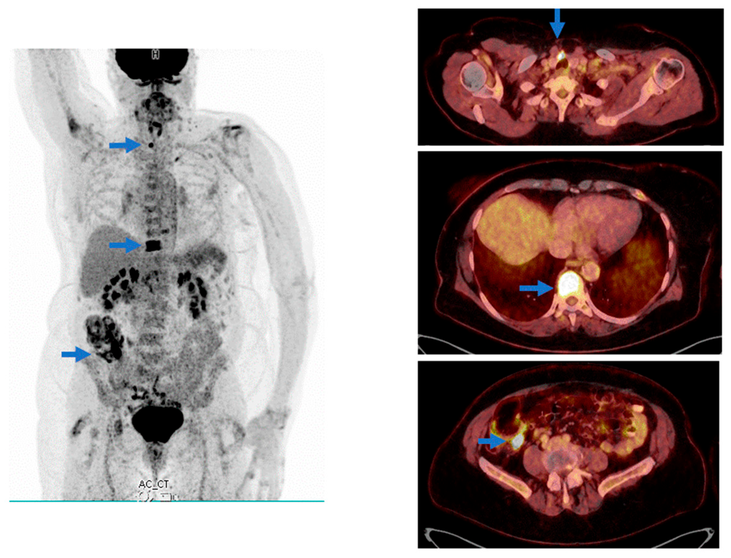
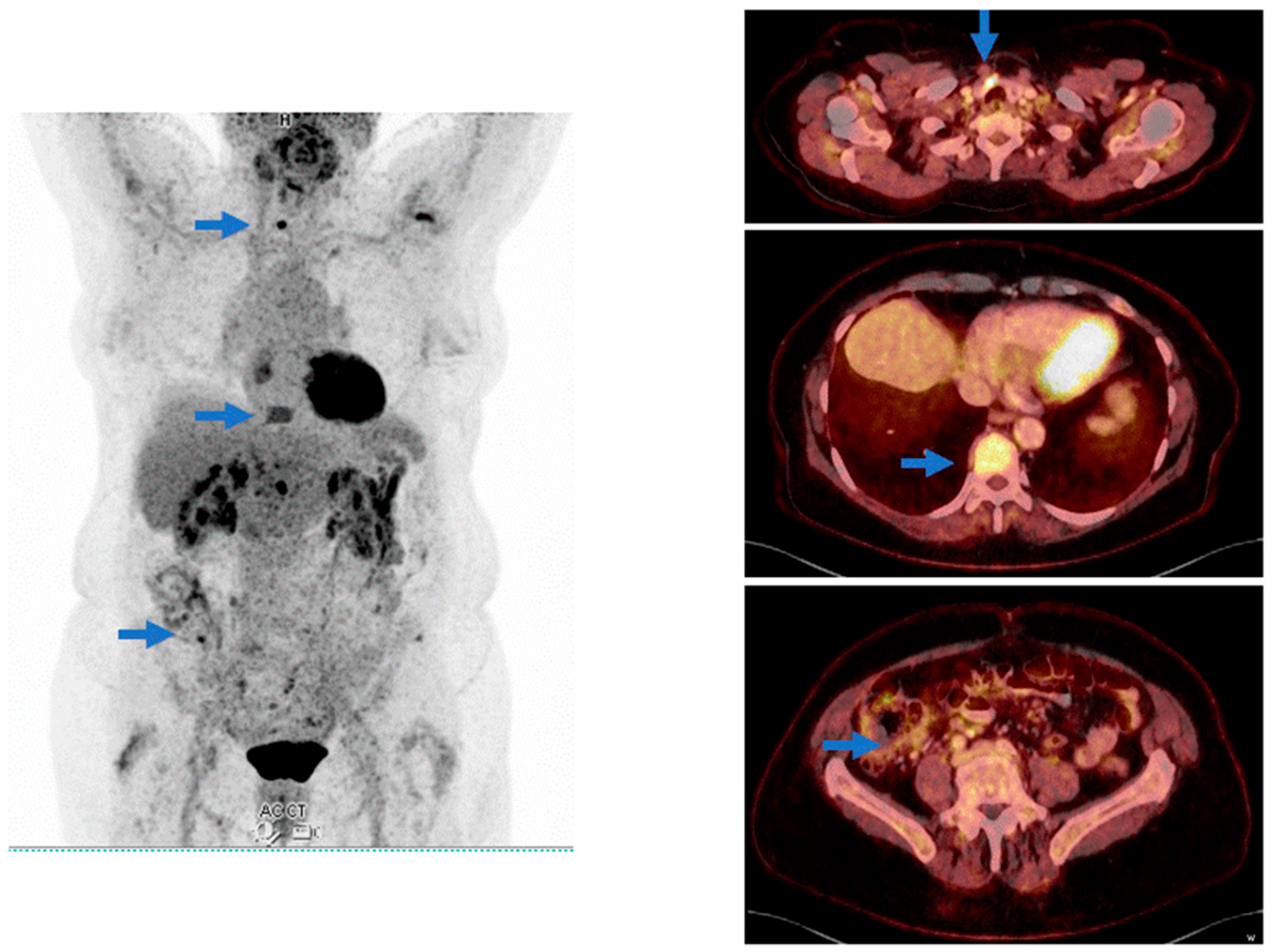
2. Case Presentation

A 77-year-old woman was evaluated at a local dermatology department for chronic pruritus. She was a former smoker (10 pack years), obese (BMI 33), and received medication for paroxysmal atrial fibrillation (anti-coagulant), hypercholesterolemia, and hypertension. Blood samples showed an elevated Fibrosis-4 (FIB-4) index for Liver Fibrosis (an index for cirrhosis based on aspartate transaminase, alanine transaminase, platelet count, and age). An upper abdominal ultrasound showed a normal liver, yet identified an incidental finding showing a 30 × 45 mm process near the right atrium that was confirmed with echocardiography.
A month later, she was referred to a secondary hospital for evaluation of a possible myxoma. Magnetic resonance imaging (MRI) of the heart localized the process behind both atria, but the FDG PET/CT of the heart did not show any metabolic activity; hence, the process was considered benign. However, during the diagnostic work-up, a whole-body FDG PET/CT scan was performed, which showed increased focal uptake in the right thyroid gland, the spinal column (most pronounced in the corpus of vertebra Th10), the portal lymph nodes, the terminal ileum, and surrounding lymph nodes (Figure 1).
Due to the suspicion of advanced malignant disease, a search for the primary tumor was initiated. Extensive blood samples were taken, including cancer, immunologic, and rheumatologic markers, which showed normal markers, except for a slight increase in the monoclonal bands (M-component).
Two months later, biopsy specimens were taken from the terminal ileum and the spinal column. During colonoscopy, the terminal ileum had a polypoid appearance; however, the biopsies showed only a non-specific reactive pattern. The CT component of the thoracic column did not show any changes corresponding to the increased activity, but an additional MRI showed that the most prominent signal changes were seen in the corpus of vertebra 12 and were more diffuse in vertebra 10. Hence, the biopsy was taken from this localization. During the biopsy procedure of the corpus of the vertebra, the bone had a normal macroscopic appearance without signs of osteolysis, and the pathologic evaluation included only normally differentiated cells without signs of malignancy, including a sparse number of T lymphocytes and very few B lymphocytes.
Due to the elevated M-component in the blood samples, whole-body low-dose CT, spinal magnetic resonance imaging (MRI), and bone marrow biopsy were performed to examine for the neoplastic proliferation of monoclonal plasma cells within the bone marrow. The radiologic examinations showed no signs of myelomatosis or myeloma. The biopsy specimen showed only a reactive pattern with a slight number of T lymphocytes and almost no B lymphocytes with a normal phenotype.
Gynecological assessment, including vaginal ultrasound, cervical smear test, and mammography, was normal.
A month later, thyroid scintigraphy (99m-Tc-pertechnetate) showed a hypofunctioning area corresponding to the site of activity in the FDG PET/CT. The ultrasonic re-examination revealed a normally sized thyroid with benign nodules, some showing calcifications, notably in the left lobe. In the isthmus, a 7 × 6 mm hypoechoic alteration with coarse calcification was observed. A small 5 × 5 × 5 mm hypoechoic nodule with abundant Doppler signals was also present nearby. FDG PET/CT suggested the most intense signal came from the hypoechoic area; a fine-needle aspiration (FNA) was conducted from the mentioned 5 mm nodule with no immediate complications. However, the FNA was inconclusive due to a lack of material.
A month later, four months after the initial FDG PET/CT was performed, the examination was repeated (Figure 2).
The blue arrows illustrate unchanged focal activity in the thyroid gland, lower activity in vertebra 10, and no activity in the terminal ileum.
Now, the previous findings had mostly resolved—completely in the terminal ileum and surrounding lymph nodes—and otherwise lowered significantly to a reactive or benign level. The only exception was the thyroid gland, in which a repeated biopsy indicated a very low-risk papillary thyroid adenocarcinoma (T1aN0M0) for which she had a successful total thyroidectomy.
In the aftermath of this extensive evaluation, the woman recalled having had a mild SARS-CoV-2 infection seven days before the first FDG PET/CT was performed but reported having recovered fully at the time of the scan. The infection was diagnosed with a lateral flow test, and she received no medical treatment. During this period, the Omicron variant of SARS-CoV-2 was dominant. She was triple-vaccinated against SARS-CoV-2 with an mRNA-based vaccination (Comirnaty) but had not received a booster for the following winter season.

3. Discussion

The present case story involved numerous PET-positive areas mimicking cancer following a period of COVID-19 disease. However, it also revealed a very low-risk papillary thyroid adenocarcinoma, prompting a successful thyroidectomy. SARS-CoV-2 infection is known to affect the results of medical imaging; this influence has predominantly been observed in cases involving the more pathogenic variants before the Omicron variant. The most common findings have been seen in the lungs and upper respiratory lymph nodes [5], but diffuse activity has also been seen in other organs [6]. Also, changes in FDG PET/CT have been observed following COVID-19 vaccination, especially in the regional lymph nodes of the area of vaccination [7]. This has interfered with the evaluation of oncologic patients [8,9,10], especially where pulmonary metastases have been suspected. If the case was different, i.e., without the excessive FDG PET/CT changes, one might have considered monitoring the very low-risk papillary thyroid adenocarcinoma, given the positive outcomes. Active surveillance has been increasingly embraced globally as an alternative to immediate surgery [11].

4. Conclusions

Our findings indicate that inflammation due to some infectious agent, presumably SARS-CoV-2, could lead to widespread radiologic findings mimicking metastatic cancer. To this day, we have not yet found comparable results to our findings in the literature.
These findings could contribute to the understanding of post-acute COVID-19, but it is equally important to evaluate whether imaging should be postponed if the patient has had recent infectious diseases such as COVID-19, or at least be interpreted with caution when performing oncologic assessment, as these findings could be misinterpreted, preventing unnecessary examinations, invasive procedures such as biopsies, excessive radiation exposure from repeated CT scans and PET/CT, and, in extreme cases, unwarranted surgeries.

Author Contributions

Conceptualization: G.H. and H.L.J.; image analysis: S.C. and J.M.; writing—original draft preparation: G.H.; writing—review and editing: all the authors. All authors have read and agreed to the published version of the manuscript.

Funding

This research received no external funding.

Institutional Review Board Statement

Ethical review and approval were waived for this study as defined by the “Faroese Act on Research Ethics Review of Health Research Projects” section 2, as the project does not constitute a health research project. Approval number: 25/13024-1.

Informed Consent Statement

Written informed consent has been obtained from the patient to publish this paper.

Data Availability Statement

The data that support the findings of this study are available from the corresponding author upon reasonable request.

Conflicts of Interest

The authors declare no conflicts of interest.

References

  1. World Health Organization. WHO Coronavirus (COVID-19) Dashboard|WHO Coronavirus (COVID-19) Dashboard with Vaccination Data; World Health Organization: Geneva, Switzerland, 2025. [Google Scholar]
  2. Guan, W.J.; Ni, Z.Y.; Hu, Y.; Liang, W.H.; Ou, C.Q.; He, J.X.; Liu, L.; Shan, H.; Lei, C.L.; Hui, D.S.C.; et al. China Medical Treatment Expert Group for Covid-19. Clinical Characteristics of Coronavirus Disease 2019 in China. N. Engl. J. Med. 2020, 382, 1708–1720. [Google Scholar] [CrossRef] [PubMed]
  3. Zhang, J.J.; Dong, X.; Liu, G.H.; Gao, Y.D. Risk and Protective Factors for COVID-19 Morbidity, Severity, and Mortality. Clin. Rev. Allergy Immunol. 2023, 64, 90–107. [Google Scholar] [CrossRef] [PubMed]
  4. Davis, H.E.; McCorkell, L.; Vogel, J.M.; Topol, E.J. Long COVID: Major findings, mechanisms and recommendations. Nat. Rev. Microbiol. 2023, 21, 133–146. [Google Scholar] [CrossRef] [PubMed]
  5. Zou, S.; Zhu, X. FDG PET/CT of COVID-19. Radiology 2020, 296, E118. [Google Scholar] [CrossRef] [PubMed]
  6. Vaz, N.; Franquet, E.; Heidari, P.; Chow, D.Z.; Jacene, H.A.; Ng, T.S.C. COVID-19: Findings in nuclear medicine from head to toe. Clin. Imaging 2023, 99, 10–18. [Google Scholar] [CrossRef] [PubMed]
  7. Cohen, D.; Krauthammer, S.H.; Wolf, I.; Even-Sapir, E. Hypermetabolic lymphadenopathy following administration of BNT162b2 mRNA Covid-19 vaccine: Incidence assessed by [18F]FDG PET-CT and relevance to study interpretation. Eur. J. Nucl. Med. Mol. Imaging 2021, 48, 1854–1863. [Google Scholar] [CrossRef] [PubMed]
  8. Pai, T.S.; Rojas, C.; Wasserman, M.C.; Parent, E.E.; Cornell, L.; Chumsri, S. Asymptomatic coronavirus disease 2019 mimicking metastatic breast cancer on positron emission tomography/computed tomography imaging. Radiol. Case Rep. 2021, 16, 2226–2230. [Google Scholar] [CrossRef] [PubMed]
  9. Kavanal, A.J.; Jena, S.R.; Kumar, R.; Das, C.K.; Kumar, S.; Mittal, B.R. Effect of COVID-19 on 18F-FDG PET/CT: Is There a Need to Consider COVID-19 Status Before Planning 18F-FDG PET/CT for Oncologic Evaluation? J. Nucl. Med. Technol. 2021, 49, 284–285. [Google Scholar] [CrossRef] [PubMed]
  10. SNMMI Statement: Possible Effect of Omicron Infection on FDG PET/CT Scans 2022. J. Nucl. Med. 2022, 63, 11N. [PubMed]
  11. Sugitani, I. Active surveillance of low-risk papillary thyroid microcarcinoma. Best. Pract. Res. Clin. Endocrinol. Metab. 2023, 37, 101630. [Google Scholar] [CrossRef] [PubMed]
Figure 1. Whole-body MIP projection of FDG PET and transaxial slices of fused FDG PET/CT at three localizations at the first examination. The blue arrows illustrate focal activity in the thyroid gland, the corpus of vertebra Th10, and the terminal ileum. The patient was on a ketogenic diet prior to the examination due to the interest in evaluating the location of a suspected myxoma; hence, no activity is seen corresponding to the heart.
Covid 05 00098 g001
Figure 2. Whole-body MIP projection of FDG PET and transaxial slices of fused FDG PET/CT at three localizations at the second examination.
Covid 05 00098 g002
Disclaimer/Publisher’s Note: The statements, opinions and data contained in all publications are solely those of the individual author(s) and contributor(s) and not of MDPI and/or the editor(s). MDPI and/or the editor(s) disclaim responsibility for any injury to people or property resulting from any ideas, methods, instructions or products referred to in the content.

