Article

Superinfections in Hospitalized COVID-19 Patients (Super COVID-19): Data from the Multicentric Retrospective CH-SUR Cohort Study in Switzerland

1
Division of Infectious Diseases, Infection Prevention and Travel Medicine, HOCH Health Ostschweiz, Cantonal Hospital of St. Gallen, CH-9001 St. Gallen, Switzerland
2
Division of Infectious Diseases and Hospital Epidemiology, University Hospital Basel, CH-4051 Basel, Switzerland
3
Cantonal Hospital of Lucerne, Children’s Hospital, Pediatric Infectious Diseases, CH-6000 Lucerne, Switzerland
4
Faculty of Health Sciences and Medicine, University of Lucerne, CH-6000 Lucerne, Switzerland
5
Paediatric Pharmacology and Pharmacometrics, University Children′s Hospital Basel, University of Basel, CH-4051 Basel, Switzerland
6
Department of General Medicine, Cantonal Hospital of Chur, CH-7000 Chur, Switzerland
7
Service of Prevention and Infection Control, Directorate of Medicine and Quality, University Hospital Geneva, CH-1205 Geneva, Switzerland
8
Institute of Global Health, University of Geneva, CH-1202 Geneva, Switzerland
9
Faculty of Health Sciences and Medicine, Clinic St. Anna, University of Lucerne, CH-6000 Lucerne, Switzerland
10
Department of Infectious Diseases, Bern University Hospital, University of Bern, CH-3010 Bern, Switzerland
*
Author to whom correspondence should be addressed.
Current address: Department of General Medicine, Canton Hospital of Glarus, CH-8750 Glarus, Switzerland.
These authors contributed equally to this work.
COVID 2025, 5(6), 86; https://doi.org/10.3390/covid5060086
Submission received: 9 April 2025 / Revised: 17 May 2025 / Accepted: 19 May 2025 / Published: 30 May 2025
(This article belongs to the Section COVID Clinical Manifestations and Management)

Abstract

Background: The epidemiology, characteristics and outcomes of coinfections in COVID-19 are still poorly understood. Methods: We investigated the prevalence of coinfections in COVID-19 patients hospitalized in Switzerland over the first three epidemic waves between 1 March 2020 and 1 June 2021, as well as risk factors and outcomes. Patients were identified from six hospitals of the Swiss prospective surveillance system database (CH-SUR). Details of the type and treatment of coinfections were retrieved retrospectively from medical charts. We assessed the proportion of patients with suspected coinfections and analyzed risk factors and 90-day in-hospital survival using logistic and Cox regression. Results: Of 13,265 identified patients, 36.6% (4859/13,625) had suspected coinfections, and 44.8% (5941/13,625) received antibiotics. Respiratory coinfections (25.6%) were the most common, followed by bloodstream (19.8%) and urinary tract infections (14.6%). Escherichia coli (14.8%), Staphylococcus aureus (10.7%) and Klebsiella pneumoniae (6.1%) were the most frequently isolated pathogens. The risk factors for coinfections included increasing age, male gender, certain underlying medical conditions and immunosuppression. Suspected coinfections were associated with a longer hospital stay (13 vs. 7 days, p < 0.001), more frequent ICU admission (26% vs. 6.7%, p < 0.001) and higher rates of in-hospital death (24% vs. 9.5%, p < 0.001). Hospitalization in the ICU at the time of COVID-19 diagnosis had the strongest association with coinfections. Conclusions: A high proportion of COVID-19 patients had coinfections, particularly respiratory infections, and received antibiotics. Coinfections were associated with severe illness and worse outcomes.

1. Introduction

COVID-19, caused by severe acute respiratory syndrome coronavirus 2 (SARS-CoV-2), has been challenging healthcare systems throughout the world. In analogy to severe influenza infections [1], there was initially a great concern for bacterial coinfections (COIs) in patients with COVID-19. Many patients early in the pandemic received antibiotics regardless of evidence of bacterial COIs [2].
The prevalence of microbiologically confirmed bacterial community-acquired COIs is generally low, ranging from 4–8% in the overall population [3,4] to 8–12% in critically ill patients [3]. These include respiratory COIs [5,6], followed by bloodstream infections (BSIs) and urinary tract infections (UTIs) [6]. Bacterial hospital-acquired COIs are more frequent, especially in critically ill patients in the ICU, with incidence rates of up to 40% [4]; these are mostly healthcare- and ventilator-associated pneumonias, BSIs and UTIs [5,6].
Among fungi, Aspergillus, Mucorales and Candida species have been the most common fungal pathogens reported to cause invasive fungal disease (IFI) in hospitalized COVID-19 patients, in particular those with critical illness in the ICU [7]. COVID-19-associated aspergillosis (CAPA) affects about 10% of invasively ventilated COVID-19 patients and is associated with a high mortality rate of approximately 50%, even with antifungal treatment [8].
How the epidemiology of COIs changed during the initial waves of the pandemic is not well characterized. In particular, the introduction of corticosteroids and immunomodulators as the standard treatment for severe COVID-19 infections has raised concerns about increased susceptibility to bacterial and fungal COIs.
In Switzerland, systematic data on the epidemiology of community-acquired and hospital-acquired COIs in COVID-19 patients are scarce [9]. The CH-SUR, a prospective national surveillance system of patients hospitalized with COVID-19 and/or influenza, collected clinical and epidemiological data on about 80% of hospitalized SARS-CoV-2/COVID-19 patients from March 1st 2020 until September 1st 2024. Although it has proved to be a powerful surveillance tool [10], the CH-SUR was not designed to capture coinfections.
In this multicentric retrospective study of hospitalized COVID-19 patients, we aimed to report the frequency and microbiological details of confirmed COIs with a focus on respiratory infections, BSIs and UTIs, to investigate the clinical characteristics and potential risk factors associated with COIs and to assess their impact on survival.

2. Materials and Methods

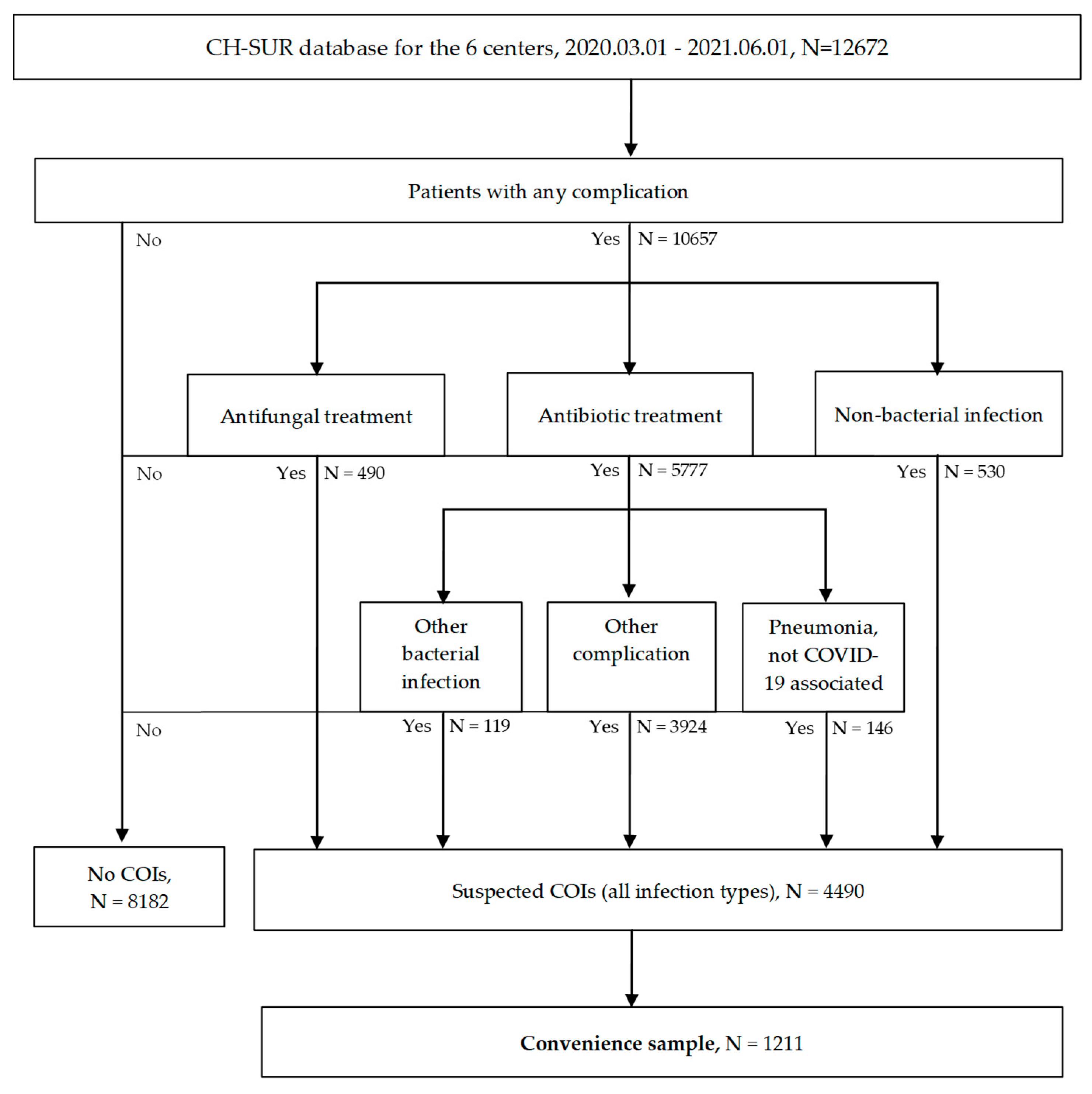
2.1. Study Setting, Design and Population

This multicentric, retrospective, observational cohort study was performed in six Swiss hospitals actively participating in CH-SUR, including three university hospitals and three teaching hospitals. The reporting of this study followed the STROBE criteria [11].
All adult patients (i.e., 18 years or older) admitted with COVID-19 for ≥24 h to one of these six hospitals between 1 March 2020 and 1 June 2021 were considered. COVID-19 infection was diagnosed by polymerase chain reaction (PCR), by antigen testing or by fulfilling clinical diagnostic criteria provided by the Swiss Federal Office of Public Health (FOPH) [12]. Patients were excluded if they declined to give their general consent.

2.2. Outcome Measures

Our primary aim was to assess the prevalence and the nature (i.e., causative microorganisms, resistance patterns) of selected COIs (i.e., respiratory COIs, BSIs, UTIs) among COVID-19 patients and compare their changes between the first, second and third COVID-19 waves, defined as the period 1 March–30April 2020, the period 1 October 2020–14 February 2021 and the period 15 February–20 June 2021, respectively. The secondary aims were to describe the time-dependent pathogen distribution of respiratory COIs, stratified for early-onset COIs and late-onset COIs, during hospitalization upon their time of onset. Finally, we investigated the clinical characteristics of coinfected patients, possible risk factors for COIs and their impact on survival.

2.3. Data Collection

The CH-SUR database provided data on patient characteristics (age, sex, obesity, history of smoking and comorbidities, e.g., immunosuppression), as well as on COVID-19 infections (e.g., date of diagnosis, laboratory and radiological findings, type of treatment, non-infectious complications), treatments for COVID-19 (i.e., antiviral or immunomodulatory therapy) and outcomes. Also, it was used to identify patients with COIs (see Section 2.4). As the CH-SUR database does not focus specifically on infectious complications, lacking information on COIs (e.g., type, timing of onset, symptoms and signs, laboratory and radiological findings, pathogens) were retrieved retrospectively by medical chart reviews. Susceptibility profiles for selected antibiotics (see Supplementary Materials) were collected for bacterial isolates.

2.4. Data Analysis

The analysis was conducted on two distinct levels. First, among all COVID-19 patients from the six participating centers, we identified those with any type of suspected COI. To achieve this, we filtered all patients for whom at least one complication was documented in the CH-SUR database (Figure 1). We included patients who received an antibiotic treatment either for a pneumonia not related to COVID-19 or for any other type of bacterial infection, which were found in the database under the fields “other complication” and “other bacterial complication”. In addition, we included patients who received an antifungal therapy, as well as patients for whom a “non-bacterial infection” was recorded. All patients of the 6 participating hospitals who fell outside our algorithm were considered as having no COIs. Comparative analyses were conducted across these groups, focusing on demographics, comorbidities, complications, treatments administered for COVID-19 or its associated complications, and patient outcomes. Also, we analyzed the survival rates in the two groups.
In the next step, using a convenience sample (all patients with suspected COIs from four sites and every 2nd or 3rd patient with a suspected COI from the remaining sites), patients with suspected COIs according to the CH-SUR database were assessed for respiratory COIs (suspected or microbiologically confirmed), BSIs and UTIs according to the definitions provided below (see Section 2.5). We analyzed the causative pathogens, their resistance profiles and antibiotic use. A multivariable analysis was performed to identify predictors for COIs and mortality.

2.5. Definitions

For the analysis of the convenience sample, the following definitions were used.
A confirmed respiratory infection was considered to be the presence of clinical signs and the detection of (I) a clinically relevant bacterial, viral or fungal pathogen in respiratory specimens, or (II) a positive pneumococcal or legionella urinary antigen or Pneumocystis jirovecii antigen. Clinical signs without pathogen detection were referred to as a suspected respiratory infection. CAPA was defined by clinical, radiological and mycological criteria according to the consensus criteria of CAPA by the ECCM/ISHAM Study Group [13] (see Supplementary Materials). Respiratory infections were further classified depending on the time of diagnosis after hospitalization into early-onset (<3 days) and late-onset (>7 days).
A bloodstream infection (BSI) was defined as the growth of a non-skin flora commensal in one or more blood cultures. To define a bloodstream infection as that caused by a common skin colonizer such as Coagulase-negative staphylococci, we required two or more blood cultures drawn from different sites and the presence of at least one clinical sign of infection (e.g., fever > 38 °C, chills associated with hypotension). A line-associated BSI was defined as the growth of bacteria or fungi in one or more blood cultures drawn from a patient with clinical signs of infection and no apparent other source of infection (see Supplementary Materials).
A urinary infection was defined as the presence of typical symptoms and signs regardless of microbial growth in a urine sample according to the ECDC criteria [14] (see Supplementary Materials).

2.6. Statistical Analysis

A descriptive analysis was conducted to summarize the characteristics of the patient population and the distribution of key variables, such as comorbidities, treatments, complications, death, BMI and ICU length of stay (ICU-LOS). Additionally, the resistance profiles and characteristics of the detected COIs were investigated. Demographics, superinfection rates, lengths of hospital stay (LOSs) and outcomes were compared across groups of patients with different coinfection status and across COVID-19 waves. The results from the descriptive analysis are presented in the form of summary tables and graphs. Categorical variables were summarized using counts and percentages, while continuous variables are reported with medians and interquartile ranges. Chi-square tests were used to assess differences between groups for categorical variables, and ANOVAs or Mann–Whitney U tests were employed for continuous variables, depending on the normality of the data distribution.
To analyze survival rates in relation to coinfection status, a Kaplan–Meier survival model was constructed using the “survival” and “survminer” R packages. The survival time was calculated from the corrected hospital entry date (or diagnosis date for hospital-acquired cases) to the discharge or in-hospital death date. Patients were right-censored at 90 days, ensuring consistency in the analysis. The survival function was stratified based on coinfection status, with groups for “no COI” and “suspected COI”. The log-rank test was used to assess differences in survival rates across these groups. p-values of <0.05 were considered significant.
Additionally, a Cox proportional hazards model was fitted to the data to account for potential confounders with in-hospital death or discharge as the outcome and coinfection status, age and gender as covariates. For missing variables, no imputation was made apart from unknown being considered negative for binary variables.
A Poisson regression was conducted to model the weekly rate of confirmed coinfections. The dependent variable was the count of confirmed coinfections per week, with time as the main independent variable and the weekly number of cases included as an offset. The model was stratified by epidemic wave to assess differences in coinfection trends across distinct time periods. A generalized linear model (GLM) was used to identify risk factors associated with coinfection status (no COI or suspected COI), employing logistic regression. The dependent variable was the coinfection status (binary outcome). The covariates included in the model were age (as a continuous variable), BMI, gender, COVID-19 wave, comorbidities (respiratory, asthma, diabetes, hypertension, cardiovascular, renal, liver, neuro-impairment, dementia, HIV-positive status, hematological/immunological disorders, transplant, immunosuppressive treatment, oncology), hospitalization origin, community-acquired or hospital-acquired status, and COVID-19 severity (based on CURB-65 score).
Covariates were preprocessed to ensure consistency: categorical variables were converted to factors, missing values were replaced with “No” or the appropriate reference level, and continuous variables such as severity scores were scaled and treated numerically. The GLM was applied to estimate odds ratios (ORs) and their 95% confidence intervals (CIs).
The model output included a table summarizing the odds ratios, confidence intervals and p-values for all covariates. Significant covariates (p-value < 0.05) were highlighted and visualized using a sorted forest plot. This forest plot displayed the odds ratios and their 95% confidence intervals, allowing for intuitive interpretation of the significant predictors of coinfection status.
All analyses were performed using R (version 4.2.3) and various associated packages such as “survival”, “survminer”, “broom” and “dplyr”. Plots were generated using “ggplot2” and “survminer”.

3. Results

3.1. Comparison Between Suspected COI and No COI Groups at CH-SUR Level

Of all COVID-19 episodes, 36.6% (4859/13,625) had suspected COIs (Supplementary Figure S1).
The factors significantly associated with suspected COIs included older age, male gender and underlying medical conditions such as chronic respiratory disease, diabetes, cardiovascular disease and renal disease, as well as neurological, oncological and hematological comorbidities, dementia and immunosuppression. Additionally, a greater percentage of individuals in the suspected COI group were treated with steroids or immunomodulators for COVID-19 and suffered from non-infectious COVID-19-related complications (Table 1).
Patients with suspected COIs had worse outcomes. They had a longer hospital stay (13 vs. 7 days, p < 0.001) and were more likely to require ICU care (26.2% vs. 6.7%, p < 0.001) or to die (23.7% vs. 9.5%, p < 0.001) compared to those without COIs.

3.2. Survival Analysis from CH-SUR

In the Kaplan–Meier survival analysis, there was a statistically significantly lower survival rate among COVID-19 patients with suspected COIs than those without COIs (p < 0.0001). The 90-day in-hospital survival probabilities were 0.52 for patients with suspected COIs (95% CI: 0.49, 0.55) and 0.64 for patients without COIs (95% CI: 0.60, 0.68) (Figure 2).

3.3. Convenience Sample Data: Type of Infection and Microbial Spectrum

In total, 1211 medical charts of patients with suspected COIs were reviewed. The proportion of confirmed COIs out of the total of suspected COIs progressively increased across the three waves (35.1% vs. 43.9% vs. 55.0%, p = 0.003) (Supplementary Table S1). As shown in Supplementary Figure S2, the Poisson regression indicates that the incidence of confirmed COIs rose during the first two waves, then declined in the third, likely reflecting improvements in treatment management.
Among these patients, the most common type of infection was respiratory COI (310/1211, 25.6%) followed by UTI (240/1211, 19.8%) and BSI (177/1211, 14.6%). This ranking remained consistent over the three waves, although the proportion of respiratory COIs out of all types of COI decreased slightly across the three waves (51.0% vs. 39.0% vs. 47.1%, p = 0.081) (Supplementary Table S2).
Overall, E. coli was the most commonly isolated pathogen (179/1211, 14.8%), followed by S. aureus (130/1211, 10.7%), K. pneumoniae (74/1211, 6.1%), P. aeruginosa (55/1211, 4.5%), S. pneumoniae and Haemophilus influenzae (each 40/1211, 3.3%) (Table 2). The resistance profiles of the ten most commonly isolated pathogens are shown in Supplementary Table S3. For E. coli, a trend toward more frequent resistance to ceftriaxone and ESBL carriers was observed across the three waves.
Among the respiratory COIs, 64.7% (650/1004) were categorized as suspected COIs (i.e., without microbiological confirmation), and 28.2% (284/1004) represented confirmed COIs (Supplementary Table S4). The CAPA prevalence was 4.6% (46/1004). In most cases, CAPA was classified as probable.
The most common pathogens isolated in early-onset respiratory COIs (i.e., onset during the first 3 days of hospitalization) were S. aureus (35/100, 35.0%), S. pneumoniae (15/100, 15.0%) and P. aeruginosa (17/100, 17.0%). After 7 days of hospitalization, the most common pathogens were S. aureus (28/164, 17.1%), K. pneumoniae (22/164, 13.4%) and E. coli (18/164, 11.0%) (Table 3).

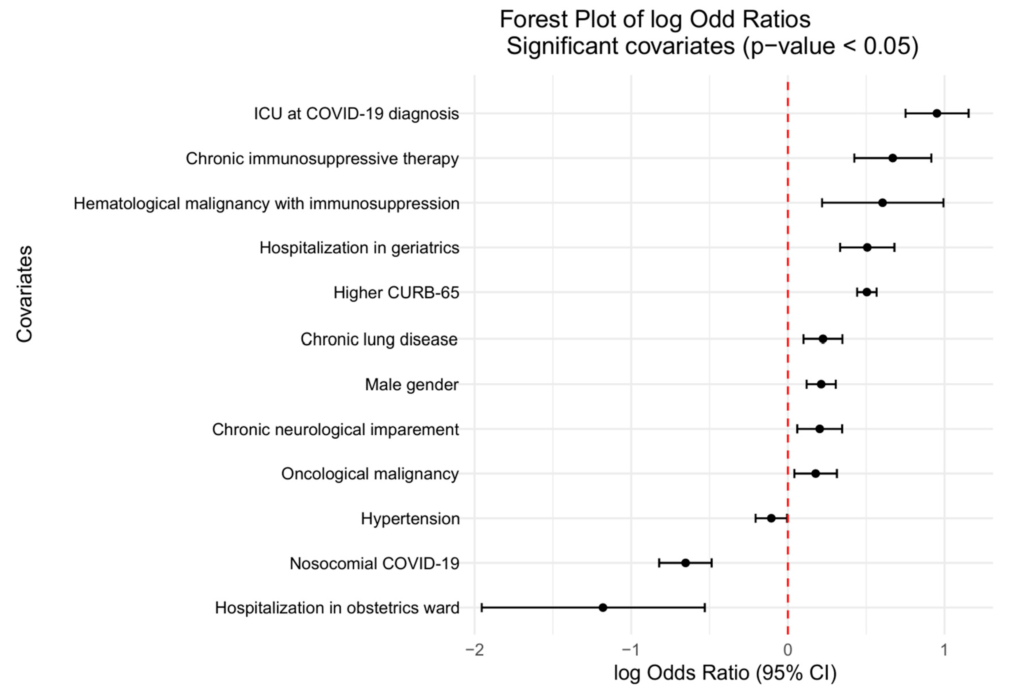
3.4. Risk Factors for COIs

The variables in the GLM model assessing risk factors for COIs included age, gender, comorbidities and baseline immunosuppression, disease severity (CURB-65 score at admission), hospitalization ward, place of origin before hospitalization and COVID-19 wave. Independent risk factors for COIs included male gender, chronic lung disease, neurological, oncological and hematological comorbidities and baseline immunosuppression (Figure 3). Additionally, patients with higher CURB-65 scores and those hospitalized in the ICU at the time of COVID-19 diagnosis had higher odds of developing COIs. In contrast, patients with hypertension, those with nosocomial COVID-19 and women hospitalized in the obstetrics ward showed lower odds of COIs.

3.5. Antibiotic Use

Nearly half of the patients (5941/13625, 44.8%) received antibiotics. Among the suspected COI group, the majority were treated with antibiotics (4536/4859, 93.4%); however, approximately one-sixth of patients without COIs also received antibiotics (1405/8406, 16.7%) (Table 1).
Amoxicillin/clavulanate was the most common antibiotic given empirically for a suspected COI (2379/3977, 59.9%), followed by piperacillin/tazobactam (1090/3977, 27.4%) and ceftriaxone (652/3977, 16.4%). Conversely, in confirmed COIs, piperacillin/tazobactam was the most common antibiotic given (269/539, 49.9%), followed by amoxicillin/clavulanate (234/539, 43.4%) and ceftriaxone (161/539, 29.9%) (Supplementary Table S5). The number of different antibiotics used was higher in confirmed COIs than in suspected COIs throughout the study and in the first wave compared to the subsequent waves (p < 0.001; Supplementary Figure S3).
Overall, across the three waves, there was a trend toward reduced antibiotic use (p < 0.001; Supplementary Table S6). We analyzed the use of a selection of commonly prescribed antibiotics in clinical practice. This trend for a decrease over time was particularly evident for the most frequently used antibiotics, i.e., amoxicillin/clavulanate and piperacillin/tazobactam, as well as for macrolides (p < 0.001; Supplementary Table S7).

3.6. Immunomodulatory Treatment for COVID-19

Steroids were used more frequently in patients with suspected COIs than in those without suspicion of COIs (19.7% vs. 33.0%, p < 0.001, Table 1). In the second wave, their use was higher than in the first and third waves (second wave: 39.1% vs. first wave: 12.4% vs. third wave: 26.6%, p < 0.001) (Supplementary Table S6).
Tocilizumab use, as assessed in the convenience sample only, was more frequent in the confirmed COI group than in the suspected COI group (8.0% vs. 1.2%, p < 0.001) (Supplementary Table S8) and was significantly lower in the second wave compared to the first and third waves (second wave: 0.1% vs. first wave: 5.1% vs. third wave: 6.0%, p < 0.001) (Supplementary Table S9).

4. Discussion

To our knowledge, this multicenter retrospective study is the first to systematically describe the nature, microbiological spectrum and antibiotic treatment of COIs in COVID-19 patients across the first three COVID-19 waves in Switzerland.
More than one-third of the patients (36.6%) had at least a suspected COI, with 93.4% of these patients receiving antibiotics. Although we were unable to assess the prevalence of confirmed COIs in the entire sample at the CH-SUR level, this aligns well with current literature indicating that antibiotic use was significantly higher than the estimated prevalence of confirmed bacterial COIs in COVID-19 patients in the early pandemic [15]. In the convenience sample, the proportion of confirmed COIs among suspected cases increased across the three waves. There might be several explanations for this. This trend may be due to limited testing due to resource constraints in healthcare settings during the early pandemic, as well as initial concerns regarding healthcare workers’ safety arising from limited knowledge about COVID-19 transmission. Alternatively, the increased use of immunomodulators [16], such as the increased use of tocilizumab observed in our analysis, and a greater number of patients at risk, such as those with chronic immunosuppression or malignancies, might have predisposed patients to more coinfections over the later waves.
Respiratory COIs were the most common types of COIs. S. aureus was the leading Gram-positive pathogen, while E. coli, K. pneumoniae and P. aeruginosa were the most common Gram-negative pathogens [4]. A meta-analysis by Langford et al. on antimicrobial resistance in COVID-19 patients, including isolates from 130 studies across 40 countries, identified the same pathogens as the most common ones.
Regarding antimicrobial resistance, we found a relatively low prevalence of resistance among the ten most common bacterial pathogens, reflecting the resistance landscape in Switzerland [17]. Despite the limited number of isolates, no increase in resistance rates was detected across the three waves, except for a significant rise in ESBL-producing strains in E. coli.
Another key finding is the characterization of the microbiological spectrum of respiratory COIs based on their timing of onset. The spectrum of bacterial pathogens differed between early-onset and late-onset respiratory COIs. In line with previous studies [18], early-onset respiratory COIs were dominated by community pathogens such as S. aureus and S. pneumoniae. However, P. aeruginosa also played a significant role. In late-onset respiratory COIs, we observed the bacterial spectrum typically found in nosocomial pneumonia [19], characterized by a diversity of Gram-negative pathogens, including Enterobacteriaceae and P. aeruginosa, in addition to S. aureus. Our findings have significant implications for empirical therapy for respiratory COIs. In COVID-19 patients, S. aureus should be treated empirically in both early-onset and late-onset respiratory COIs, while optimal Gram-negative coverage seems to be necessary at later stages. In Switzerland, given the low prevalence of methicillin-resistant S. aureus, amoxicillin/clavulanate seems to a reasonable choice in early-onset respiratory COIs, provided there are no specific risk factors for P. aeruginosa. For late-onset respiratory COIs, piperacillin/tazobactam or cefepime is recommended to cover both S. aureus and Gram-negative pathogens. Given that the prevalence of ESBL-producing bacteria in Switzerland is below 15% (www.anresis.ch), carbapenems are not first-line treatments in our local guidelines, except in cases of known colonization, strong risk factors for carriage (based on epidemiology, e.g., transfer from endemic areas) or severe illness such as septic shock.
As widely reported [20], we found that the overall antibiotic use in COVID-19 patients was high (5941/13,525, 43.9%), regardless of whether bacterial COIs were ultimately confirmed. Of note, 17% of patients without suspected COIs received antibiotics. This result may be due to an underreporting of COIs in the database, as the Red Cap database was not designed to detect COIs, but—more likely—it could also be partly explained by inappropriate antibiotic use. Interestingly, we observed a significant drop in consumption of the two most commonly used antibiotics (amoxicillin/clavulanate and piperacillin/tazobactam) and of macrolides from wave to wave. This likely reflects the accumulated knowledge on COIs in COVID-19 patients throughout the pandemic. Our study aligns with another Swiss study [21] on the impact of the COVID-19 pandemic on antibiotic consumption, showing significantly higher antibiotic consumption during the first wave compared to the same periods before the pandemic and to the second wave, particularly evident for broad-spectrum antibiotics (including piperacillin/tazobactam) and macrolides.
Our study adds to the literature associating COIs with a higher severity of illness [22,23] and higher risk for unfavorable outcomes [24]. Compared to non-infected patients, those with suspected COIs presented with more severe disease at admission, as indicated by their higher CURB-65 scores and greater need for ICU care. They had more COVID-19-related complications and were more often treated with steroids/immunomodulators for COVID-19 (in the univariable analysis). Additionally, patients in the suspected COI group had longer hospital stays and were more likely to require ICU care.
Furthermore, we sought to identify possible risk factors for COIs at admission. Several studies, mainly conducted in the ICU setting and focused on respiratory COIs, have tried to identify predictors for COIs, with heterogeneous results. The most commonly reported factors include increasing age [22,25], male sex [22,26], comorbidities [22,25,27], baseline immunosuppression, being unvaccinated [25] and different clinical and laboratory markers (e.g., mechanical ventilation, higher C-reactive protein levels at admission, neutropenia, lymphopenia) [23]. ICU admission itself has also been linked to increased risk of COIs [28,29].
In our analysis, the strongest predictor for COIs was hospitalization in the ICU at the time of COVID-19 diagnosis, which, beyond being a risk factor for hospital-acquired infections (due to, e.g., invasive procedures and devices), may indicate a higher COVID-19 severity. Additionally, a higher CURB-65 score was associated with an increased risk of COIs. Other factors recognized as risk factors for severe COVID-19 disease [30]—such as male sex; pulmonal, hemato-oncological and neurological comorbidities; and baseline use of immunosuppressive medications—were also linked to a higher risk of COIs. Our findings suggest that the severity of COVID-19 itself is likely a primary indicator of the risk of COIs. This conclusion supports previous reports that identified severe COVID-19 as an independent risk factor for COIs [22,27,31].
We did not include the use of immunomodulators in our multivariable analysis, since we could not assess the temporal relationship between the use of immunomodulators and diagnoses of COIs. Evidence on the impact of immunomodulatory treatments for COVID-19 on the risk of bacterial and fungal COIs is inconclusive [23]. A meta-analysis by Peng et al. failed to show a significant association between immunosuppressive treatment and the risk of developing a coinfection in their overall analysis, but increased fungal infections were observed in COVID-19 patients treated with tocilizumab and corticosteroids [32,33].
This study’s strengths include the large number of patients, as well as the complete collection of microbiologic data for respiratory COIs, BSIs and UTIs in the convenience sample. However, our study had several limitations. First, the number of suspected COIs at the CH-SUR level might have been overestimated. Screening for COIs was performed using surrogate fields in the CH-SUR database, which was not designed to collect COIs specifically. This was finally achieved through manual data collection from hospital records in the convenience sample. Second, despite the use of established criteria for the main infectious disease syndromes from surveillance studies, these were retrospectively applied and therefore contain some uncertainty regarding COI classification, especially in patients without pathogen confirmation. On the other hand, confirmed COIs were likely underestimated since, due to the retrospective nature of the study, there was no structured diagnostic algorithm including viral testing in place. Last, we used convenience sampling due to limited resources.

5. Conclusions

In conclusion, this large multicenter study offers valuable insights into the epidemiology of COIs in hospitalized COVID-19 patients in Switzerland. The differentiation between early-onset and late-onset respiratory COIs, along with the characterization of their microbial spectrum, provides crucial information for the empirical treatment of respiratory COIs in COVID-19 patients. It is essential to be aware of the risk of COIs in patients with severe COVID-19, particularly those in the ICU. This study further suggests considerable potential for improved antibiotic stewardship during viral pandemics.

Supplementary Materials

The following supporting information can be downloaded at https://www.mdpi.com/article/10.3390/covid5060086/s1, Figure S1: 7-day count of COVID-19 cases by coinfection status from March 2020 to June 2021; Figure S2: Temporal trends in coinfection rates during COVID-19 waves.; Figure S3: Median number of antibiotics used in the two groups (suspected COIs, confirmed COIs) across the three waves.; Table S1: Proportion of confirmed COIs out of the total suspected and confirmed COIs of the convenience sample across the three waves.; Table S2: Proportion of respiratory COIs, BSI and UTI out of the confirmed COIs of the convenience sample across the three waves (patient level).; Table S3: Resistance profiles of the 10 most common pathogens.; Table S4: Respiratory COIs over the three waves in patients included in the convenience sample from the 6 participating hospitals (pathogen level). Table S5: Antibiotic use according to the coinfection status for a selection of frequently used antibiotics in patients included in the convenience sample from the 6 participating hospitals.; Table S6: Overall use of antibiotic and steroids in the 6 participating hospitals across the three waves.; Table S7: Antibiotic use for a selection of frequently used antibiotics in all patients from the 6 participating hospitals.; Table S8: Tocilizumab use according to the coinfection status in patients included in the convenience sample from the 6 participating hospitals.; Table S9: Tocilizumab use over the three waves in patients included in the convenience sample from the 6 participating hospitals.

Author Contributions

Conceptualization, W.C.A. and G.S.; methodology, W.C.A., G.S., A.B. and J.S.; software, J.S.; validation, W.C.A., G.S., A.B. and J.S.; formal analysis, J.S., G.S. and A.B.; investigation, G.S., A.B., W.C.A., V.B., M.B., A.C., A.I., O.K. and R.S.; resources, W.C.A., G.S., V.B., M.B., A.C., A.I., O.K. and R.S.; data curation, A.B. and G.S.; writing—original draft preparation, G.S., W.C.A., A.B. and J.S.; writing—review and editing, all authors; visualization, G.S., A.B. and J.S.; supervision, W.C.A.; project administration, W.C.A.; funding acquisition, W.C.A. and G.S. All authors have read and agreed to the published version of the manuscript.

Funding

This research was partially funded by Gilead, grant number Grant Swiss Fellowship 2022. Gilead had no influence on the data acquisition, analysis or writing of the manuscript. The Swiss Federal Office of Public Health (FOPH) funded the Swiss hospital surveillance program, including the CH-SUR database.

Institutional Review Board Statement

The COVID-19 surveillance as part of the CH-SUR system was approved on 07.05.2020 by the Ethics Committee of the Canton of Geneva, Switzerland (CCER 2020-00827). Data collection was also approved by all local ethics committees.

Informed Consent Statement

The requirement for individual consent was waived as this study used anonymized data gathered for epidemiological purposes already falling under a previous approved study protocol.

Data Availability Statement

The datasets presented in this article are not readily available because they are part of the Swiss surveillance system. Requests to access the datasets should be directed to the Swiss Federal Office of Public Health.

Acknowledgments

We would like to thank the patients and their treating physicians, and all the staff involved in the CH-SUR data collection. We thank Stephan Harbarth, University Hospital Geneva, for generously providing resources for statistical support.

Conflicts of Interest

None of the co-authors report any conflicts of interest.

Abbreviations

The following abbreviations are used in this manuscript:
COVID-19Coronavirus disease 2019
COICoinfection
CH-SURCOVID-19 Hospital-Based Surveillance
BSIBloodstream infection
UTIUrinary tract infection
ICUIntensive care unit
IFIInvasive fungal disease
CAPACOVID-19-associated aspergillosis
PCRPolymerase chain reaction
ICU-LOSIntensive care unit length of stay
LOSLength of stay
BMIBody mass index
HIVHuman immunodeficiency virus
ARDSAcute respiratory distress syndrome
LTCFLong-term care facility
ESBLExtended-spectrum beta lactamase
ANOVAAnalysis of variance
GLMGeneralized linear model
ANRESISSwiss Centre for Antibiotic Resistance
BALBronchoalveolar lavage
GMGalactomannan
CFUColony-forming unit

References

  1. Morens, D.M.; Taubenberger, J.K.; Fauci, A.S. Predominant role of bacterial pneumonia as a cause of death in pandemic influenza: Implications for pandemic influenza preparedness. J. Infect. Dis. 2008, 198, 962–970. [Google Scholar] [CrossRef] [PubMed]
  2. Guan, W.J.; Ni, Z.Y.; Hu, Y.; Liang, W.H.; Ou, C.Q.; He, J.X.; Liu, L.; Shan, H.; Lei, C.L.; Hui, D.S.C.; et al. Clinical Characteristics of Coronavirus Disease 2019 in China. N. Engl. J. Med. 2020, 382, 1708–1720. [Google Scholar] [CrossRef] [PubMed]
  3. Calderon, M.; Gysin, G.; Gujjar, A.; McMaster, A.; King, L.; Comandé, D.; Hunter, E.; Payne, B. Bacterial co-infection and antibiotic stewardship in patients with COVID-19: A systematic review and meta-analysis. BMC Infect. Dis. 2023, 23, 14. [Google Scholar] [CrossRef] [PubMed]
  4. Langford, B.J.; So, M.; Simeonova, M.; Leung, V.; Lo, J.; Kan, T.; Raybardhan, S.; Sapin, M.E.; Mponponsuo, K.; Farrell, A.; et al. Antimicrobial resistance in patients with COVID-19: A systematic review and meta-analysis. Lancet Microbe 2023, 4, e179–e191. [Google Scholar] [CrossRef]
  5. Garcia-Vidal, C.; Sanjuan, G.; Moreno-García, E.; Puerta-Alcalde, P.; Garcia-Pouton, N.; Chumbita, M.; Fernandez-Pittol, M.; Pitart, C.; Inciarte, A.; Bodro, M.; et al. Incidence of co-infections and superinfections in hospitalized patients with COVID-19: A retrospective cohort study. Clin. Microbiol. Infect. 2021, 27, 83–88. [Google Scholar] [CrossRef]
  6. Kubin, C.J.; McConville, T.H.; Dietz, D.; Zucker, J.; May, M.; Nelson, B.; Istorico, E.; Bartram, L.; Small-Saunders, J.; Sobieszczyk, M.E.; et al. Characterization of Bacterial and Fungal Infections in Hospitalized Patients With Coronavirus Disease 2019 and Factors Associated With Health Care-Associated Infections. Open Forum Infect. Dis. 2021, 8, ofab201. [Google Scholar] [CrossRef]
  7. Hoenigl, M.; Seidel, D.; Sprute, R.; Cunha, C.; Oliverio, M.; Goldman, G.H.; Ibrahim, A.S.; Carvalho, A. COVID-19-associated fungal infections. Nat. Microbiol. 2022, 7, 1127–1140. [Google Scholar] [CrossRef]
  8. Singh, S.; Verma, N.; Kanaujia, R.; Chakrabarti, A.; Rudramurthy, S.M. Mortality in critically ill patients with coronavirus disease 2019-associated pulmonary aspergillosis: A systematic review and meta-analysis. Mycoses 2021, 64, 1015–1027. [Google Scholar] [CrossRef]
  9. Søgaard, K.K.; Baettig, V.; Osthoff, M.; Marsch, S.; Leuzinger, K.; Schweitzer, M.; Meier, J.; Bassetti, S.; Bingisser, R.; Nickel, C.H.; et al. Community-acquired and hospital-acquired respiratory tract infection and bloodstream infection in patients hospitalized with COVID-19 pneumonia. J. Intensive Care 2021, 9, 10. [Google Scholar] [CrossRef]
  10. Thiabaud, A.; Iten, A.; Balmelli, C.; Senn, L.; Troillet, N.; Widmer, A.; Flury, D.; Schreiber, P.W.; Vázquez, M.; Damonti, L.; et al. Cohort profile: SARS-CoV-2/COVID-19 hospitalised patients in Switzerland. Swiss Med. Wkly. 2021, 151, w20475. [Google Scholar] [CrossRef]
  11. Von Elm, E.; Altman, D.G.; Egger, M.; Pocock, S.J.; Gøtzsche, P.C.; Vandenbroucke, J.P. The Strengthening the Reporting of Observational Studies in Epidemiology (STROBE) statement: Guidelines for reporting observational studies. Lancet 2007, 370, 1453–1457. [Google Scholar] [CrossRef] [PubMed]
  12. Bundesamt für Gesundheit, (BAG). Neues Coronavirus (COVID-19). In Verdachts-, Beprobungs- und Meldekriterien vom 01.04.2022. 2022. Available online: https://www.bag.admin.ch/dam/bag/de/dokumente/mt/msys/covid-19-verdachts-beprobungs-meldekriterien.pdf.download.pdf/BAG_Verdachts_Beprobungs_und_Meldekriterien.pdf (accessed on 18 May 2025).
  13. Koehler, P.; Bassetti, M.; Chakrabarti, A.; Chen, S.C.A.; Colombo, A.L.; Hoenigl, M.; Klimko, N.; Lass-Flörl, C.; Oladele, R.O.; Vinh, D.C.; et al. Defining and managing COVID-19-associated pulmonary aspergillosis: The 2020 ECMM/ISHAM consensus criteria for research and clinical guidance. Lancet Infect. Dis. 2021, 21, e149–e162. [Google Scholar] [CrossRef] [PubMed]
  14. European Centre for Disease Prevention and Control, (ECDC). Point prevalence survey of healthcare-associated infections and antimicrobial use in European acute care hospitals. In Protocol Version 5.3. 2016. Available online: https://www.ecdc.europa.eu/sites/default/files/media/en/publications/Publications/PPS-HAI-antimicrobial-use-EU-acute-care-hospitals-V5-3.pdf (accessed on 18 May 2025).
  15. Langford, B.J.; So, M.; Raybardhan, S.; Leung, V.; Soucy, J.R.; Westwood, D.; Daneman, N.; MacFadden, D.R. Antibiotic prescribing in patients with COVID-19: Rapid review and meta-analysis. Clin. Microbiol. Infect. 2021, 27, 520–531. [Google Scholar] [CrossRef] [PubMed]
  16. Slim, M.A.; Appelman, B.; Peters-Sengers, H.; Dongelmans, D.A.; de Keizer, N.F.; Schade, R.P.; de Boer, M.G.J.; Müller, M.C.A.; Vlaar, A.P.J.; Wiersinga, W.J.; et al. Real-world Evidence of the Effects of Novel Treatments for COVID-19 on Mortality: A Nationwide Comparative Cohort Study of Hospitalized Patients in the First, Second, Third, and Fourth Waves in the Netherlands. Open Forum Infect. Dis. 2022, 9, ofac632. [Google Scholar] [CrossRef]
  17. University of Bern; Institute for Infectious Diseases. Anresis.ch–Sentinel Surveillance of Antibiotic Resistance in Switzerland. Available online: https://www.anresis.ch/ (accessed on 1 January 2025).
  18. Westblade, L.F.; Simon, M.S.; Satlin, M.J. Bacterial Coinfections in Coronavirus Disease 2019. Trends Microbiol. 2021, 29, 930–941. [Google Scholar] [CrossRef]
  19. Jones, R.N. Microbial etiologies of hospital-acquired bacterial pneumonia and ventilator-associated bacterial pneumonia. Clin. Infect. Dis. 2010, 51 (Suppl. S1), S81–S87. [Google Scholar] [CrossRef]
  20. Khan, S.; Hasan, S.S.; Bond, S.E.; Conway, B.R.; Aldeyab, M.A. Antimicrobial consumption in patients with COVID-19: A systematic review and meta-analysis. Expert. Rev. Anti Infect. Ther. 2022, 20, 749–772. [Google Scholar] [CrossRef]
  21. Friedli, O.; Gasser, M.; Cusini, A.; Fulchini, R.; Vuichard-Gysin, D.; Halder Tobler, R.; Wassilew, N.; Plüss-Suard, C.; Kronenberg, A. Impact of the COVID-19 Pandemic on Inpatient Antibiotic Consumption in Switzerland. Antibiotics 2022, 11, 792. [Google Scholar] [CrossRef]
  22. Duan, Y.; Wang, J.; Wang, S.; Zhang, R.; Hu, J.; Li, W.; Chen, B. Risk factors, outcomes, and epidemiological and etiological study of hospitalized COVID-19 patients with bacterial co-infection and secondary infections. Eur. J. Clin. Microbiol. Infect. Dis. 2024, 43, 577–586. [Google Scholar] [CrossRef]
  23. Falcone, M.; Tiseo, G.; Giordano, C.; Leonildi, A.; Menichini, M.; Vecchione, A.; Pistello, M.; Guarracino, F.; Ghiadoni, L.; Forfori, F.; et al. Predictors of hospital-acquired bacterial and fungal superinfections in COVID-19: A prospective observational study. J. Antimicrob. Chemother. 2021, 76, 1078–1084. [Google Scholar] [CrossRef]
  24. Musuuza, J.S.; Watson, L.; Parmasad, V.; Putman-Buehler, N.; Christensen, L.; Safdar, N. Prevalence and outcomes of co-infection and superinfection with SARS-CoV-2 and other pathogens: A systematic review and meta-analysis. PLoS ONE 2021, 16, e0251170. [Google Scholar] [CrossRef] [PubMed]
  25. Murray, H.C.; Muleme, M.; Cooper, D.; McNamara, B.J.; Hussain, M.A.; Bartolo, C.; O’Brien, D.P.; Athan, E. Prevalence, risk factors, and outcomes of secondary infections among hospitalized patients with COVID-19 or post-COVID-19 conditions in Victoria, 2020–2023. Int. J. Infect. Dis. 2024, 145, 107078. [Google Scholar] [CrossRef] [PubMed]
  26. López-Herrero, R.; Sánchez-de Prada, L.; Tamayo-Velasco, A.; Lorenzo-López, M.; Gómez-Pesquera, E.; Sánchez-Quirós, B.; de la Varga-Martínez, O.; Gómez-Sánchez, E.; Resino, S.; Tamayo, E.; et al. Epidemiology of bacterial co-infections and risk factors in COVID-19-hospitalized patients in Spain: A nationwide study. Eur. J. Public Health 2023, 33, 675–681. [Google Scholar] [CrossRef] [PubMed]
  27. Santus, P.; Danzo, F.; Signorello, J.C.; Rizzo, A.; Gori, A.; Antinori, S.; Gismondo, M.R.; Brambilla, A.M.; Contoli, M.; Rizzardini, G.; et al. Burden and Risk Factors for Coinfections in Patients with a Viral Respiratory Tract Infection. Pathogens 2024, 13, 993. [Google Scholar] [CrossRef]
  28. Ripa, M.; Galli, L.; Poli, A.; Oltolini, C.; Spagnuolo, V.; Mastrangelo, A.; Muccini, C.; Monti, G.; De Luca, G.; Landoni, G.; et al. Secondary infections in patients hospitalized with COVID-19: Incidence and predictive factors. Clin. Microbiol. Infect. 2021, 27, 451–457. [Google Scholar] [CrossRef]
  29. Gudiol, C.; Durà-Miralles, X.; Aguilar-Company, J.; Hernández-Jiménez, P.; Martínez-Cutillas, M.; Fernandez-Avilés, F.; Machado, M.; Vázquez, L.; Martín-Dávila, P.; de Castro, N.; et al. Co-infections and superinfections complicating COVID-19 in cancer patients: A multicentre, international study. J. Infect. 2021, 83, 306–313. [Google Scholar] [CrossRef]
  30. CDC–U.S. Centers For Disease Control And Prevention. Underlying Conditions and the Higher Risk for Severe COVID-19. Available online: https://www.cdc.gov/covid/hcp/clinical-care/underlying-conditions.html (accessed on 20 December 2024).
  31. Bardi, T.; Pintado, V.; Gomez-Rojo, M.; Escudero-Sanchez, R.; Azzam Lopez, A.; Diez-Remesal, Y.; Martinez Castro, N.; Ruiz-Garbajosa, P.; Pestaña, D. Nosocomial infections associated to COVID-19 in the intensive care unit: Clinical characteristics and outcome. Eur. J. Clin. Microbiol. Infect. Dis. 2021, 40, 495–502. [Google Scholar] [CrossRef]
  32. Bartoletti, M.; Pascale, R.; Cricca, M.; Rinaldi, M.; Maccaro, A.; Bussini, L.; Fornaro, G.; Tonetti, T.; Pizzilli, G.; Francalanci, E.; et al. Epidemiology of Invasive Pulmonary Aspergillosis Among Intubated Patients With COVID-19: A Prospective Study. Clin. Infect. Dis. 2021, 73, e3606–e3614. [Google Scholar] [CrossRef]
  33. Fekkar, A.; Lampros, A.; Mayaux, J.; Poignon, C.; Demeret, S.; Constantin, J.M.; Marcelin, A.G.; Monsel, A.; Luyt, C.E.; Blaize, M. Occurrence of Invasive Pulmonary Fungal Infections in Patients with Severe COVID-19 Admitted to the ICU. Am. J. Respir. Crit. Care Med. 2021, 203, 307–317. [Google Scholar] [CrossRef]
Figure 1. Algorithm for patient selection. N corresponds to the number of patients. Note: Patients with complications may fulfil more than 1 of the entry criteria (i.e., antifungal treatment, antibiotic treatment subcategories: other bacterial infection, other complication, pneumonia which was not COVID-19-associated, and non-bacterial infection). Thus, the sum of each is larger than the number of patients with suspected COIs.
Covid 05 00086 g001
Figure 2. Ninety-day survival: Kaplan–Meier survival curves showing overall survival of COVID-19 patients by coinfection status.
Covid 05 00086 g002
Figure 3. A forest plot showing the multivariable, adjusted associations between selected variables and coinfections.
Covid 05 00086 g003
Table 1. A comparison of all COVID-19 hospitalizations included in the CH-SUR from the 6 participating hospitals according to their coinfection status.
General InformationTotalNo COI 1Suspected COI 1p-Value 2
No. of hospitalizations13,26584064859
Readmission13,265 <0.001
First admission 7970 (97.4%)4159 (92.6%)
Readmission 212 (2.6%)331 (7.4%)
Demographic characteristics
Age13,26569 (56,80)73 (62, 82)<0.001
Male gender13,2654623 (55.0%)2999 (61.7%)<0.001
BMI (kg/m2)10,72326.9 (23.7, 30.7)26.6 (23.5, 30.8)0.4
Comorbidities
Chronic respiratory disease11,0271131 (16.9%)950 (22.3%)<0.001
Asthma11,013485 (7.2%)315 (7.4%)0.9
Diabetes11,0271894 (28.4%)1361 (32.0%)<0.001
Hypertension11,0274224 (62.4%)2741 (64.4%)0.052
Cardiovascular disease11,0272580 (38.1%)1842 (43.3%)<0.001
Chronic kidney disease11,0271338 (19.7%)1007 (23.7%)<0.001
Chronic liver disease11,027285 (4.2%)199 (4.7%)0.3
Chronic neurological
impairment
11,027886 (13.1%)664 (15.6%)<0.001
Dementia11,027706 (10.4%)494 (11.6%)0.040
HIV11,02722 (0.3%)20 (0.5%)0.063
Hematological or immunological disease11,01091 (1.3%)94 (2.2%)<0.001
Oncological disease11,027904 (13.3%)694 (16.3%)<0.001
Immunosuppressive treatment11,010233 (3.4%)244 (5.7%)<0.001
Solid organ transplant11,01076 (1.1%)54 (1.3%)0.13
Risk characteristics
Pregnancy5642215 (5.7%)19 (1.0%)<0.001
Smoking13,246978 (11.7%)558 (11.5%)<0.001
Angiotensin-converting enzyme inhibitor13,2461826 (21.8%)1226 (25.5%)<0.001
Charlson Comorbidity Score12,9933.0 (1.0, 5.0)4.0 (2.0, 6.0)<0.001
Severity of illness at admission
CURB-65 score13,265 <0.001
0–1 5942 (71%)2469 (51%)
2 1853 (22%)1553 (32%)
≥3 611 (7.3%)837 (17%)
Complications
COVID-19 pneumonia97564626 (86.2%)3951 (90.1%)<0.001
ARDS9756475 (8.8%)1449 (33.0%)<0.001
Cardiac and cardiovascular
diseases
11,134767 (12.1%)1350 (28.0%)<0.001
Thrombosis/Embolism11,134317 (5.0%)528 (11.0%)<0.001
Neurological complication11,134319 (5.0%)650 (13.5%)<0.001
Encephalitis/Encephalopathy780235 (0.8%)98 (2.8%)<0.001
Treatments
COVID-19
Corticosteroids13,2601657 (19.7%)1602 (33.0%)<0.001
Immunomodulators937130 (0.5%)44 (1.2%)<0.001
Coinfections
Antibiotics13,2611405 (16.7%)4536 (93.4%)<0.001
Outcomes
Length of stay (LOS) in hospital (days)13,2477 (4, 12)13 (7, 25)<0.001
Intensive care unit (ICU) stay (no. (%))13,261564 (6.7%)1275 (26.2%)<0.001
ICU LOS (days)18373 (1, 7)12 (5, 21)<0.001
In-hospital death13,259800 (9.5%)1,149 (23.7%)<0.001
Discharge destination11,399 <0.001
Domicile 5959 (77.8%)2363 (63.1%)
Another hospital 250 (3.3%)308 (8.2%)
Rehabilitation clinic 404 (5.3%)419 (11.2%)
Long-term care facility (LTCF) 693 (9.1%)429 (11.5%)
Other 327 (4.3%)212 (5.7%)
1 Median (IQR); n (%). 2 One-way ANOVA; Pearson’s Chi-squared test. The category “suspected COI” includes all patients with a suspected coinfection, regardless of definitive confirmation.
Table 2. The 10 most commonly isolated pathogens among the confirmed COIs.
PathogenN (%) *
Escherichia coli179 (14.8%)
Staphylococcus aureus130 (10.7%)
Klebsiella pneumoniae74 (6.1%)
Pseudomonas aeruginosa55 (4.5%)
Haemophilus influenzae40 (3.3%)
Streptococcus pneumoniae40 (3.3%)
Coagulase-negative staphylococci36 (2.9%)
Enterobacter cloacae35 (2.8%)
Enterococcus faecalis30 (2.5%)
Proteus mirabilis28 (2.3%)
* N = number of isolates. % = percentage of the total number of the 1211 patients tested microbiologically.
Table 3. The time-dependent distribution of the ten most commonly isolated pathogens in respiratory COIs.
PathogenN = 343<3 Days
After Hosp., N = 100 1
3 to 7 Days
After Hosp., N = 79 1
>7 Days
After Hosp.,
N = 164 1
p-Value 2
Aspergillus fumigatus264 (4.0%)6 (7.6%)16 (9.8%)0.2
Citrobacter spp.61 (1.0%)3 (3.8%)2 (1.2%)0.4
Escherichia coli243 (3.0%)3 (3.8%)18 (11.0%)0.021
Enterobacter cloacae165 (5.0%)2 (2.5%)9 (5.5%)0.6
Haemophilus influenzae126 (6.0%)3 (3.8%)3 (1.8%)0.2
Klebsiella pneumoniae291 (1.0%)6 (7.6%)22 (13.4%)0.002
Pseudomonas aeruginosa3210 (10.0%)8 (10.1%)14 (8.5%)0.9
Serratia spp.143 (3.0%)3 (3.8%)8 (4.9%)0.8
Staphylococcus aureus8735 (35.0%)24 (30.4%)28 (17.1%)0.003
Streptococcus pneumoniae2015 (15.0%)3 (3.8%)2 (1.2%)<0.001
Other 37717 (17.0%)18 (22.8%)42 (25.6%)0.3
1 N (%); 2 Pearson’s Chi-squared test; Fisher’s exact test. 3 Other bacterial, fungal or viral pathogens isolated in fewer than 6 cases each.
Disclaimer/Publisher’s Note: The statements, opinions and data contained in all publications are solely those of the individual author(s) and contributor(s) and not of MDPI and/or the editor(s). MDPI and/or the editor(s) disclaim responsibility for any injury to people or property resulting from any ideas, methods, instructions or products referred to in the content.

