Article

Long-Term Myocardial Involvement and Outcome in the Post-COVID-19 Condition

1
Department of Cardiology, Helios University Hospital Wuppertal, Witten/Herdecke University, 42117 Wuppertal, Germany
2
Department of Cardiology I, Division of Cardiovascular Imaging, University Hospital Münster, Von-Esmarch-Str. 48, 48149 Münster, Germany
3
University Medical Center for Radiology, Helios University Hospital Wuppertal, University Witten/Herdecke, 58455 Witten, Germany
*
Author to whom correspondence should be addressed.
COVID 2025, 5(11), 193; https://doi.org/10.3390/covid5110193
Submission received: 27 July 2025 / Revised: 5 November 2025 / Accepted: 11 November 2025 / Published: 20 November 2025
(This article belongs to the Section Long COVID and Post-Acute Sequelae)

Abstract

After SARS-CoV-2 infection, a subset of patients experience persistent cardiac symptoms, yet data on long-term cardiac involvement and clinical outcomes in the post-COVID-19 condition remain limited. This study aimed to investigate myocardial abnormalities using advanced cardiovascular magnetic resonance (CMR) imaging techniques in patients with ongoing cardiac symptoms for at least three months following COVID-19 diagnosis, and to assess their clinical outcomes. Between January 2021 and March 2022, 94 post-COVID-19 patients were examined at a median of 99 days (IQR 92–110) after diagnosis and compared to 100 controls. The CMR assessment included the left ventricular ejection fraction (LVEF), myocardial T2 signal, late gadolinium enhancement (LGE), and myocardial strain parameters. Follow-up for major adverse cardiac events (MACEs) was conducted at a median of 269 days (IQR 144–352). While no significant differences in LVEF were observed, post-COVID-19 patients demonstrated significantly reduced peak radial and circumferential strain values, suggesting subclinical myocardial dysfunction. Additionally, these patients exhibited a higher event rate compared to controls (0.063 vs. 0; p = 0.029). These findings indicate that patients with cardiac symptoms following COVID-19 may exhibit subtle but measurable myocardial changes and an increased risk of adverse outcomes. The observed alterations in myocardial strain most likely reflect mild, subclinical myocardial involvement within the spectrum of post-COVID-19 effects, rather than a direct cause of persistent symptoms. Further research is warranted to determine the prognostic significance of these findings.

1. Introduction

With over 775 million confirmed cases reported worldwide as of May 2025, the COVID-19 pandemic is still challenging clinicians all over the world [1]. While SARS-CoV-2 was initially thought to primarily affect the respiratory system, growing evidence suggests that it may also impact multiple organ systems, including the cardiovascular system [2].
An increasing number of patients appear to experience persistent symptoms following recovery from the acute infection. However, there is currently limited agreement on a globally standardized definition for post-COVID-19 (PC) symptoms. In an international Delphi consensus, experts proposed the term “post-COVID-19 condition”, which is currently accepted by the World Health Organization as the main definition. This refers to symptoms occurring in individuals with a probable or confirmed history of SARS-CoV-2 infection, typically emerging within three months of onset, lasting for at least two months, and not explained by an alternative diagnosis. Notably, these symptoms can fluctuate or relapse over time [3].
Persistent symptoms are often referred to as long COVID if they continue beyond four weeks. If they last more than two months, they are commonly described as post-COVID-19 (PC) syndrome [3]. Reports suggest that up to 27.8% of mildly affected, non-hospitalized individuals may develop such symptoms [4].
Some studies have indicated that even patients with initially mild infections might show signs of cardiac involvement during follow-up [3,5,6]. However, the reported prevalence varies, and the exact mechanisms remain incompletely understood.
Cardiovascular magnetic resonance imaging (CMR) is a well-established, non-invasive technique that can help detect myocardial tissue abnormalities and assess functional impairments with high precision [5,6,7,8]. Current clinical recommendations advocate for CMR evaluation in individuals who present with ongoing cardiac symptoms—such as exertional dyspnea, palpitations, chest discomfort, or heart failure—persisting beyond four weeks after SARS-CoV-2 infection [2].
The aim of this study was to analyze cardiac function, morphology, and tissue characterization in PC patients who had persistent symptoms, with a focus on long-term cardiac alterations.

2. Materials and Methods

2.1. Study Population and Design

This study was designed as a prospective, observational, case–control study conducted at a tertiary care center between January 2021 and March 2022. All participants underwent a standardized evaluation by their primary care physician and a cardiologist due to persistent symptoms following a COVID-19 infection. This assessment included high-sensitivity troponin, N-terminal pro-B-type natriuretic peptide (NT-proBNP), C-reactive protein (CRP), and transthoracic echocardiography. All referred patients had normal results with no identifiable cardiac explanation for their persistent symptoms.
Based on the severity and type of symptoms and the results of initial testing, the patients were referred for and underwent CMR. Patients over 18 years of age were eligible for inclusion if they met the following criteria:
  • Confirmed prior SARS-CoV-2 infection by reverse transcription polymerase chain reaction (RT-PCR), not requiring hospitalization;
  • Persistent cardiac symptoms (e.g., chest pain, dyspnea, palpitations, or exercise intolerance) for at least three months after COVID-19 diagnosis, with negative RT-PCR at the time of CMR assessment;
  • No history of cardiac symptoms prior to the COVID-19 infection.
Exclusion criteria included a history of myocardial infarction, coronary artery bypass graft surgery, coronary artery disease, congenital heart disease and any history or suspicion of cardiomyopathy, prior myocarditis, or contraindications to CMR or gadolinium-based contrast agents. Patients meeting these criteria were prospectively enrolled and underwent CMR at a median of 99 days (interquartile range 92–110 days) after COVID-19 diagnosis.
In addition, we included existing CMR data from 100 control subjects who underwent imaging during the same study period as part of routine clinical evaluation. The control group consisted of individuals matched by age and sex frequency, without history or suspicion of COVID-19, using the same exclusion criteria as the post-COVID-19 group. Propensity-score or exact-matching techniques were not applied due to the observational nature of the study. A flowchart outlining the study selection process is shown in Figure 1.
Major adverse cardiac events (MACEs) were defined as a composite of heart failure, myocardial infarction, myocarditis, stroke, and all-cause mortality. Follow-up was performed via standardized interviews approximately six months after the initial examination.

2.2. Cardiovascular Magnetic Resonance Protocol

We performed contrast-enhanced cardiovascular magnetic resonance (CMR) imaging using a 1.5 T MRI scanner (Intera Achieva, Philips Medical Systems, Best, The Netherlands) to evaluate global and regional left ventricular (LV) function and myocardial strain patterns, and to assess myocardial injury through tissue characterization. The CMR protocol included two-dimensional turbo gradient-echo sequences and a state-of-the-art steady-state free precession (SSFP) technique. Imaging was conducted in short-axis views, as well as in standard two-chamber, three-chamber, and four-chamber long-axis views of the left ventricle. To assess myocardial edema, T2-weighted black-blood turbo spin-echo sequences with a fat-saturation pre-pulse were acquired in short-axis views covering the entire left ventricle. These sequences have previously been validated for this purpose [9]. For late gadolinium enhancement (LGE) imaging, a three-dimensional inversion-recovery turbo gradient-echo sequence was used to image both the left and right ventricles in long- and short-axis orientations. LGE images were acquired 10 to 15 min after intravenous administration of 0.2 mmol/kg gadoteridol (ProHance®, Bracco Imaging, Konstanz, Germany). Patients with CMR-confirmed perimyocarditis-like injury underwent repeat CMR imaging approximately two months later to reassess left ventricular function and the extent of myocardial injury.

2.3. Image Analysis

A commercially available software tool (CMR/CVI 42, Version 5.14.2, Circle Cardiovascular Imaging Inc., Calgary, AB, Canada) was used for CMR image analysis by two independent cardiologists with specific certified CMR training and highest-level certification on the dedicated CMR curriculum. The assessment of LV functional parameters included quantification of the global ejection fraction (LVEF), LV end-diastolic and -systolic volumes, and parameters for reversible and irreversible myocardial injury. As previously described, areas with myocardial edema were designated as having a signal intensity threshold > 2 standard deviations (SDs) above that of remote normal myocardium in T2-weighted sequences [9]. Myocardial edema volumes (T2-size) were expressed as a percentage of the total LV mass.
LGE was defined as hyperenhancement of myocardium that was ≥5 SDs above that of the signal intensity threshold of remote normal myocardium, as previously described [10]. A semi-automated quantification in each short-axis slice of the entire left ventricle and right ventricle was used for LGE image analysis. Myocardial scarring volumes (LGE size) were expressed as percentages of the total LV mass.
We applied previously described feature-tracking tools for the evaluation of left ventricular 2D strain [11,12]. Traditional short-axis and two- and four-chamber view cine images of the complete left ventricle were used for analyses of the global longitudinal (GLS), circumferential (GCS), and radial (GRS) strain. Both endocardial and epicardial contours of myocardium were traced using an AI algorithm during the end-diastolic phase. Contours were manually corrected if necessary. The software automatically propagated the endocardial and epicardial contours and tracked the motion of the in-plane tissue voxels through the entire cardiac cycle. We documented left ventricular peak values for GLS, GCS, and GRS.

2.4. Statistical Analysis

Statistical analysis was performed using STATA/IC 16.1 for Unix (Stata Corp, 4905 Lakeway Drive, College Station, TX, USA). Categorical variables were described by frequencies; continuous data were expressed as means, standard deviations, and either medians and IQRs or minima and maxima. Fisher’s exact test was used to test for independence between a group and a categorical variable. The distribution of continuous data was compared by the Mann–Whitney U test. For myocardial strain parameters, mean group differences with 95% confidence intervals were estimated using stepwise linear regression (p < 0.05), keeping the group difference in the linear regression model (lockterm), and by selecting from the following variables: hyperlipidemia, diabetes, smoking, family history, obesity. The quadratic approximation to the Poisson log likelihood for the log-rate parameter was used to calculate the 95% confidence intervals of the annual MACE rate. Estimates of MACE-free survival were determined by Kaplan–Meier analyses and shown as a Kaplan–Meier plot. All statistical tests were two-sided. A p-value of less than 0.05 was considered statistically significant.

2.5. Ethics Approval and Study Registration

This study was performed in line with the principles of the Declaration of Helsinki. Approval was granted by the Ethics Committee of University Witten/Herdecke, Germany. Written informed consent was obtained from all individual participants included in the study. The study was registered prior to patient recruitment in the German Clinical Trials Registry (DRKS) and assigned the clinical trial number DRKS00032954.

3. Results

Out of the 429 patients with suspected myocarditis, we excluded 255 patients due to a prior history of myocarditis or secondary to no history of COVID-19 infection. We also excluded 35 patients due to a history of prior cardiovascular disease or cardiac surgery, and another 45 patients were excluded due to the presence of metal devices/gadolinium contrast agent intolerance or allergy/claustrophobia. Ultimately, we evaluated 94 patients with PC, who fulfilled the inclusion criteria and presented at our tertiary care center from January 2021 until March 2022. All participants were unvaccinated at the time of the baseline evaluation.

3.1. Baseline Characteristics and Initial Symptoms

Patients with PC had a mean age of 48.7 ± 16.5 years, whereas controls had a mean age of 44.9 ± 16.0 years (p = 0.093). The majority in both groups were female patients, with 62.8% in the PC group and 60% in the control group. The majority of PC patients had a mild clinical course during the acute infection, with shortness of breath in 49 patients (55.1%), with NYHA-I in 9 (10.1%), NYHA-II in 16 (18.0%), NYHA-III in 14 (15.7%), and NYHA-IV in 10 (11.2%). In 49 (55.1%) patients, no chest distress was reported; however, 12 (13.5%) patients complained of chest pain CCS-I, 13 (14.6%) patients of CCS-II, 3 (3.4%) patients of chest pain CCS-III, and 12 (13.5%) of CCS-IV. None of these symptoms were reported prior to SARS-CoV-2 infection. None of the included patients with PC had prior CMR imaging during the acute SARS-CoV-2 infection. The patients’ baseline characteristics are presented in Table 1.

3.2. Functional and Myocardial Tissue Parameters

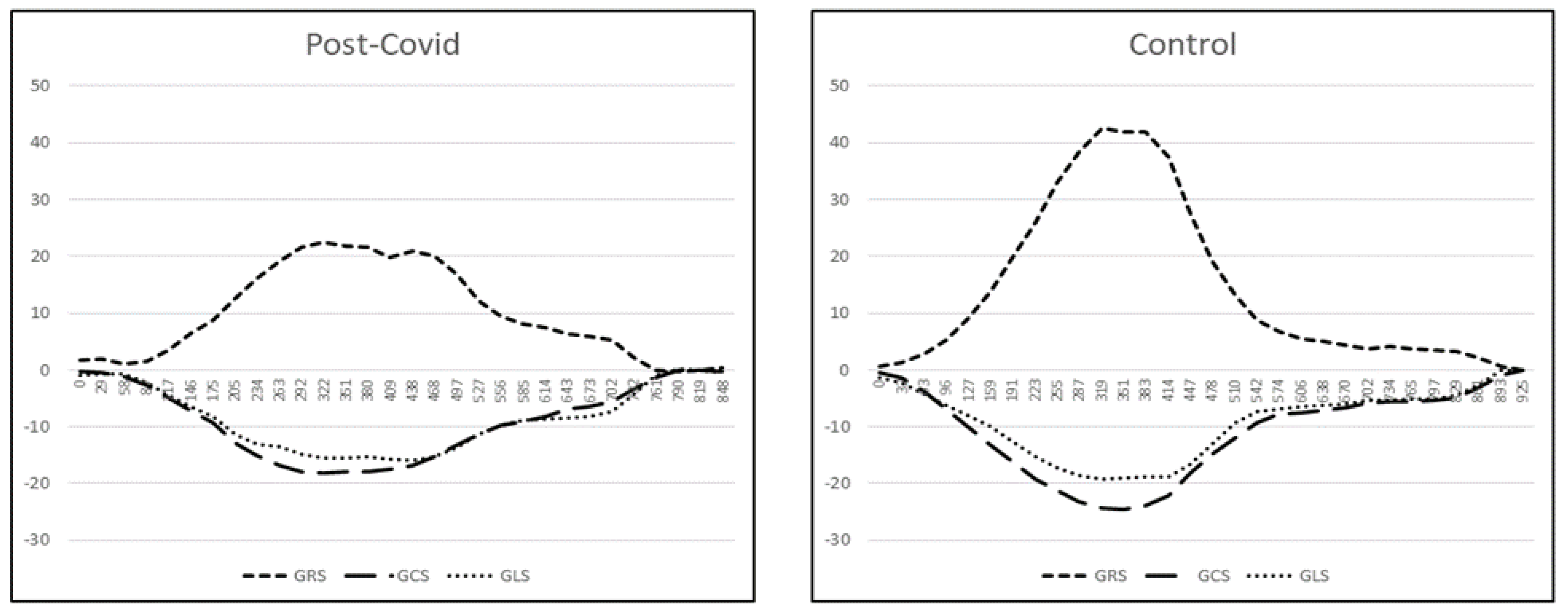
The initial CMR scan was performed a median of 99 days (IQR 92-110) after the COVID-19 infection. There was no significant difference in LVEF (63.6% vs. 63.2%, p = 0.85) or LV morphology between the groups in the initial CMR scan (Table 2). However, the initial CMR revealed significant group differences in reduced strain parameters for the PC group compared to the control group (GRS 32.6 ± 8.9 vs. 34.9 ± 8.4, p = 0.049; GCS −20.5 ± 2.8 vs. −21.5 ± 2.6, p = 0.015; radial systolic strain rate 1.9 ± 0.8 vs. 2.1 ± 0.6, p = 0.025; circumferential systolic strain rate −1.1 ± 0.3 vs. −1.2 ± 0.2, p= 0.022). These significant differences were confirmed in the multivariate analysis by stepwise linear regression for GRS (−2.9% (−5.4; −0.4), p = 0.025), GCS (1.2% (0.4; 2.0), p = 0.003), the systolic circumferential strain rate (0.08/s (0.01; 0.15), p = 0.02), and the systolic longitudinal strain rate (0.08/s (0.01; 0.15), p = 0.035) (Table 3).
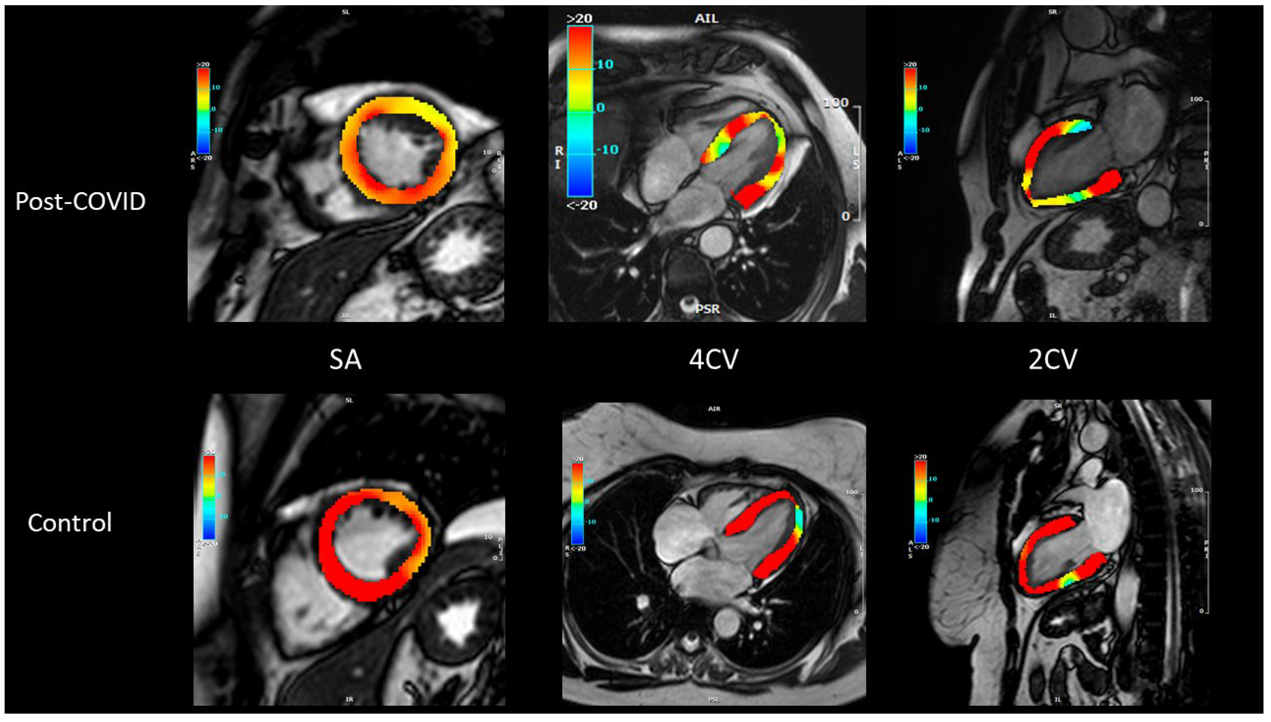
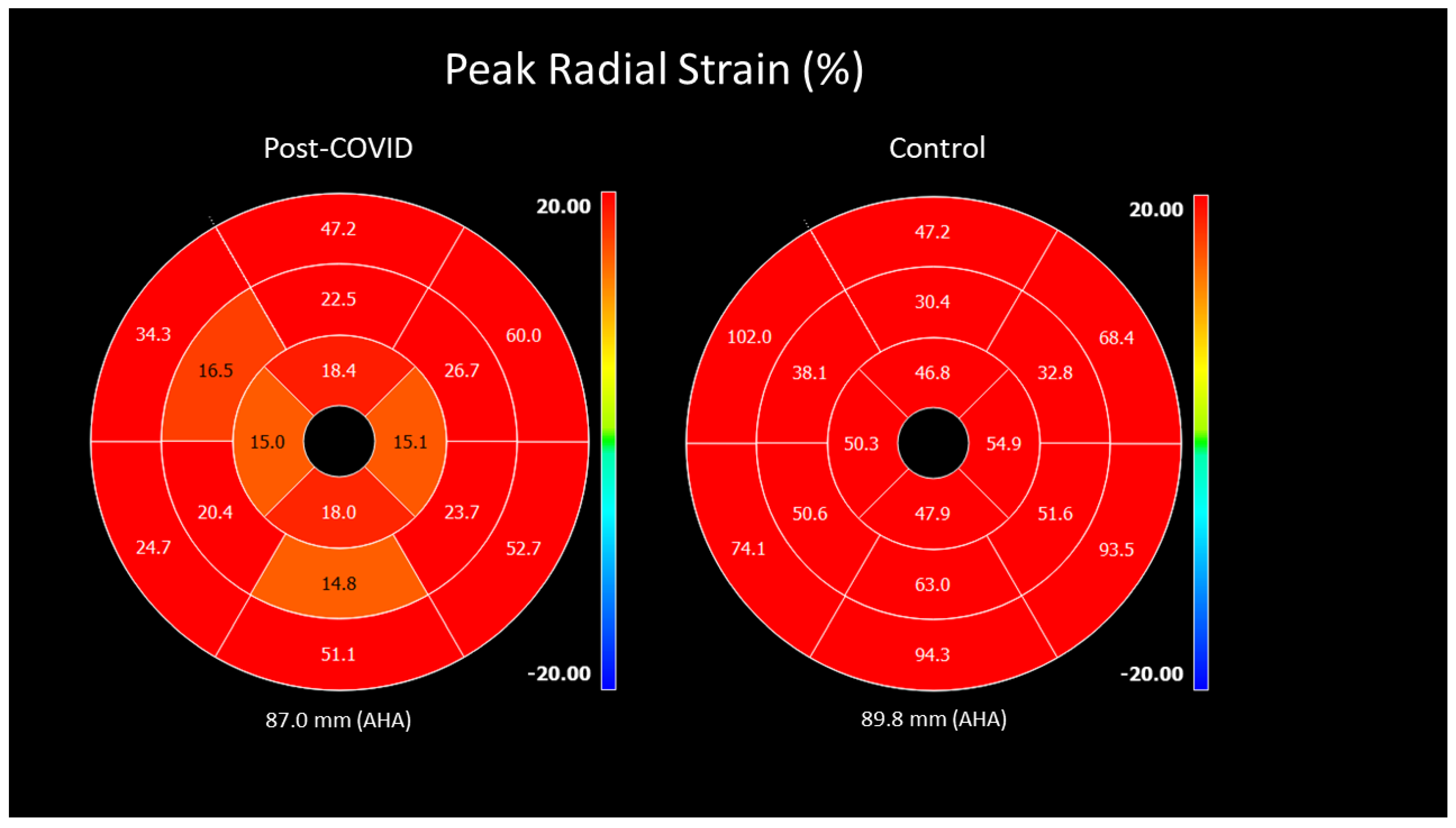
Additionally, we adjusted the statistical evaluation of strain differences for cardiovascular risk factors, family history, and obesity. A representative comparison of peak radial strain values using color-coded CMR is shown in Figure 2. A corresponding segmental polar map is demonstrated in Figure 3. The differences in radial and circumferential strain between the study groups are shown in Figure 4.
We detected myocardial LGE in eight (8.5%) patients and pericardial LGE in four patients (4.3%). Myocardial LGE had a myocarditis-typical pattern with intramural, subepicardial extension, mostly of the lateral walls. No LGE was found in the control group (p = 0.357; p = 0.581). A significant T2 signal representing myocardial edema in four (4.3%) patients and representing pericardial edema in two (2.1%) patients was detected. A normal T2 signal was found in the control group (p = 1.0). Pericardial effusion was detected in four (4.3%) PC patients and in none in the control group (p = 0.053).

3.3. Follow-Up Scan and Clinical Course

The median time between the initial CMR and follow-up scan in the 11 patients with perimyocarditis-like injury on initial examination was 90 days, IQR (82–113). There was no significant difference in LVEF between the initial and follow-up CMR scans (61.9 ± 6.7 vs. 59.25.9, p = 0.258). We found strain parameters to still be impaired, with no statistically significant difference for the strain parameters in the initial CMR scan compared to the follow-up scan: GRS 31.8 ± 7.8% vs. 30.4 ± 7.1, p = 0.638; GCS −19.5 ± 2.7% vs. −18.2 ± 3.0, p = 0.102; GLS −14.5 ± 2.5% vs. −13.5 ± 2.1, p = 0.465. Myocardial edema, LGE, and pericardial effusion resolved completely on the follow-up scan for all patients (3.8 ± 2.6% relative LGE size vs. 0%, p = 0.016, and 0.1 ± 0.2% relative T2 size vs. 0%, p = 0.5). The reported persistent clinical symptoms were also improved at follow-up. Six patients (54%) reported no shortness of breath and ten (91%) were free of chest discomfort at the time of follow-up. One patient developed newly diagnosed myocarditis during follow-up, fulfilling Lake Louise criteria on repeat CMR with positive edema and a typical late gadolinium enhancement pattern. No biopsy was performed, as the diagnosis was supported by typical imaging and clinical findings.

3.4. Prognosis

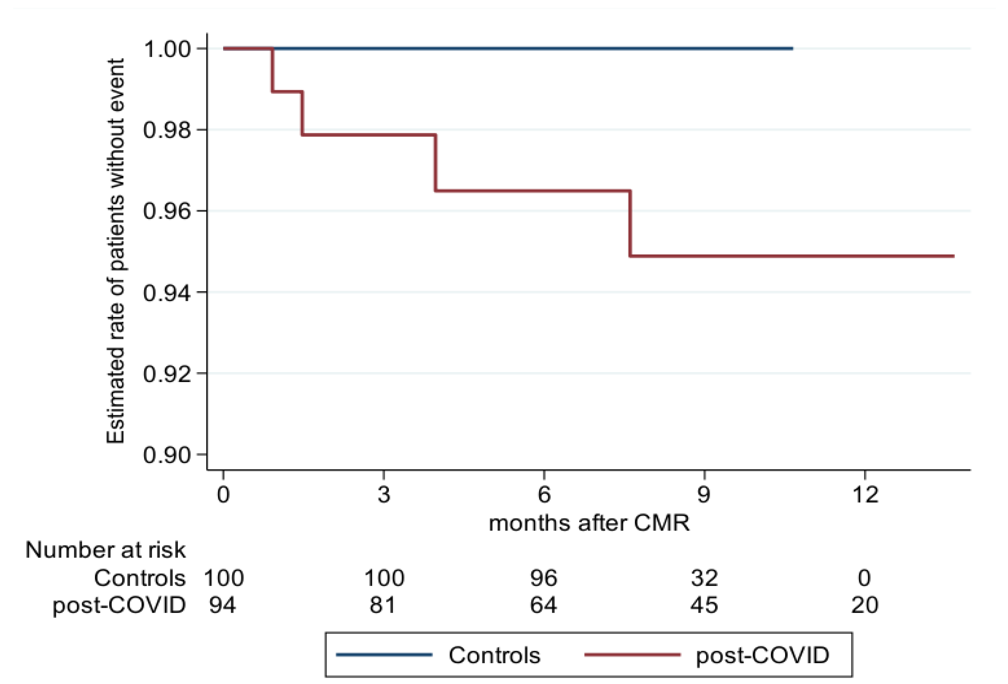
Patient MACE outcomes were assessed at a median of 269 days (IQR 146–351 days) after initial COVID-19 diagnosis in 91 (96.8%) patients of the PC group and in 100 (100%) of the control group (p = 0.112). We observed significantly different annual MACE rates of 0.063 (0.024; 0.167, p = 0.029) in the PC group and 0.000 in the controls (shown in Figure 5). The PC group had one patient with stroke, one patient with myocarditis, and two patients with myocardial infarction (Table 4).

4. Discussion

Healthcare systems worldwide are increasingly confronted with patients experiencing persistent cardiac symptoms following SARS-CoV-2 infection. The underlying pathophysiological mechanisms, associated clinical outcomes, and resulting healthcare burden remain poorly defined. In this study, we employed cardiovascular magnetic resonance (CMR) imaging to non-invasively and precisely characterize myocardial injury in individuals with the post-COVID-19 condition presenting with ongoing cardiovascular symptoms [2,13,14].

4.1. First Post-COVID-19 Assessment

We observed a modified strain pattern in the PC patients, with significantly reduced GRS and GCS 2D strain compared to a control group in the first assessment at three months post-infection.
Strain rate analysis reflects myocardial deformation and provides a sensitive marker of sub-clinical myocardial dysfunction. Reduced myocardial strain precedes impaired left ventricular functional parameters like the ejection fraction. Pathological myocardial deformation is a prognostic parameter in various cardiovascular diseases. Specifically, GLS is an independent predictor of outcome in non-ischemic diseases [15,16], while GCS is a predictor of segmental contractile recovery in ischemic diseases [17]. The observed reductions in global radial and circumferential strain should be interpreted as evidence of subtle, subclinical myocardial involvement rather than a direct cause of symptoms. Myocardial strain derived from feature-tracking CMR is a sensitive imaging biomarker capable of detecting early functional changes even when the ejection fraction remains normal. It is relatively less dependent on loading conditions and demonstrates good inter- and intra-observer reproducibility. Nevertheless, vendor-specific variability and the absence of universally defined normal reference ranges limit its diagnostic specificity. These factors were considered in the interpretation of our results.
Our results of significant strain changes in PC patients are in line with the findings of Ulloa et al., who observed reduced GRS and GCS in an initially hospitalized PC cohort. However, differences in this study were only significant in a subgroup analysis of seven patients [18]. In addition, Li et al. [19] also found altered GLS in 40 patients at 90 days after COVID-19 diagnosis. In contrast to our results, they found no significant changes in GCS in their smaller cohort. However, whether detected alterations in systolic strain patterns can be correlated with ongoing cardiac symptoms is unclear. Unlike other reports of early CMR evaluations of patients recently recovered from COVID-19 [14], we observed no significant impairment of LV function 99 days after infection in PC patients compared to a control group. Similar findings were recently reported by Salatzki et al. [20], who observed no evidence of persistent cardiac impairment and normal functional and morphological parameters in long-term follow-up after SARS-CoV-2 infection, despite early reports of minor strain alterations. Our results are consistent with these observations, indicating that small strain differences may reflect subtle myocardial changes without overt dysfunction. Similarly, Huang et al. found no significant alteration in LV function in patients recovered for one month from SARS-CoV-2 infection [6]. These seemingly inconsistent findings regarding left ventricular function in post-COVID-19 (PC) patients may be partly explained by several factors: (i) heterogeneity in study populations, including the presence of pre-existing conditions; (ii) varying severity of the initial SARS-CoV-2 infection; (iii) differences in the time interval between infection and CMR evaluation; and (iv) a lack of standardized CMR acquisition and analysis protocols.
In addition to functional assessment, CMR has high diagnostic value in the non-invasive evaluation of myocarditis [21]. A case series of 10 patients with acute COVID-19 identified diffuse myocardial edema in 8 and LGE in 3 patients [22]. Previous studies also found subclinical myocarditis, identified by myocardial edema and myocardial necrosis, in patients recovered from initial infection [6,23]. In addition, Wang and colleagues revealed LGE in 30% of initially hospitalized PC patients with a longer infection-to-CMR interval of 102.5 ± 20.6 days [5].
Our initial CMR evaluation of persons who were never hospitalized but who had the post-COVID-19 condition at three months after diagnosis revealed myocardial edema in 4.3% and myocardial necrosis as detected by LGE in 12.8%. In summary, myocardial inflammation and myocardial necrosis are not present in all PC patients, and this variability may be due to disease severity and the time interval since infection [2].
In addition, myocarditis is an infectious, inflammatory condition; however, the individual genetic background plays an important role in predisposition to inflammation and in its possible evolution towards subclinical cardiomyopathies [24]. Therefore, to date it cannot be excluded that the changes we observed are an expression of a subclinical cardiomyopathy with certain cardiomyopathy genes predisposing the development of an inflammatory condition. Despite the high prevalence of infections in the general population, it is not completely clear why only a small portion of them develop myocarditis, and only a portion of them will go on to cardiomyopathy, emphasizing the role of genetic variance in this setting. Our understanding of this problem is less solid, and further genetic investigation will be necessary.

4.2. Follow-Up Assessment

All patients with myocardial edema found in their initial CMR scan showed complete remission of the elevated T2 signal in the follow-up scan at six months post-infection. Of note, in a longer-term study, Puntmann et al. observed diffuse myocardial edema in symptomatic PC patients 329 days after COVID-19 diagnosis [7]. As alluded to previously, this may be due to differences in disease severity and patient selection.
LGE indicating myocardial injury in the initial CMR scan likewise vanished in our follow-up assessment after 189 days. The long-term cardiac effects due to persistent myocardial injury are still debated, and data on long-term cardiac involvement are patchy [2]. However, a study of 74 health care workers at six months after having COVID-19, but not requiring hospitalization, showed no significant changes in LV structure, LV function, or myocardial tissue [25]. Interestingly, subtle, long-term cardiac changes in terms of increased metabolic demand were still detectable more than twelve years after recovery in SARS patients [26].
Our observations are concordant with early case reports in hospitalized patients showing a frequent presence of LGE [27,28] and diffuse inflammatory involvement [29]. Unlike these previous reports, our findings reveal that significant cardiac involvement occurs independently of the severity of original presentation and persists beyond the period of acute presentation. Taken together, it is clear that the long-term effects of COVID-19 on the myocardium still require further investigation.

4.3. Prognostic Considerations

We observed a significantly higher annual MACE rate in the PC patients compared to a control group approximately one year after diagnosis. Currently, prognostic data are lacking after recovery from COVID-19. Only a few studies with a short follow-up interval have examined cardiovascular outcomes in previously hospitalized individuals. An unselected Italian follow-up study, with a median period of 60 days since symptom onset, revealed fatigue and persistent dyspnea as the primary symptoms after COVID-19, but no MACE evaluation was performed [30].
Our cardiac findings confirm the results of a recent cohort study on US veterans that described an increased risk and one-year burden for cardiovascular diseases in patients after COVID-19 [31]. Another recent cohort study observed substantial pulmonary and extra-pulmonary health impairments more than six months after a COVID-19 diagnosis [32]. In addition to traditional cardiovascular risk factors, other mechanisms may underlie the observed long-term effects. The elevated MACE rate observed in the post-COVID-19 cohort likely reflects a multifactorial process. Baseline cardiovascular risk factors such as obesity, hyperlipidemia, and family history were more prevalent in this group, and although these were adjusted for in regression analyses, residual confounding cannot be excluded. A potential selection bias—where individuals with higher cardiovascular risk may have been more susceptible to symptomatic infection—may also contribute. Beyond these conventional risks, emerging evidence points toward persistent endothelial dysfunction, thrombo-inflammation, subclinical microvascular ischemia, and autonomic imbalance as mechanisms increasing long-term cardiovascular risk after SARS-CoV-2 infection. A recent CMR study by Weberling et al. (2025) [33] demonstrated abnormal coronary vascular responses in patients with long COVID syndrome, suggesting that microvascular dysfunction and impaired coronary vasoreactivity may contribute to ongoing symptoms and increased cardiovascular risk despite otherwise normal ventricular morphology and function. These findings support our interpretation that subtle strain changes might represent part of a broader post-COVID-19 myocardial or vascular involvement process.
The long-term prognosis of the post-COVID-19 condition is not well understood. Therefore, large-scale longitudinal studies are needed to better understand the medium-term and long-term implications of the post-COVID-19 condition and remaining cardiac changes for accurate risk stratification of patients for adverse cardiovascular events.

4.4. Study Limitations

The single-center study design and relatively small patient number are limitations of our study. Our study deliberately concentrated on a selection of non-hospitalized patients who had persistent cardiac symptoms after recovering from COVID-19. Our results may not be transferrable to the global PC population, and we cannot determine whether our findings are causal, since correlation does not imply causation. In addition, some other subtle CMR findings may not correlate with clinical symptoms. Thus, conclusions that are drawn from our study have to be interpreted carefully and, therefore, prospective multi-center studies need to be conducted to confirm our findings.
Furthermore, mapping techniques for tissue characterization were not included due to a lack of protocol sequences at the time of data acquisition; therefore, the full 2018 Lake Louise criteria for myocarditis could not be applied. Diffuse tissue changes may have been missed. In addition, asymptomatic SARS-CoV-2 infections in the control cohort cannot be completely excluded. Further limitations include potential residual confounding due to group differences in cardiovascular risk profiles and the inability to confirm infection-naïve status with serological testing. These factors should be considered when interpreting our findings and underscore the need for multi-center longitudinal studies to validate and expand on these results.

5. Conclusions

In our long-term observation of PC patients, we found a reversible perimyocarditis-like injury and a persistent altered strain pattern six months after diagnosis with COVID-19. Persistent myocardial deformation may be one explanation for the ongoing cardiac symptoms in PC patients. Verification of these findings is required in larger multi-center trials with a broader symptom range, including asymptomatic individuals and those with severe COVID-19 disease.
The COVID-19 pandemic led to increased research in the field of acute infection. However, the number of affected patients suffering from the post-COVID-19 condition is currently rising. In our study, we evaluated cardiac involvement in post-COVID-19 conditions lasting for more than three months. Research data on post-COVID-19 are lacking. Here we observed subtle mechanical changes in the strain pattern of symptomatic post-COVID-19 patients, although the prevalence of classical myocarditis-like injury in this population was low. Clinical outcome analysis showed significantly increased event rates in the post-COVID-19 condition compared to a control group.

Author Contributions

Conceptualization, N.A.-K. and N.A.; methodology, N.A.-K.; software, N.A.; data curation, N.A.; writing—original draft preparation, N.A.-K. and N.A.; writing—review and editing, L.K., N.N.-B., M.G. and A.Z.; supervision, N.A.-K.; project administration, P.H. and M.S.; funding acquisition, N.A.-K. and N.A. All authors have read and agreed to the published version of the manuscript.

Funding

This research was funded by University Witten/Herdecke internal Research Program IFF-2021. N.A.-K. and N.A. received funds from the aforementioned Program.

Institutional Review Board Statement

This study was conducted in accordance with the Declaration of Helsinki and approved by the Ethics Committee of University Witten/Herdecke, Germany, Code IFF-2021, Date: l January 2021.

Informed Consent Statement

Informed consent was obtained from all subjects involved in this study.

Data Availability Statement

All data used in this study are securely stored in encrypted form to ensure data protection and confidentiality. Prior to analysis, all datasets were anonymized to remove any personally identifiable information. The anonymized data are available from the corresponding author upon reasonable request.

Acknowledgments

We especially thank Hiltrud Niggemann, certified biometrician, for the excellent collaboration and statistical analysis. We are extraordinarily thankful to Lisa Costello-Boerrigter, for critically important revisions and important intellectual and linguistic content. We gratefully thank the entire staff of technologists of the cardiovascular magnetic imaging department of the Helios University Hospital Wuppertal for data acquisition.

Conflicts of Interest

The authors declare no conflicts of interest.

Abbreviations

The following abbreviations are used in this manuscript:
SARS-CoV-2Severe Acute Respiratory Syndrome Coronavirus 2
COVID-19Coronavirus Disease 2019
MRMagnetic resonance
CMRCardiovascular magnetic resonance
PCPost-COVID-19
RT-PCRReverse transcription polymerase
MRIMagnetic resonance imaging
LGELate gadolinium enhancement
LV-EFLeft ventricular ejection fraction
GLSGlobal longitudinal strain
GCSGlobal circumferential strain
GRSGlobal radial strain
MACEMajor adverse cardiac event
NYHA ClassNew York Heart Association classification
CCS ClassCanadian Cardiovascular Society classification
LVEDVLeft ventricular end-diastolic volume
LVESVLeft ventricular end-systolic volume
LVSVLeft ventricular stroke volume

References

  1. Available online: https://covid19.who.int/ (accessed on 1 August 2025).
  2. Petersen, S.E.; Friedrich, M.G.; Leiner, T.; Elias, M.D.; Ferreira, V.M.; Fenski, M.; Flamm, S.D.; Fogel, M.; Garg, R.; Halushka, M.K.; et al. Cardiovascular Magnetic Resonance for Patients With COVID-19. JACC Cardiovasc. Imaging 2022, 15, 685–699. [Google Scholar] [CrossRef]
  3. Soriano, J.B.; Murthy, S.; Marshall, J.C.; Relan, P.; Diaz, J.V. A clinical case definition of post-COVID-19 condition by a Delphi consensus. Lancet Infect. Dis. 2022, 22, e102–e107. [Google Scholar] [CrossRef] [PubMed]
  4. Augustin, M.; Schommers, P.; Stecher, M.; Dewald, F.; Gieselmann, L.; Gruell, H.; Horn, C.; Vanshylla, K.; Di Cristanziano, V.; Osebold, L.; et al. Post-COVID syndrome in non-hospitalised patients with COVID-19, a longitudinal prospective cohort study. Lancet Reg. Health Eur. 2021, 6, 100122. [Google Scholar] [CrossRef] [PubMed]
  5. Wang, H.; Li, R.; Zhou, Z.; Jiang, H.; Yan, Z.; Tao, X.; Li, H.; Xu, L. Cardiac involvement in COVID-19 patients: Mid-term follow up by cardiovascular magnetic resonance. J. Cardiovasc. Magn. Reson. 2021, 23, 14. [Google Scholar] [CrossRef] [PubMed]
  6. Huang, L.; Zhao, P.; Tang, D.; Zhu, T.; Han, R.; Zhan, C.; Liu, W.; Zeng, H.; Tao, Q.; Xia, L. Cardiac Involvement in Patients Recovered From COVID-2019 Identified Using Magnetic Resonance Imaging. JACC Cardiovasc. Imaging 2020, 13, 2330–2339. [Google Scholar] [CrossRef]
  7. Puntmann, V.O.; Martin, S.; Shchendrygina, A.; Hoffmann, J.; Ka, M.M.; Giokoglu, E.; Vanchin, B.; Holm, N.; Karyou, A.; Laux, G.S.; et al. Long-term cardiac pathology in individuals with mild initial COVID-19 illness. Nat. Med. 2022, 28, 2117–2123. [Google Scholar] [CrossRef]
  8. Kramer, C.M.; Barkhausen, J.; Bucciarelli-Ducci, C.; Flamm, S.D.; Kim, R.J.; Nagel, E. Standardized cardiovascular magnetic resonance imaging (CMR) protocols: 2020 update. J. Cardiovasc. Magn. Reson. 2020, 22, 17. [Google Scholar] [CrossRef]
  9. Wright, J.; Adriaenssens, T.; Dymarkowski, S.; Desmet, W.; Bogaert, J. Quantification of Myocardial Area at Risk With T2-Weighted CMR. JACC Cardiovasc. Imaging 2009, 2, 825–831. [Google Scholar] [CrossRef]
  10. Schulz-Menger, J.; Bluemke, D.A.; Bremerich, J.; Flamm, S.D.; Fogel, M.A.; Friedrich, M.G.; Kim, R.J.; von Knobelsdorff-Brenkenhoff, F.; Kramer, C.M.; Pennell, D.J.; et al. Standardized image interpretation and post processing in cardiovascular magnetic resonance: Society for Cardiovascular Magnetic Resonance (SCMR) Board of Trustees Task Force on Standardized Post Processing. J. Cardiovasc. Magn. Reson. 2013, 15, 35. [Google Scholar] [CrossRef]
  11. Goto, Y.; Ishida, M.; Takase, S.; Sigfridsson, A.; Uno, M.; Nagata, M.; Ichikawa, Y.; Kitagawa, K.; Sakuma, H. Comparison of Displacement Encoding With Stimulated Echoes to Magnetic Resonance Feature Tracking for the Assessment of Myocardial Strain in Patients With Acute Myocardial Infarction. Am. J. Cardiol. 2017, 119, 1542–1547. [Google Scholar] [CrossRef]
  12. Liu, B.; Dardeer, A.M.; Moody, W.E.; Hayer, M.K.; Baig, S.; Price, A.M.; Leyva, F.; Edwards, N.C.; Steeds, R.P. Reference ranges for three-dimensional feature tracking cardiac magnetic resonance: Comparison with two-dimensional methodology and relevance of age and gender. Int. J. Cardiovasc. Imaging 2017, 34, 761–775. [Google Scholar] [CrossRef]
  13. Bavishi, C.; Bonow, R.O.; Trivedi, V.; Abbott, J.D.; Messerli, F.H.; Bhatt, D.L. Special Article—Acute myocardial injury in patients hospitalized with COVID-19 infection: A review. Prog. Cardiovasc. Dis. 2020, 63, 682–689. [Google Scholar] [CrossRef] [PubMed]
  14. Puntmann, V.O.; Carerj, M.L.; Wieters, I.; Fahim, M.; Arendt, C.; Hoffmann, J.; Shchendrygina, A.; Escher, F.; Vasa-Nicotera, M.; Zeiher, A.M.; et al. Outcomes of Cardiovascular Magnetic Resonance Imaging in Patients Recently Recovered From Coronavirus Disease 2019 (COVID-19). JAMA Cardiol. 2020, 5, 1265. [Google Scholar] [CrossRef] [PubMed]
  15. Buss, S.J.; Breuninger, K.; Lehrke, S.; Voss, A.; Galuschky, C.; Lossnitzer, D.; Andre, F.; Ehlermann, P.; Franke, J.; Taeger, T.; et al. Assessment of myocardial deformation with cardiac magnetic resonance strain imaging improves risk stratification in patients with dilated cardiomyopathy. Eur. Heart J. Cardiovasc. Imaging 2015, 16, 307–315. [Google Scholar] [CrossRef]
  16. Houard, L.; Benaets, M.B.; de Meester de Ravenstein, C.; Rousseau, M.F.; Ahn, S.A.; Amzulescu, M.S.; Roy, C.; Slimani, A.; Vancraeynest, D.; Pasquet, A.; et al. Additional Prognostic Value of 2D Right Ventricular Speckle-Tracking Strain for Prediction of Survival in Heart Failure and Reduced Ejection Fraction. JACC Cardiovasc. Imaging 2019, 12, 2373–2385. [Google Scholar] [CrossRef]
  17. Wong, D.T.; Leong, D.P.; Weightman, M.J.; Richardson, J.D.; Dundon, B.K.; Psaltis, P.J.; Leung, M.C.; Meredith, I.T.; Worthley, M.I.; Worthley, S.G. Magnetic resonance-derived circumferential strain provides a superior and incremental assessment of improvement in contractile function in patients early after ST-segment elevation myocardial infarction. Eur. Radiol. 2014, 24, 1219–1228. [Google Scholar] [CrossRef]
  18. Ulloa, J.U.; de Vega, V.M.; Montañés, O.S.; Vázquez, A.Á.; Sánchez-Enrique, C.; Jiménez, S.H.; García, F.D.S.; Ruiz, L.L.; Rodríguez, M.R.; Pizarro, G.; et al. Cardiac magnetic resonance in recovering COVID-19 patients. Feature tracking and mapping analysis to detect persistent myocardial involvement. IJC Heart Vasc. 2021, 36, 100854. [Google Scholar] [CrossRef]
  19. Li, X.; Wang, H.; Zhao, R.; Wang, T.; Zhu, Y.; Qian, Y.; Liu, B.; Yu, Y.; Han, Y. Elevated Extracellular Volume Fraction and Reduced Global Longitudinal Strains in Participants Recovered from COVID-19 without Clinical Cardiac Findings. Radiology 2021, 299, E230–E240. [Google Scholar] [CrossRef] [PubMed]
  20. Salatzki, J.; Ochs, A.; Weberling, L.D.; Heins, J.; Zahlten, M.; Whayne, J.G.; Stehning, C.; Giannitsis, E.; Denkinger, C.M.; Merle, U.; et al. Absence of cardiac impairment in patients after severe acute respiratory syndrome coronavirus type 2 infection: A long-term follow-up study. J. Cardiovasc. Magn. Reson. 2024, 26, 101124. [Google Scholar] [CrossRef]
  21. Kotanidis, C.P.; Bazmpani, M.-A.; Haidich, A.-B.; Karvounis, C.; Antoniades, C.; Karamitsos, T.D. Diagnostic Accuracy of Cardiovascular Magnetic Resonance in Acute Myocarditis. JACC Cardiovasc. Imaging 2018, 11, 1583–1590. [Google Scholar] [CrossRef]
  22. Esposito, A.; Palmisano, A.; Natale, L.; Ligabue, G.; Peretto, G.; Lovato, L.; Vignale, D.; Fiocchi, F.; Marano, R.; Russo, V. Cardiac Magnetic Resonance Characterization of Myocarditis-Like Acute Cardiac Syndrome in COVID-19. JACC Cardiovasc. Imaging 2020, 13, 2462–2465. [Google Scholar] [CrossRef] [PubMed]
  23. Ng, M.Y.; Ferreira, V.M.; Leung, S.T.; Yin Lee, J.C.; Ho-Tung Fong, A.; To Liu, R.W.; Man Chan, J.W.; Wu, A.K.L.; Lung, K.C.; Crean, A.M.; et al. Patients Recovered From COVID-19 Show Ongoing Subclinical Myocarditis as Revealed by Cardiac Magnetic Resonance Imaging. JACC Cardiovasc. Imaging 2020, 13, 2476–2478. [Google Scholar] [CrossRef]
  24. Cannata’, A.; Artico, J.; Gentile, P.; Merlo, M.; Sinagra, G. Myocarditis evolving in cardiomyopathy: When genetics and offending causes work together. Eur. Heart J. Suppl. 2019, 21, B90–B95. [Google Scholar] [CrossRef]
  25. Joy, G.; Artico, J.; Kurdi, H.; Seraphim, A.; Lau, C.; Thornton, G.D.; Oliveira, M.F.; Adam, R.D.; Aziminia, N.; Menacho, K.; et al. Prospective Case-Control Study of Cardiovascular Abnormalities 6 Months Following Mild COVID-19 in Healthcare Workers. JACC Cardiovasc. Imaging 2021, 14, 2155–2166. [Google Scholar] [CrossRef]
  26. Wu, Q.I.; Zhou, L.; Sun, X.; Yan, Z.; Hu, C.; Wu, J.; Xu, L.; Li, X.; Liu, H.; Yin, P.; et al. Altered Lipid Metabolism in Recovered SARS Patients Twelve Years after Infection. Sci. Rep. 2017, 7, 9110. [Google Scholar] [CrossRef]
  27. Inciardi, R.M.; Lupi, L.; Zaccone, G.; Italia, L.; Raffo, M.; Tomasoni, D.; Cani, D.S.; Cerini, M.; Farina, D.; Gavazzi, E.; et al. Cardiac Involvement in a Patient With Coronavirus Disease 2019 (COVID-19). JAMA Cardiol. 2020, 5, 819. [Google Scholar] [CrossRef]
  28. Gravinay, P.; Issa, N.; Girard, D.; Camou, F.; Cochet, H. CMR and serology to diagnose COVID-19 infection with primary cardiac involvement. Eur. Heart J. Cardiovasc. Imaging 2021, 22, 133. [Google Scholar] [CrossRef]
  29. Luetkens, J.A.; Isaak, A.; Zimmer, S.; Nattermann, J.; Sprinkart, A.M.; Boesecke, C.; Rieke, G.J.; Zachoval, C.; Heine, A.; Velten, M.; et al. Diffuse Myocardial Inflammation in COVID-19 Associated Myocarditis Detected by Multiparametric Cardiac Magnetic Resonance Imaging. Circ. Cardiovasc. Imaging 2020, 13, e010897. [Google Scholar] [CrossRef]
  30. Carfì, A.; Bernabei, R.; Landi, F. Persistent Symptoms in Patients After Acute COVID-19. JAMA 2020, 324, 603. [Google Scholar] [CrossRef] [PubMed]
  31. Xie, Y.; Xu, E.; Bowe, B.; Al-Aly, Z. Long-term cardiovascular outcomes of COVID-19. Nat. Med. 2022, 28, 583–590. [Google Scholar] [CrossRef] [PubMed]
  32. Al-Aly, Z.; Xie, Y.; Bowe, B. High-dimensional characterization of post-acute sequelae of COVID-19. Nature 2021, 594, 259–264. [Google Scholar] [CrossRef] [PubMed]
  33. Weberling, L.D.; Hillier, E.; Friedrich, M.G.; Zahlten, M.; Frey, N.; André, F.; Steen, H. Abnormal coronary vascular response in patients with long COVID syndrome—A case-control study using oxygenation-sensitive cardiovascular magnetic resonance. J. Cardiovasc. Magn. Reson. 2025, 27, 101890. [Google Scholar] [CrossRef] [PubMed]
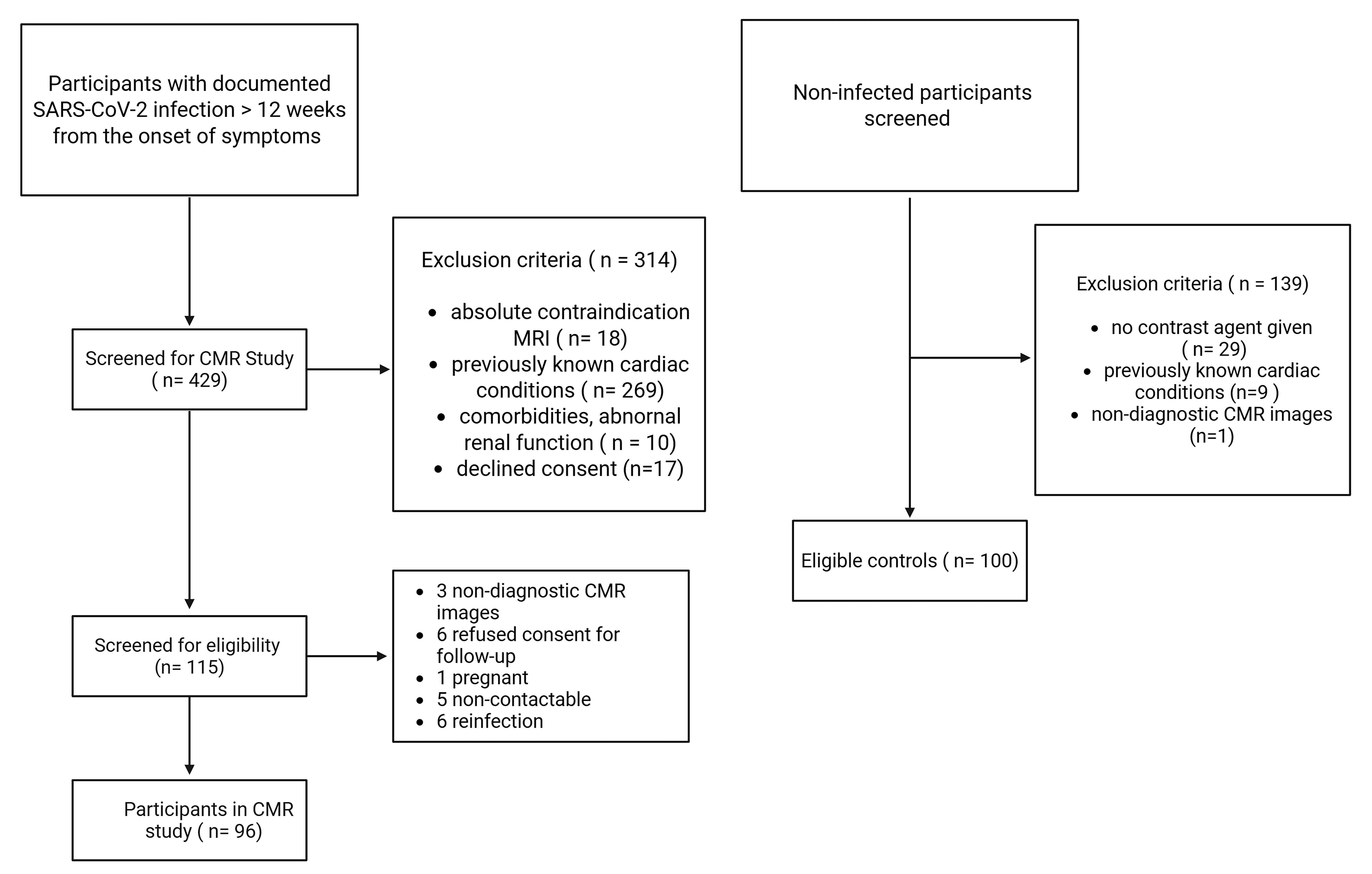
Figure 1. Study inclusion flowchart.
Covid 05 00193 g001
Figure 2. Images showing short-axis (SAX) view, four-chamber view (4CV), and two-chamber view (2CV) show reduced strain in post-COVID-19 patients compared to control patients.
Covid 05 00193 g002
Figure 3. Polar maps of peak radial strain: Representative polar maps of peak radial strain showing significant reduction in the apical to mid-ventricular strain pattern of a PC patient (left) compared to a control patient (right).
Covid 05 00193 g003
Figure 4. Strain parameter comparison: These diagrams of the three strain parameters show the reduction in GRS and GCS between the PC and control groups.
Covid 05 00193 g004
Figure 5. Kaplan–Meier plot with estimated rates of event-free patients 12 months after CMR among PC patients and controls.
Covid 05 00193 g005
Table 1. Comparison of baseline characteristics between post-COVID-19 patients and controls.
PC
n = 94
C
n = 100
p-Value *
Gendermale35 (37.2%)40 (40.0%)
female59 (62.8%)60 (60.0%)0.768
Age 44.9 ± 16.048.7 ± 16.50.093
BMI 26.3 ± 4.926.0 ± 4.20.801
Positive COVID-19 Testno0 (0%)100 (100%)
yes94 (100%)0 (0%)<0.001
Hypertensionno60 (73.2%)66 (66.0%)
yes22 (26.8%)34 (34.0%)0.335
Hyperlipidemiano58 (70.7%)93 (93.0%)
yes24 (29.3%)7 (7.0%)<0.001
Diabetesno77 (93.9%)96 (96.0%)
yes5 (6.1%)4 (4.0%)0.733
Smokingno71 (86.6%)90 (90.9%)
yes11 (13.4%)9 (9.1%)0.476
Family History of Cardiovascular Diseaseno47 (58.0%)87 (87.0%)
yes34 (42.0%)13 (13.0%)<0.001
Obesityno61 (74.4%)94 (94.0%)
yes21 (25.6%)6 (6.0%)<0.001
Clinical Symptoms in The Acute Phaseno
yes
3 (3.2%)
91 (96.8%)
-
-
Dyspneano
yes
40 (44.9%)
49 (55.1%)
-
-
NYHA Class0
1
2
3
4
40 (44.9%)
9 (10.1%)
16 (18.0%)
14 (15.7%)
10 (11.2%)
-
-
-
-
-
Angina CCS Class0
1
2
3
4
49 (55.1%)
12 (13.5%)
13 (14.6%)
3 (3.4%)
12 (13.5%)
-
-
-
-
-
Feverno
yes
25 (28.1%)
64 (71.9%)
-
-
Diarrheano
yes
64 (71.9%)
25(28.1%)
-
-
Loss of Smell/Tasteno
yes
29 (32.6%)
60 (67.4%)
-
-
PC = post-COVID-19 patients; C = controls; BMI = body mass index; CCS = Canadian Cardiovascular Society (CCS) angina class; NYHA = New York Heart Association functional class. (-) = no symptoms * Fisher’s exact test.
Table 2. Mean values and SDs of the CMR parameters in the initial CMR scan between post-COVID-19 patients and controls.
PC
n = 94
C
n = 100
p-Value *
LVEF [%]63.4 ± 4.963.4 ± 5.10.850
LVEDV [mL]152.9 ± 38.8149.3 ± 36.20.558
LVESV [mL]56.7 ± 17.655.5 ± 16.90.627
LVSV [mL]96.2 ± 23.794.8 ± 23.40.732
LGE-Volume [mL]0.4 ± 1.300.168
T2-Volume [mL]0.1 ± 1.300.485
* Mann–Whitney U test. PC = post-COVID-19 patients; C = controls; LVEF = left ventricular ejection fraction; LVEDV = left ventricular end-diastolic volume; LVESV = left ventricular end-systolic volume; LVSV = left ventricular stroke volume; LGE = late gadolinium enhancement.
Table 3. Mean values and SDs together with distributions in brackets of CMR feature-tracking strain parameters—differences between post-COVID-19 patients and control group.
Post-COVID-19
n = 94
Control
n = 100
p-Value *95% CI,
p-Value **
Peak strain% radial
(GRS)
32.6 ± 8.9
[18.1–72.3]
34.9 ± 8.4
[20.5–58.0]
0.049−2.9 (−5.4; −0.4), p = 0.025
Peak strain% circumferential−20.5 ± 2.8−21.5 ± 2.60.0151.2 (0.4; 2.0), p = 0.003
(GCS)[−26.4–11.8][−27.2–15.5]
Peak strain% longitudinal−14.7 ± 2.3−15.1 ± 2.20.3790.4 (−0.3; 1.1), p = 0.292
(GLS)[−19.6–9.2][−20.1–9.0]
Time to peak strain (ms)326.1 ± 45.9315.4 ± 44.0.06811.8 (−1.3; 25.0), p = 0.078
radial[239.2–423.2][213.2–442.4]
Time to peak strain (ms)317.7 ± 44.5308.2 ± 38.40.1536.8 (−5.5; 19.1), p = 0.277
circumferential[232.8–445.2][222.0–401.8]
Time to peak strain (ms)355.7 ± 59.8340.1 ± 54.90.1066.5 (−9.9; 22.9), p = 0.437
longitudinal[222.2–508.8][213.6–460.6]
Systolic strain rate (/s)1.9 ± 0.82.1 ± 0.60.025−0.17 (−0.37; 0.02), p = 0.084
radial[1.0–5.5][1.1–4.1]
Systolic strain rate (/s)−1.1 ± 0.3−1.2 ± 0.20.0220.08 (0.01; 0.15), p = 0.020
circumferential[−2.7–0.7][−2.0–0.7]
Systolic strain rate (/s)−0.8 ± 0.3−0.9 ± 0.20.0990.08 (0.01; 0.15), p = 0.035
longitudinal[−2.7–0.6][−1.9–0.5]
Peak diastolic strain rate (/s)−2.5 ± 1.0−2.6 ± 0.80.2370.16 (−0.11; 0.43), p = 0.242
radial[−8.2–1.1][−5.2–1.0]
Peak diastolic strain rate (/s)1.4 ± 0.51.4 ± 0.30.8190.06 (−0.03; 0.14), p = 0.186
circumferential[−1.8–2.6][0.7–2.5]
Peak diastolic strain rate (/s)1.0 ± 0.31.0 ± 0.30.588−0.02 (−0.10; 0.05), p = 0.535
longitudinal[0.5–2.3][0.6–2.1]
* p-value of Mann–Whitney U test (bivariate analysis). ** Estimated difference (95% CI), p-value. Estimation by stepwise linear regression (p < 0.05) keeping the group difference in the linear regression model (lockterm) and selecting from the following cardiovascular risk factors: hyperlipidemia, diabetes, smoking, family history, obesity. PC = post-COVID-19 patients; C = controls.
Table 4. Frequency of major adverse clinical events: comparison between post-COVID-19 patients and controls.
PC
n = 91
C
n = 100
p-Value *
MACE40
Stroke1
Myocarditis1
MI2
Death0
Years63.768.3
Rate per year (95% CI)0.063 (0.024; 0.167)0 0.029
Estimated MACE rate ** after 12 months (95% CI)5.2 (2.0; 13.5)0
* Log-rank test for equality of survivor functions. ** Kaplan–Meier estimation. PC = Post-COVID-19 patients; C = controls; MACE = major adverse clinical event; MI = myocardial infarction.
Disclaimer/Publisher’s Note: The statements, opinions and data contained in all publications are solely those of the individual author(s) and contributor(s) and not of MDPI and/or the editor(s). MDPI and/or the editor(s) disclaim responsibility for any injury to people or property resulting from any ideas, methods, instructions or products referred to in the content.