Article

Survey of the Trunk Wood Mycobiome of an Ancient Tilia × europaea L.

1
Mendeleum—Institute of Genetics, Faculty of Horticulture, Mendel University in Brno, Valtická 334, 691 44 Lednice, Czech Republic
2
Department of Planting Design and Maintenance, Faculty of Horticulture, Mendel University in Brno, Valtická 686, 691 44 Lednice, Czech Republic
*
Author to whom correspondence should be addressed.
Appl. Microbiol. 2025, 5(4), 131; https://doi.org/10.3390/applmicrobiol5040131
Submission received: 25 September 2025 / Revised: 25 October 2025 / Accepted: 5 November 2025 / Published: 13 November 2025

Abstract

The genus Tilia (Malvaceae) comprises long-lived broadleaf trees of considerable ecological, cultural, and historical importance in temperate Europe and Asia. Among these, Tilia × europaea L. (common European linden) is a key native species in Central and Northern Europe, with individuals documented to live for several centuries. While the phyllosphere and soil-associated microbiomes of linden have been studied, the internal fungal communities inhabiting ancient trees remain poorly understood. In this study, the complete mycobiome of linden tree wood was analyzed. Wood-inhabiting fungi (the wood mycobiome) include endophytes, saprotrophs, and potential pathogens that can strongly influence host vitality and ecosystem processes. Advances in high-throughput amplicon sequencing (HTAS) now provide unprecedented opportunities to characterize these hidden communities. In this study, we investigated the trunk wood mycobiome of an ancient T. × europaea L. individual using a culture-independent HTAS approach. The results reveal a diverse fungal assemblage, including taxa like Arthinium or Phialemonium not previously reported from living linden wood, and highlight potential implications for tree health and longevity. This work provides a first baseline characterization of the internal mycobiome of the ancient Tilia tree and contributes to broader efforts to conserve its biological and cultural value.

1. Introduction

The genus Tilia L. (Malvaceae), commonly referred to as linden trees, comprises long-lived broadleaf species widely distributed across temperate regions of Europe and Asia. T. × europaea L. is a prominent native hybrid species in Central and Northern Europe because it originates from two native parental species and holds significant ecological importance, cultural symbolism, and historical relevance in both urban and rural landscapes. Dendrological studies using DBH-based models have shown a strong correlation (r2 = 0.926) between trunk diameter and age, indicating that Tilia sp. individuals can live for several centuries with high estimation accuracy (<10% error) [1]. Genetic analyses using SSR markers further revealed that some historically valuable specimens of T. cordata are aged between 400 and 1000 years [2].
Fungal communities inhabiting the internal woody tissues of trees, the so-called wood mycobiome, play critical roles in host physiology, wood decay, and broader ecosystem function. These communities include both saprotrophic and endophytic fungi; they are particularly dangerous because symptoms appear only after the host has been subjected to stress [3]. For instance, pyrotag sequencing of ITS2 has been successfully applied to characterize the deadwood mycobiome, demonstrating a correlation between fungal community composition, host species, and wood properties [4]. Moreover, long-read PacBio sequencing of fungal ITS regions in deadwood revealed rich taxonomic and functional diversity, including endophytes, saprotrophs, and pathogens, reinforcing the complexity of wood-inhabiting mycobiomes [5].
Despite the growing interest in tree-associated microbiomes, knowledge of the internal mycobiome of ancient linden trees remains sparse. Most studies to date have emphasized foliar pathogens or soil fungal communities, whereas endophytic and wood-colonizing fungi within long-lived linden trees are largely unexplored. This knowledge gap persists even as methodological advances, particularly high-throughput amplicon sequencing (HTAS) of the ITS ribosomal DNA region, offer unprecedented resolution in characterizing fungal diversity in woody substrates [6]. There are also different sequencing approaches used for fungal identification, such as whole-genome sequencing, RNA sequencing, long-read amplicon sequencing, multi-locus sequence typing, and classical Sanger sequencing.
In this study, we surveyed the wood mycobiome of the trunk of an ancient linden tree using a culture-independent high-throughput sequencing approach. Our objectives were (i) to characterize the taxonomic composition of the internal fungal community of linden wood, (ii) to document the presence of fungal taxa not previously reported from living linden wood, and (iii) to assess whether these fungi could represent potential threats to the health and longevity of the ancient tree. This work provides a baseline for understanding the hidden fungal diversity of old-growth Tilia trees and contributes to broader efforts aimed at conserving their ecological and cultural value.

2. Materials and Methods

2.1. Linden Tree

T. × europaea L. is located in Lednice in front of the Mendeleum area (Faculty of Horticulture, Mendel University in Brno, Czech Republic), GPS 48.7936175 N, 16.7984311 E. This tree is the first tree of Bezručova Alley. Bezručova Alley is a straight, over 6-kilometre-long linden and chestnut tree avenue that connects the towns of Valtice and Lednice in the Břeclav District. The alley was planted in 1715 as a link between the Liechtenstein chateaux of Valtice and Lednice, and it is part of the Lednice-Valtice Cultural Landscape, a UNESCO World Heritage Site. The tree is approximately 150 years old, based on the trunk circumference of the linden tree, which is 3.5 m, and with a height of 18 m. We used this formula: age (in years) ≈ trunk circumference (in cm) ÷ average annual circumference increment [7].

2.2. Sampling

Wood samples, specifically sawdust, were collected from the tree trunk. A sample of sawdust was taken using an increment borer with a diameter of 1.0 cm and a length of 70 cm. The drilled holes in the trunk are depicted in (Figure 1). To ensure the highest possible reliability of the sample from infected tissues, sawdust was collected from three points of the trunk at a height of approximately 1 m above ground on 27 March 2023. One sample thus consisted of sawdust from three boreholes, perpendicular to each other. Each borehole was treated with a grafting balm. The tools used were disinfected between each sampling. The collected wood samples were cooled on site.

2.3. High-Throughput Amplicon Sequencing Methodology

Samples were homogenized to a fine powder using a pestle and subsequently used for DNA extraction following the protocol of the commercial NucleoSpin Tissue kit (Macherey-Nagel, Düren, Germany). DNA concentration was measured using the SPECTROstar Nano (BMG Labtech, Offenburg, Germany), and we obtained a total DNA concentration of 12.52 ng/µL−1. Genomic libraries were prepared by amplifying a fragment of the internal transcribed spacer (ITS) region (~500–600 bp) using the primers gITS7 (GTG AAT CAT CGA ATC TTT G) and ITS4 (TCC TCC GCT TAT TGA TAT GC). The thermal cycling conditions included an initial denaturation at 95 °C for 10 min, followed by 35 cycles of 94 °C for 20 s, 47 °C for 30 s, and 72 °C for 20 s, with a final elongation step at 72 °C for 7 min. PCR was carried out in 50 μL reaction volumes containing 25 μL of Q5® High-Fidelity 2× Master Mix (NEB, Ipswich, UK), 2.5 μL of each primer (10 μM), 2 μL of template DNA, and 18 μL of nuclease-free water. Library preparation followed the Nextera XT protocol (Illumina, San Diego, CA, USA). The final library was subjected to a quality check using a Fast qPCR Library Quantification Kit (MCLAB, San Francisco, CA, USA) and sequenced using MiniSeq (Illumina, San Diego, CA, USA) (2 × 150 paired-end reads) with a MiniSeq Mid Output Kit (300 cycles) (Illumina, San Diego, CA, USA). To evaluate potential contamination, negative controls were included during the extraction, amplification, and sequencing to evaluate potential contamination throughout the entire process.

2.4. Bioinformatics and Data Evaluation

The quality of the sequences was assessed using FastQC v0.12.1 [8]. Trimming and merging of paired-end reads were performed with CLC Genomics Workbench v6.5.1 (CLC Bio, Aarhus, Denmark), applying a quality score threshold of Q30. Only reads longer than 100 nucleotides were retained for further analysis. The expected fragment size for trimming and merging was set between 150 and 300 nucleotides. Primer and Illumina adapter sequences were removed during trimming. The resulting FASTQ files were clustered using the SCATA pipeline (https://scata.mykopat.slu.se/, accessed on 7 June 2023). Clustering parameters were as follows: clustering distance, 0.015; minimum alignment for clustering, 0.95; mismatch penalty, 0.1; gap open penalty, 0; gap extension penalty, 1; end gap weight, 0; homopolymers collapsed at 3; downsample sample size, 0; removal of low-frequency genotypes, 0; Tag-by-Cluster Max, 10,000,000; BLAST E-value cutoff, 1 × 10−60; clustering engine, USERACH; number of representative sequences reported: 50. CBS isolates were used as reference sequences. Singleton operational taxonomic units (OTUs) were discarded. Non-singleton OTUs were represented by a consensus sequence and identified using the BLASTn algorithm against the NCBI GenBank reference database (version 2.2.30+). OTUs that matched mitochondrial, Viridiplantae, or chloroplast sequences, or those lacking kingdom-level classification, were excluded. Also, the mapping of the total raw reads was performed with CLC Genomics Workbench v6.5.1 to see the percentage of linden tree sequences, and the mapping was completed with parameters of “No masking”, “Linear gap cost”, “Global alignment”, and “Map randomly”. Additionally, OTUs represented by fewer than 50 reads in total were removed from the dataset [9]. Richness and diversity metrics were calculated from the final curated sample.

2.5. Fungal Diversity and Statistical Analysis

Alpha diversity was calculated by analyzing the Chao1 richness and Shannon diversity in PAST version 2.17c. Good’s coverage values were also calculated.

3. Results

3.1. High-Throughput Amplicon Sequencing

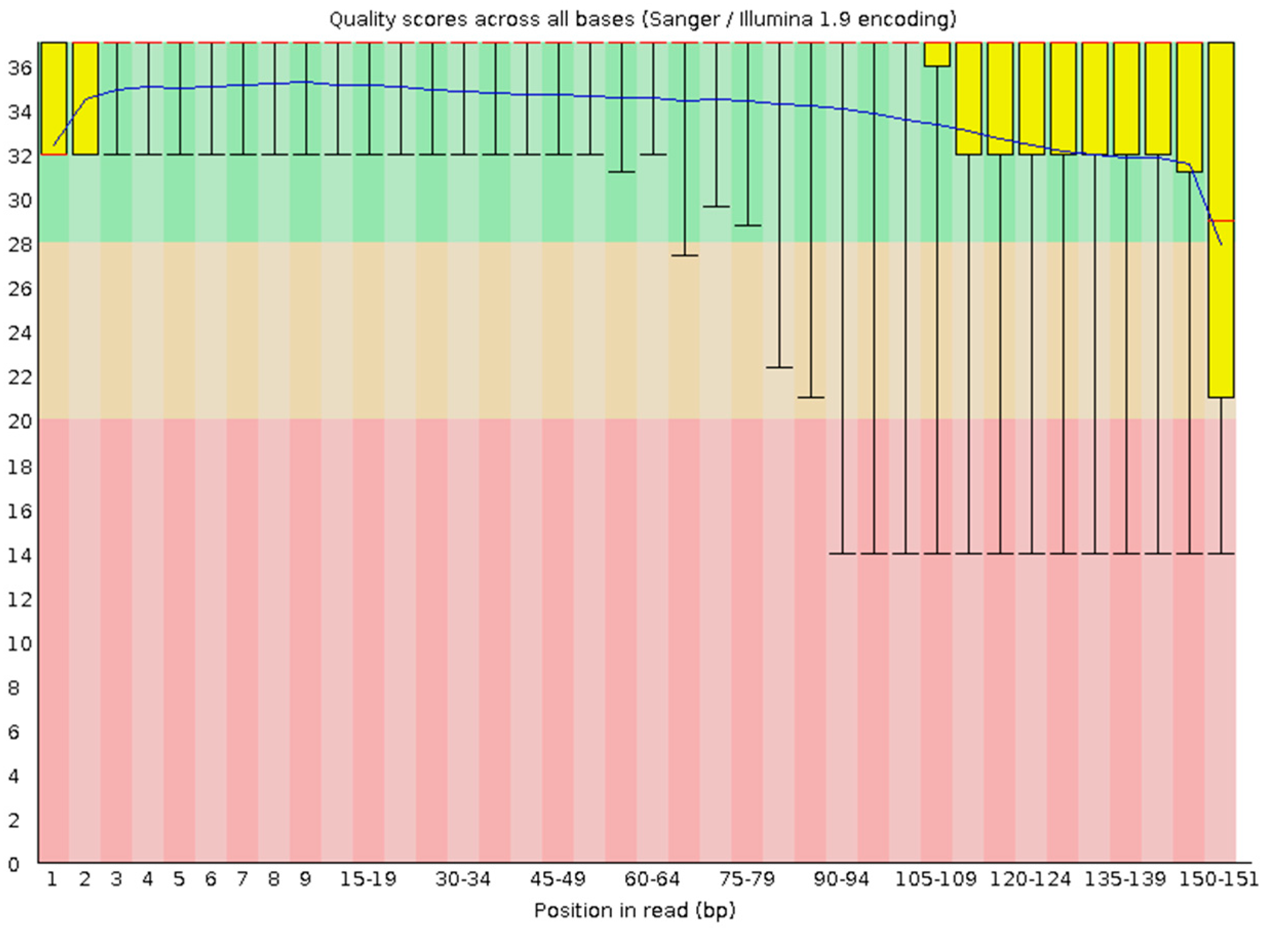
After paired-end alignments, quality filtering, and deletion of chimeras and singletons, a total of 6,102,452 sequences were generated per read from one direction, R1 reads, an example of which is depicted in (Figure 2), wherein 47% GC bases were generated. The R2 dataset also comprised 6,102,452 reads. In general, we obtained 12,204,904 reads. We mapped the reads on the ITS of the linden tree (Acc. No. KF897521), and 3,642,227 reads were successfully mapped on this sequence, which is approximately 30% of the total reads. Eight genera were identified in the DNA sample. According to Good’s coverage, 94.2% of the total genus richness was represented in the fungal communities (Table 1). The Chao1 richness estimator was 8, and the Shannon diversity estimator was 1.813. High-throughput amplicon sequencing (HTAS) data were deposited in NCBI GenBank under BioProject accession number PRJNA1270958, BioSample: SAMN48846729.

3.2. Taxonomy

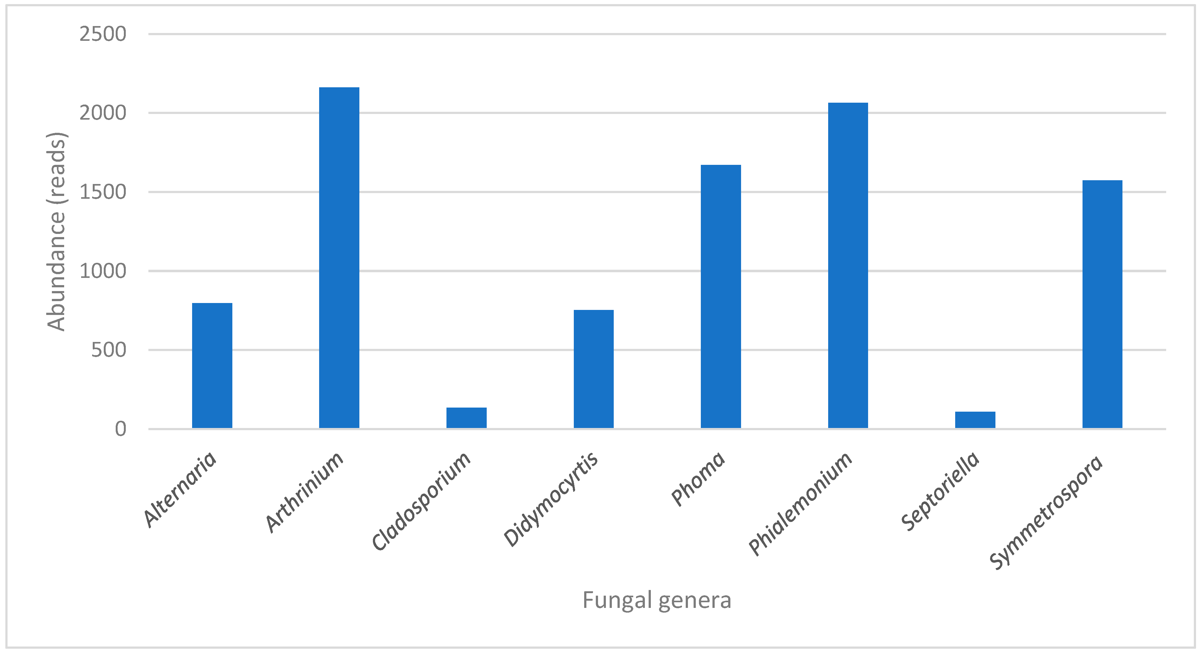
Ascomycota dominated the fungal phyla (seven from eight genera), one genus was identified as Basidiomycota, Symmetrospora—Basidiomycota (Cystobasidiomycetes) that reached 17% abundance. The rest belongs to Ascomycota: Alternaria (Dothideomycetes), Arthrinium (Sordariomycetes), Cladosporium (Dothideomycetes), Didymocyrtis (Dothideomycetes), Phoma (Dothideomycetes), Phialemonium (Sordariomycetes), and Septoriella (Dothideomycetes). The most abundant genus was Arthrinium, which reached 23% abundance, followed by Phialemonium with 22% (Figure 3 and Figure 4).

3.3. Morphological Evaluation of the Linden Tree

The linden tree was estimated to be 150 years old, with a trunk circumference of 3.5 m and a height of 18 m. It is a solitary specimen, with a crown height of 15 m and an average crown diameter of 13 m. Its overall health was good, although about 10% of the young branches were dried out. The tree was infested with Viscum album L. but generally appeared healthy (Figure 1). Several younger branches extending from the main crown show visible signs of drying shrinkage. No visible damage was observed. Operational safety was assessed as average, and the tree’s maintenance state was unkempt. From a biological perspective, the estimated remaining lifespan is likely more than 15 years.

4. Discussion

This study provides the first comprehensive, culture-independent characterization of the internal wood mycobiome of an ancient T. × europaea tree, reveals unexpectedly high fungal diversity including previously unreported taxa, and offers new insights into how these hidden communities may influence tree health, longevity, and conservation.
To the best of our knowledge, we do not know of any study using the HTAS analysis of the living linden wood mycobiome. We detected the Arthrinium genus in abundance at 23%. Apiospora was previously known as the sexual morph of the genus Arthrinium [10,11]. According to the International Code of Nomenclature for Algae, Fungi, and Plants (ICN) [12], Apiospora was a synonym of Arthrinium due to the early introduction of Arthinium, and it was more commonly used in the literature [13]. As endophytes, plant pathogens, and humus, Apiospora is ubiquitous in a wide range of terrestrial environments, such as soil, atmosphere, and even marine substrates, but its main hosts are still plants, especially Poaceae [14]. Apiospora is widely distributed throughout the world and is usually identified as an endophyte, pathogen, or saprobe. Ai et al. [15] described six strains that were isolated from Bambusaceae sp., Prunus armeniaca L., Salix babylonica L., and saprophytic leaves in Shandong Province, China. These fungi were isolated from the leaves, not from the wooden tissues. According to Gerin et al. [16], Arthrinium marii is capable of colonizing the wood of olive trees and has been identified as a widespread fungal species involved in severe dieback of olive trees in southern Italy. Phialemonium was detected at the genus level in abundance of 22%, as the second most abundant genus. Phialemonium is a genus of ascomycetous fungi, morphologically intermediate between Acremonium and Phialophora [17]. According to Manici et al. [18], who investigated fungal communities colonizing the wood of poplar, black locust, and willow, Phialemonium was most frequently isolated from willow. It was grouped within the complex of tree decay fungi, together with Neocucurbitaria, Neofusicoccum, and Botryosphaeria. Daldinia childiae co-colonized undecomposed willow wood samples alongside the endophytes Botryosphaeria dothidea, Neofusicoccum parvum, and Phialemonium, suggesting that these ascomycetes were not antagonistic. Members of the Botryosphaeriaceae family and Phialemonium colonized willow at early stages and subsequently shifted to a pathogenic phase, compromising plant health and facilitating further colonization by Daldinia as the main ligninolytic taxon. The potential use of P. curvatum as a biological control agent against Ophiostoma crassivaginatum, a wood pathogen on aspen, was previously investigated [19]. The growth of the pathogen Ophiostoma crassivaginatum was significantly suppressed in vitro [19]. Phialemonium was identified as the most abundant species according to the references discussed. Phoma species are environmentally widespread fungi, frequently associated with soil and aquatic habitats, primarily recognized as plant pathogens. Phoma spp. have been isolated from various sources, including water, food, and agricultural crops. It is a well-known saphrophyte or opportunistic pathogen, but an endophyte too. Phoma spp. was isolated from the wood of Taxus wallachiana, and the fungus was present among the plant cells of xylem [20]. But it is not understood as a typical colonizing fungus of the wood. On the other hand, interveinal chlorosis, presence of ash-grey areas on withered trees, and wilting with salmon-pink or orange-red discolouration of the wood of citrus trees is a characteristic feature of the mal secco disease caused by Phoma tracheiphila [21,22]. The ash-grey or lead-grey areas indicate the presence of pycnidia produced underneath the trees. To the best of our knowledge, we did not find any information about the Phoma fungus in living wooden tissues of Tilia spp. The Symmetrospora genus was found to be in abundance, at 17% in the linden wood. The Symmetrospora genus is a red yeast from the subphylum Pucciniomycotina in the phylum Basidiomycota [23]. Symmetrospora pseudomarina SA42 was isolated from decaying wood, Louisiana, USA [24]. Symmetrospora was also detected in high abundance on the bamboo samples [25] and in citrus tissue samples, but not in the wood [26]. Our study proved that the Symmetrospora genus could colonize linden tree wood. Alternaria was identified in 9% abundance. Alternaria malorum was identified as a causal agent of bark canker on walnut trees [27]. Alternaria tenuissima was identified in the wood of Cedrus atlantica L. in Morocco. Several studies have identified A. tenuissima as an aggressive pathogen of apple trees in South Africa and pecan in China, and as the causal agent of leaf spot in Datura metel L. and black chokeberry (Aronia melanocarpa L.) in Korea. The fungus also produces metabolites, including mycotoxins, that can degrade various plant tissues [28]. Didymocyrtis was identified in abundance of 8%. The fungal genus Didymocyrtis, long overlooked since its original description, has recently been resurrected and now encompasses lichenicolous species that were previously placed under Diederichia, Diederichomyces, Leptosphaeria, and Phoma [29,30], but also some foliicolous [31,32] and terricolous species [33]. The genus belongs to the family Phaeosphaeriaceae. It was identified on Banksia sessilis var. cygnorum or Brachylaena discolor. Cladosporium has been reported as an endophyte in the wood (xylem, sapwood, and heartwood) of numerous tree species. In contrast to Alternaria, which is primarily recognized as a foliar pathogen, Cladosporium species are commonly associated with the internal woody tissues of both healthy and stressed trees. These fungi exhibit a remarkably broad ecological amplitude, occurring on diverse substrates and a wide range of hosts, either as biotrophs or colonizers of dead and senescing tissue. Contrary to earlier assumptions, only a few species are truly plurivorous, cosmopolitan saprobes, such as C. herbarum, C. cladosporioides, and C. oxysporum, which show little evidence of strong environmental preferences [34]. This fungus was detected in abundance of 2%. Septoriella was detected in abundance of 1%. This genus is usually found as saprobic on dead wood. However, the wood that was extracted from the old trunk of the linden tree was not dead. The agents of leaf spot disease Mycosphaerella millegrana and Apiognomonia errabunda are also widespread on the leaves of Tilia trees in urban plantings of many Eastern and Central European countries: Belarus [35], Estonia [36], Croatia [37], Slovakia [38], Poland [39], Hungary [40], and Russia [41].
This study also has a limitation, as the analysis was conducted on a single ancient linden tree, which may not fully represent the diversity and variability that could be observed across different individuals or locations. Nevertheless, it provides a valuable contribution by establishing and demonstrating a methodological framework for the in-depth exploration of the wooden tree mycobiome. This protocol can serve as a reference for similar studies aiming to investigate the microbial communities inhabiting the woody tissues of long-lived trees. In future research, it would be particularly useful to compare these findings with results obtained through culture-dependent identification methods, in order to validate and complement the culture-independent approach and gain a more comprehensive understanding of the fungal community structure.

5. Conclusions

Based on the analytical pipeline, high-throughput sequencing of the ITS amplicon revealed eight fungal genera; however, this approach does not provide sufficient resolution for confident species-level identification. The most abundant genera detected were Arthrinium and Phialemonium, both commonly associated with wood but generally regarded as non-pathogenic under normal conditions. Remarkably, all eight identified fungal taxa were documented in living linden wood for the first time, highlighting the novelty of this study and expanding current knowledge about endophytic fungal diversity in old trees. Considering the ecological characteristics of the detected genera, none of the fungi are currently believed to pose a significant risk to the health or stability of this ancient linden tree. The described diagnostic pipeline, therefore, represents a rapid, reliable, and non-destructive approach for plant diagnostics that can be used to monitor the microbial health of valuable or protected trees. Beyond this specific case, the methodology has broad potential applications in studying the wood mycobiota of other tree species, providing new insights for forest pathology, biodiversity monitoring, and conservation-oriented microbiome research.

Author Contributions

Conceptualization, A.E. and M.S.; methodology, A.E.; software, L.F.; validation, J.C., P.S. and L.S.; formal analysis, J.P.; investigation, L.F.; resources, L.F.; data curation, A.E.; writing—original draft preparation, J.P.; writing—review and editing, L.S.; visualization, P.S.; supervision, P.S.; project administration, J.C.; funding acquisition, L.S. All authors have read and agreed to the published version of the manuscript.

Funding

This work was supported by the Ministry of Culture of the Czech Republic, from the NAKI III programme (program to support applied research in the field of national and cultural identity for the years 2023 to 2030); project name: Important and Unresolved Topics of Landscape Architecture; identification code: DH23P03OVV053. The work was also supported by the internal project of MENDELU No. IGA-FFWT-23-IP-028.

Data Availability Statement

High-throughput amplicon sequencing (HTAS) data were deposited in NCBI GenBank under BioProject accession number PRJNA1270958, BioSample: SAMN48846729. [GenBank] [https://www.ncbi.nlm.nih.gov/search/all/?term=PRJNA1270958] [PRJNA1270958].

Acknowledgments

We would like to thank Miloš Pejchal for his valuable comments on the surveyed linden tree.

Conflicts of Interest

The authors declare no conflicts of interest.

References

  1. Lukaszkiewicz, J.; Kosmala, M.; Chrapka, M.; Borowski, J. Determining the age of streetside Tilia cordata trees with a DBH-based model. J. Arboric. 2005, 31, 280–284. [Google Scholar] [CrossRef]
  2. Bilous, S.; Prysiazhniuk, L.M. DNA analysis of centuries-old linden trees using SSR-markers. Ukr. J. For. Wood Sci. 2020, 11, 4–14. [Google Scholar] [CrossRef]
  3. Stravinskienė, V.; Snieškienė, V.; Stankevičienė, A. Health condition of Tilia cordata Mill. trees growing in the urban environment. Urban For. Urban Green. 2015, 14, 115–122. [Google Scholar] [CrossRef]
  4. Purahong, W.; Pietsch, K.A.; Lentendu, G.; Schöps, R.; Bruelheide, H.; Wirth, C.; Buscot, F.; Wubet, T. Characterization of unexplored deadwood mycobiome in highly diverse subtropical forests using culture-independent molecular technique. Front. Microbiol. 2017, 8, 574. [Google Scholar] [CrossRef]
  5. Purahong, W.; Mapook, A.; Wu, Y.-T.; Chen, C.-T. Characterization of the Castanopsis carlesii deadwood mycobiome by PacBio sequencing of the full-length fungal nuclear ribosomal internal transcribed spacer (ITS). Front. Microbiol. 2019, 10, 983. [Google Scholar] [CrossRef]
  6. Eichmeier, A.; Pečenka, J.; Peňázová, E.; Baránek, M.; Català-García, S.; León, M.; Armengol, J.; Gramaje, D. High-throughput amplicon sequencing based analysis of active fungal communities inhabiting grapevine after hot-water treatments reveals unexpectedly high fungal diversity. Fungal Ecol. 2018, 36, 26–38. [Google Scholar] [CrossRef]
  7. Ricker, M.; Gutiérrez-García, G.; Juárez-Guerrero, D.; Evans, M.E.K. Statistical age determination of tree rings. PLoS ONE 2020, 15, e0239052. [Google Scholar] [CrossRef]
  8. Andrews, S. FastQC: A Quality Control Tool for High Throughput Sequence Data; Babraham Bioinformatics: Cambridge, UK, 2010. [Google Scholar]
  9. Glynou, K.; Nam, B.; Thines, M.; Maciá-Vicente, J.G. Facultative root-colonizing fungi dominate endophytic assemblages in roots of nonmycorrhizal Microthlaspi species. New Phytol. 2018, 217, 1190–1202. [Google Scholar] [CrossRef]
  10. Ellis, M.B. Dematiaceous Hyphomycetes: IV; Commonwealth Mycological Institute: Surrey, UK, 1963; Volume 89, pp. 1–33. [Google Scholar]
  11. Seifert, K.; Morgan-Jones, G.; Gams, W.; Kendrick, B. The Genera of Hyphomycetes; CBS Biodiversity Series 9; CBS-KNAW Fungal Biodiversity Centre: Utrecht, The Netherlands, 2011. [Google Scholar]
  12. McNeill, J.; Barrie, F.R.; Buck, W.R.; Demoulin, V.; Greuter, W.; Hawksworth, D.L.; Herendeen, P.S.; Knapp, S.; Marhold, K.; Prado, J.; et al. International Code of Nomenclature for Algae, Fungi, and Plants (Melbourne Code); Koeltz Scientific Books: Königstein, Germany, 2012. [Google Scholar]
  13. Crous, P.W.; Groenewald, J.Z. A phylogenetic re-evaluation of Arthrinium. IMA Fungus 2013, 4, 133–154. [Google Scholar] [CrossRef] [PubMed]
  14. Yan, H.; Jiang, N.; Liang, L.Y.; Yang, Q.; Tian, C.M. Arthrinium trachycarpum sp. nov. from Trachycarpus fortunei in China. Phytotaxa 2019, 400, 203–210. [Google Scholar] [CrossRef]
  15. Ai, C.; Dong, Z.; Yun, J.; Zhang, Z.; Xia, J.; Zhang, X. Phylogeny, taxonomy and morphological characteristics of Apiospora (Amphisphaeriales, Apiosporaceae). Microorganisms 2024, 12, 1372. [Google Scholar] [CrossRef]
  16. Gerin, D.; Nigro, F.; Faretra, F.; Pollastro, S. Identification of Arthrinium marii as causal agent of olive tree dieback in Apulia (Southern Italy). Plant Dis. 2020, 104, 694–701. [Google Scholar] [CrossRef]
  17. Gams, W.; McGinnis, M.R. Phialemonium, a new anamorph genus intermediate between Phialophora and Acremonium. Mycologia 1983, 75, 977–987. [Google Scholar] [CrossRef]
  18. Manici, L.M.; Bonora, P. The relationship between tree species and wood-colonising fungi and fungal interactions influences wood degradation. Ecol. Indic. 2023, 151, 110312. [Google Scholar] [CrossRef]
  19. Hiratsuka, Y.; Chakravarty, P. Role of Phialemonium curvatum as a potential biological control agent against a blue stain fungus on aspen. Eur. J. For. Pathol. 1999, 29, 305–310. [Google Scholar] [CrossRef]
  20. Yang, X.; Strobel, G.A.; Stierle, A.; Hess, W.M.; Lee, J.; Clardy, J. A fungal endophyte–tree relationship: Phoma sp. in Taxus wallachiana. Plant Sci. 1994, 102, 1–9. [Google Scholar] [CrossRef]
  21. Migheli, Q.; Cacciola, S.O.; Balmas, V.; Pane, A.; Ezra, D.; Di San Lio, G.M. Mal secco disease caused by Phoma tracheiphila: A potential threat to lemon production worldwide. Plant Dis. 2009, 93, 852–867. [Google Scholar] [CrossRef]
  22. Nigro, F.; Ippolito, A.; Salerno, M.G. Mal secco disease of citrus: A journey through a century of research. J. Plant Pathol. 2011, 93, 523–560. Available online: https://www.jstor.org/stable/41999031 (accessed on 4 November 2025).
  23. Wang, Q.-M.; Yurkov, A.M.; Göker, M.; Lumbsch, H.T.; Leavitt, S.D.; Groenewald, M.; Theelen, B.; Liu, X.-Z.; Boekhout, T.; Bai, F.-Y. Phylogenetic classification of yeasts and related taxa within Pucciniomycotina. Stud. Mycol. 2015, 81, 149–189. [Google Scholar] [CrossRef]
  24. Haelewaters, D.; Toome-Heller, M.; Albu, S.; Aime, M.C. Red yeasts from leaf surfaces and other habitats: Three new species and a new combination of Symmetrospora (Pucciniomycotina, Cystobasidiomycetes). Fungal Syst. Evol. 2020, 5, 187–196. [Google Scholar] [CrossRef]
  25. An, X.; Han, S.; Ren, X.; Sichone, J.; Fan, Z.; Wu, X.; Zhang, Y.; Wang, H.; Cai, W.; Sun, F. Succession of Fungal Community during Outdoor Deterioration of Round Bamboo. J. Fungi 2023, 9, 691. [Google Scholar] [CrossRef]
  26. Xi, M.Y.; Deyett, E.; Stajich, J.E.; El-Kereamy, A.; Roper, M.C.; Rolshausen, P.E. Microbiome diversity, composition and assembly in a California citrus orchard. Front. Microbiol. 2023, 14, 1100590. [Google Scholar] [CrossRef]
  27. Bagherabadi, S.; Zafari, D. Isolation and characterization of Alternaria malorum as a causal agent of bark canker on walnut trees. J. Plant Prot. Res. 2022, 62, 102–106. [Google Scholar] [CrossRef]
  28. Chauiyakh, O.; El Fahime, E.; Ninich, O.; Aarabi, S.; Bentata, F.; Chaouch, A.; Ettahir, A. Short notes: First report of the phytopathogenic fungus Alternaria tenuissima in cedarwood (Cedrus atlantica M.) in Morocco. Wood Res. 2023, 68, 619–626. [Google Scholar] [CrossRef]
  29. Trakunyingcharoen, T.; Lombard, L.; Groenewald, J.Z.; Cheewangkoon, R.; Toanun, C.; Alfenas, A.C.; Crous, P.W. Mycoparasitic species of Sphaerellopsis, and allied lichenicolous and other genera. IMA Fungus 2014, 5, 391–414. [Google Scholar] [CrossRef] [PubMed]
  30. Ertz, D.; Diederich, P.; Lawrey, J.D.; Berger, F.; Freebury, C.E.; Coppins, B.J.; Gardiennet, A.; Hafellner, J. Phylogenetic insights resolve Dacampiaceae (Pleosporales) as polyphyletic: Didymocyrtis (Pleosporales, Phaeosphaeriaceae) with Phoma-like anamorphs resurrected and segregated from Polycoccum (Trypetheliales, Polycoccaceae fam. nov.). Fungal Divers. 2015, 74, 53–89. [Google Scholar] [CrossRef]
  31. Crous, P.W.; Wingfield, M.J.; Burgess, T.I.; Hardy, G.E.S.J.; Barber, P.A.; Alvarado, P.; Barnes, C.W.; Buchanan, P.K.; Heykoop, M.; Moreno, G. Fungal Planet description sheets: 558–624. Persoonia Mol. Phylogeny Evol. Fungi 2017, 38, 240–384. [Google Scholar] [CrossRef] [PubMed]
  32. Crous, P.W.; Wingfield, M.J.; Burgess, T.I.; Hardy, G.E.S.J.; Barber, P.A.; Alvarado, P.; Barnes, C.W.; Buchanan, P.K.; Heykoop, M.; Moreno, G. Fungal Planet description sheets: 716–784. Persoonia Mol. Phylogeny Evol. Fungi 2018, 40, 240–393. [Google Scholar] [CrossRef]
  33. Das, K.; Lee, S.-Y.; Jung, H.-Y. Morphology and phylogeny of two novel species within the class Dothideomycetes collected from soil in Korea. Mycobiology 2020, 49, 15–23. [Google Scholar] [CrossRef] [PubMed]
  34. Bensch, K.; Braun, U.; Groenewald, J.Z.; Crous, P.W. The genus Cladosporium. Stud. Mycol. 2012, 72, 1–401. [Google Scholar] [CrossRef]
  35. Gorlenko, S.V.; Panko, N.A. Formirovanie Mikoflory i Entomofauny Gorodskikh Zelenykh Nasazhdenii: The Formation of Microflora and Entomofauna of Urban Green Plantations. Avtory. Nauka I Tekhnika. 1972. Available online: https://elib.bsu.by/bitstream/123456789/319880/1/Journal%20of%20the%20Belarusian%20State%20University.%20Biology_2023_No_2.pdf (accessed on 4 November 2025).
  36. Kalamees, K.; Saar, I. Mycobiota of the Naissaar Nature Park (Estonia). Folia Cryptog. Estonica. Fasc. 2006, 42, 25–41. [Google Scholar]
  37. Tomiczek, C.; Diminić, D.; Cech, T.; Hrašovec, B.; Krehan, H.; Pernek, M.; Perny, B. Pests and Diseases of Urban Trees; Šumarski Institute: Zagreb, Croatia; Šumarski Fakultet Sveučilišta u Zagrebu: Jastrebarsko, Croatia, 2008; p. 370. [Google Scholar]
  38. Bernadovičová, S.; Ivanová, H. Leaf spot disease on Tilia cordata caused by the fungus Cercospora microsora. Biologia 2008, 63, 44–49. [Google Scholar] [CrossRef]
  39. Mulenko, W.M.T.; Ruszkiewicz-Michalska, M. A Preliminary Checklist of Micromycetes in Poland; W. Szafer Institute of Botany, Polish Academy of Sciences: Krakow, Poland, 2008. [Google Scholar]
  40. Szabo, I. Leaf Pathogenic Fungi of Forest Trees and Shrubs in Hungary; Mycologische Dreiländert: Graz, Austria, 2002; pp. 67–70. [Google Scholar]
  41. Kolemasova, N.N.; Kovalevskaja, N.V. Fungous Leaf Diseases of Trees and Shrubs in Parks and Gardens; Lesnoj Vestnik: Saint Petersburg, Russia, 2000; Volume 6, pp. 119–124. [Google Scholar]
Figure 1. Linden tree (T. × europaea L.), (A) habitus of the tree; (B) sampling using increment borer; (C) drilled holes in the trunk of the linden tree.
Applmicrobiol 05 00131 g001
Figure 2. Quality scores across all nucleotide bases (Sanger/Illumina 1.9 encoding), R1 reads, generated by FastQC v0.10.1.
Applmicrobiol 05 00131 g002
Figure 3. Graph of the numbers of paired Illumina reads associated with the fungal genera.
Applmicrobiol 05 00131 g003
Figure 4. Percentage of the Illumina reads abundance associated with the particular fungal genera.
Applmicrobiol 05 00131 g004
Table 1. Estimates of sample coverage and diversity indices at the genus level for fungal profiles.
Diversity IndicesValue
Taxa_S8
Individuals9262
Dominance_D0.1798
Simpson_1-D0.8202
Shannon_H1.813
Evenness_e^H/S0.7662
Brillouin1.81
Menhinick0.08313
Margalef0.7664
Equitability_J0.8719
Fisher_alpha0.8618
Berger-Parker0.2333
Chao-18
Disclaimer/Publisher’s Note: The statements, opinions and data contained in all publications are solely those of the individual author(s) and contributor(s) and not of MDPI and/or the editor(s). MDPI and/or the editor(s) disclaim responsibility for any injury to people or property resulting from any ideas, methods, instructions or products referred to in the content.