Article

Survey of Thirteen Novel Pseudomonas putida Bacteriophages

1
College of Medicine, University of Central Florida, Tampa, FL 33620, USA
2
Biotechnology Program, James Madison University, Harrisonburg, VA 22807, USA
3
Department of Biomedical Sciences and Pathobiology, Virginia-Maryland College of Veterinary Medicine, Blacksburg, VA 24061, USA
4
Johns Hopkins Hospital, Baltimore, MD 21231, USA
5
Sapporo-Stone Brewing, Richmond, VA 23224, USA
6
Department of Defense, Fredericksburg, VA 22406, USA
7
Department of Biology, James Madison University, Harrisonburg, VA 22807, USA
8
Center for Biologics Evaluation and Research, US Food and Drug Administration, 10903 New Hampshire Ave, Silver Spring, MD 20993, USA
9
Biotechnology and Bioengineering, Sandia National Laboratories, 7011 East Avenue, Livermore, CA 94550, USA
10
School of Integrated Sciences, James Madison University, Harrisonburg, VA 22807, USA
*
Author to whom correspondence should be addressed.
Appl. Microbiol. 2025, 5(4), 108; https://doi.org/10.3390/applmicrobiol5040108
Submission received: 30 August 2025 / Revised: 25 September 2025 / Accepted: 26 September 2025 / Published: 7 October 2025

Abstract

Bacteriophages have been widely investigated as a promising treatment of food, medical equipment, and humans colonized by antibiotic-resistant bacteria. Phages pose particular interest in combating those bacteria which form biofilms, such as the medically important human pathogen Pseudomonas aeruginosa and several plant pathogens, including P. syringae. In an undergraduate lab course, P. putida was used as the host to isolate novel anti-pseudomonal bacteriophages. Environmental samples of soil and water were collected, and purified phage isolates were obtained. After Illumina sequencing, genomes of these phages were assembled de novo and annotated. Assembled genomes were compared with known genomes in the literature and GenBank to identify taxonomic relations and to refine their functional annotations. The thirteen phages described are sipho-, myo-, and podoviruses in several families of Caudoviricetes, spanning several novel genera, with genomes ranging from 40,000 to 96,000 bp. One phage (DDSR119) is unique and is the first reported P. putida siphovirus. The remaining 12 can be clustered into four distinct groups. Six are highly related to each other and to previously described Autotranscriptaviridae phages: Waldo5, PlaquesPlease, and Laces98 all belong to the Waldovirus genus, whereas Stalingrad, Bosely, and Stamos belong to the Troedvirus genus. Zuri was previously classified as the founding member of a new genus Zurivirus within the family Schitoviridae. Ebordelon and Holyagarpour each represent different species within Zurivirus, whereas Meara is a more distantly related member of the Schitoviridae. Dolphis and Jeremy are similar enough to form a genus but have only a few distant relatives among sequenced phages and are notable for being temperate. We identified the lysis cassettes in all 13 phages, compared tail spike structures, and found auxiliary metabolic genes in several. Studies like these, which isolate and characterize infectious virions, enable the identification of novel proteins and molecular systems and also provide the raw materials for further study, evaluation, and manipulation of phage proteins and their hosts.

1. Introduction

Bacteria of the genus Pseudomonas are ubiquitous in soil, water, and the surfaces of living organisms and inanimate objects [1,2,3,4]. The medically important pseudomonad, P. aeruginosa, causes severe, often fatal infections, primarily in hospital environments [3,5,6,7]. P. syringae poses a serious threat in agriculture, including over 60 pathovars that infect and destroy economically important crops [8]. Both P. aeruginosa and P. syringae are notorious for their ability to form biofilms, which can serve to increase infectivity and allow bacterial cells to resist damage from antibiotics [9,10].
The host used in this discovery-based study, P. putida, is a fast-growing bacterium found mostly in temperate soil and water habitats. It colonizes plant roots and provides benefits to the root microbiome [11]. For many years, P. putida has been used for bioremediation of environmental contaminants [12,13,14,15,16,17]. P. putida possesses a broad spectrum of metabolic pathways and can tolerate a wide range of stressors, making it an organism of interest for practical applications in biomanufacturing and biotechnology [18]. Proposals have included manipulating P. putida populations in the rhizosphere to benefit agriculture [19] and bioremediation strategies such as using P. putida proteins to degrade aromatic pollutants captured via nanoparticles [20,21]. Some of the molecular and genetic tools that could enable and support these types of applications include using bacteriophage-derived depolymerase enzymes to degrade P. putida biofilms [22] and using bacteriophage vectors for delivery of large genetic cassettes into P. putida genomes [17,23,24].
Bacteriophages (“phages”) are among the most genetically diverse organisms on the planet, serving as a reservoir of unique DNA sequences [25]. They are being investigated as possible antibacterial agents to help address the rising problem of antibiotic-resistant bacterial pathogens [26,27,28,29] and have been administered directly to humans in clinical trials and as experimental treatment for individual patients [26,30,31]. Some phage products are marketed for decontamination of food products or management of plant diseases, including plant diseases caused by pseudomonads [23,27,28,32]. In addition, individual phage components such as endolysins are being developed for use as antibacterial drugs [33,34].
Relatively few bacteriophages have been isolated that infect P. putida, especially when compared to human pathogens such as P. aeruginosa [35,36]. The first reported phage to be isolated on P. putida, gh-1, was published in 1966 [37] and the genome sequence reported decades later [38]. A review of the literature prior to 2024 in PubMed and DNA sequences in GenBank yielded only nine publications describing phages of P. putida that also had sequences available through GenBank, and four additional papers describing approximately 30 phages were not accessible. The diversity within this small group of phages is remarkable; finding two or more phages similar enough to constitute a genetically related group was unusual. Only myovirus and podovirus morphologies were seen, all are double-stranded DNA phages, and no temperate phages were reported. As of 14 April 2025, GenBank contained a total of 91 submitted dsDNA genomes from P. putida bacteriophages, of which 72 belong to the broadly T7-like Autographivirales (Supplemental Table S1), mostly from a single study that reported a collection of phages infecting P. putida KT2440 [35]. Although the number of phages substantially increased, the same trends hold true. Further, according to previous reports, P. putida phages infection is mostly limited to P. putida host strains, but there have been reports of infection in P. syringae, and P. fluorescens [39].
Collecting new viruses expands the repertoire of unique P. putida phages, not only for genomic information but also for potential biotechnology applications such as engineering broader host ranges [40], manipulating P. putida populations in the rhizosphere [19], degrading biofilms through phage-encoded depolymerase enzymes [22], and serving as vectors for delivery of large genetic cassettes into P. putida genomes [23]. Importantly, at the time of writing, over 259 prophages have been identified in P. putida isolates, but few have been isolated as free phages [36]. It is unknown how many of these annotated prophages can be induced to yield whole virions. Indeed, it is not known whether these even represent complete phage genomes. Thus, many relevant aspects of their biology cannot be studied [41].
The goal of this study was to isolate and analyze phages using a locally isolated strain of P. putida. Here we describe the discovery and characterization by undergraduates in a research course of thirteen novel P. putida phages, including two temperate phages, the first shown to infect P. putida. We provide in-depth analysis of these phage genomes including comparative genomics between our phages and other published phages, lytic and lysogenic cassettes, spike proteins that may help with degradation of biofilms common for pseudomonads, and auxiliary metabolic genes that would be useful for many environmental remediation applications.

2. Materials and Methods

2.1. Phage Isolation, Culturing, and Imaging

Phages were discovered from random environmental samples (see Table 1 for locations and type) through enrichment of extracts with P. putida strain ISAT203. P. putida ISAT203 was sequenced and confirmed to be a novel strain using PacBio sequencing (Supplemental Methods). The closest relative in GenBank is P. putida ISoF (CP072013.1, ref. [42]). Bacterial suspensions for initial phage propagation consisted of approximately ½ cup of soil/water mixed with 50 mL Luria–Bertani (LB) broth (10 g/L peptone, 5 g/L yeast extract, 10 g/L NaCl). After overnight shaking incubation at 37 °C, enriched cultures were sterilized with 0.22 µm filters and 5 μL of filtrate was spotted on a lawn of the host bacteria in 0.7% LB agar. Phage populations were purified by standard SEA-PHAGES methods (https://seaphagesphagediscoveryguide.helpdocsonline.com/home, accessed on 3 February 2021) [43], using SM buffer (100 mM NaCl, 8 mM MgSO4·7H2O, 50 mM pH 7.5 Tris-HCl), serially diluting, infecting into culture aliquots and plating with LB top agar. After purification, a single isolated plaque was picked into SM buffer and the sample diluted to a concentration predicted to produce near confluent lysis after infection of the appropriate host. Phage lysates were harvested by flooding confluent plates with SM buffer, held at 4 °C for 4 to 12 h stationary, then collected and filtered. Suspensions of phage were stored at 4 °C in SM buffer.
Phages from lysates were negatively stained with 1% uranyl acetate on formvar-coated copper grids and photographed by a FEI Morgagni 268 transmission electron microscope (TEM) (Hillsboro, OR, USA), Copenhagen, Denmark, at Mary Washington University, Fredericksburg, VA, USA.

2.2. DNA Isolation

DNA was purified from 0.2 micron-filtered lysates of ≥1 × 10⁹ plaque-forming units (PFU)/mL that had been treated with 1 unit/μL of DNAse (Ambion cat. #AM2224, Austin, TX, USA) for 1 h at room temperature to degrade residual bacterial DNA. DNA for PlaquesPlease, Stalingrad, DDSR119, and Dolphis was prepared using a protocol developed at JMU. Briefly, 100 μL of DNA prep solution (10 mM EDTA, 2.5% Ficoll® 400, 3.3 mM pH 8.0 Tris-HCl, 0.08% SDS) was added to 100 μL of each lysate, and the suspension was mixed and held at room temperature for 10 min. Next, 600 μL of cold 100% isopropanol was added, and the solution was kept on ice for 5 min. The mixture was centrifuged at 4 °C at 13,000 RPM for 30 min, the supernatant was removed, and the pellet was allowed to dry. DNA was resuspended in 100 μL of diH2O. DNA for Waldo5 and Zuri was prepared with a DNA purification kit (Promega, Madison WI, USA) [44] using a modified procedure as described in the SEA-PHAGES manual (https://seaphagesphagediscoveryguide.helpdocsonline.com/home, accessed on 3 February 2021) [43]. DNA for Laces98, Meara, Stamos, Jeremy, Ebordelon, Bosely, and Holyagarpour was isolated using the Norgen Biotek Phage DNA Isolation kit (cat. #46800, Thoroid, ON, Canada) following the manufacturer’s protocol.

2.3. DNA Sequencing and Assembly

DNA libraries were prepared using Illumina DNA prep fragmentation kit (cat. #20060059, San Diego, CA, USA) with Illumina Nextera DNA Unique Dual index adapters (set D, cat. #20091660, San Diego, CA, USA). DNA libraries were pooled and sequenced using Illumina MiSeq or HiSeq 2000 technology through core facilities located at Admera Health Genome Center (South Plainfield, NJ, USA), the North Carolina State Genomic Sciences Laboratory (Raleigh, NC, USA), or Sandia National Laboratories (Livermore, CA, USA).
Raw reads were processed to remove low quality reads with BBDuk (https://sourceforge.net/projects/bbmap/, accessed on 3 February 2021) using the following parameters: ktrim  =  r, k  =  21, mink  =  11, hdist  =  1. Paired-end reads (150 or 300 bp depending on platform and kit) were assembled into whole genome consensus sequences using Newbler v2.9 [45] or SPAdes v3.12 [46] at ~50X coverage for each assembly. Each genome assembled into a linear sequence, most with defined ends. Genomes were manually examined for coverage and integrity of sequence using Consed v.14 [47], consistent with SEA-PHAGES standards for genome completeness (https://seaphagesphagediscoveryguide.helpdocsonline.com/home, accessed on 3 February 2021) [43].
Short inverted repeat ends were determined by direct inspection of genome sequences. Terminal repeats on phages with defined ends were determined by observing pileups in read alignments, using the PhageTerm program [48], or by comparison to related phages in GenBank.

2.4. Genome Annotation and Other Analyses

Laces98, Stamos, Meara, Holyagarpour, Jeremy, Ebordelon, and Bosely were annotated for ORFs and functions using DRAM-v (version 0.1.2) [49] at KBASE (version 1.2.0) [50]. Additional functional annotation was performed using web based BLASTp (version 2.17.0) [51]. All thirteen phage genomes were run through DRAM-v at KBase to determine AMGs.
Genomes for Zuri, PlaquesPlease, Waldo5, Dolphis, Stalingrad, and DDSR119 were annotated using DNA Master 5 (https://phagesdb.org/DNAMaster/, accessed on 10 January 2019) [52], and PECAAN (https://seaphages.org/media/docs/PECAAN_User_Guide_Dec7_2016.pdf, accessed on 3 February 2021). ORFs were identified using Glimmer [53], GeneMark.hmm [54], and features built into DNA Master 5 and PECAAN, and were refined through homology to ORFs in previously discovered phages using the current version of BLASTn (version 2.17.0). The function of each gene product was classified using homology to known proteins in the current versions of BLASTp, HHpred (version 3.3.0) [55], and Protein Data Bank [56]. Prediction of transmembrane domains (TMDs) with TMpred (version 1.0) [57] and DeepTMHMM (version 1.0.24) [58] was used to refine the calling of protein functions. Using the default parameters, TMHMM (version 2.0) [59] and lipoP (version 1.0) [60] were used to evaluate all the phage coding sequences for potential holin, endolysin, and spanin candidates based on sequence homology, presence of TMDs, and lipoprotein membrane anchor signals. Aragorn was used to identify tRNA genes [61]. Phage genome maps were prepared using web based Proksee and colored according to gene type [62].

2.5. Comparative Genomics

Taxonomic information is based on Master Species List #40v.1, released by the International Committee on Taxonomy of Viruses (ICTV) on 3 March 2025 (https://ictv.global/taxonomy, accessed on 5 April 2025). Initially, each phage’s closest relatives were identified using nucleotide-level BLASTn comparisons in GenBank, with comparisons limited to phages with contractile tails (taxid:2731619), long non-contractile tails (taxid:2731619), and short tails (taxid:2731619). Subsequently, VIRIDIC [63] and ViPTree [64] were run with the default parameters to explore possible evolutionary relationships among our phages and previously described phages. VIRIDIC generates percent intergenomic similarity values for all possible pairwise genome comparisons based on genome identity and coverage. ViPTree builds phylogenetic trees based on genome-wide protein-level similarity generated with tBLASTx [51]. More detailed comparisons and alignments of related genomes were performed using Phamerator [65]. In addition to displaying nucleotide sequence similarities, Phamerator displays protein phamily (group) assignments calculated using PhaMMseqs (version 1.0.4) and can derive shared proteins [66].

2.6. Lifestyle Prediction

Bacphlip was used to predict temperate versus virulent phage lifestyles [67]. Bacphlip is a random forest classifier that was trained primarily on tailed phages and uses a broad array of protein signatures associated with temperate phages to predict lifestyle.

2.7. Protein Structure Predictions

Prediction of secondary structure was performed with the SABLE program (http://sable.cchmc.org/, accessed on 10 March 2025). Prediction of tertiary structure of all available spike proteins in the new collection and several from Pseudomonas, Escherichia coli, and Salmonella was performed using AlphaFold3 (https://alphafold.com/, accessed on 20 January 2025) [68,69], which uses a trained machine-learning architecture to model intra- and inter-protein interactions. Visualizations of these structures were prepared in MolStar (https://molstar.org/, accessed on 6 April 2025) [70], which includes an option to overlay multiple proteins. For pairwise comparisons, we highlighted the amino acids on each protein that correspond to the desired region (C- or N- terminal regions in this study) and superimposed. This rotates one of the two figures to be situated where the highlighted regions are as close as possible without altering the individual structures.

2.8. Phages Added from GenBank

Eight phages related by genome or proteome similarity to some of our thirteen newly isolated phages were added to the analyses described herein, for the purpose of situating ours within new or existing classifications and observing conservation of proteins, lifestyles, and morphology (Table 2). When identifying these relatives, we excluded sequences that were only deposited as MAGs (metagenome-assembled sequences) and typically excluded genomes with regions of nucleotide identity covering less than 10% of our query genomes.

2.9. Sequencing and Analysis of P. putida Host

P. putida ISAT203 was grown overnight at 30 °C in LB broth. Genomic DNA was isolated following manufacturers’ protocols using the Pacbio Nanobind CBB kit (PN-301-102-900, Menlo Park, CA, USA). PacBio HiFi DNA library was prepared following the manufacturer’s protocol. In brief, DNA was sheared using COVARIS g-tubes and cleaned up using the SMRTbell cleanup beads (PacBio 102-158-300). The HMW library was prepared using SMRTbell Express Template prep kit 3.2 with SMRTbell Adapters (PacBio) for barcoding. Size selection was performed with AMPure PB beads, and the final library was pooled with other barcoded samples. The HMW DNA library size was measured with an Agilent Technologies (Santa Clara, CA, USA) 4200 Tape Station to be 11.1 kbp. The concentration of the HMW DNA library was 82.6 ng/µL determined by the Qubit Flex fluorometer (Thermo Fisher, Waltham, MA, USA). DNA libraries were run on a single 8 M SMRT cell on a PacBio Sequel IIe.
There were 74 HMW resultant reads from the long-read sequencing effort ranging in size from 1506 bp to 16,217 bp. Whole genome assemblies were attempted with Canu v2.2 (https://github.com/marbl/canu, accessed on 12 April 2025) (genome size = 6.2 m) but failed due to low coverage. To determine the host strain taxonomy, we used BLASTn against the core_nt database for each read larger than 5000 bp [51]. We further analyzed through bowtie alignment of raw reads to the published P. putida ISoF genome (Genbank Accession: GCA_023101365.1) and found a 97.3% alignment rate.

3. Results

3.1. Genome Features and Relatedness to Other Phages

In this study, we report the discovery and analysis of thirteen novel phages that infect P. putida strain ISAT203, a new bacterium isolated from soil in the same geographical region as the phages. “Novel” denotes that there are no identical phage genomes present in the GenBank non-redundant nucleic acid database. Major genomic features are summarized in Table 3 and genome maps are provided in Supplemental Figures S1 and S2. While we did not conduct extensive host-range testing of our phages, our preliminary data showed that none of these phages were able to plaque on P. putida strains KT2440, S12, or eight other strains belonging to a different clade of P. putida than ISAT203.
Figure 1A is a dotplot comparison of the thirteen phages that we isolated and characterized in this study. We also used VIRIDIC to calculate the percent intergenomic similarity of all of our phages plus their closest relatives in GenBank. Figure 1B,C correspond to the two related groups of phages boxed in blue in the upper left of the dotplot (the Waldovirus and Troedvirus genera). Waldo5 and PlaquesPlease each represent different ICTV-recognized species within the genus Waldovirus (ICTV MSL#40v.1). Based on the commonly accepted thresholds of 70% and 95% intergenomic similarity for genus and species inclusion, respectively [75], Laces98 belongs to the same species as PlaquesPlease (Figure 1B). Similarly, Stalingrad (Figure 1C) was recently found to represent new species within the Troedvirus genus. The only other ICTV-recognized species within Troedvirus is represented by phage phi15. One of the two closely related phages in our collection, Bosely, belongs to the same species as Stalingrad, whereas Stamos represents a new species within Troedvirus that is distinct from both Stalingrad and phi15. Collectively, all of these phages belong to the Studiervirinae subfamily in the Autotranscriptaviridae family of lytic podoviruses that encode their own single-subunit RNA polymerases.
The four phages boxed in pink in the center of the dotplot are related to Zuri. Zuri is the founding member of the genus Zurivirus (ICTV MSL#36), within the Schitoviridae family of lytic N4-like podoviruses [76]. We propose that Ebordelon and Holyagarpour each represent new species within Zurivirus (Figure 1D). Based on BLASTn comparisons, Zuri is also the closest relative for our novel phage Meara. Only one other complete phage genome in GenBank, Arace01 (Table 2), shares any significant nucleotide-level identity with Zuri across >10% of the genome. Based on a whole-proteome tree, Meara and Arace01 cluster within the Schitoviridae family (Supplemental Figure S3A), and we therefore propose that they each represent a novel genus within that family.
We propose that Dolphis and Jeremy represent two new species within a new genus (Figure 1E, boxed in green in Figure 1A), based on the 95% and 70% intergenomic similarity thresholds described above. No higher-level classification seems possible within the Caudoviricetes due to a dearth of close relatives. In a tBLASTx search of Caudoviricetes genomes, we only identified three phages with any notable protein-level similarity to Dolphis or Jeremy: Bordetella phage PHB04, P. syringae phage Touem01, and Aeromonas phage ST4. Using ViPTree proteome trees, these five phages cluster together, separately from other phages. ViPTree can be useful to define higher-order taxa such as Families and Subfamilies but in this case, the available phages are too few and too distantly related to clearly support such intermediate taxa (Supplemental Figure S3B).
DDSR119 is a singleton, with no close relatives in GenBank. A recent study [72] reported finding a Pseudomonas poae siphovirus called Torfinnsbu (PQ464596.1) that shares 20% intergenomic similarity with DDSR119, mostly involving structural genes.

3.2. Phage Morphology

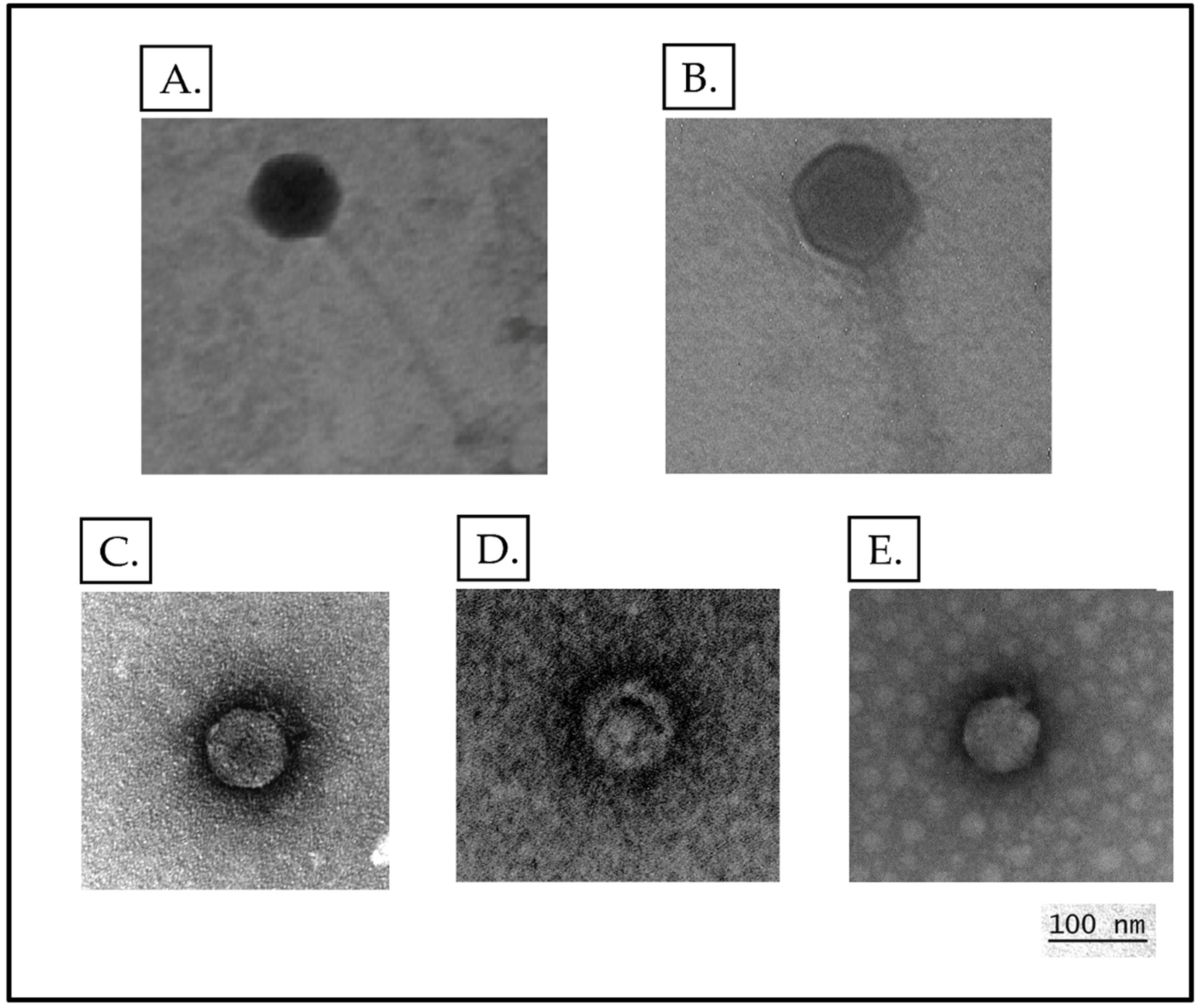
We obtained electron micrographs of several of our phages. Representative images are shown in Figure 2. Virion dimensions are approximately 78 nm capsid and 170 nm tail for DDSR119 (Figure 2A) and 94 nm capsid and 145 nm tail for the Dolphis-like phages (Figure 2B). For the Waldovirus (Figure 2C), Troedvirus (Figure 2D) and Zuri-like (Figure 2E) podoviruses, capsid diameters are approximately 52 nm, 52 nm, and 69 nm, respectively, correlating with the genome lengths ranging from 40,504 bp to 75,873 bp. The virion morphologies shown in these TEMs are all consistent with expectations based on the taxonomic comparisons described above (e.g., Autrotranscriptoviridae phages have the expected podovirus morphology). DDSR119 appears to be a siphovirus, like Torfinnsbu, and thus would be the first reported P. putida siphovirus.

3.3. Protein Features of our Phages and Selected Relatives Using Phamerator

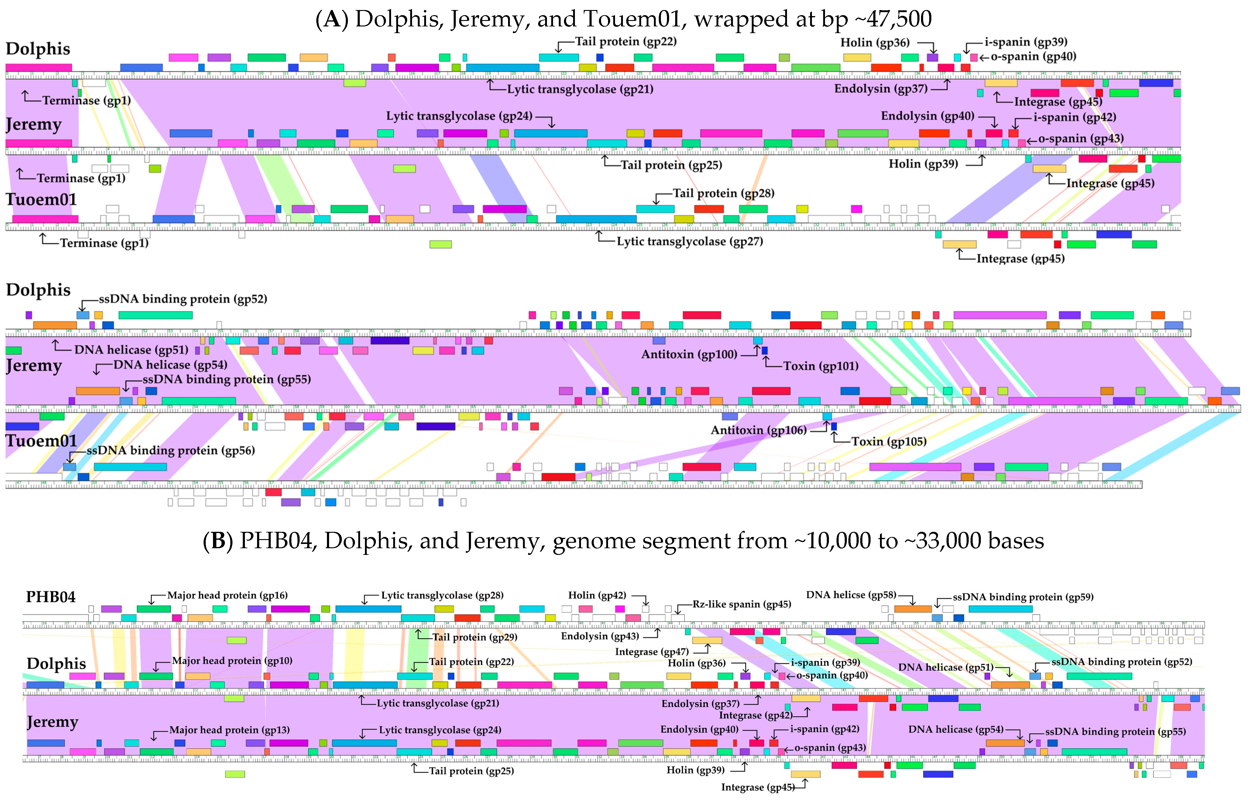
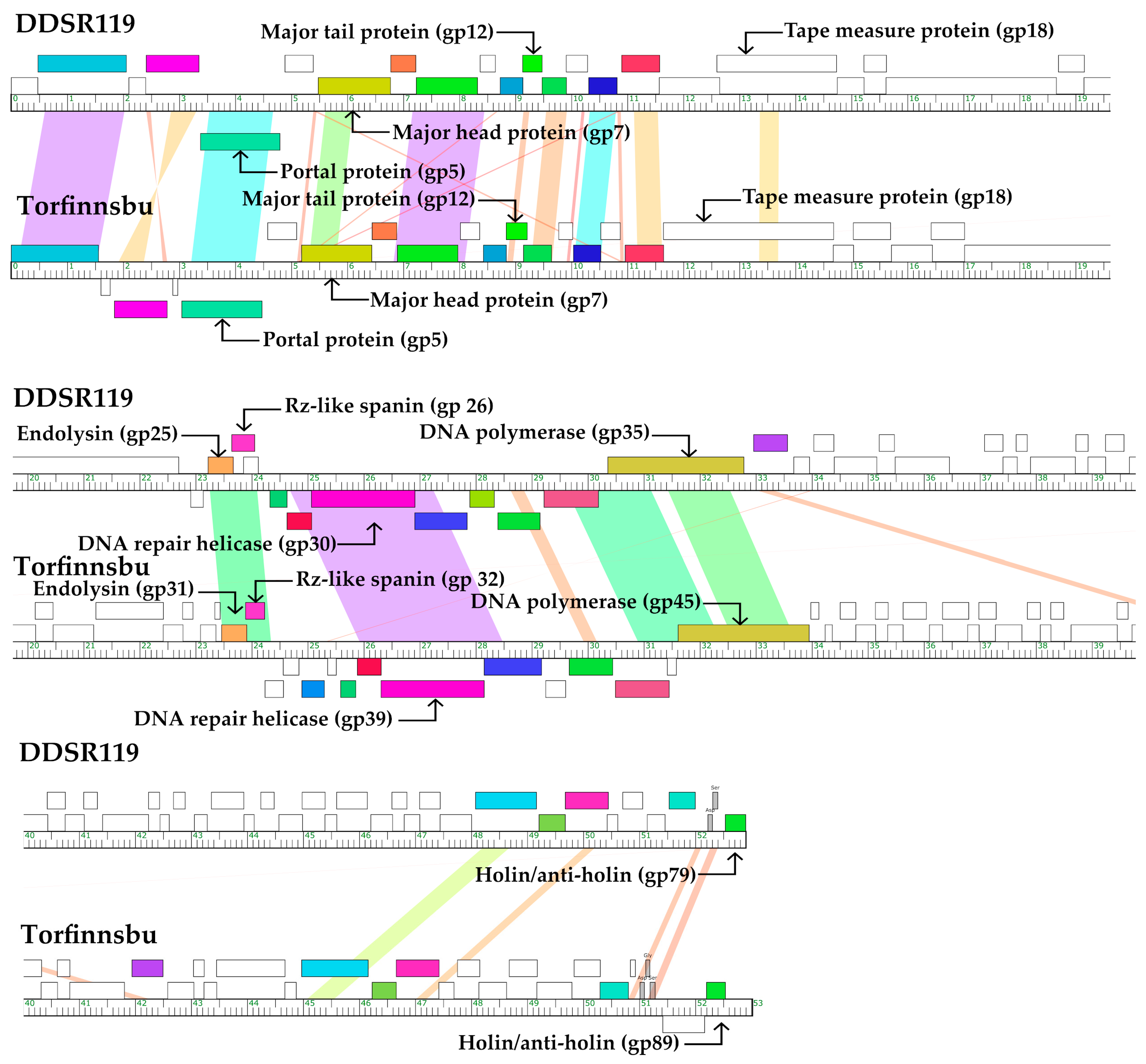
Having identified the closest relatives of our phages, we conducted more detailed genome-wide comparisons and alignments using Phamerator [65]. The phages are then sorted into clusters based on those phamily assignments. In Figure 3, Figure 4, Figure 5 and Figure 6, DNA sequence similarity was determined using pairwise BLASTn with E-value visualized according to the color spectrum from violet (E-value of 0) to red (E-value of 10−4). Protein phamilies are shown in color-coded rectangles. Selected proteins with predicted functions are shown above or below the boxed and colored predicted proteins. All other proteins with predicated functions assigned can be seen in the associated GenBank files. Typical in phage genomes, fewer than 40% of genes can be assigned a function, even though they can be organized into Phamilies. Those who cannot be assigned a function with confidence are named “hypothetical protein.” White boxes, “orphams,” represent predicted proteins not shared with any other in the specific database used to generate the Phamerator output. The more unique the phage is, the fewer functions can be defined; note phage DDSR117 in Figure 6. Consistent coloration of proteins across these genomes indicates conserved protein functions, even when nucleic acid sequences may not be conserved; many examples of these are seen in Figure 3, Figure 4, Figure 5 and Figure 6 and are most obvious in comparisons of distantly related phages (Figure 5 and Figure 6). In the image, the ruler represents the DNA and is divided into segments of 100 bp (small ticks) and 1000 bp (numbered ticks), allowing visual estimates of gene sizes.
The results of genome clustering matched the taxonomic groupings that we inferred from genomic data and are grouped this way throughout Figure 3, Figure 4, Figure 5 and Figure 6. In Figure 3, we compared our three Waldovirus phages, our three Troedvirus phages along with phi15, and two phages from other genera in the Autotranscriptaviridae family that were chosen somewhat randomly to help visualize the higher-level relatedness within the family. Overall, 25 phams were shared by all nine phages in Figure 3, encompassing morphogenetic proteins and essential phage enzymes; see examples identified by function name. The Waldovirus phages alone share 43 phams and the Troedvirus phages share 36 phams. Despite each belonging to different genera, phages gh-1 and Henninger are more similar to the three Waldovirus phages than any of them are to the Troedvirus phages, such that these five phages share 29 phams.
Our three Zurivirus phages were highly conserved at the DNA level, whereas Meara, a member of our new collection within Schitoviridae, and Arace01 from GenBank, were highly dissimilar over >50% of the genome (Figure 4). However, many of the proteins (68 phams) were still shared, most notably structural genes, lysis cassette proteins, and genes for nucleotide metabolism. Only one, Holyagarpour, contains an identifiable spike protein gene. All 5 phages in this group each have 2 tape measure genes encoding proteins involved in the length of tails [77]. The larger one (Pham57, 401 AA) is at location ~gp76. The smaller one (Pham43, 109 AA) is at location ~gp82. There is no similarity with these tape measure proteins and those of Waldoviruses or Troedviruses. One possible explanation is that the tape measure proteins are performing some other function than annotated. In E. coli phage HK97, the protein annotated as a tape measure was shown to be involved in the genome injection process [78]. In Mycobacterium phages, tail tape measure proteins may act as signaling molecules that promote growth of dormant bacterial hosts [79].
As described in Section 3.1, Dolphis and Jeremy do not have close relatives within our collection or in GenBank, but we did find three distantly related phages with protein-level similarity: Bordetella phage PHB04, Aeromonas phage ST4, and P. syringae phage Touem01 (Phage ST4 was not included in the comparative genomics studies). The three Pseudomonas phages, Dolphis, Jeremy, and Touem01, share 46 phams. These include some structural genes, as would be expected among related myoviruses, and also include several DNA-interacting proteins distributed across the lengths of all three genomes (see examples in Figure 5A). In contrast, Bordetella phage PHB04, which is more distantly related, shares only 26 phams with Dolphis and Jeremy, and these are limited to a much smaller segment of the genome (Figure 5B). The Jeremy genome contains a 715 bp tract of randomly ordered purine bases at coordinates 70,489–71,204, a highly unusual phenomenon. We confirmed the presence of this sequence by read walking to rule out an assembly artifact. A protein predicted within this region would contain glycine, glutamic acid, lysine, and arginine, similarly in random order. A similar phenomenon has been reported in Bacillus podoviruses, where a 150 bp run of A’s was observed in Bacillus phage Stitch (KX349901.1) [79] and a 100 bp run of purines in Bacillus phage Porch (unpublished data). The significance of these is unknown and nothing else resembling such DNA or protein sequences is found in the GenBank NR database. However, there is one report of Mycobacterium phage Bxz1 having a run of 150 GC pairs (not included in the GenBank entry) [80].
While DDSR119 has no close relatives in GenBank, we compared it to Torfinnsbu, a P. poae phage that was recently reported to share 20% intergenomic similarity based on a VIRIDIC analysis of nucleotide sequence. The DNA similarities and shared proteins are largely clustered in two rations (Figure 6). In our analysis, these two phages share 26 phams, reflecting similar protein sequences for the terminase, some structural proteins, endolysin, spanin, DNA primase and DNA helicase; however, most of the shared genes are hypothetical (Figure 6).

3.4. Phage Lifestyle

Phage lifestyle prediction (virulent vs temperate) can be challenging. The presence of unambiguous integrase and/or repressor genes is reliably predictive of a temperate life history, even if a phage appears to undergo only lytic replication under a particular set of experimental conditions or carries other mutations that prevent lysogenic replication. However, bioinformatic identification of integrases, and especially repressors, is imperfect and therefore the apparent absence of these genes is not a reliable indicator of an obligately lytic lifestyle. In addition, some temperate phages are maintained as extrachromosomal elements in plasmidial or episomal form and do not encode integrases at all [81,82]. We therefore used multiple approaches to predict the lifestyles of these six phages: identification of specific lysogeny-associated genes such as integrases, and proteomic comparisons to well-characterized phages using ViPTree and Bacphlip.
Bacphlip predicted Dolphis and Jeremy to be temperate (p-values = 0.90, 0.837, respectively). We identified tyrosine integrases in both phages: gp42 in Dolphis and gp46 in Jeremy (Figure 5A,B, and Supplemental Figure S1). The two predicted tyrosine integrases are 99.3% similar over 98% of the protein suggesting they are the same integrase. We mapped Jeremy (gp46) to a Phage Integrase pFAM (PF00589; e-value 3.9 × 10⁻⁹), which also contains E. coli phage Lambda and Salmonella phage P22. The predicted protein also contains both the arm DNA binding domain (Jeremy 4–93aa, PFAM13356, e-value 2.7 × 10⁻¹¹) and the integrase catalytic core (Jeremy 222-411aa; e-value 7 × 10⁻²¹) [83]. We also identified highly related integrases in the distantly related phages PHB04 and Tuoem01 (~60% similar over 95% of the protein; Figure 5A,B).
Bacphlip predicted that our other 11 phages are obligately lytic (Zuri p = 1.0, Ebordelon p = 1.0, Holyagarpour p = 0.995, Meara p = 0.975, Waldo5 p = 0.873, PlaquesPlease p = 0.925, Lace98 p = 0.925, Stalingrad p = 0.997, Stamos p = 0.999, Bosely p = 0.997, DDSR119 p = 0.85) and no genes were annotated as integrases. The Bacphlip prediction for Zuri is both highly confident (p-value ≥ 0.95) [67] and consistent with the known lifestyle of other Schitoviridae phages. Bacphlip prediction confidences for Waldo5, PlaquesPlease, and Stalingrad were variable, but again the obligately lytic lifestyle is consistent with what is known about other Autotranscriptaviridae phages. DDSR119 does not have any close characterized relatives, but the Bacphlip results, and the lack of identifiable integrase genes support a tentative prediction that DDSR119 is obligately lytic.

3.5. Lysis Systems

Phages infecting Gram-negative hosts are known to encode three classes of lysis proteins: holins, endolysins, and spanins [84]. Each is responsible for subversion of one of the three layers of the cell envelope during host lysis: inner membrane (IM), peptidoglycan (PG) layer, and outer membrane (OM), respectively. We were able to identify all three lysis elements in our thirteen phages. These are identified in the whole genome maps in Figure 3, Figure 4, Figure 5 and Figure 6.
In Zuri-like phages (Zuri, Ebordelon, Holyagarpour, and Meara), all the lysis genes were grouped together as a lysis cassette (Figure 7A). Zuri encodes a class II holin (“N-in, C-in topology”, where both termini are cytoplasmic) with 2 predicted TMDs, followed by an endolysin with an N-terminal signal anchor release (SAR) domain and an embedded two-component spanin system, where the o-spanin gene is entirely embedded in the +1 reading frame of the i-spanin gene. The equivalent genes were easily identifiable based on sequence similarity in Ebordelon and Holyagarpour. Despite having lower overall sequence similarity, Meara appears to use the same embedded o-spanin arrangement, yielding a strong lipoprotein membrane anchor signal in the predicted protein. Like many embedded or overlapping o-spanins, the genes in these four phages use a non-canonical translation start codon.
Waldo5, PlaquesPlease, Bosely, Stamos, Laces98, and Stalingrad also encode class II holins with two TMDs and embedded two-component spanins, but their endolysins did not seem to contain any N-terminal SAR domains. Furthermore, the endolysin gene was located far upstream from the holin and spanin genes (Figure 7B). The lysis proteins in Waldo5, Laces98, and PlaquesPlease are 100% identical and share substantial sequence identity with those from Stalingrad: holin (45%), endolysin (61%), i-spanin (40%) and o-spanin (51%).
In Dolphis (Figure 7C), we identified gp37 as an endolysin based on sequence similarity. The upstream gene gp36 and downstream gene pair of gp39–gp40 have all the necessary features to be holin and the spanin pair, respectively, but the predicted holin has no sequence similarity to previously annotated holins. Based on our analyses, Jeremy appears to have essentially the same lysis cassette as its close relative, Dolphis. Similarly, in DDSR119 (Figure 7D), gp25 was annotated as the endolysin based on sequence homology. A potential i-spanin/o-spanin pair (gp26 and 26a) could be found immediately downstream with identity of 50% to other Rz-like spanins, but the best holin candidate (gp58) is far away from other lysis proteins and is not similar to other holin sequences.

3.6. Auxiliary Metabolic Genes

AMGs are important components of phage genomes useful for enhancing the host metabolism when integrated or altering the host metabolism when virulent to increase phage infection [85]. We used the standard workflow to predict AMGs in these thirteen phage genomes. We found AMGs in seven of the phages (Supplemental Figures S1 and S2). Dolphis (gp73) and Jeremy (gp76) contain a DNA (cytosine-5-)-methyltransferase which is likely used in epigenetic regulation of the phage genome to prevent host nuclease degradation [86]. Zuri (gp45), DDSR119 (gp31), Meara (gp37), Holyagarpour (gp42), and Ebordelon (gp43) all contain a dCTP deaminase which enhances DNA metabolism of the phage genome.

3.7. Podovirus Tail Spike Proteins

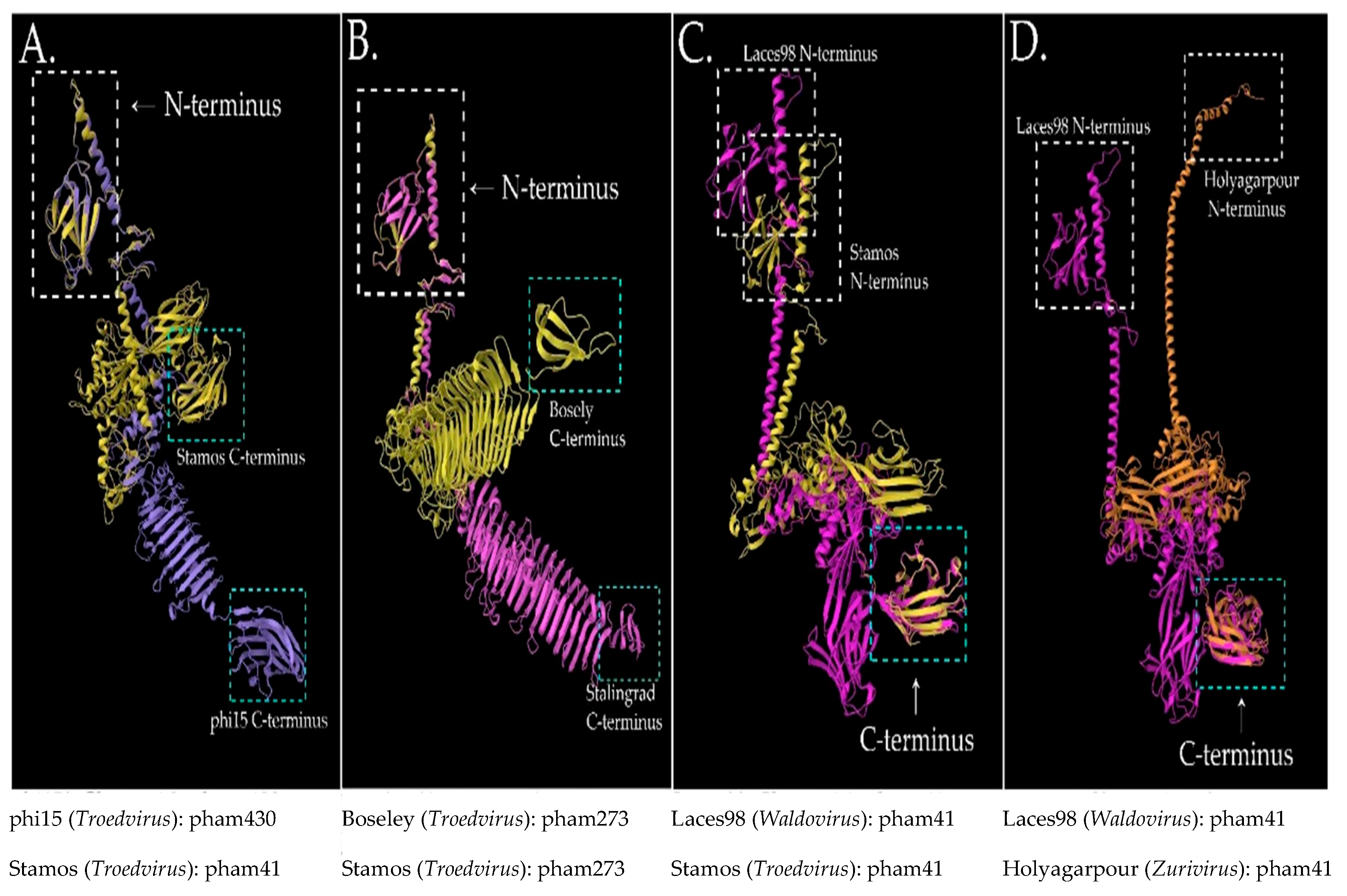
Tail spike proteins in podoviruses play several important roles in the early stages of infection, and extensive studies have been done with the spike proteins of E. coli phage T7 [87] and Salmonella phage P22 [88]. However, structural studies in P. putida are limited, and therefore we are limited to in silico analyses for this study [41]. Using Phamerator, we identified spike proteins in seven of the ten podoviruses in our new collection (the six Autotranscriptaviridae phages and Holyagarpour) and four previously isolated phages (gh-1 [38], phi15 [71], tf [89], and AF [90]) for comparison. The spike proteins from this dataset fall into 3 phams (41, 430, and 273) which are derived from amino acid comparisons and domain identification. Note that the pham numbers are derived within a given Phamerator dataset, so while they differentiate our tail spikes, they are otherwise arbitrary numbers that are not relevant beyond our dataset. We found that spike protein phams correlated with our phage taxonomic clusters, with one exception: Holyagarpour belongs to the Zurivirus genus, but its spike protein is more closely related to the spike proteins from the Waldovirus phages (Pham41), possibly indicating recombination among their ancestral phages. Also, while the spike proteins of singleton AF (protein: YP_007237194.1) [90] and tf (protein: YP_006382521.1) [89] are similar to the annotated spike proteins in our other podoviruses; no other proteins are shared among these phages.
In addition to the primary and secondary structure elements incorporated by Phamerator, we used SABLE to predict secondary structures in the N- and C- termini of several phage tail spike proteins and AlphaFold to predict tertiary structures (Figure 8; Supplemental Figures S4 and S5). In both datasets, we found that the first ~150 amino acids form 7–8 β-strands with an α-helix at the N-terminus. The exception to this was Holyagarpour, where the entire segment was primarily α-helices (and see below regarding tertiary structure). The last ~150 amino acids were not as conserved as the N-termini, but β-strands and β-sheets predominated. At the tertiary structure level, both the N-terminus and C-terminus of the spike proteins appear to be conserved among these P. putida phages regardless of the tail spike pham or the overall phage relatedness (Figure 8). The middle portions of the proteins show variability, but often contain an extended α-helix, a β-barrel, and a region of disorder. Using the overlay capability of MolStar, we conducted pairwise comparisons of tail spike proteins within phams and between phams (Figure 8). For each comparison, we prioritized aligning specific sections of the N- or C-terminus based on apparently conserved secondary structures. In most cases, there was good alignment of the N- and C-termini, even across phams. This is noteworthy because pham assignment is based on whole-protein similarity, not just conserved domains within them. When both termini of the pair could not be simultaneously aligned, this might be due to the flexibility of linker regions rather than some major difference in the structure itself. In addition, the N-terminus of the Holyagarpour tail spike was associated with a poor AlphaFold confidence score, so it is unclear how much of the apparent N-terminal differences in Figure 8D are due to a low-confidence prediction versus actual differences between the tail spike in this Zurivirus phage versus the tail spikes in the more closely related Waldovirus and Troedvirus phages. In Salmonella phage P22, and presumably in other podoviruses with related tail spike proteins, these proteins exist in a homotrimer in the virion (crystal structure shown in Supplemental Figure S4).

4. Discussion

The variety of P. putida phages described in this paper and in Brauer et al. [35] is unusual amongst other large phage collections in which a single bacterial strain has been used to isolate large numbers of phages [35]. Based on our proposed classifications, our thirteen phages include representatives of six different genera across at least four higher-order taxonomic groups, all isolated using a single P. putida strain. Similarly, Brauer et al. isolated 67 phages belonging to 22 groups, also on a single strain. Two examples show the opposite experience. From 2008 to 2011 a locally (Harrisonburg, VA, USA)-isolated Bacillus pumilus host was used for phage isolation. Some 120 phages were isolated, of which 27 were sequenced and all found to belong to one genetic cluster [91]. Also, Bacillus thuringiensis Kurstaki was used as a host in four different universities in Maryland and Virginia, in the United States, and several dozen phages were sequenced over the first five years and are continuing to be isolated. In this case, there is one cluster of B. thuringiensis phages with three highly related subclusters regardless of the year, the location, the type of sample, etc. [92]. While changing phage taxonomy over time makes it difficult to directly compare the number of genetic clusters reported by each study, it still seems clear that these examples from Bacillus phage discovery projects reflect a general theme of repeatedly isolating larger numbers of closely related phages that infect the same strain. This is in stark contrast to the findings of Brauer et al. [35] and to our findings in this work, where we see a wide diversity of phages infecting one strain of P. putida, even on the comparatively small scale of 13 phages. The reasons for this dramatic difference are unknown but could potentially reflect a greater diversity of P. putida phages in the sampled environments. Interestingly, our study used a host strain that is from a different clade of P. putida than the strain used by Brauer et al., so, at a minimum, it does not seem to be a strain-dependent phenomenon within P. putida [35]. In a whole genome BLASTp analysis, none of the Brauer study phages showed detectable similarities to any of the 13 we report here.
Two of the novel phages that we isolated, Dolphis and Jeremy, are predicted to be temperate. It is estimated that ~90% of phages are temperate or capable of undergoing lysogeny [93,94]. While prophages are evident in P. putida bacterial sequences, isolated temperate phages infecting P. putida are rare [95]. One of Dolphis and Jeremy’s distant relatives, PHB04, was previously predicted to be lytic [73]. However, we question this conclusion for several reasons. First, the authors identified a credible integrase gene in PHB04. Second, they concluded that PHB04 is not temperate based on a failure to amplify certain PHB04 genes from the genomes of 40 PHB04-resistant mutants of one particular host strain, which is insufficient to determine that PHB04 is not a temperate phage. Third, an intermediate relative, Touem01, was also predicted to be temperate based on multiple lines of evidence [72]. Therefore, while the creators of Bacphlip recommend that predictions with P-values lower than 95% be treated with caution, and future experimental work to verify them would be valuable, the preponderance of evidence points to Dolphis, Jeremy, and their three relatives (Touem01, ST4, and PHB04) being temperate phages.
The lysis cassettes of our phages have some interesting features. While the four basic types of endolysins (glycosylases, endopeptidases, amidases, and transglycosylases) are sufficiently conserved that they can usually be identified based on nucleotide sequence similarity [96], the holin and spanin sequences are much less well-conserved and are usually identified based on a combination of gene size, position relative to the endolysin, and predicted features such as transmembrane domains in the spanins [96]. In Zuri and its relatives, the lysis proteins are grouped together as a lysis cassette and manual annotation of the embedded i-spanin was straightforward. In all of our T7-like Waldovirus and Troedvirus phages, both the primary amino acid sequences and gene spacing (i.e., substantial genome distance between the endolysin gene and the other lysis genes) are similar to what is seen in phage T7. Interestingly, the T7 lysozyme has also been shown to play a role in regulating the balance between phage gene transcription and phage replication and packaging, which may explain its location in the genome among nucleic acid-processing enzymes, and its distance from the other lysis proteins [97]. In Dolphis and Jeremy, we proposed holins and spanins based on gene location, protein size, and some predicted secondary structures, but they have little or no sequence homology to known lysis proteins. If their function can be experimentally confirmed, their novel sequences should facilitate the rapid discovery of holins and spanins in other phages. In DDSR119, we note that the lysis gene organization is similar to that of some Pseudomonas pyocins. Specifically, when the DDSR119 genome is viewed in the circularly permuted form that we predict, the holin gene is located upstream of the endolysin and spanin genes, separated by the tail morphogenesis genes. This is analogous to the gene order of R-type pyocins in several Pseudomonas species, including P. putida [98]. The R-type pyocins are also called tailocins for their resemblance to myovirus tails, whereas DDSR119 has a siphovirus morphology. The R-type pyocins are also thought to be evolutionarily related to temperate phages [99] whereas we tentatively predict DDSR119 to be lytic. The similarity of gene order to tailocins could be coincidental, but with DDSR119 having no close known relatives among either lytic or temperate phages, it remains an intriguing observation.
When considering the fact that all thirteen of our phages infect the same host strain (and that they do not infect P. putida KT2440 or S12 strains) we wondered whether this might correlate with any obvious similarities in phage proteins that are typically associated with receptor binding. For example, if the bacterial receptor and the phage receptor binding proteins are conserved, this observation could be explained. The conserved lipopolysaccharide in Gram-negative bacteria has been shown to be the receptor for many phages [100], and specifically P. putida phages [35]. However, we could only identify tail spike proteins in some of our podoviruses (primarily among the T7-like phages) and even these were not highly conserved at the amino acid sequence level, nor did they cluster in a single pham, despite the fact that phams typically incorporate more predictive information than just primary amino acid sequence. Nevertheless, our AlphaFold predictions suggest that the conserved tertiary structure in P. putida spike proteins among even distantly related phages could play a role in the diversity of pairings in the genus and species of bacterial hosts. Both the N- and C-termini of spike proteins have attachment and enzymatic function, based on varying functional experimental data [101,102,103]. Bacteriophage hosts often have primary and secondary attachment sites, so both spike protein termini might be active simultaneously or in sequence,111 but this would need to be tested in future work. It has also been hypothesized that the tertiary structure of these proteins would allow the N- and C-termini to come together physically to exhibit multiple functions [104]. The AlphaFold-predicted structure shows that the flexible and varied middle section of these proteins might allow such a conformational change.
All Pseudomonads are able to form substantial biofilms that protect them from antibiotics, environmental chemicals, and infection by bacteriophages. In the search for novel anti-biofilm agents that might be used in conjunction with antibiotics in multidrug-resistant infections, there is a rising interest in the biofilm-degrading properties of phages. A number of studies have suggested that spike proteins have such activity, using genetic manipulation and mutant analyses [22,41]. Building on the bioinformatic analysis of the spike proteins presented here, future molecular experiments may elucidate specific mechanisms that could be used in biofilm remediation studies.
The phages explored in this study were isolated and characterized in an undergraduate laboratory course, one of some 200 similar courses across the US and abroad (https://seaphages.org/, accessed on 3 February 2021). The extraordinary educational benefits of this combined educational and discovery-based model have been well characterized; see work by Hanauer et al. [105,106]. At James Madison University, a two-semester sequence, phage discovery and genomic analysis, is taken mostly by 1st or 2nd year students and open to any major, with no prerequisites. Our students have isolated upwards of thirteen hundred phages since 2009, using members of Mycobacterium, Microbacterium, Bacillus, and Pseudomonas genera as hosts. Other examples of how these research experiences are offered at different types of institutions and resources for getting started are included in the Supplemental Materials, “Resources and comments on using phage discovery and genomics in undergraduate settings.”
The sheer number of phages being collected has exploded and bacterial hosts expanded over the last 15 years, creating a large reservoir of phages with potential to address problems facing medicine, agriculture, and industry. In just the one study described herein, our students discovered over two dozen phages that infect the Pseudomonas genus, including the ones described here. Collectively, thousands of phages have been added to existing collections isolated as free phages, where the functional viral particles can be used for basic biology and biotechnology applications.

Supplementary Materials

The following supporting information can be downloaded at: https://www.mdpi.com/article/10.3390/applmicrobiol5040108/s1: Figure S1: Genome Maps for Dolphis-like, Zuri-like, and Meara. Genome maps were created using the proksee web version. Genes are color coded according to function. A 5 kbp marker is included for reference. Dolphis and Jeremy are oriented to begin with the terminase gene. Zuri, Ebordelon, and Holyagarpour were oriented based on the empirically determined direct terminal repeats and Meara was adjusted to be collinear with them; Figure S2: Genome Maps for PlaquesPlease-like, Waldo5-like and DDSR119. Genome maps were created using the proksee web version. Genes are color coded according to function. A 5 kbp marker is included for reference. DDSR119 was oriented to begin with the terminase gene. All other phages were oriented based on the empirically determined direct terminal repeats; Figure S3: ViPTree proteome trees of (A) a subset of the Schitoviridae Family that includes the Zuri-like phages isolated in this study and their closest relatives, and (B) Dolphis-like phages isolated in this study and their closest relatives, all of which cluster distantly from other defined genera. Branch lengths are log-scaled.; Figure S4: AlphaFold3 structural predictions of seven tailspike proteins from our phages and three from previously published phages, grouped by pham. Also shown is the crystal structure of trimeric P22 tailspike (PDB: 8U1O). AlphaFold3 prediction confidences are shown in the legend. The tail spike in phage gh-1 (a Waldovirus phage) did not belong to any of these phams.; Figure S5: Secondary structure predictions from a representative of each pham. JPred results show extended (E), helical (H) and other (-) types of secondary structure from (in order) jnet, HMMer2, and PSI-BLAST-PSSM; Document S1: Resources for teaching phage discovery; Table S1: P. putida phages in GenBank as of 25 April, or in this study.

Author Contributions

Conceptualization, S.A., R.P., A.P., S.M.L., R.K. and L.T.; Data curation, S.A., R.P., S.M.L., R.K., J.D.J., S.G.C. and L.T.; Formal analysis, S.A., R.P., A.P., E.R., J.O., K.I.C., S.M.L., R.K., J.D.J., C.M.M., S.G.C. and L.T.; Investigation, S.A., R.P., A.P., E.R., J.O., K.I.C., S.M.L., R.K. and L.T.; Methodology, S.A., R.P., S.M.L., R.K., J.D.J., C.M.M., S.G.C. and L.T.; Resources, S.M.L. and L.T.; Software, K.I.C. and S.G.C.; Supervision, S.M.L. and L.T.; Validation, A.P.; Visualization, S.A. and K.I.C.; Writing—original draft, S.A., R.P., A.P., S.M.L., R.K., C.M.M. and L.T.; Writing—review & editing, S.A., R.P., A.P., E.R., J.O., K.I.C., S.M.L., R.K., C.M.M. and L.T. All authors have read and agreed to the published version of the manuscript.

Funding

Research by C.M.M. and J.D.J. was supported by the Laboratory Directed Research and Development (LDRD) program of Sandia National Laboratories, which is a multi-mission laboratory managed and operated by National Technology and Engineering Solutions of Sandia, LLC, a wholly owned subsidiary of Honeywell International Inc., for the U.S. Department of Energy’s National Nuclear Security Administration under contract DE-NA0003525. The DOE Systems Biology Knowledgebase (KBase) is funded by the U.S. Department of Energy, Office of Science, Office of Biological and Environmental Research under Award Numbers DE-AC02-05CH11231, DE-AC02-06CH113.

Data Availability Statement

All data are either included in the paper or can be independently verified using the listed tools and primary data from public databases such as GenBank.

Acknowledgments

We are grateful to Bordelon, Emily, Needs, Robert, McAndrew, Miles, Staples, Lacey, Meara, William, Dimitris, James, O’Donnell, Jake, Bose, Lily, Treml, Alex, Patel, Akaash, Rupe, Easton, Anderson, Simon, Daniel, Deborah for their support during the study.

Conflicts of Interest

Author Easton Rupe was employed by the company Sapporo-Stone Brewing. The remaining authors declare that the research was conducted in the absence of any commercial or financial relationships that could be construed as a potential conflict of interest.

References

  1. Lyng, M.; Kovács, Á.T. Frenemies of the soil: Bacillus and Pseudomonas interspecies interactions. Trends Microbiol. 2023, 31, 845–857. [Google Scholar] [CrossRef]
  2. Iglewski, B.H. Pseudomonas. In Medical Microbiology, 4th ed.; Baron, S., Ed.; University of Texas Medical Branch at Galveston: Galveston, TX, USA, 1996. Available online: http://www.ncbi.nlm.nih.gov/books/NBK8326/ (accessed on 2 April 2025).
  3. Kramer, A.; Schwebke, I.; Kampf, G. How long do nosocomial pathogens persist on inanimate surfaces? A systematic review. BMC Infect. Dis. 2006, 6, 130. [Google Scholar] [CrossRef] [PubMed]
  4. Rohmer, L.; Hocquet, D.; Miller, S.I. Are pathogenic bacteria just looking for food? Metabolism and microbial pathogenesis. Trends Microbiol. 2011, 19, 341–348. [Google Scholar] [CrossRef]
  5. Ahearn, D.G.; Borazjani, R.N.; Simmons, R.B.; Gabriel, M.M. Primary adhesion of Pseudomonas aeruginosa to inanimate surfaces including biomaterials. Methods Enzym. 1999, 310, 551–557. [Google Scholar] [CrossRef]
  6. Perkins, K.M.; Reddy, S.C.; Fagan, R.; Arduino, M.J.; Perz, J.F. Investigation of healthcare infection risks from water-related organisms: Summary of CDC consultations, 2014–2017. Infect. Control. Hosp. Epidemiol. 2019, 40, 621–626. [Google Scholar] [CrossRef]
  7. Sukhum, K.V.; Newcomer, E.P.; Cass, C.; Wallace, M.A.; Johnson, C.; Fine, J.; Sax, S.; Barlet, M.H.; Burnham, C.D.; Dantas, G.; et al. Antibiotic-resistant organisms establish reservoirs in new hospital built environments and are related to patient blood infection isolates. Commun. Med. 2022, 2, 62. [Google Scholar] [CrossRef]
  8. Xin, X.F.; Kvitko, B.; He, S.Y. Pseudomonas syringae: What it takes to be a pathogen. Nat. Rev. Microbiol. 2018, 16, 316–328. [Google Scholar] [CrossRef]
  9. Moreau-Marquis, S.; Stanton, B.A.; O’Toole, G.A. Pseudomonas aeruginosa biofilm formation in the cystic fibrosis airway. Pulm. Pharmacol. Ther. 2008, 21, 595–599. [Google Scholar] [CrossRef]
  10. Han, Q.; Feng, L.; Zhang, Y.; Zhang, R.; Wang, G.; Zhang, Y. Effect of Juglone against Pseudomonas syringae pv actinidiae Planktonic Growth and Biofilm Formation. Molecules 2021, 26, 7580. [Google Scholar] [CrossRef]
  11. Planchamp, C.; Glauser, G.; Mauch-Mani, B. Root inoculation with Pseudomonas putida KT2440 induces transcriptional and metabolic changes and systemic resistance in maize plants. Front. Plant Sci. 2015, 5, 719. [Google Scholar] [CrossRef]
  12. Timmis, K.N.; Steffan, R.J.; Unterman, R. Designing microorganisms for the treatment of toxic wastes. Annu. Rev. Microbiol. 1994, 48, 525–557. [Google Scholar] [CrossRef] [PubMed]
  13. Lehrbach, P.R.; Zeyer, J.; Reineke, W.; Knackmuss, H.J.; Timmis, K.N. Enzyme recruitment in vitro: Use of cloned genes to extend the range of haloaromatics degraded by Pseudomonas sp. strain B13. J. Bacteriol. 1984, 158, 1025–1032. [Google Scholar] [CrossRef] [PubMed]
  14. Dejonghe, W.; Boon, N.; Seghers, D.; Top, E.M.; Verstraete, W. Bioaugmentation of soils by increasing microbial richness: Missing links. Environ. Microbiol. 2001, 3, 649–657. [Google Scholar] [CrossRef] [PubMed]
  15. Gong, T.; Xu, X.; Dang, Y.; Kong, A.; Wu, Y.; Liang, P.; Wang, S.; Yu, H.; Xu, P.; Yang, C. An engineered Pseudomonas putida can simultaneously degrade organophosphates, pyrethroids and carbamates. Sci. Total Environ. 2018, 628–629, 1258–1265. [Google Scholar] [CrossRef]
  16. Qi, X.; Gao, X.; Wang, X.; Xu, P. Harnessing Pseudomonas putida in bioelectrochemical systems. Trends Biotechnol. 2024, 42, 877–894. [Google Scholar] [CrossRef]
  17. de Lorenzo, V.; Pérez-Pantoja, D.; Nikel, P.I. Pseudomonas putida KT2440: The long journey of a soil-dweller to become a synthetic biology chassis. J. Bacteriol. 2024, 206, e0013624. [Google Scholar] [CrossRef]
  18. Weimer, A.; Kohlstedt, M.; Volke, D.C.; Nikel, P.I.; Wittmann, C. Industrial biotechnology of Pseudomonas putida: Advances and prospects. Appl. Microbiol. Biotechnol. 2020, 104, 7745–7766. [Google Scholar] [CrossRef]
  19. Campbell, J.I.A.; Albrechtsen, M.; Sørensen, J. Large Pseudomonas phages isolated from barley rhizosphere. FEMS Microbiol. Ecol. 2006, 18, 63–74. [Google Scholar] [CrossRef]
  20. Il Kim, M.; Park, C.Y.; Seo, J.M.; Kang, K.S.; Park, K.S.; Kang, J.; Hong, K.S.; Choi, Y.; Lee, S.Y.; Park, J.P.; et al. In Situ Biosynthesis of a Metal Nanoparticle Encapsulated in Alginate Gel for Imageable Drug-Delivery System. ACS Appl. Mater. Interfaces 2021, 13, 36697–36708. [Google Scholar] [CrossRef]
  21. Avendaño, R.; Muñoz-Montero, S.; Rojas-Gätjens, D.; Fuentes-Schweizer, P.; Vieto, S.; Montenegro, R.; Salvador, M.; Frew, R.; Kim, J.; Chavarría, M.; et al. Production of selenium nanoparticles occurs through an interconnected pathway of sulphur metabolism and oxidative stress response in Pseudomonas putida KT2440. Microb. Biotechnol. 2023, 16, 931–946. [Google Scholar] [CrossRef]
  22. Shaburova, O.V.; Krylov, S.V.; Veĭko, V.P.; Pleteneva, E.A.; Burkal’tseva, M.V.; Miroshnokov, K.A.; Kornelissen, A.; Lavogne, R.; Sykilinda, N.N.; Kadykov, V.A.; et al. Search for destruction factors of bacterial biofilms: Comparison of phage properties in a group of Pseudomonas putida bacteriophages and specificity of their halo-formation products. Genetika 2009, 45, 185–195. [Google Scholar] [CrossRef]
  23. Fage, C.; Lemire, N.; Moineau, S. Delivery of CRISPR-Cas systems using phage-based vectors. Curr. Opin. Biotechnol. 2021, 68, 174–180. [Google Scholar] [CrossRef]
  24. Nikel, P.I.; de Lorenzo, V. Pseudomonas putida as a functional chassis for industrial biocatalysis: From native biochemistry to trans-metabolism. Metab. Eng. 2018, 50, 142–155. [Google Scholar] [CrossRef] [PubMed]
  25. Wommack, K.E.; Colwell, R.R. Virioplankton: Viruses in Aquatic Ecosystems. Microbiol. Mol. Biol. Rev. 2000, 64, 69–114. [Google Scholar] [CrossRef] [PubMed]
  26. Vandenheuvel, D.; Lavigne, R.; Brüssow, H. Bacteriophage Therapy: Advances in Formulation Strategies and Human Clinical Trials. Annu. Rev. Virol. 2015, 2, 599–618. [Google Scholar] [CrossRef] [PubMed]
  27. Endersen, L.; O’Mahony, J.; Hill, C.; Ross, R.P.; McAuliffe, O.; Coffey, A. Phage therapy in the food industry. Annu. Rev. Food Sci. Technol. 2014, 5, 327–349. [Google Scholar] [CrossRef]
  28. Jones, J.B.; Vallad, G.E.; Iriarte, F.B.; Obradović, A.; Wernsing, M.H.; Jackson, L.E.; Balogh, B.; Hong, J.C.; Momol, M.T. Considerations for using bacteriophages for plant disease control. Bacteriophage 2012, 2, 208–214. [Google Scholar] [CrossRef]
  29. Di Lallo, G.; Evangelisti, M.; Mancuso, F.; Ferrante, P.; Marcelletti, S.; Tinari, A.; Superti, F.; Migliore, L.; D’Addabbo, P.; Frezza, D.; et al. Isolation and partial characterization of bacteriophages infecting Pseudomonas syringae pv. actinidiae, causal agent of kiwifruit bacterial canker. J. Basic Microbiol. 2014, 54, 1210–1221. [Google Scholar] [CrossRef]
  30. Dedrick, R.M.; Smith, B.E.; Cristinziano, M.; Freeman, K.G.; Jacobs-Sera, D.; Belessis, Y.; Whitney Brown, A.; Cohen, K.A.; Davidson, R.M.; van Duin, D.; et al. Phage Therapy of Mycobacterium Infections: Compassionate Use of Phages in 20 Patients With Drug-Resistant Mycobacterial Disease. Clin. Infect. Dis. 2023, 76, 103–112. [Google Scholar] [CrossRef]
  31. Slopek, S.; Kucharewicz-Krukowska, A.; Weber-Dabrowska, B.; Dabrowski, M. Results of bacteriophage treatment of suppurative bacterial infections. V. Evaluation of the results obtained in children. Arch. Immunol. Ther. Exp. 1985, 33, 241–259. [Google Scholar]
  32. Rabiey, M.; Roy, S.R.; Holtappels, D.; Franceschetti, L.; Quilty, B.J.; Creeth, R.; Sundin, G.W.; Wagemans, J.; Lavigne, R.; Jackson, R.W. Phage biocontrol to combat Pseudomonas syringae pathogens causing disease in cherry. Microb. Biotechnol. 2020, 13, 1428–1445. [Google Scholar] [CrossRef] [PubMed]
  33. Lee, C.; Kim, H.; Ryu, S. Bacteriophage and endolysin engineering for biocontrol of food pathogens/pathogens in the food: Recent advances and future trends. Crit. Rev. Food Sci. Nutr. 2023, 63, 8919–8938. [Google Scholar] [CrossRef] [PubMed]
  34. Liu, H.; Hu, Z.; Li, M.; Yang, Y.; Lu, S.; Rao, X. Therapeutic potential of bacteriophage endolysins for infections caused by Gram-positive bacteria. J. Biomed. Sci. 2023, 30, 29. [Google Scholar] [CrossRef] [PubMed]
  35. Brauer, A.; Rosendahl, S.; Kängsep, A.; Lewańczyk, A.C.; Rikberg, R.; Hõrak, R.; Tamman, H. Isolation and characterization of a phage collection against Pseudomonas putida. Environ. Microbiol. 2024, 26, e16671. [Google Scholar] [CrossRef]
  36. Lammens, E.M.; Nikel, P.I.; Lavigne, R. Exploring the synthetic biology potential of bacteriophages for engineering non-model bacteria. Nat. Commun. 2020, 11, 5294. [Google Scholar] [CrossRef]
  37. Lee, L.F.; Boezi, J.A. Characterization of bacteriophage gh-1 for Pseudomonas putida. J. Bacteriol. 1966, 92, 1821–1827. [Google Scholar] [CrossRef]
  38. Kovalyova, I.V.; Kropinski, A.M. The complete genomic sequence of lytic bacteriophage gh-1 infecting Pseudomonas putida—Evidence for close relationship to the T7 group. Virology 2003, 311, 305–315. [Google Scholar] [CrossRef]
  39. Ha, A.D.; Denver, D.R. Comparative Genomic Analysis of 130 Bacteriophages Infecting Bacteria in the Genus Pseudomonas. Front. Microbiol. 2018, 9, 1456. [Google Scholar] [CrossRef]
  40. Yehl, K.; Lemire, S.; Yang, A.C.; Ando, H.; Mimee, M.; Torres, M.T.; de la Fuente-Nunez, C.; Lu, T.K. Engineering Phage Host-Range and Suppressing Bacterial Resistance through Phage Tail Fiber Mutagenesis. Cell 2019, 179, 459–469.e9. [Google Scholar] [CrossRef]
  41. Mageeney, C.M.; Trubl, G.; Williams, K.P. Improved Mobilome Delineation in Fragmented Genomes. Front. Bioinform. 2022, 2, 866850. [Google Scholar] [CrossRef]
  42. Purtschert-Montenegro, G.; Cárcamo-Oyarce, G.; Pinto-Carbó, M.; Agnoli, K.; Bailly, A.; Eberl, L. Pseudomonas putida mediates bacterial killing, biofilm invasion and biocontrol with a type IVB secretion system. Nat. Microbiol. 2022, 7, 1547–1557. [Google Scholar] [CrossRef] [PubMed]
  43. Poxleitner, M.; Pope, W.; Jacobs-Sera, D.; Sivanathan, V.; Hatfull, G. Phage Discovery Guide. Published Online 2018. Available online: https://seaphagesphagediscoveryguide.helpdocsonline.com/home (accessed on 27 March 2025).
  44. Promega. Wizard® DNA Clean-Up System Protocol. 2010. Available online: https://www.promega.com/resources/protocols/technical-bulletins/0/wizard-dna-cleanup-system-protocol/ (accessed on 7 July 2020).
  45. Fryslie, B. Newbler. 2012. Available online: https://swes.cals.arizona.edu/maier_lab/kartchner/documentation/index.php/home/docs/newbler (accessed on 26 March 2019).
  46. Bankevich, A.; Nurk, S.; Antipov, D.; Gurevich, A.A.; Dvorkin, M.; Kulikov, A.S.; Lesin, V.M.; Nikolenko, S.I.; Pham, S.; Prjibelski, A.D.; et al. SPAdes: A new genome assembly algorithm and its applications to single-cell sequencing. J. Comput. Biol. 2012, 19, 455–477. [Google Scholar] [CrossRef] [PubMed]
  47. Gordon, D.; Green, P. Consed: A graphical editor for next-generation sequencing. Bioinformatics 2013, 29, 2936–2937. [Google Scholar] [CrossRef] [PubMed]
  48. Garneau, J.R.; Depardieu, F.; Fortier, L.C.; Bikard, D.; Monot, M. PhageTerm: A tool for fast and accurate determination of phage termini and packaging mechanism using next-generation sequencing data. Sci. Rep. 2017, 7, 8292. [Google Scholar] [CrossRef]
  49. Shaffer, M.; Borton, M.A.; McGivern, B.B.; Zayed, A.A.; La Rosa, S.L.; Solden, L.M.; Liu, P.; Narrowe, A.B.; Rodríguez-Ramos, J.; Bolduc, B.; et al. DRAM for distilling microbial metabolism to automate the curation of microbiome function. Nucleic Acids Res. 2020, 48, 8883–8900. [Google Scholar] [CrossRef]
  50. Arkin, A.P.; Cottingham, R.W.; Henry, C.S.; Harris, N.L.; Stevens, R.L.; Maslov, S.; Dehal, P.; Ware, D.; Perez, F.; Canon, S.; et al. KBase: The United States Department of Energy Systems Biology Knowledgebase. Nat. Biotechnol. 2018, 36, 566–569. [Google Scholar] [CrossRef]
  51. Boratyn, G.M.; Camacho, C.; Cooper, P.S.; Coulouris, G.; Fong, A.; Ma, N.; Madden, T.L.; Matten, W.T.; McGinnis, S.D.; Merezhuk, Y.; et al. BLAST: A more efficient report with usability improvements. Nucleic Acids Res. 2013, 41, W29. [Google Scholar] [CrossRef]
  52. Pope, W.H.; Jacobs-Sera, D. Annotation of Bacteriophage Genome Sequences Using DNA Master: An Overview. In Bacteriophages: Methods and Protocols; Clokie, M.R.J., Kropinski, A.M., Lavigne, R., Eds.; Springer: New York, NY, USA, 2018; pp. 217–229. [Google Scholar] [CrossRef]
  53. Delcher, A.L.; Bratke, K.A.; Powers, E.C.; Salzberg, S.L. Identifying bacterial genes and endosymbiont DNA with Glimmer. Bioinformatics 2007, 23, 673–679. [Google Scholar] [CrossRef]
  54. Lukashin, A.V.; Borodovsky, M. GeneMark. hmm: New solutions for gene finding. Nucleic Acids Res. 1998, 26, 1107–1115. [Google Scholar] [CrossRef]
  55. Söding, J.; Biegert, A.; Lupas, A.N. The HHpred interactive server for protein homology detection and structure prediction. Nucleic Acids Res. 2005, 33 (Suppl. 2), W244–W248. [Google Scholar] [CrossRef]
  56. Berman, H.M.; Westbrook, J.; Feng, Z.; Gilliland, G.; Bhat, T.N.; Weissig, H.; Shindyalov, I.N.; Bourne, P.E. The Protein Data Bank. Nucleic Acids Res. 2000, 28, 235–242. [Google Scholar] [CrossRef]
  57. Hofmann, K.; TMbase—A Database of Membrane Spanning Protein Segments. TMpred Documentation. 1993. Available online: https://sbcb.bioch.ox.ac.uk/TM_noj/TM_noj.html (accessed on 19 September 2020).
  58. Hallgren, J.; Tsirigos, K.D.; Pedersen, M.D.; Almagro Armenteros, J.J.; Marcatili, P.; Nielsen, H.; Krogh, A.; Winther, O. DeepTMHMM predicts alpha and beta transmembrane proteins using deep neural networks. bioRxiv 2022, 2202–2204. [Google Scholar] [CrossRef]
  59. Kahsay, R.Y.; Gao, G.; Liao, L. An improved hidden Markov model for transmembrane protein detection and topology prediction and its applications to complete genomes. Bioinformatics 2005, 21, 1853–1858. [Google Scholar] [CrossRef] [PubMed]
  60. Juncker, A.S.; Willenbrock, H.; Von Heijne, G.; Brunak, S.; Nielsen, H.; Krogh, A. Prediction of lipoprotein signal peptides in Gram-negative bacteria. Protein Sci. 2003, 12, 1652–1662. [Google Scholar] [CrossRef] [PubMed]
  61. Laslett, D.; Canback, B. ARAGORN, a program to detect tRNA genes and tmRNA genes in nucleotide sequences. Nucleic Acids Res. 2004, 32, 11–16. [Google Scholar] [CrossRef]
  62. Grant, J.R.; Enns, E.; Marinier, E.; Mandal, A.; Herman, E.K.; Chen, C.Y.; Graham, M.; Van Domselaar, G.; Stothard, P. Proksee: In-depth characterization and visualization of bacterial genomes. Nucleic Acids Res. 2023, 51, W484–W492. [Google Scholar] [CrossRef]
  63. Moraru, C.; Varsani, A.; Kropinski, A.M. VIRIDIC-A Novel Tool to Calculate the Intergenomic Similarities of Prokaryote-Infecting Viruses. Viruses 2020, 12, 1268. [Google Scholar] [CrossRef]
  64. Nishimura, Y.; Yoshida, T.; Kuronishi, M.; Uehara, H.; Ogata, H.; Goto, S. ViPTree: The viral proteomic tree server. Bioinformatics 2017, 33, 2379–2380. [Google Scholar] [CrossRef]
  65. Cresawn, S.G.; Bogel, M.; Day, N.; Jacobs-Sera, D.; Hendrix, R.W.; Hatfull, G.F. Phamerator: A bioinformatic tool for comparative bacteriophage genomics. BMC Bioinform. 2011, 12, 395. [Google Scholar] [CrossRef]
  66. Gauthier, C.H.; Hatfull, G.F. A Bioinformatic Ecosystem for Bacteriophage Genomics: PhaMMSeqs, Phamerator, pdm_utils, PhagesDB, DEPhT, and PhamClust. Viruses 2024, 16, 1278. [Google Scholar] [CrossRef]
  67. Hockenberry, A.J.; Wilke, C.O. BACPHLIP: Predicting bacteriophage lifestyle from conserved protein domains. PeerJ 2021, 9, e11396. [Google Scholar] [CrossRef]
  68. Jumper, J.; Evans, R.; Pritzel, A.; Green, T.; Figurnov, M.; Ronneberger, O.; Tunyasuvunakool, K.; Bates, R.; Žídek, A.; Potapenko, A.; et al. Highly accurate protein structure prediction with AlphaFold. Nature 2021, 596, 583–589. [Google Scholar] [CrossRef] [PubMed]
  69. Varadi, M.; Anyango, S.; Deshpande, M.; Nair, S.; Natassia, C.; Yordanova, G.; Yuan, D.; Stroe, O.; Wood, G.; Laydon, A.; et al. AlphaFold Protein Structure Database: Massively expanding the structural coverage of protein-sequence space with high-accuracy models. Nucleic Acids Res. 2022, 50, D439–D444. [Google Scholar] [CrossRef] [PubMed]
  70. Sehnal, D.; Bittrich, S.; Deshpande, M.; Svobodová, R.; Berka, K.; Bazgier, V.; Velankar, S.; Burley, S.K.; Koča, J.; Rose, A.S. Mol* Viewer: Modern web app for 3D visualization and analysis of large biomolecular structures. Nucleic Acids Res. 2021, 49, W431–W437. [Google Scholar] [CrossRef]
  71. Cornelissen, A.; Ceyssens, P.J.; T’Syen, J.; Van Praet, H.; Noben, J.P.; Shaburova, O.V.; Krylov, V.N.; Volckaert, G.; Lavigne, R. The T7-related Pseudomonas putida phage φ15 displays virion-associated biofilm degradation properties. PLoS ONE 2011, 6, e18597. [Google Scholar] [CrossRef]
  72. Feltin, C.; Garneau, J.R.; Morris, C.E.; Bérard, A.; Torres-Barceló, C. Novel phages of Pseudomonas syringae unveil numerous potential auxiliary metabolic genes. J. Gen. Virol. 2024, 105, 001990. [Google Scholar] [CrossRef]
  73. Chen, Y.; Yang, L.; Sun, E.; Song, J.; Wu, B. Characterisation of a newly detected bacteriophage infecting Bordetella bronchiseptica in swine. Arch. Virol. 2019, 164, 33–40. [Google Scholar] [CrossRef]
  74. Dougherty, P.E.; Pedersen, M.S.; Forero-Junco, L.M.; Carstens, A.B.; Raaijmakers, J.M.; Riber, L.; Hansen, L.H. Novel bacteriophages targeting wheat phyllosphere bacteria carry DNA modifications and single-strand breaks. Virus Res. 2025, 352, 199524. [Google Scholar] [CrossRef]
  75. Adriaenssens, E.; Brister, J.R. How to Name and Classify Your Phage: An Informal Guide. Viruses 2017, 9, 70. [Google Scholar] [CrossRef]
  76. Wittmann, J.; Turner, D.; Millard, A.D.; Mahadevan, P.; Kropinski, A.M.; Adriaenssens, E.M. From orphan phage to a proposed new family–the diversity of N4-like viruses. Antibiotics 2020, 9, 663. [Google Scholar] [CrossRef]
  77. Belcaid, M.; Bergeron, A.; Poisson, G. The evolution of the tape measure protein: Units, duplications and losses. BMC Bioinform. 2011, 12 (Suppl. 9), S10. [Google Scholar] [CrossRef] [PubMed]
  78. Cumby, N.; Reimer, K.; Mengin-Lecreulx, D.; Davidson, A.R.; Maxwell, K.L. The phage tail tape measure protein, an inner membrane protein and a periplasmic chaperone play connected roles in the genome injection process of E. coli phage HK97. Mol. Microbiol. 2015, 96, 437–447. [Google Scholar] [CrossRef] [PubMed]
  79. Ismail, A.; Saini, T.; Al Qaffas, A.; Erill, I.; Caruso, S.M.; Temple, L.; Johnson, A.A. Evidence of a Set of Core-Function Genes in 16 Bacillus Podoviral Genomes with Considerable Genomic Diversity. Viruses 2023, 15, 276. [Google Scholar] [CrossRef] [PubMed]
  80. Pedulla, M.L.; Ford, M.E.; Houtz, J.M.; Karthikeyan, T.; Wadsworth, C.; Lewis, J.A.; Jacobs-Sera, D.; Falbo, J.; Gross, J.; Pannunzio, N.R.; et al. Origins of highly mosaic mycobacteriophage genomes. Cell 2003, 113, 172–182. [Google Scholar] [CrossRef]
  81. Wetzel, K.S.; Aull, H.G.; Zack, K.M.; Garlena, R.A.; Hatfull, G.F. Protein-Mediated and RNA-Based Origins of Replication of Extrachromosomal Mycobacterial Prophages. mBio 2020, 11, e00385-20. [Google Scholar] [CrossRef]
  82. Deutsch, D.R.; Utter, B.; Verratti, K.J.; Sichtig, H.; Tallon, L.J.; Fischetti, V.A. Extra-Chromosomal DNA Sequencing Reveals Episomal Prophages Capable of Impacting Virulence Factor Expression in Staphylococcus aureus. Front. Microbiol. 2018, 9, 1406. Available online: https://www.frontiersin.org/articles/10.3389/fmicb.2018.01406 (accessed on 25 December 2023). [CrossRef]
  83. Landy, A. The λ Integrase Site-specific Recombination Pathway. Microbiol. Spectr. 2015, 3, MDNA3-0051-2014. [Google Scholar] [CrossRef]
  84. Cahill, J.; Young, R. Phage Lysis: Multiple Genes for Multiple Barriers. Adv. Virus Res. 2019, 103, 33–70. [Google Scholar] [CrossRef]
  85. Pratama, A.A.; Bolduc, B.; Zayed, A.A.; Zhong, Z.P.; Guo, J.; Vik, D.R.; Gazitúa, M.C.; Wainaina, J.M.; Roux, S.; Sullivan, M.B. Expanding standards in viromics: In silico evaluation of dsDNA viral genome identification, classification, and auxiliary metabolic gene curation. PeerJ 2021, 9, e11447. [Google Scholar] [CrossRef]
  86. Murphy, J.; Mahony, J.; Ainsworth, S.; Nauta, A.; van Sinderen, D. Bacteriophage orphan DNA methyltransferases: Insights from their bacterial origin, function, and occurrence. Appl. Environ. Microbiol. 2013, 79, 7547–7555. [Google Scholar] [CrossRef]
  87. Steven, A.C.; Trus, B.L.; Maizel, J.V.; Unser, M.; Parry, D.A.; Wall, J.S.; Hainfeld, J.F.; Studier, F.W. Molecular substructure of a viral receptor-recognition protein. The gp17 tail-fiber of bacteriophage T7. J. Mol. Biol. 1988, 200, 351–365. [Google Scholar] [CrossRef]
  88. Steinbacher, S.; Miller, S.; Baxa, U.; Budisa, N.; Weintraub, A.; Seckler, R.; Huber, R. Phage P22 tailspike protein: Crystal structure of the head-binding domain at 2.3 A, fully refined structure of the endorhamnosidase at 1.56 A resolution, and the molecular basis of O-antigen recognition and cleavage. J. Mol. Biol. 1997, 267, 865–880. [Google Scholar] [CrossRef] [PubMed]
  89. Glukhov, A.S.; Krutilina, A.I.; Shlyapnikov, M.G.; Severinov, K.; Lavysh, D.; Kochetkov, V.V.; McGrath, J.W.; de Leeuwe, C.; Shaburova, O.V.; Krylov, V.N.; et al. Genomic Analysis of Pseudomonas putida Phage tf with Localized Single-Strand DNA Interruptions. PLoS ONE 2012, 7, e51163. [Google Scholar] [CrossRef] [PubMed]
  90. Cornelissen, A.; Ceyssens, P.J.; Krylov, V.N.; Noben, J.P.; Volckaert, G.; Lavigne, R. Identification of EPS-degrading activity within the tail spikes of the novel Pseudomonas putida phage AF. Virology 2012, 434, 251–256. [Google Scholar] [CrossRef] [PubMed]
  91. Lorenz, L.; Lins, B.; Barrett, J.; Montgomery, A.; Trapani, S.; Schindler, A.; Christie, G.E.; Cresawn, S.G.; Temple, L. Genomic characterization of six novel Bacillus pumilus bacteriophages. Virology 2013, 444, 374–383. [Google Scholar] [CrossRef]
  92. Sauder, A.B.; Quinn, M.R.; Brouillette, A.; Caruso, S.; Cresawn, S.; Erill, I.; Lewis, L.; Loesser-Casey, K.; Pate, M.; Scott, C.; et al. Genomic characterization and comparison of seven Myoviridae bacteriophage infecting Bacillus thuringiensis. Virology 2016, 489, 243–251. [Google Scholar] [CrossRef]
  93. Zhang, M.; Zhang, T.; Yu, M.; Chen, Y.L.; Jin, M. The Life Cycle Transitions of Temperate Phages: Regulating Factors and Potential Ecological Implications. Viruses 2022, 14, 1904. [Google Scholar] [CrossRef]
  94. Freifelder, D. A Comprehensive Introduction to Prokaryotes and Eukaryotes. In More DNA Molecular Biology; Biosicence; Jones and Bartlett Publishers, Inc.: Burlington, MA, USA, 1984; p. 660. [Google Scholar]
  95. Yu, S.L.; McClain, H.M.; Torrance, E.L.; Schoeniger, J.S.; Mageeney, C.M.; Williams, K.P. The TIGER/Islander resource for genomic islands. Microbiol. Resour. Announc. 2025, 14, e0049225. [Google Scholar] [CrossRef]
  96. Oliveira, H.; Melo, L.D.; Santos, S.B.; Nóbrega, F.L.; Ferreira, E.C.; Cerca, N.; Azeredo, J.; Kluskens, L.D. Molecular Aspects and Comparative Genomics of Bacteriophage Endolysins. J. Virol. 2013, 87, 4558–4570. [Google Scholar] [CrossRef]
  97. Zhang, X.; Studier, F.W. Multiple roles of T7 RNA polymerase and T7 lysozyme during bacteriophage T7 infection. J. Mol. Biol. 2004, 340, 707–730. [Google Scholar] [CrossRef]
  98. Scholl, D. Phage Tail–Like Bacteriocins. Annu. Rev. Virol. 2017, 4, 453–467. [Google Scholar] [CrossRef] [PubMed]
  99. Ghequire, M.G.K.; Dillen, Y.; Lambrichts, I.; Proost, P.; Wattiez, R.; De Mot, R. Different Ancestries of R Tailocins in Rhizospheric Pseudomonas Isolates. Genome Biol. Evol. 2015, 7, 2810–2828. [Google Scholar] [CrossRef] [PubMed]
  100. Bertozzi Silva, J.; Storms, Z.; Sauvageau, D. Host receptors for bacteriophage adsorption. FEMS Microbiol. Lett. 2016, 363, fnw002. [Google Scholar] [CrossRef] [PubMed]
  101. Hardy, J.M.; Dunstan, R.A.; Lithgow, T.; Coulibaly, F. Tall tails: Cryo-electron microscopy of phage tail DNA ejection conduits. Biochem. Soc. Trans. 2022, 50, 459-22W. [Google Scholar] [CrossRef]
  102. Johnson, J.E.; Chiu, W. DNA packaging and delivery machines in tailed bacteriophages. Curr. Opin. Struct. Biol. 2007, 17, 237–243. [Google Scholar] [CrossRef]
  103. Knecht, L.E.; Veljkovic, M.; Fieseler, L. Diversity and Function of Phage Encoded Depolymerases. Front. Microbiol. 2020, 10, 2949. [Google Scholar] [CrossRef]
  104. Ouyang, R.; Ongenae, V.; Muok, A.; Claessen, D.; Briegel, A. Phage fibers and spikes: A nanoscale Swiss army knife for host infection. Curr. Opin. Microbiol. 2024, 77, 102429. [Google Scholar] [CrossRef]
  105. Hanauer, D.I.; Graham, M.J.; SEA-PHAGES; Betancur, L.; Bobrownicki, A.; Cresawn, S.G.; Garlena, R.A.; Jacobs-Sera, D.; Kaufmann, N.; Pope, W.H.; et al. An inclusive Research Education Community (iREC): Impact of the SEA-PHAGES program on research outcomes and student learning. Proc. Natl. Acad. Sci. USA 2017, 114, 13531–13536. [Google Scholar] [CrossRef]
  106. Hanauer, D.I.; Graham, M.J.; Jacobs-Sera, D.; Garlena, R.A.; Russell, D.A.; Sivanathan, V.; Asai, D.J.; Hatfull, G.F. Broadening Access to STEM through the Community College: Investigating the Role of Course-Based Research Experiences (CREs). CBE Life Sci. Educ. 2022, 21, ar38. [Google Scholar] [CrossRef]
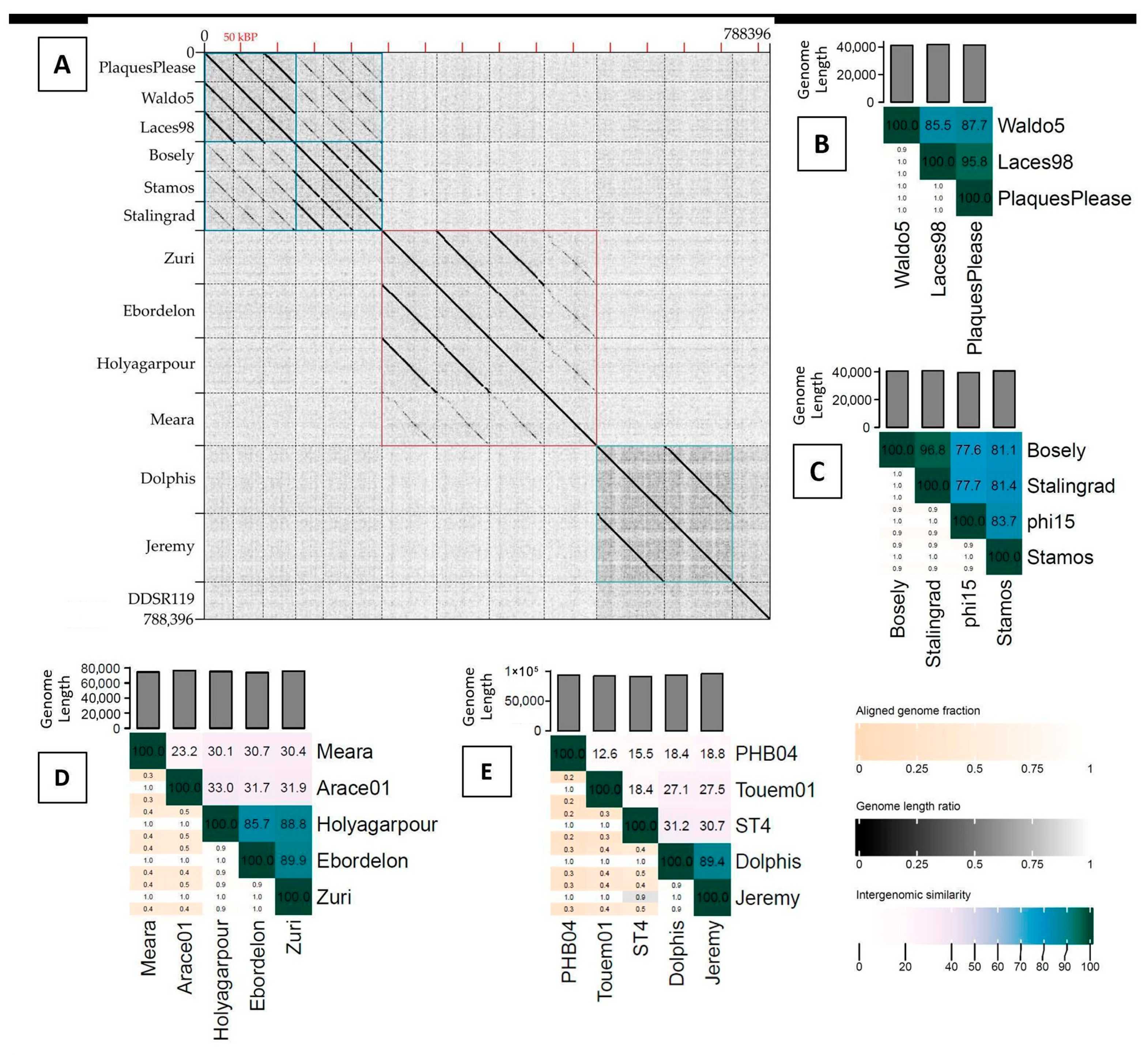
Figure 1. Genomic relatedness of our thirteen phages and selected relatives. (A) Dotplot comparing the genomes of our thirteen phages. (BE) VIRIDIC heatmaps of intergenomic similarity among our phages and their closest relatives. (B) All phages that formally belong to the Waldovirus genus (Waldo5, PlaquesPlease) or that we propose for inclusion (Laces98). These correspond to the first three phages in the dot plot (boxed in blue). (C) All phages that formally belong to the Troedvirus genus (Stalingrad, phi15) or that we propose for inclusion (Bosely, Stamos). These correspond to the next three phages in the dotplot (also boxed in blue) and are related to the Waldovirus phages. (D) Zuri, along with two other phages (Ebordelon, Holyagarpour) that we propose for inclusion in Zurivirus and another phage from our collection (Meara). These correspond to the portion of the dotplot boxed in pink. The other notably related phage currently in GenBank is Arace01. (E) Two novel phages (Dolphis, Jeremy) and their only notable relatives currently in GenBank (ST4, Touem01, PHB04).
Applmicrobiol 05 00108 g001
Figure 2. Transmission electron micrographs. (A) DDSR119, a taxonomic singleton, and representatives of (B) the Dolphis-like phages (Jeremy), (C) the Waldovirus phages (Waldo5), (D) the Troedvirus phages (Bosely), and (E) the Zuri-like phages (Holyagarpour).
Applmicrobiol 05 00108 g002
Figure 3. A Phamerator map depicting the relationships among the Waldovirus phages (Laces98, PlaquesPlease, Waldo5), the Troedvirus phages (Bosely, Stalingrad, Stamos, phi15), and two representatives from other genera within the Autotranscriptaviridae (gh-1, Henninger). The top five genomes are gh-1, Henninger, and the Waldoviruses. The bottom four genomes are the Troedviruses. Proteins (shown as boxes) that have the same color across multiple genomes belong to the same pham. Colored lines connecting different genomes reflect nucleotide-level similarity (if BLASTn score is 10−4 or better), with violet indicating very similar sequences, blues indicating less similarity, and oranges/reds being the least similar. Note that the genome termini are based on the direct terminal repeats that were empirically determined for our phages (see Section 2). Other genomes were re-oriented to be collinear as needed and might therefore be oriented differently than they appear in GenBank.
Applmicrobiol 05 00108 g003
Figure 4. A Phamerator map depicting the relationships among the three Zurivirus phages (Zuri, Ebordelon, Holyagarpour), our other Zuri-like phage (Meara), and the other closest relative in GenBank (Arace01). The sequences are divided into 2 panels at approximately bp 38,500 for better readability. Note that the genome termini for our three Zurivirus phages are based on the direct terminal repeats that were empirically determined for our phages. Other genomes were re-oriented to be collinear, as needed, and might therefore be oriented differently than they appear in GenBank. The largest gene in all five genomes is the RNA polymerase and the virion structural genes appear to the right of that.
Applmicrobiol 05 00108 g004
Figure 5. A Phamerator map of Dolphis, Jeremy, and selected relatives. (A) shows the complete genomes of Dolphis, Jeremy, and P. syringae phage Touem01 (wrapped into two lines, for better visualization). (B) compares Dolphis, Jeremy, and Bordetella phage PHB04. This segment of the aligned genomes corresponds to approximately coordinates 10,000 through 33,000. The shared terminase genes on the left of this alignment were cropped out of the diagram to improve visualization of the major conserved modular sections. To support collinear comparisons of these circularly permuted genomes, all genomes were oriented to begin with the terminase gene. As a result, proteins encoded in the left region are primarily structural, and those in the right region have enzymatic functions.
Applmicrobiol 05 00108 g005
Figure 6. Shared phams between DDSR119 and its distant relative, Torfinnsbu. Genomes are wrapped at approximately 20,000 and 40,000 for clarity of visualization. Note that structural genes are on the left and enzymes on the right.
Applmicrobiol 05 00108 g006
Figure 7. Schematic representation of lysis cassette architecture. Genes predicted to encode the holin (blue), endolysin (orange), i-spanin (dark green), and o-spanin (gray) in each phage genome are shown. Nearby proteins not known to be associated with lysis are shown in white. (A) Zuri (representative of the group), Ebordelon, and Holyagarpour have an embedded o-spanin. (B) The Waldovirus and Troedvirus phages (represented here by Stalingrad) also use an embedded o-spanin but have a different overall gene order than the Zuri-like phages. (C) Our proposed holin genes in the Dolphis-like phages, including Meara, do not show sequence similarity to other holins. (D) In DDSR119, the most likely holin candidate is quite distant from the rest of the lysis cassette. The image has been designed to show lysis gene order and general spacing and is not precisely to scale.
Applmicrobiol 05 00108 g007
Figure 8. De novo tail spike structure predicted via AlphaFold. (A) Stamos and phi15 (same genus, different phams: 430 and 41). (B) Bosely and Stalingrad (same genus, same pham: 273). (C) Laces98 and Stamos (different genera, same pham: 41). (D) Laces98 and Holyagarpour (different genera, same pham: 41). Contrasting colors were chosen to highlight different domains overlapping in each pair.
Applmicrobiol 05 00108 g008
Table 1. P. putida phages isolated in this study, ordered by year of isolation and sample type. All environmental samples were collected in Harrisonburg, VA (38′ N, 78′ W) except for PlaquesPlease, which was collected in Nokesville, VA (38′ N, 77′ W).
Phage NameGenBank AccessionYear IsolatedSource
EbordelonPV565042.12017Soil
HolyagarpourPV565043.12017Soil
JeremyPV565041.12017Soil
Laces98PV477980.12017Soil
MearaPV565044.12017Soil
StamosPV565045.12017Soil
Waldo5MT711889.12017Soil
BoselyPV329693.12018Soil
ZuriMK863032.22018Soil
DolphisMT711888.12018Soil
StalingradMT711887.22018Stream water
PlaquesPleaseMT711890.12018Streambed clay
DDSR119MT663720.12019Tree root soil
Table 2. Previously isolated phages used in our analyses.
Phage NameGenBank AccessionBacterial HostPublication PMID
phi15NC_015208.1P. putida21526174 [71]
Arace01PP179312.1P. putida38833289 [72]
gh-1NC_004665.1P. putida12842620 [38]
HenningerNC_047922.1P. sp.Unpublished
PHB04NC_047861.1Bordetella bronchiseptica30229303 [73]
ST4OR261032.1Aeromonas hydrophilaunpublished
TorfinnsbuPQ464596.1P. poae39742975 [74]
Touem01PP179325.1P. syringae38833289 [72]
Table 3. Genomic features of thirteen novel Pseudomonas putida phages.
Phages *GenBank Accession Genome Length (bp) GC Content Protein-
Coding Genes
(tRNA Genes)
Proteins with Putative Functions Packaged Genome Structure **Lifestyle
Autotranscriptaviridae
Waldovirus
PlaquesPlease MT711890.141,45658.07%51 (0)30218 bp DTRVirulent
Waldo5MT711889.141,19557.73%53 (0)29219 bp DTRVirulent
Laces98PV477980.141,87058.15%50 (0)32219 bp DTRVirulent
Troedvirus
BoselyPV329693.140,50457.82%50 (0)32253 bp DTRVirulent
StamosPV565045.140,66758.38%48 (0)32253 bp DTRVirulent
Stalingrad MT711887.240,72357.85%49 (0)32253 bp DTRVirulent
Unclassified Caudoviricetes
Zurivirus
ZuriNC_049456.275,87353.53%104 (3)37457 bp DTRVirulent
EbordelonPV565042.174,04353.70%93 (3)41466 bp DTRVirulent
HolyagarpourPV565043.175,46253.55%95 (3)43467 bp DTRVirulent
Novel genus related to Zurivirus
MearaPV565044.174,70553.68%97 (3)34Circularly permuted, TRVirulent
Novel genus, no close relative
Dolphis MT711888.193,50763.71%125 (0)56Circularly permuted, TRTemperate
JeremyPV565041.195,48763.53%126 (0)54Circularly permuted, TRTemperate
Novel genus, no close relative
DDSR119MT663720.152,90560.39%79 (2)34Circularly permuted, TRVirulent
* Phages are grouped based on existing or proposed membership in ICTV-recognized taxa. ** DTR = direct terminal repeats; TR = terminally redundant.
Disclaimer/Publisher’s Note: The statements, opinions and data contained in all publications are solely those of the individual author(s) and contributor(s) and not of MDPI and/or the editor(s). MDPI and/or the editor(s) disclaim responsibility for any injury to people or property resulting from any ideas, methods, instructions or products referred to in the content.