Article

Biosynthesis of the Siderophore Desferrioxamine E in Rouxiella badensis SER3 and Its Antagonistic Activity Against Fusarium brachygibbosum

by
Luzmaria R. Morales-Cedeño
1,
Sergio de los Santos Villalobos
2,
Pedro D. Loeza-Lara
3,
Debasis Mitra
4,
Ajay Kumar
5,
Ma. del Carmen Orozco-Mosqueda
6 and
Gustavo Santoyo
1,*
1
Instituto de Investigaciones Químico-Biológicas, Universidad Michoacana de San Nicolás de Hidalgo, Morelia 58030, Michoacán, Mexico
2
Instituto Tecnológico de Sonora, 5 de Febrero 818 sur, Ciudad Obregón 85000, Sonora, Mexico
3
Department of Food Genomics, Universidad de La Ciénega del Estado de Michoacán de Ocampo (UCEMICH), Sahuayo 59103, Michoacán, Mexico
4
Department of Microbiology, Graphic Era (Deemed to be University), Dehradun 24800, Uttarakhand, India
5
Amity Institute of Biotechnology, Amity University, Noida 201313, Uttar Pradesh, India
6
Departamento de Ingeniería Bioquímica y Ambiental, Tecnológico Nacional de México en Celaya, Celaya 38010, Guanajuato, Mexico
*
Author to whom correspondence should be addressed.
Appl. Microbiol. 2025, 5(3), 91; https://doi.org/10.3390/applmicrobiol5030091
Submission received: 26 July 2025 / Revised: 19 August 2025 / Accepted: 22 August 2025 / Published: 26 August 2025
(This article belongs to the Topic New Challenges on Plant–Microbe Interactions)

Abstract

Iron is a limiting factor for plant and microbial growth because, in soil environments, it is predominantly present as oxyhydroxide minerals, rendering it unavailable to plants and microorganisms. Siderophores are chelating agents secreted to solubilize iron and facilitate its uptake. To understand the evolutionary and ecological dynamics of microbial communities, as well as the evolution of pathogens within hosts, it is essential to study the genes shared between microorganisms for environmental adaptation and survival. In this study, we conducted microbiological assays to evaluate the effect of the siderophore produced by Rouxiella badensis strain SER3 on the mycelial growth of fungal pathogens such as Fusarium brachygibbosum 4BF. Using spectrophotometric techniques and bioinformatics tools, we identified desferrioxamine E (nocardamine) in the culture supernatant, and the corresponding biosynthetic gene cluster in the SER3 genome was confirmed through antiSMASH analysis and synteny comparisons. Gene expression analysis by RT-PCR showed differential expression of biosynthetic precursors when strain SER3 was grown alone or in interaction with fungal pathogen. Finally, scanning electron microscopy revealed structural damage to F. brachygibbosum hyphae during co-culture with strain SER3. These results suggest that the production of desferrioxamine E may act as a biocontrol mechanism employed by R. badensis SER3 against F. brachygibbosum.

1. Introduction

Iron is a fundamental element in the life of organisms. In soil environments, iron is predominantly found as oxyhydroxide minerals, and the concentrations of bioavailable free Fe3+ are below the levels required for optimal plant and microbial growth, making iron a limiting factor. Under such conditions, many plants, fungi, and bacteria produce chelating agents called siderophores, which are secreted into the environment to solubilize iron and facilitate its uptake through selective membrane receptors that import the iron-bound complex [1]. Siderophores have been described as secondary metabolites that scavenge iron from environmental sources and deliver it to cells via specific receptors [2].
Siderophores are a chemically diverse group of secondary metabolites, and extensive research has been conducted to understand their chemical structures, synthesis, export, uptake, and regulation. However, their evolutionary aspects have been largely overlooked, despite their importance in understanding the evolutionary and ecological dynamics of microbial communities and the adaptation of pathogens within hosts [3]. Some microorganisms have evolved highly diffusible siderophores to solubilize iron, but in some cases, these siderophores are not exclusively recovered by their producers. This means that siderophores can make iron available not only to the producing organism but also to other individuals with a matching receptor, leading to the distribution of production costs and benefits among different individuals within the population [2]. For example, Yamanaka et al. (2005) [4] reported the isolation and characterization of a substance produced by Streptomyces griseus that stimulates the growth and development of Streptomyces tanashiensis. This substance was identified as desferrioxamine E (nocardamine), a siderophore produced by various Streptomyces species and related organisms. S. tanashiensis was deficient in siderophore production but was able to utilize siderophores secreted by other organisms. Additionally, siderophores secreted by one microorganism can influence the fitness of other microorganisms within the microbial community, including members of the same or different species. The same study observed that an exogenous supply of desferrioxamine E stimulated secondary metabolite production and morphological differentiation in various actinomycetes [4].
Alternatively, the binding of iron to siderophores can induce iron starvation in microorganisms that lack the corresponding receptor for uptake. In this competition for iron, biological control agents can capture available iron, limiting its acquisition by pathogens that do not possess the appropriate receptor for iron-siderophore complex uptake, thereby preventing their growth and pathogenesis. This mechanism has been described in numerous studies as a key antagonistic strategy employed by microorganisms against pathogens [5,6,7]. Producing siderophores incurs a significant energy cost, so non-producing microorganisms often evolve strategies to obtain siderophores without synthesizing them. This can occur through mutations in transporter proteins that enable the uptake of exogenous siderophores, or via the horizontal transfer of uptake pathways from siderophore-producing organisms. The exploitation of siderophores by non-producers exerts selective pressure on producers, driving the evolution of novel siderophores that cannot be utilized by competitors. Such new siderophores often emerge through the divergent evolution of biosynthetic pathways, resulting in structurally similar molecules with subtle chemical modifications [8]. Thus, siderophores play a crucial role in mediating interactions both within and between microbial species.
Beyond iron competition, siderophores are also involved in another biological control mechanism, as they can activate induced systemic resistance (ISR) in the host. However, the precise mechanism by which siderophores trigger this response remains poorly understood [9]. In a genomic analysis of Rouxiella badensis strain SER3, Morales-Cedeño et al., 2021 [10] identified a gene cluster responsible for the biosynthesis of the siderophore desferrioxamine E (nocardamine). Furthermore, this bacterium has been evaluated as an antagonist of postharvest fungal pathogens such as Botrytis cinerea and Fusarium brachygibbosum in strawberry fruits. Studying the siderophores produced by this bacterium is of interest, because it has not been studied as a biocontrol agent and because siderophore production could be one of its mechanisms of action against fungal pathogens.
Given these characteristics, it is of great interest to evaluate siderophores as a mechanism of action against pathogens while also investigating siderophore identification, biosynthesis pathways, and gene cluster comparisons across siderophore-producing species. Therefore, this study aimed to assess the role of desferrioxamine E (nocardamine) as a biocontrol mechanism of R. badensis strain SER3 against fungal pathogens, amplify the genes involved in its production, compare these genes with those of other species, and analyze their expression under different conditions.

2. Materials and Methods

2.1. Siderophore Production

The Chrome Azurol S (CAS)-agar assay was used to test siderophore production by Rouxiella badensis strain SER3 in triplicate, following the protocol established by Neilands, 1987 [11]. Briefly, 100 µL of an O.D. 1 culture of R. badensis SER3 was added to Petri dishes containing CAS agar and incubated for three days at 30 °C.
To prepare 1 L of blue agar, 60.5 mg of CAS was dissolved in 50 mL of water and mixed with 10 mL of iron (III) solution (1 mM FeCl3·6H2O, 10 mM HCl). While stirring, this solution was slowly added to 72.9 mg of HDTMA (Hexadecyltrimethylammonium bromide) dissolved in 40 mL of water, forming a dark blue liquid, which was then autoclaved. Separately, a mixture of 750 mL of water, Pseudomonas agar base, 30.24 g of PIPES (Piperazine-N,N′-bis(2-ethanesulfonic acid)), and 12.00 g of a 50% (w/w) NaOH solution was prepared to adjust the pH to the pK of PIPES (6.8) and autoclaved. Finally, the dye solution was added along the glass wall of the container with sufficient agitation to ensure mixing without foam formation. Each plate received 30 mL of blue agar.

2.2. Characterizing the Siderophore Produced by Rouxiella badensis SER3

To determine the presence of hydroxamate siderophores, the FeCl3 test was performed. Two milliliters of 2% FeCl3 were added to 1 mL of the filtered supernatant from R. badensis SER3. The samples were analyzed in triplicate using a NanoDrop UV–Visible spectrophotometer (Thermo Fisher Scientific, Waltham, MA, USA). The presence of a peak between 400 and 450 nm indicates the hydroxamate nature of the siderophore (Figure S1), while a peak at 495 nm suggests the presence of catecholate (Figure S2) [12].

2.3. Confrontation Assays In Vitro

Fungal antagonism by the siderophore of R. badensis SER3 against Fusarium brachygibbosum was evaluated. Briefly, a 100 µL drop of an O.D. 1 culture of SER3 was placed on Chrome Azurol S (CAS)-agar plates, and a 6 mm mycelial plug from a F. brachygibbosum culture was deposited at an equidistant point. The plates were incubated in the dark at 21 °C, and the mycelial growth diameter was measured. The following treatments were evaluated: bacteria and fungus inoculated simultaneously (0 h), bacteria inoculated 24 h before the fungus, bacteria inoculated 48 h before the fungus, and bacteria inoculated 72 h before the fungus. To confirm that fungal growth inhibition was caused by iron sequestration via siderophores, an FeCl3 solution was added to restore fungal growth. The experiments were performed in triplicate.

2.4. In Vitro Assays with Commercial Desferrioxamine E

A concentration of 500 µg/mL of desferrioxamine E was spread on potato dextrose agar plates (PDA). After drying, a 6 mm mycelial plug of Botrytis cinerea or Fusarium brachygibbosum was placed in the middle of the plate. The mycelium growth was monitored at 24 h, 48 h, and measured after 72 h post-inoculation. Experiments were performed in triplicate.

2.5. Desferrioxamine E Cluster Analysis and Phylogeny

The complete genome sequence of SER3 strain is available in the GenBank under the following accession number: NZ_CP049603.1. Coding sequences for Desferrioxamine E siderophore synthesis in Rouxiela badensis SER3 were identified through in silico analysis using AntiSMASH software version 8.0 (https://antismash.secondarymetabolites.org/, accessed on 13 April 2025). The coding sequences of the gene cluster: dfoS, dfoC, dfoA, and dfoJ for Desferrioxamine E (nocardamine) synthesis were determined. The dfoJ and dfoA genes, which encode the enzyme pyridoxal-phosphate-dependent lysine/ornithine decarboxylase, and an enzyme amine monooxygenase, respectively, were used for primer design using the Primer Design Tool from NCBI database (https://www.ncbi.nlm.nih.gov/tools/primer-blast/, accessed on 13 April 2025), and the services of Integrated DNA Technologies (https://www.idtdna.com/page, accessed on 13 April 2025) were also used. The designed primers are shown in Table S1 of the Supplementary Materials.
To establish the relationship with other species, a search of the complete Desferrioxamine E gene cluster was performed in the NCBI database, and a phylogenetic tree was constructed using the MEGA 6 software. Also, the sequence of the SER3 gen cluster for Desferrioxamine E was compared using the BLASTn tool (version 2.17.0, https://blast.ncbi.nlm.nih.gov, accessed on 13 April 2025), and a phylogenetic tree was constructed with the sequences with the most similarity.

2.6. Assays of Gene Expression and Desferrioxamine E Production

To determine the expression of the dfoJ and dfoA genes, two treatments were established. In one treatment, SER3 interacted with Fusarium brachygibbosum HBF, while in the other, the bacteria were cultured only in the CAS medium. Siderophore production was determined based on the color change in the iron-containing medium. The interaction between the bacterium and the fungus was carried out for 72 h. After 72 h, the plate was scraped to recover a bacterial pellet, from which RNA extraction was performed. The assays were performed in triplicate.
RNA extraction was carried out using TriZol according to the manufacturer’s instructions. After the extraction process, the quality and purity of the RNA were determined through a 1.2% agarose gel in 1X TAE buffer and with the NanoDrop 2000c spectrophotometer (Thermo Fisher Scientific, Waltham, MA, USA).
The obtained RNA was used for cDNA synthesis using the ImProm-II Kit (Sigma Aldritch, St. Louis, MO, USA). Briefly, 500 ng of RNA was used as the template for the reverse transcription reaction. The resulting product was diluted 1:10 and used as a cDNA template for PCR reactions.
The amplification of the dfoJ and dfoA genes was performed using Promega’s SYBR Green Supermix (Promega, Madison, WI, USA) following the manufacturer’s instructions. The amplification reaction was carried out in a Benchmark TC-Mini Thermal Cycler (Benchmark Scientific, Sayreville, NJ, USA) with an initial denaturation cycle at 95 °C for 5 min, followed by 35 cycles of denaturation at 95 °C for 30 s, annealing at 57 °C for 30 s, extension at 72 °C for 2 min, and a final polymerization step at 72 °C for 5 min.
PCR products were analyzed by electrophoresis on a 1.2% agarose gel in 1X TAE buffer.

2.7. Effect of Direct Interaction of Strain SER3 on the Mycelium of the Pathogen Fusarium brachygibbosum

Co-cultures of R. badensis SER3 with the fungal pathogen were processed to be observed under the scanning electron microscope (JSM-6400 Model, JEOL Ltd., Tokyo, Japan). Briefly, 6 mm of mycelium were placed in vials containing 3% glutaraldehyde and 2% paraformaldehyde in phosphate buffer (PBS) pH 7.4, at 4 °C for 4 hr. Then, the samples were washed with PBS (pH 7.4) for 10 min, this process was repeated three times, and the samples were dehydrated in a series of ethanol solutions (70%, 80%, 90%, and 100%) for 10 min each solution. The ethanol was extracted and the samples were allowed to dry in a laminar flow hood. Subsequently, they were metalized with gold for visualization in the scanning electron microscope.

3. Results

3.1. Production and Characterization of the Rouxiella badensis SER3 Siderophores

Siderophore production by R. badensis SER3 was observed after 24 h of incubation, as indicated by the formation of an orange halo around the colony. This halo increased in size with prolonged incubation (Figure 1).
The siderophore produced by R. badensis SER3 was identified using a spectrophotometric FeCl3 assay, which confirmed that the metabolite belongs to the hydroxamate class due to the presence of a peak at 410 nm. No peak was detected at 495 nm, indicating the absence of catecholate-type siderophores. These results are consistent with previous findings [10], where AntiSMASH analysis of the SER3 genome revealed biosynthetic gene clusters involved in siderophore synthesis (100% similarity), particularly for desferrioxamine E, a hydroxamate-type siderophore (Table 1).

3.2. Confrontation Assays

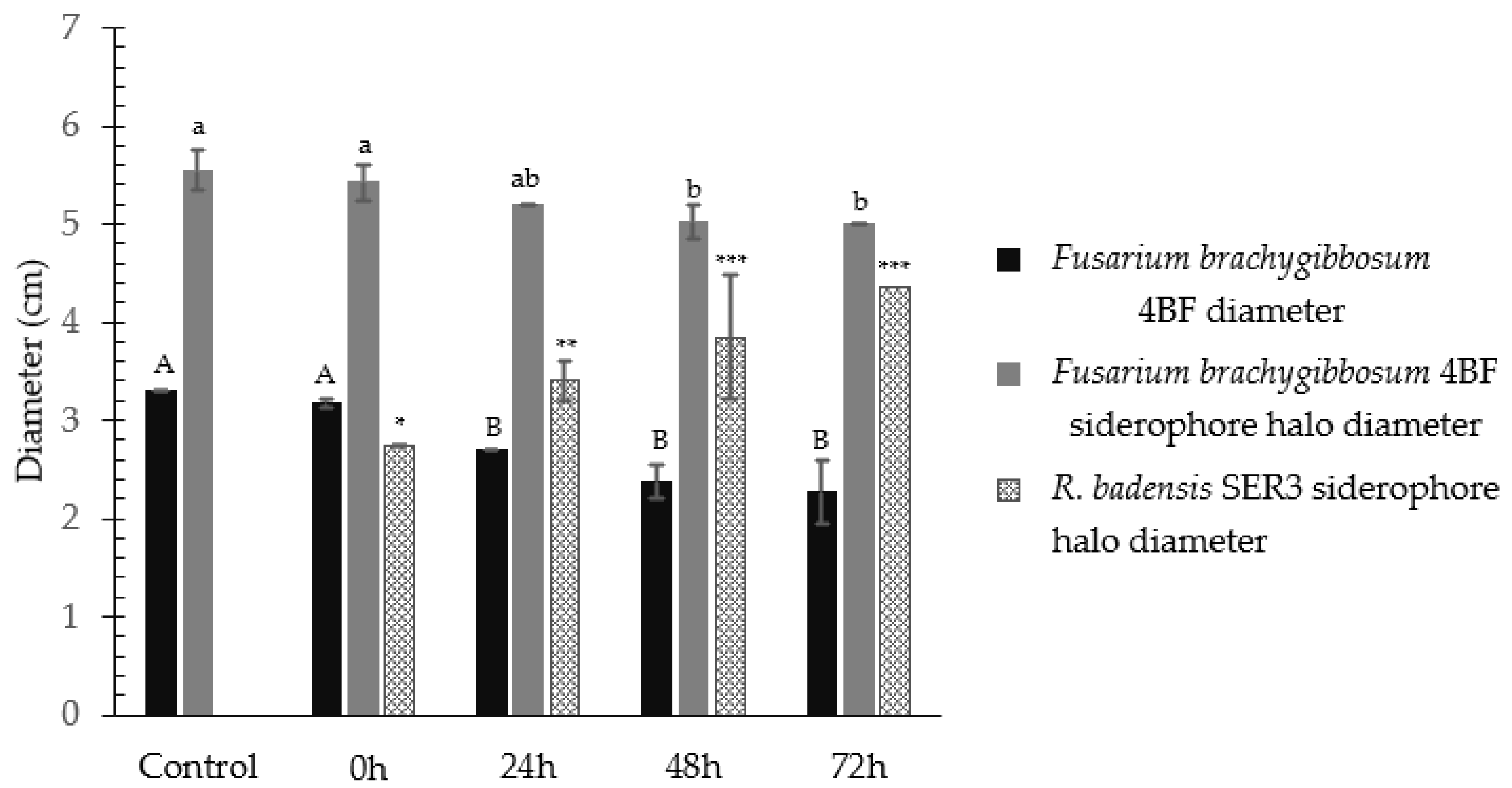
Siderophore production was detected in confrontation assays between strain SER3 and the phytopathogen Fusarium brachygibbosum 4BF in Chrome Azurol S (CAS) medium. Both the bacterium and the pathogen produced siderophores (Figure 2).
When the bacterium and fungus were inoculated simultaneously, no significant differences were observed in fungal growth or in the size of the siderophore halo produced by the pathogen. However, when the bacterium was inoculated 24 h before the fungus, fungal growth was reduced by 18%, and the siderophore halo produced by the pathogen also decreased in size, while the bacterial halo was slightly larger compared to simultaneous inoculation. When strain SER3 was inoculated 48 h before Fusarium, fungal growth inhibition increased to 28%. The siderophore halo produced by the fungus was significantly smaller compared to the control and to the treatments where the bacterium and fungus were inoculated simultaneously or with a 24 h difference.
Additionally, when strain SER3 was inoculated 72 h before the pathogen, fungal growth inhibition reached 31%, and the siderophore halo produced by the pathogen remained approximately the same size as in the 48 h treatment. However, the siderophore halo produced by the bacterium was larger compared to treatments where the bacterium and fungus were inoculated simultaneously or with 24 and 48 h intervals (Figure 2).
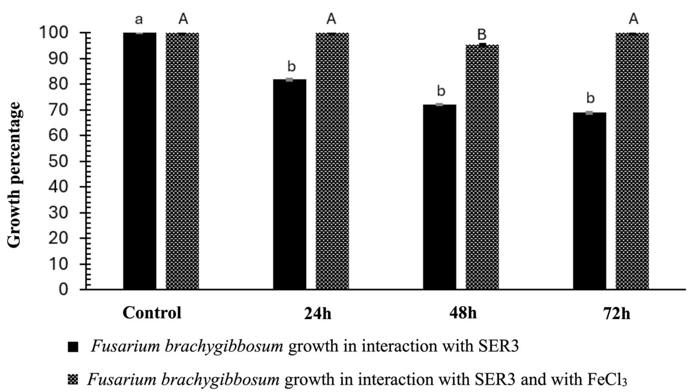
To verify that iron deficiency caused by siderophores is the cause of the inhibition of Fusarium brachygibbosum growth, the same experiments were performed but with the addition of a FeCl3 solution to see if the growth of the fungus was restored by providing iron. If we compare these results with those of the previous experiment in terms of percentage, the growth of the fungus is restored to 100% when the FeCl3 solution is administered (Figure 3).

3.3. Desferrioxamine E (Nocardamine) Cluster Analysis and Phylogeny

The cluster is composed of four genes, and their positions in the R. badensis SER3 genome are shown in Figure 4. It has been described that the putative gene dfoJ encodes the enzyme pyridoxal-phosphate-dependent lysine/ornithine decarboxylase. This enzyme decarboxylates lysine to produce cadaverine. The putative gene dfoA encodes the enzyme amine monooxygenase, which drives oxygenation at one of the terminal amino groups of cadaverine to produce 1-amino-5-(N-hydroxy)-aminopentane. The putative dfoC gene carries out the coupling reaction of succinyl-CoA with 1-amino-5-(N-hydroxy)-aminopentane to form N-5-aminopentyl-N-(hydroxyl)-succinamic acid. This compound is then trimerized and cyclized in an ATP-dependent reaction by the dfoC protein to form desferrioxamine E. The putative dfoS gene encodes a major facilitator superfamily (MFS) membrane transport protein responsible for exporting desferrioxamine E from the cell [13]. This information is summarized in Table 2.
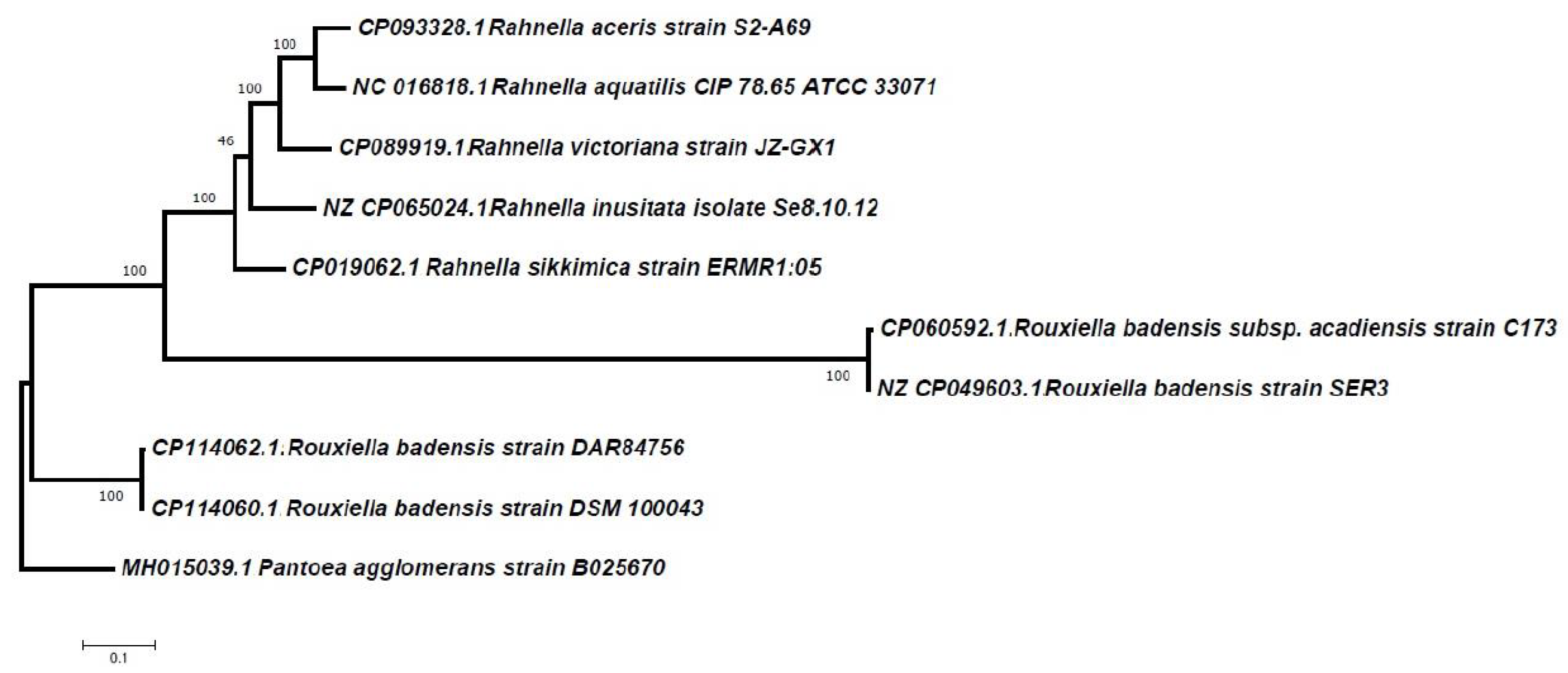
To perform a phylogenetic comparison of the desferrioxamine E gene cluster sequence of R. badensis strain SER3, we searched for sequences containing the complete desferrioxamine E cluster in the NCBI database. We found 9 sequences with which we could carry out a phylogenetic analysis (Figure 5), revealing that R. badensis SER3 is grouped with Pantoea ananatis. Additionally, by analyzing the SER3 gene cluster sequence with the BLAST tool, we found phylogenetic similarities in segments of complete genomes of Rouxiella and Rahnella species. However, these sequences are not reported as gene clusters for desferrioxamine E in these genera, likely because they have not been explored (Figure 6).
With these observations we realized that there are few reported sequences of the Desferrioxamine E gene cluster, and that there is still much to explore in bacterial genomes, so it is interesting to continue the study of this cluster to understand the relationships with other genera and if also they have biocontrol activities like R. badensis SER3.
With these results, we decided to perform a synteny analysis to compare the order of the genes that conform to the Desferrioxamine E gene cluster in the genera Pantoea agglomerans, Rahnella aceris, and Rouxiella spp. The diagram is shown in Figure 7. This allows us to observe the similarities and differences in the desferrioxamine E gene cluster between different genera, using the dfoJACS cluster in P. agglomerans as a reference. This cluster encodes enzymes involved in the biosynthesis of the hydroxamate-type siderophore desferrioxamine E. dfoJ is thought to encode an N-hydroxylase that initiates the modification of lysine residues. dfoA encodes a decarboxylase that converts modified amino acids into the siderophore backbone. dfoC encodes a synthetase that links the building blocks into the linear desferrioxamine molecule, and dfoS is proposed to function as a regulatory or transporter-associated protein that ensures efficient siderophore assembly and secretion. Together, these genes allow the bacterium to produce desferrioxamine E and acquire iron under limiting conditions.

3.4. Gene Expression Assays and Desferrioxamine E Production

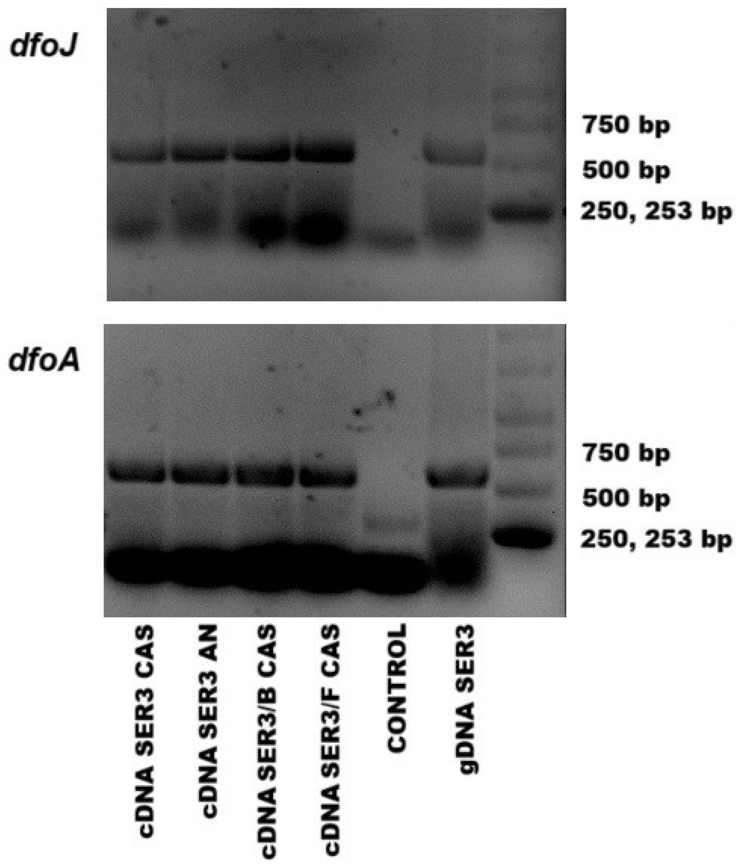
Figure 8 shows the genes dfoJ and dfoA expression. These genes are important in the synthesis of precursors of desferrioxamine E. With these results, we demonstrate that the genes are expressed in the presence of phytopathogens, such as Botrytis cinerea and Fusarium brachygibbosum, also in a medium with iron and an iron-deficient culture medium.

3.5. Effect of Direct Interaction of the SER3 Strain on the Mycelium of the Pathogens Botrytis cinerea and Fusarium brachygibbosum

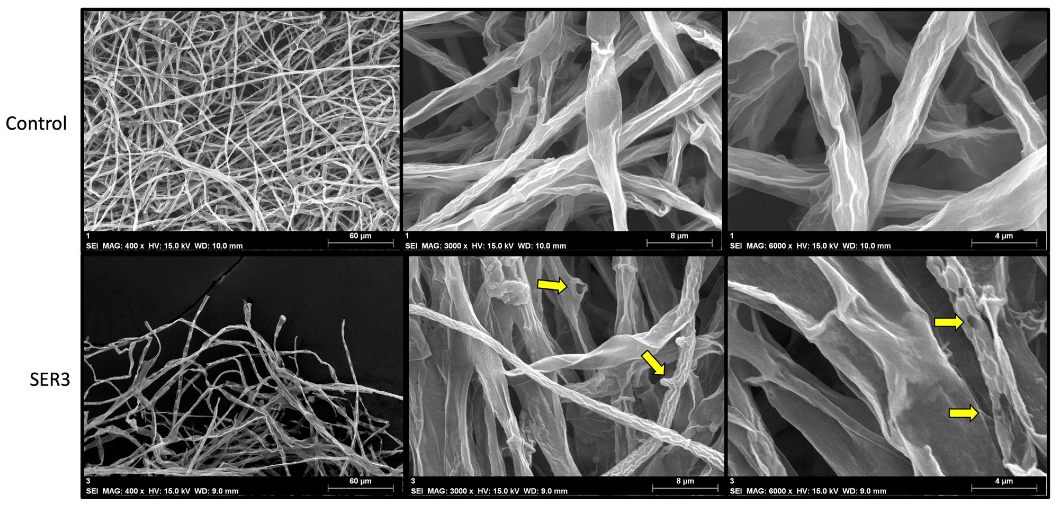
Using scanning electron microscopy (SEM), we observed that hyphae in contact with the SER3 strain exhibited noticeable deformities. As shown in Figure 9, the hyphae of Fusarium brachygibbosum in contact with SER3 display ruptures and protuberances. These observations suggest that compounds produced by the SER3 strain, which diffuse into the surrounding medium, induce structural damage to the pathogen’s hyphae, thereby inhibiting their growth.

4. Discussion

Genome mining methodologies like anti-SMASH have been widely used to identify and characterize the biosynthetic gene clusters encoding the production of secondary metabolites [14]. In this work the anti-SMASH results showed two types of siderophores in R. badensis strain SER3: one is a catecholate type, corresponding to turnerbactin, which showed a percentage of less than 70%. Despite this, it was decided to verify its presence in the supernatant of the cell-free liquid culture of strain SER3; however, it was not found in any of the liquid cultures performed, the nutrient medium, nor in the nutrient-poor medium. The other siderophore is of the hydroxamate type and corresponds to Desferrioxamine E, also known as Nocardamine. This type of siderophore was detected with Nanodrop UV-vis spectrophotometry in both culture media used. The spectrum obtained provides information about the type of siderophore present in the sample. In the work carried out by de los Santos-Villalobos et al., 2012 [15], the types of siderophores produced by Burkholderia cepacia XXVI were identified using UV-vis spectrophotometry. They describe that adding a 2% iron chloride solution to the cell-free supernatant and detecting a peak in the spectrum at a wavelength of 495 nm indicates the presence of catecholate-type siderophores, while the presence of a peak at a wavelength between 400 and 450 nm indicates the presence of hydroxamate-type siderophores. Based on these results and those provided by antiSMASH, it is suggested that the turnerbactin siderophore (catecholate type) is not produced, and that the hydroxamate-type siderophore detected with UV-vis spectrophotometry corresponds to desferrioxamine E.
Additionally, we synthesized oligonucleotides to perform a RT-PCR and demonstrate that the genes involved in the synthesis of the precursors of Desferrioxamine E are present and expressed under different conditions, such as the presence or absence of a phytopathogen, or in different culture media, either rich or poor in iron. This is important because it has been reported that the effectiveness of biocontrol agents or their action mechanisms are affected by environmental conditions, for example, the light, the amount of nutrients, the presence of other microorganisms, etc. [16] So, if a biocontrol agent or its action mechanism is intended to be used, it is important to know the conditions under which it is effective [17].
The presence of siderophores was also detected by another biochemical assay using chromeazurol medium (CAS), where a color change from blue to orange indicates the presence of siderophores [11]. Using this medium, confrontation tests were also carried out with the pathogen Fusarium brachygibbosum 4BF and strain SER3 at different times, inoculating bacteria and fungus either at the same time, 24 h before the fungus, or 48 and 72 h before the fungus. The results showed no significant difference in the growth of the fungus when both microorganisms were inoculated at the same time. However, when the bacterial strain was inoculated 24 h before the pathogen, its growth was inhibited by 18%. When comparing the production of siderophores by the pathogen and the bacteria in the treatment where they were inoculated at the same time, we observed a slight decrease in the siderophores produced by the fungus and an increase in the siderophores produced by the bacteria. When the bacterial strain was inoculated 48 h before the fungus, the percentage of inhibition increased to 28%. When comparing siderophore production by the fungus and the bacteria in this treatment with that observed in the 24 h and simultaneous inoculation treatments, a significant decrease in fungal siderophore production and an increase in bacterial siderophore production were observed. In the 72 h treatment, a similar effect was noted, with fungal inhibition reaching 31%. These results indicate that the timing of bacterial inoculation influences both fungal growth and siderophore production. With a longer inoculation time than the fungus, the bacterial strain produces more siderophores, as it has more time to adapt and consume the little iron available in the medium. Therefore, when the fungus is inoculated, there is less iron available for it, and its growth is restricted [18,19,20,21]. Furthermore, adding a solution containing iron restores the growth of the fungus, proving that the iron deficiency caused by siderophores inhibits growth, suggesting that competition for space and nutrients is one of the mechanisms of action used by the strain against this pathogen. There are numerous works than mention competition for space and nutrients and the production of siderophores as mechanisms of action against fungal pathogens [22,23,24,25].
However, when using the commercial siderophore, we did not observe any change in the growth of the pathogen mycelium compared to the control. Functionality tests performed in CAS medium prior to the trial showed that the orange color disappears from the medium after 72 h. If the compound disappears before the fungus develops, it will have no effect. Unlike the bacterial strain, which produces the siderophore continuously. Further testing is needed to determine the stability of the siderophore for use in these types of experiments [26].
Additionally, a phylogenetic analysis allows us to understand the evolutionary relationships between species [27]. Bacteria can acquire new genes through various mechanisms, such as transformation, conjugation, or transduction. This enables them to acquire new capacities to adapt to their environment and survive [28]. In this study, we searched for microorganism sequences containing the complete desferrioxamine E gene cluster. This cluster has been studied and identified in species of Streptomyces, Pantoea, and Erwinia, so we compared the cluster of strain SER3 with these genera, revealing a high percentage of similarity. We also performed a comparison using the BLAST tool in the NCBI database. These results showed that R. badensis SER3 aligns with a high percentage of similarity with genome segments from bacteria in the Rouxiella and Rahnella genera. However, these segments have not been reported as genes involved in the synthesis of desferrioxamine E, likely because they have not been studied.

5. Conclusions

Previous studies had shown siderophore production in Rouxiella badensis SER3, without specifying the type of chelating compound produced. In this work, the siderophore desferrioxamine E produced by R. badensis SER3 was identified. The complete gene cluster involved in the synthesis and export of this metabolite was determined and compared with those of other genera. Additionally, it was confirmed that the precursor genes involved in the synthesis of this siderophore are expressed under different conditions, such as varying culture media and the presence or absence of pathogens. Comparing these genes with those of other species is important for establishing phylogenetic relationships. However, further research is required to study this metabolite in other species. It was also demonstrated that the production of this siderophore limits the growth of Fusarium brachygibbosum, indicating that desferrioxamine E (nocardamine) functions as a biocontrol mechanism of R. badensis SER3 against fungal pathogens, primarily through competition for space and nutrients, particularly iron.

Supplementary Materials

The following supporting information can be downloaded at: https://www.mdpi.com/article/10.3390/applmicrobiol5030091/s1, Table S1: Supplementary material. Oligonucleotides sequences for PCR. Figure S1: Spectrum obtained by UV-vis spectrophotometry using the supernatant of strain SER3 in nutrient broth. The presence of a peak between 400 and 450 nm indicates the presence of hydroxamate-type siderophores. Figure S2: Spectrum obtained with UV-vis spectrophotometry using the supernatant of strain SER3 in nutrient broth. The presence of a peak at 495 nm would indicate the presence of catecholate-type siderophores; however, No peaks of interest were found.

Author Contributions

Conceptualization, methodology, L.R.M.-C. and G.S.; formal analysis, writing—original draft preparation, writing—review and editing, L.R.M.-C., S.d.l.S.V., P.D.L.-L., D.M., A.K., M.d.C.O.-M. and G.S. All authors have read and agreed to the published version of the manuscript.

Funding

This research was funded by CIC-UMSNH, grant number 2024–2025.

Institutional Review Board Statement

Not applicable.

Informed Consent Statement

Not applicable.

Data Availability Statement

Supplementary data is available.

Acknowledgments

We thank Aurora Flores Piña for technical support.

Conflicts of Interest

The authors declare no conflicts of interest.

References

  1. Boiteau, R.M.; Fansler, S.J.; Farris, Y.; Shaw, J.B.; Koppenaal, D.W.; Pasa-Tolic, L.; Jansson, J.K. Siderophore profiling of co-habitating soil bacteria by ultra-high resolution mass spectrometry. Metallomics 2019, 11, 166–175. [Google Scholar] [CrossRef]
  2. Kramer, J.; Özkaya, Ö.; Kümmerli, R. Bacterial siderophores in community and host interactions. Nat. Rev. Microbiol. 2020, 18, 152–163. [Google Scholar] [CrossRef]
  3. West, S.A.; Griffin, A.S.; Gardner, A.; Diggle, S.P. Social evolution theory for microorganisms. Nat. Rev. Microbiol. 2006, 4, 597–607. [Google Scholar] [CrossRef]
  4. Yamanaka, K.; Oikawa, H.; Ogawa, H.-O.; Hosono, K.; Shinmachi, F.; Takano, H.; Sakuda, S.; Beppu, T.; Ueda, K. Desferrioxamine E produced by Streptomyces griseus stimulates growth and development of Streptomyces tanashiensis. Microbiology 2005, 151, 2899–2905. [Google Scholar] [CrossRef]
  5. Dukare, A.S.; Paul, S.; Nambi, V.E.; Gupta, R.K.; Singh, R.; Sharma, K.; Vishwakarma, R.K. Exploitation of microbial antagonists for the control of postharvest diseases of fruits: A review. Crit. Rev. Food Sci. Nutr. 2019, 59, 1498–1513. [Google Scholar] [CrossRef]
  6. Carmona-Hernandez, S.; Reyes-Pérez, J.J.; Chiquito-Contreras, R.G.; Rincon-Enriquez, G.; Cerdan-Cabrera, C.R.; Hernandez-Montiel, L.G. Biocontrol of Postharvest Fruit Fungal Diseases by Bacterial Antagonists: A Review. Agronomy 2019, 9, 121. [Google Scholar] [CrossRef]
  7. del Carmen Orozco Mosqueda, M.; Pizano, G.S. Bacterias Promotoras del Crecimiento Vegetal; Editorial Fontamara: Coyoacán, Mexico, 2020. [Google Scholar]
  8. Fischbach, M.A.; Walsh, C.T.; Clardy, J. The evolution of gene collectives: How natural selection drives chemical innovation. Proc. Natl. Acad. Sci. USA 2008, 105, 4601–4608. [Google Scholar] [CrossRef]
  9. Orozco-Mosqueda, M.d.C.; Santoyo, G.; Glick, B.R. Recent Advances in the Bacterial Phytohormone Modulation of Plant Growth. Plants 2023, 12, 606. [Google Scholar] [CrossRef]
  10. Morales-Cedeño, L.R.; Santos-Villalobos, S.d.L.; Santoyo, G. Functional and Genomic Analysis of Rouxiella badensis SER3 as a Novel Biocontrol Agent of Fungal Pathogens. Front. Microbiol. 2021, 12, 709855. [Google Scholar] [CrossRef]
  11. Schwyn, B.; Neilands, J. Universal chemical assay for the detection and determination of siderophores. Anal. Biochem. 1987, 160, 47–56. [Google Scholar] [CrossRef]
  12. Neilands, J.B. Microbial Iron Compounds. Annu. Rev. Biochem. 1981, 50, 715–731. [Google Scholar] [CrossRef]
  13. Smits, T.H.M.; Duffy, B. Genomics of iron acquisition in the plant pathogen Erwinia amylovora: Insights in the biosynthetic pathway of the siderophore desferrioxamine E. Arch. Microbiol. 2011, 193, 693–699. [Google Scholar] [CrossRef]
  14. Blin, K.; Wolf, T.; Chevrette, M.G.; Lu, X.; Schwalen, C.J.; Kautsar, S.A.; Duran, H.G.S.; de Los Santos, E.L.C.; Kim, H.U.; Nave, M.; et al. antiSMASH 4.0—Improvements in chemistry prediction and gene cluster boundary identification. Nucleic Acids Res. 2017, 45, W36–W41. [Google Scholar] [CrossRef]
  15. Santos-Villalobos, S.d.L.; Barrera-Galicia, G.C.; Miranda-Salcedo, M.A.; Peña-Cabriales, J.J. Burkholderia cepacia XXVI siderophore with biocontrol capacity against Colletotrichum gloeosporioides. World J. Microbiol. Biotechnol. 2012, 28, 2615–2623. [Google Scholar] [CrossRef]
  16. Karlsson, M.E.; Hellström, M.; Flöhr, A.; Bergstrand, K.-J.; Alsanius, B.W. The power of light: Impact on the performance of biocontrol agents under minimal nutrient conditions. Front. Microbiol. 2023, 14, 1087639. [Google Scholar] [CrossRef]
  17. Hawkes, C.V.; Connor, E.W. Translating Phytobiomes from Theory to Practice: Ecological and Evolutionary Considerations. Phytobiomes J. 2017, 1, 57–69. [Google Scholar] [CrossRef]
  18. Gu, S.; Yang, T.; Shao, Z.; Wang, T.; Cao, K.; Jousset, A.; Friman, V.-P.; Mallon, C.; Mei, X.; Wei, Z.; et al. Siderophore-Mediated Interactions Determine the Disease Suppressiveness of Microbial Consortia. mSystems 2020, 5, e00811-19. [Google Scholar] [CrossRef]
  19. Miethke, M.; Marahiel, M.A. Siderophore-Based Iron Acquisition and Pathogen Control. Microbiol. Mol. Biol. Rev. 2007, 71, 413–451. [Google Scholar] [CrossRef]
  20. Ghosh, S.K.; Bera, T.; Chakrabarty, A.M. Microbial siderophore—A boon to agricultural sciences. Biol. Control 2020, 144, 104214. [Google Scholar] [CrossRef]
  21. Lambrese, Y.; Guiñez, M.; Calvente, V.; Sansone, G.; Cerutti, S.; Raba, J.; Sanz, M.I. Production of siderophores by the bacterium Kosakonia radicincitans and its application to control of phytopathogenic fungi. Bioresour. Technol. Rep. 2018, 3, 82–87. [Google Scholar] [CrossRef]
  22. Nunes, C.A. Biological control of postharvest diseases of fruit. Eur. J. Plant Pathol. 2012, 133, 181–196. [Google Scholar] [CrossRef]
  23. Hosseini, A.; Saba, M.K.; Watkins, C.B. Microbial Antagonists to Biologically Control Postharvest Decay and Preserve Fruit Quality. Crit. Rev. Food Sci. Nutr. 2023, 7330–7342. [Google Scholar] [CrossRef]
  24. Verma, S.; Azevedo, L.C.B.; Pandey, J.; Khusharia, S.; Kumari, M.; Kumar, D.; Kaushalendra; Bhardwaj, N.; Teotia, P.; Kumar, A. Microbial Intervention: An Approach to Combat the Postharvest Pathogens of Fruits. Plants 2022, 11, 3452. [Google Scholar] [CrossRef]
  25. Ram, R.M.; Keswani, C.; Bisen, K.; Tripathi, R.; Singh, S.P.; Singh, H.B. Biocontrol technology: Eco-friendly approaches for sustainable agriculture. In Omics Technologies and Bio-Engineering: Volume 2: Towards Improving Quality of Life; Elsevier: Amsterdam, The Netherlands, 2018; pp. 177–190. [Google Scholar] [CrossRef]
  26. Soares, E.V. Perspective on the biotechnological production of bacterial siderophores and their use. Appl. Microbiol. Biotechnol. 2022, 106, 3985–4004. [Google Scholar] [CrossRef]
  27. Lang, A.S.; Buchan, A.; Burrus, V. Interactions and evolutionary relationships among bacterial mobile genetic elements. Nat. Rev. Microbiol. 2025, 23, 423–438. [Google Scholar] [CrossRef]
  28. Yin, X.; Stotzky, G. Gene Transfer Among Bacteria in Natural Environments; Academic Press Inc.: Cambridge, MA, USA, 1997. [Google Scholar] [CrossRef]
Figure 1. Rouxiella badensis SER3 siderophore production in chrome-azurol (CAS) medium. Panel (A) shows an orange halo at 24 h of incubation. Panel (B), 48 h incubation, and panel (C), 72 h incubation.
Applmicrobiol 05 00091 g001
Figure 2. Confrontation assays between Rouxiella badensis SER3 and Fusarium brachygibbosum on CAS medium. Hours represent the inoculation times of the bacteria before inoculation of the pathogen. Bars represent the means of the treatments ± standard deviation (n = 3). Letters and asterisks indicate that the means differ significantly according to Duncan’s multiple range test (* p < 0.05; ** p < 0.01; *** p < 0.001).
Applmicrobiol 05 00091 g002
Figure 3. Confrontation assays between Rouxiella badensis SER3 and Fusarium brachygibbosum in CAS medium. Hours represent the inoculation times of the bacteria before inoculation of the pathogen. After 72 h of interaction between the bacteria and the fungus in each treatment, a FeCl3 solution was added. The bars represent the means of the treatments ± standard deviation (n = 3). The letters indicate that the means differ significantly according to Duncan’s multiple range test (p < 0.05).
Applmicrobiol 05 00091 g003
Figure 4. Gene cluster involved in the synthesis and excretion of the siderophore desferrioxamine E, also known as nocardamine, and its position in the genome of strain SER3.
Applmicrobiol 05 00091 g004
Figure 5. Phylogenetic analysis of the complete Desferrioxamine E gene cluster of R. badensis SER3 with the Desferrioxamine E sequences identified and reported in the NCBI database.
Applmicrobiol 05 00091 g005
Figure 6. Phylogenetic tree with closer species to R. badensis SER3. The tree was constructed using sequences from BLAST alignment to identify the Desferrioxamine E gene cluster in the complete genomes of Rouxiella and Rahnella species.
Applmicrobiol 05 00091 g006
Figure 7. Synteny analysis that compares the genes that conform to the Desferrioxamine E gene cluster in the genera Pantoea, Rahnella, and Rouxiella.
Applmicrobiol 05 00091 g007
Figure 8. dfoJ and dfoA gene expression in the presence or absence of the pathogens Botrytis cinerea (B) and Fusarium brachygibbosum (F), also in a medium with iron (nutrient agar AN) and an iron-deficient culture medium (chromo-azurol agar CAS).
Applmicrobiol 05 00091 g008
Figure 9. Fusarium brachygibbosum 4BF hyphae. Upper panels show hyphae under control conditions observed at 400× (left), 3000× (middle), and 6000× (right). Lower panels show hyphae co-cultured with strain SER3 observed at 400× (left), 3000× (middle), and 6000× (right).
Applmicrobiol 05 00091 g009
Table 1. Secondary metabolite regions identified in R. badensis SER3 genome using antiSMASH software.
RegionTypeFromToMost Similar Known ClusterSimilarity
Region 1NRPS10,16871,398TurnerbactinNRP38%
Region 2Siderophore873,555885,909Desferrioxamine EOther100%
Region 3Thiopeptide1,138,8411,165,158O-antigenSaccharide14%
Region 4Arylpolyene1,962,0992,005,722Aryl polyenesOther77%
Region 5T1PKS2,289,7922,332,482
Region 6T3PKS2,719,3642,760,449VenemycinPolyketide18%
Region 7NRPS, TransAT-PKS3,528,5363,604,918Tolaasin I/tolaasin FNRP: Lipopeptide40%
Region 8Hserlactone4,818,6824,839,332
Region 9Redox-cofactor4,905,4524,927,635Lankacidin CNRP+Polyketide13%
Table 2. Composition of the Desferrioxamine E gene cluster in Pantoea, its products, and database accession numbers.
NCBI Accession NumberGeneProduct
AWD37884.1dfoSPutative siderophore MFS transporter DfoS
AWD37885.1dfoCPutative siderophore biosynthesis protein DfoC
AWD37886.1dfoAPutative lysine/ornithine N-monooxygenase DfoA
AWD37887.1dfoJPutative pyridoxal-dependent decarboxylase DfoJ
Disclaimer/Publisher’s Note: The statements, opinions and data contained in all publications are solely those of the individual author(s) and contributor(s) and not of MDPI and/or the editor(s). MDPI and/or the editor(s) disclaim responsibility for any injury to people or property resulting from any ideas, methods, instructions or products referred to in the content.