Article

16S rRNA Gene Sequencing-Based Identification and Comparative Analysis of the Fecal Microbiota of Five Syntopic Lizard Species from a Low-Mountain Area in Western Bulgaria

1
The Stephan Angeloff Institute of Microbiology, Bulgarian Academy of Sciences, Acad. Georgi Bonchev Str., Bl. 26, 1113 Sofia, Bulgaria
2
Institute of Biodiversity and Ecosystem Research, Bulgarian Academy of Sciences, 1 Tsar Osvoboditel Blvd., 1000 Sofia, Bulgaria
3
Faculty of Biology, Sofia University “St. Kliment Ohridski”, 8 Dragan Tsankov Blvd., 1164 Sofia, Bulgaria
*
Author to whom correspondence should be addressed.
Appl. Microbiol. 2024, 4(1), 181-193; https://doi.org/10.3390/applmicrobiol4010013
Submission received: 22 December 2023 / Revised: 12 January 2024 / Accepted: 15 January 2024 / Published: 18 January 2024

Abstract

:
Studies on the gut microbiome of free-living reptiles in Europe are generally fragmentary and still missing in Bulgaria. We aimed to identify and compare the fecal microbiota profiles of five syntopic lizard species from three families: the European green lizard (Lacerta viridis), the common wall lizard (Podarcis muralis), the meadow lizard (Darevskia praticola) (Lacertidae), the European snake-eyed skink (Ablepharus kitaibelii) (Scincidae), and the European slow worm (Anguis fragilis) (Anguidae), which coinhabit a low mountainous area in the western part of the country. A high-throughput sequencing of the hypervariable V3-V4 region of the 16S rRNA gene, performed on the Illumina HiSeq2500 platform, was used. The core microbiota of lizard hosts seems to be species-specific. A dynamic phyla proportion between hosts was found. The richest alpha diversity was observed in D. praticola, and the lowest alpha diversity was observed in P. muralis and A. fragilis. Within the three lacertids, the microbiota of D. praticola and L. viridis were more closely related to each other than they were to those of P. muralis. Sharing a largely common trophic resource (all species except A. fragilis are mainly insectivorous) was not an indication of similarity in their gut microbial communities.

1. Introduction

The development of high-throughput, next-generation sequencing (NGS) technologies and DNA metabarcoding over the past two decades has greatly facilitated the rapid and reliable identification of multiple species from a single sample at the depth required to adequately characterize diverse microbial communities in a given environment/ecological niche, including fastidious and unculturable taxa [1,2,3]. A metagenomic approach has allowed for a comprehensive identification of the microbiota in different groups of vertebrates [4]. However, most research has focused on mammals [5,6,7,8,9], especially humans [10,11,12,13,14], and less than 10% have been conducted on fish [15,16,17], amphibians [18,19,20], reptiles [21,22,23,24,25], and birds [26,27,28]. Furthermore, the majority of studies are based on fecal samples from captive animals, often from laboratories or zoos [29,30], which tend to exhibit distinct microbiotas compared to their wild counterparts; therefore, the effect of artificial perturbations is well known [30,31,32,33,34]. Host diet and phylogeny are thought to be important predictors of the gut microbiota composition [5,21,35], and host genotype could affect the abundance of some microbial genera [36]. More investigations need to be conducted to characterize the microbiome in non-model systems to achieve a better understanding of how the environment and host alike shape endogenous microbial communities in wild animal populations [36,37].
Reptiles comprise 17% of all vertebrate species, representing an ancient group with approximately 12,000 extant species, of which ~60% belong to the clade Sauria, known as lizards [38]. To date, investigations on the gut microbiota of some lizard species from different parts of the world have been reported [39,40,41,42,43]. In Europe, although lizards are important components of ecosystems and are among the dominant reptile species in terms of numbers [44], studies are generally scarce [45,46], except for more intensive studies on some Podarcis species [47,48,49,50]. Five lizard species belonging to three families: the European green lizard (Lacerta viridis Laurenti, 1768), the common wall lizard (Podarcis muralis Laurenti, 1768), the meadow lizard (Darevskia praticola Eversmann, 1834) (Lacertidae), the European snake-eyed skink (Ablepharus kitaibelii Bibron and Bory de Saint-Vincent, 1833) (Scincidae), and the European slow worm (Anguis fragilis Linnaeus, 1758) (Anguidae). These lizard species coinhabit a low-mountain area in the western part of Bulgaria. They exhibited different preferences in terms of microhabitat selection, food spectrum, and trophic niche width [44]. The aim of this study was to identify and compare the fecal microbiota of those syntopic lizard species by sequencing the hypervariable V3–V4 region of the 16S rRNA gene. So far, research on the herpetofauna with an aspect on the microbiome has not been conducted in Bulgaria. Therefore, this study could illuminate factors shaping the gut microbiota in still understudied reptile hosts.

2. Materials and Methods

2.1. Study Area and Sample Collection

The study area was located in western Bulgaria, along the valley of the Dalbochitsa River in Ihtimanska Sredna Gora Mountains, northeast of the village of Gabrovitsa (42°15′12″ N 23°53′59″ E), 430–580 m above sea level. Fieldwork was conducted in May and June 2022. A total of 86 specimens were captured: L. viridis n = 15; P. muralis n = 17; D. praticola n = 26; A. kitaibelii n = 26; and A. fragilis n = 2. Lizards were caught by hand, and the animals were placed in individual sterile plastic containers and transported to the laboratory for a period of 1–3 days. After defecation, feces were collected using sterile tweezers, transferred in sterile microcentrifuge tubes (Eppendorf® 2 mL), and immediately frozen at −20 °C until they were used for further DNA extraction. After sampling, the lizards were released at their place of capture (the location of each individual was recorded using a GPS device). The handling of animals was performed according to the necessary regulations and ethics requirements.

2.2. Genomic DNA Extraction

The total DNA from fecal samples was extracted using HiPurA®Stool DNA Purification Kit (HiMedia, Mumbai, India) according to the manufacturer’s protocol. Approximately equal portions of feces from several individuals were transferred into a sterile microcentrifuge tube and mixed to obtain over 250 mg of content per tube. DNA concentration and purity were measured using a NanoDrop 3300 fluorospectrometer (Thermo Fisher Scientific, Waltham, MA, USA) and 1% agarose gel electrophoresis, respectively. Extracted DNA from all probes for a given species was pooled together and processed further as a single sample.

2.3. PCR Amplification and Sequencing

The hypervariable V3-V4 region of the 16S rRNA gene was amplified through polymerase chain reaction (PCR) using the universal bacterial primers 341F and 805R. Libraries of the amplicons were generated using a Herculase II Fusion DNA Polymerase Nextera XT Index V2 Kit and sequenced using an Illumina MiSeq platform (2 × 300 bp PE) by Macrogen Inc. (DNA Sequencing Service, Seoul, Republic of Korea).

2.4. Sequence Assembly and Taxonomic Identification

The processing of the raw sequencing reads obtained (including overlapping, filtration by sequence quality (Q30), trimming of the primers) was performed using Bioinformatic Software Tools (Illumina, San Diego, CA, USA). The Quantitative Insights Into Microbial Ecology 2 (QIIME2) platform, version 2019.10 [51] was used for the quality control and analysis of data. Assembly of the paired end reads from the original DNA fragments were merged using FLASH (1.2.11). Raw read filtering and trimming to remove the low-quality sequences and chimeras, and operational taxonomic units (OTU) clustering at different distance cutoffs (0.03) were performed using de novo CD-HIT-OUT. Sequences with ≥97% similarity were assigned to the same OTUs using NCBI_16S_20230103 (BLAST). Bacterial taxa with a relative abundance lower than 0.5% were combined into an “Others” class.

2.5. Bioinformatics and Statistical Analysis

Alpha diversity (calculated intrasample) and beta diversity (calculated as the dissimilarity between species) were measured using QIIME 2, version 2019.10 [51]. The alpha diversity was based on the number of observed OTUs, community richness (Chao1), diversity (Shannon), evenness (Gini–Simpson), and Good’s coverage indexes. To compare the significance of the differences between species, a diversity permutation test based on the Shannon and Simpson indices was performed via PAST 4.07 [52]. Beta diversity was measured using principal coordinate analysis (PCoA) based on both weighted and unweighted UniFrac distance matrices. A vegan package in R software was used for visualization.

3. Results

3.1. Description of the Sequencing Data

A total of 433 093 read counts (Q30 > 93) ranging from 71,927 to 99,437 across species were obtained. All rarefaction curves asymptotically approached a plateau, suggesting an accurate reflection of the microbial community and indicating a satisfactory level of diversity sampling (Figure 1a). A total of 721 OTUs at the 97% similarity level from all samples were extracted.

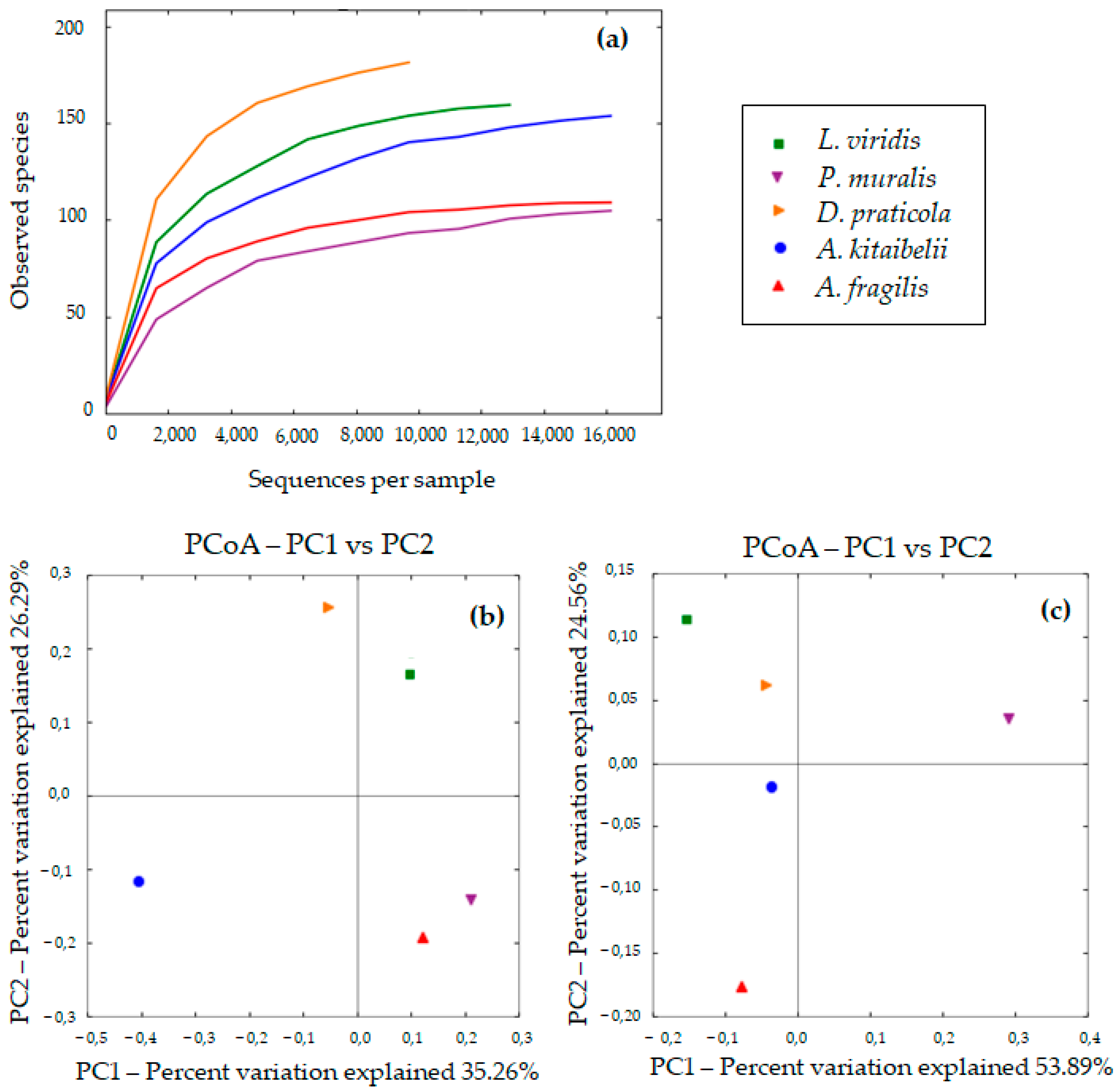
3.2. Diversity of Bacterial Communities

The alpha diversity of the gut microbiota of L. viridis, P. muralis, D. praticola, A. kitaibelii, and A. fragilis was characterized by the number of OTUs, Chao1, Shannon’s, and Gini–Simpson indices, as summarized in Table 1. Good’s coverage index (0.999) indicated that optimally, 16S rRNA gene sequences were extracted from the fecal samples and sufficient sequencing depth was achieved to evaluate bacterial diversity in all five species. The results of the diversity permutation test in terms of the Shannon index, as well as the Simpson index, showed that for all combinations, the differences between species were of a high degree of statistical significance (p < 0.001).
The largest number of OTUs and the highest Chao1 and Shannon index values were found in D. praticola. The least alpha diversity of the gut bacterial community was observed in P. muralis and A. fragilis.
Beta diversity in the fecal samples of the five lizard species was compared by referencing unweighted and weighted UniFrac distances. Unweighted UniFrac (qualitative), which is based on presence or absence of observed taxa, is more sensitive to differences in low-abundance features. Weighted UniFrac (quantitave) takes into account the relative abundance of taxa shared between samples, and the impact of low-abundance features is diminished; therefore, it is useful for examining differences in community structure. Both are useful to interpret together. Principal coordinate analysis (PCoA) based on unweighted UniFrac revealed that L. viridis and D. praticola formed a cluster with the highest level of similarity. A. fragilis appeared to be more closely related to the lacertid cluster than A. kitaibelii, which represents a separate clade (Figure 1b). In terms of relative abundance, D. praticola and A. kitaibelii were found to be closest. P. muralis was distant from all, not only from the other lacertids (Figure 1c).

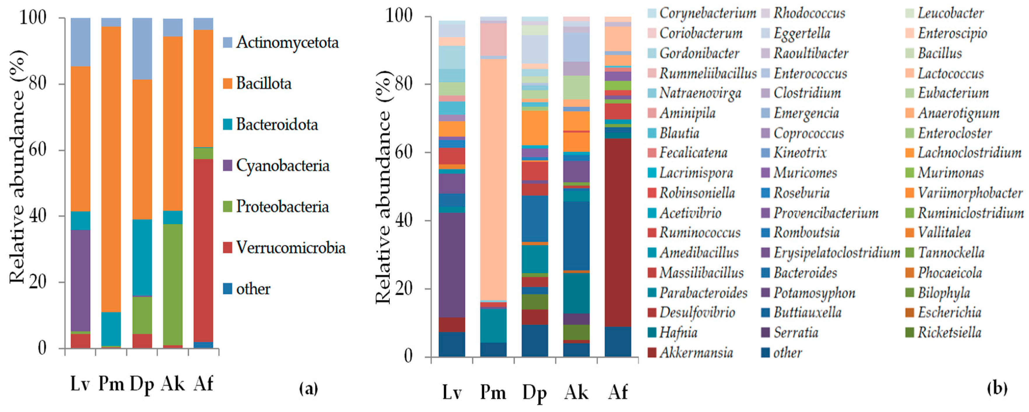
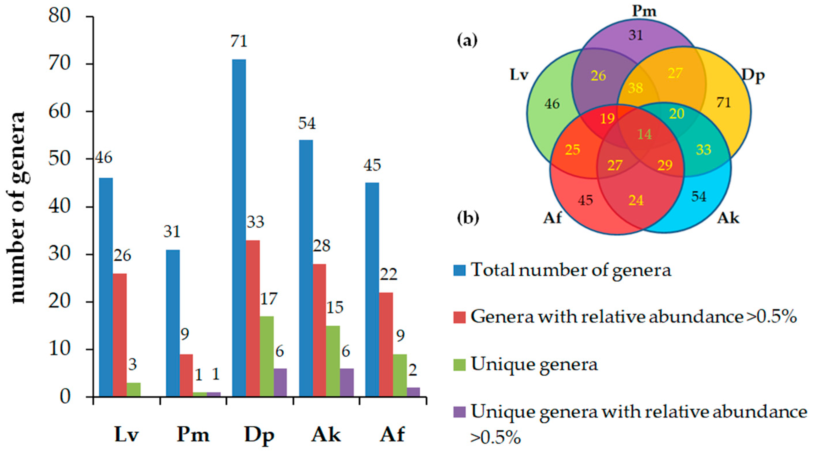
3.3. Taxonomic Composition and Abundance of Gut Microbiota

A total of six phyla, 12 classes, 21 orders, 43 families, and 106 genera were taxonomically assigned in the overall dataset. The proportion of phyla varied greatly by species (Figure 2a). The most dominant phylum in all lizard species was Bacillota, which ranged between 35.4 and 86.4%, followed by Proteobacteria (0.4–36.8%), Bacteroidota (0.2–23%), Actinomycetota (2.5–18.7%), and Veruccomicrobia (0.4–4.4%), except in A. fragilis (55.2%). Cyanobacteria were present only in L. viridis (30.8%) (and a negligible incidence < 0.5% in D. praticola). Each phylum was at its highest abundance in a different species of lizard: Bacillota in P. muralis, Bacteroidota in D. praticola, Proteobacteria in A. kitaibelii, Verruccomicrobia in A. fragilis, and Cyanobacteria in L. viridis.
The largest share in the gut microbiota of L. viridis was occupied by Bacillota (43.8%), Cyanobacteria (30.8%), and Actinomycetota (14.7%), whereas Bacteroidota (5.6%) and Verrucomicrobia (4.3%) were less represented. A huge portion of the intestinal microbiota of P. muralis was occupied by Bacillota (86,4%), followed by Bacteroidota (10.2%) and Actinomycetota (2.5%). Bacillota (42.3%), Bacteroidota (23%), Actinomycetota (18.7%), Proteobacteria (11.3%), and Verrucomicrobia (4.4%) constituted the gut microbial community at D. praticola. Bacillota (52.7%) and Proteobacteria (36.8%) were the dominant phyla in A. kitaibelii, followed by Actinomycetota (5.4%) and Bacteroidota (4%). In A. fragilis, Verrucomicrobia (55.2%) and Bacillota (35.4%) prevailed, whereas Actinomycetota (3.6%), Proteobacteria (3.6%), and unidentified (2%) were less common.
At the class level, overall, the most abundant were Clostridia (ranging between 24 and 34.5%, except in P. muralis: 3.9%), Bacteroidia (4–23%, except in A. fragilis <0.5%), and Coriobacteriia (2.4–13.4%). Gammaproteobacteria (36.3%) was a prevalent class in A. kitaibelii.
At the order level, the most common were Eubacteriales (3.9–34.5%), Bacteroidales (4–23%), Eggerthellales (2.4–13.4%) and Erysipelotrichales (1.2–9.8%).
At the family level, Lachnospiraceae (14.4–20.4%, only in P. muralis: 1.6%) and Eggerthellaceae (3.1–13.4%) were represented with the highest abundance overall.
At the genus level, as a whole, Lachnoclostridium, Ruminococcus, Eubacterium, Erysipelatoclostridium, Parabacteroides, Bacteroides, Eggerthella, and Akkermansia were the most abundant genera. Amongst the prominent genera in L. viridis were Potamosyphon (30.8%), Gordonibacter (6.7%), Erysipelatoclostridium (5.9%), Ruminococcus (4.9%). In P. muralis, Lactococcus (70.9%), Rummeliibacillus (9.4%), and Parabacteroides (9.9%) were the most prominent. In D. praticola, Bacteroides (13.6%), Lachnoclostridium (10.1%), and Parabacteroides (8.2%) were found in the most abundance. In A. kitaibelii, the most prominent were Buttiauxella (20.2%), Hafnia (11.9%), and Enterococcus (8.6%). In A. fragilis, the most abundant were Akkermansia (55.2%), Lactococcus (7.2%), and Ruminococcus (4.6%) (Figure 2b).
The taxa common to the five lizard species encompassed nine classes, nine orders and 12 families. A Venn diagram depicts the number of shared genera between lizard species (Figure 3a). A total of 14 out of 106 recorded genera occurred in all species. The largest number of common genera was between D. praticola/L. viridis (38) and D. praticola/A. kitaibelii (33). Differences in terms of the total number of genera, genera with significant presence, and unique genera found in each lizard species are presented in Figure 3b. The underrepresented genera with a relative abundance less than 0.5% occupied between 43.5% and 53.5% of the intestinal microbiota in each species, reaching 71% in P. muralis. The microbial diversity of A. kitaibelii, D. praticola, and A. fragilis featured a high proportion of unique genera (27.8%, 23.9%, and 20%, respectively), followed by L. viridis (6.5%) and P. muralis (only one genera). However, the participation of unique genera with a relative abundance >0.5% was much lower: A. kitaibelii (11.1%), D. praticola (8.4%), A. fragilis (4.4%), and L. viridis (0%). The number of unique species also varied greatly: twenty-two in D. praticola (26.5% of the total number of species found in this lizard species), seventeen in A. kitaibelii (29.3%), sixteen in A. fragilis (34%), seven in L. viridis (11.4%), and only one (2.9%) in P. muralis.

4. Discussion

4.1. Composition and Diversity of Lizard Gut Microbiota

Previous reports have indicated that the core gut microbiota of lizards is dominated by the phyla Bacillota (formerly known as Firmicutes) (33.2–73%), Bacteroidota (Bacteroidetes) (6.2–45.7%), and Proteobacteria (5.7–62.3%) [23,31,33,39,40]. We found a dynamic phyla proportion between hosts. Moreover, the core microbiota of each lizard species was dominated by a different phylum. Consistent with other studies, Bacillota (Firmicutes) represented a prevailing phylum in all five lizard species. Firmicutes can encode enzymes related to energy metabolism and the degradation of proteins and other macromolecules [4,31]. Preponderance (except for P. muralis) had families Lachnospiraceae (14.4–20.4%), Oscillospiraceae (6–7.2%), and Erysipelothrichaceae (2.2–8.4%), whose members play an essential role in intestinal metabolism. Lachnospiraceae are important producers of butyrate [41], which is involved in maintaining energy homeostasis [53]. Ruminococcus sp., a member of Oscillospiraceae, is one of the mutualists adapted to use mucin glycan epitopes, such as fucose or sialic acid, that possesses a unique sialic acid metabolism pathway [54]. Erysipelotrichaceae is probably involved in the host’s lipid metabolism [43]. The Bacillota membership in A. kitaibelii was distinguished by a higher relative abundance than in other lizard species of Enterococcus (8.6%), Eubacterium (7.1%), and Variimorphobacter (5.6%) and the unique presence of Clostridium (4%). Whereas Eubacterium (Eubacteriaceae) and Variimorphobacter (Lachnospiraceae) produce butyrate, propanol, and acetic acid [55,56], Enterococcus (Enterococcaceae) exhibits proteolytic, hydrolytic, and lipolytic activity and produces bacteriocins with antimicrobial properties [57].
Bacteroidota are thought to complement eukaryotic genomes with carbohydrate-processing enzymes (CAZymes) covering a wide range of substrates, in particular polysaccharides, and are responsible for the production of volatile, short-chain fatty acids (SCFAs) (mainly acetate, propionate, and butyrate), which can be reabsorbed by the host, thereby supporting the total caloric supply [53,58]. This appears to be the phylum with the greatest number of predicted B vitamin producers, as well [59]. Indeed, both Firmicutes and Bacteroidetes contain many different subgroups of bacteria with a variety of properties [21], and the ratio between them (F/B) as the two dominant phyla in gut microbiota has been studied in both humans and animals [60,61,62,63]. In general, in reptiles, an increased F/B ratio is associated with hosts having a greater capacity to harvest energy [34]. However, the change in relative abundance of these phyla may be influenced by multiple factors, such as periods of active digestion/fasting [64], free-living/captivity [34], and altitude dependence [65]. F/B ratio could be compared in P. muralis and D. praticola (in the rest, Bacteroidota was inferior in relative abundance to other phyla). Both species have a similar size and weight, as well as diets close in composition [44]. The high relative abundance of Bacillota in P. muralis results in a numerical superiority of the F/B ratio but scarce diversity at the genus level (19 genera vs. 47 in D. praticola, 15 shared). Extreme predominance of Lactococcus formosensis (70.9%) and Rumeliibacillus stabekisii (9.4%), which are both unique to P. muralis, probably partially compensates for the lack of diversity. Regarding their possible role in energy balance, Lactococcus as a member of lactic acid bacteria, can produce lactic acid and SCFAs with a proven positive effect on the energy metabolism of animals [66], whereas some strains of R. stabekisii are potent protease producers [67]. A better-balanced microbial consortium was supposed in D. praticola. Therefore, there was more efficient energy acquisition due to the high relative abundance of Lachnospiraceae members Bacteroides and Desulfovibrio. The latter two genera largely contribute to hydrogen elimination [59]. The removal of hydrogen allows for the more complete oxidation of organic substrates and, therefore, a higher energy yield from anaerobic fermentation [59].
Proteobacteria represents a physiologically and metabolically assorted group of facultative anaerobes relevant for maintaining gut pH and producing carbon dioxide and nutrients for further colonization by strict anaerobes [25]. Its members are able to grow on a range of organic compounds, including proteins, carbohydrates, and lipids, and despite their relatively lower abundance, they contribute to much of the functional variation [68]. It is the dominant phylum in the gastrointestinal tract of some fish [69], snakes [21], and birds [70]. We found the highest relative abundance of Proteobacteria in D. praticola (11.3%) and especially, in A. kitaibelii (36.8%). Most prevalent in A. kitaibelii was Enterobacteriaceae (21.1%), whose members are involved in glucose fermentation and the reduction of nitrates to nitrites [43]. Those with the highest relative abundance were Buttiauxella warmboldiae (20.2%), Hafnia paralvei (11.9%), and uniquely present Serratia liquefaciens (3.3%). H. alvei has been reported in various animals, but in reptiles, it has been identified mostly in snakes and skinks [71].
Proteobacteria in D. praticola were represented by the Desulfovibrionaceae and Coxiellaceae families. Desulfovibrionaceae have a role in the production of hydrogen sulfide through sulfate reduction, which is an important process for reducing H2 byproducts associated with anaerobic fermentation [39,43]. The main genus of gut sulphate-reducing bacteria is Desulfovibrio [59]. It can obtain sulfate from the host via cross-feeding mediated by Bacteroides-encoded sulfatase [72]. D. praticola harbored a higher relative abundance of both genera (>4%) than the other lizard species (<0.5%). Coxiellaceae belongs to the order Legionellales, which contains several clinically important microorganisms that have been well-studied for their pathogenesis [73]. Rickettsiella spp. are obligate intracellular symbionts residing in Ixodes ticks and other arthropods, including insects, arachnids, and crustaceans [74]. Rickettsiella massiliensis was uniquely present in D. praticola.
Verrucomicrobia appeared to be the dominant phylum in the fecal microbiota of A. fragilis (55.2%). The only species that has been reported in the gastrointestinal tract—Akkermansia muciniphila—relies on mucin as a sole source of carbon, nitrogen, and energy [75]. As a part of specialization to mucin-degradation, it possesses a variety of enzymes for utilizing mucin oligosaccharides, such as proteases, sulfatases, and glycosyl hydrolases, including sialidases [76] and β-galactosidases [77,78]. A. muciniphila is a keystone species within the gut microbial community in the mucosal environment because it increases the availability of mucin sugars and produces acetate and propionate, which serve as substrates for other bacteria and the host [79,80]. The huge proportion of A. muciniphila in the gut microbiota of A. fragilis probably corresponds to the high mucus content of its diet. Akkermansia has also been reported as one of the prevalent genera in the gut microbiota of the mollusk-eating terrapin Batagur affinis [81] and in the cold climate-adapted lizards of the genus Phrynocephalus [43]. Some studies have indicated that the relative abundance of Akkermansia increases under calorie restriction [65]. A mucin-rich environment favors the growth of several other bacteria, except Akkermansia, which have the enzymatic capacity to partially or completely degrade mucin, including Clostridium, Lactobacillus, Enterococcus, Ruminococcus, and Bacteroides [82]. In A. fragilis, L. viridis, and D. praticola, there was higher relative abundance of Ruminococcus, as well as members of Lachnospiraceae correlated with the prevalence of Akkermansia, which suggests cross-feeding interactions.
Phylum Cyanobacteria was present exclusively in L. viridis. A non-photosynthetic group of Cyanobacteria (Melainabacteria) has been regularly identified as a minor (below 1%) component of the microbial populations inhabiting the digestive tract of humans and some animals [9,83,84,85]. A greater amount of Cyanobacteria has been found to be present in organisms associated with aquatic environments, such as some tadpoles [86]. Cyanobacteria in wild lizards may be related to the ingestion of insects they feed on [31] or those that have a plant-rich diet [39]. Our finding of Potamosiphon australiensis (Oscillatoriales) [87] was puzzling because of its high proportion and non-Melainabacteria affiliation. In the specific case of L. viridis, however, no environmental or dietary explanation seems plausible. We found no evidence in the available literature of such a widespread intestinal colonization by Cyanobacteria in other reptiles; therefore, this phenomenon remains unclear and demands further attention.

4.2. Relationship between Gut Microbiotas of Lizard Species and Diet

In the selected model territory, the syntopic lizard species rely on the same potentially available food resources. The food base and diet of each species have been previously established [44]. We also had this background in mind to compare their fecal microbiotas. The food spectrum of A. fragilis is very limited, consisting mainly of snails, insect larvae, earthworms, and centipedes. The remaining four species are insectivorous and, despite their different taxonomic status and body size, largely share a common food base. The trophic niches of the three lacertids and the skink widely overlap, to the greatest extent between A. kitaibelii and D. praticola, to a lesser extent between A. kitaibelii and P. muralis, and to the least extent between L. viridis and A. kitaibelii. According to similarity in food spectrum, A. kitaibelii and D. praticola formed a cluster and L. viridis represented a separate clade [44]. A weighted UniFrac analysis of the gut microbiota accounting for the relative abundance of taxa shared between species also showed the highest relatedness between D. praticola and A. kitaibelii.
Variety in diet is thought to imply richer microbial diversity, but we did not observe a strict correlation. D. praticola had the highest microbial alpha diversity, but it did not have the widest trophic niche. Conversely, P. muralis had the least alpha diversity but did not have the narrowest trophic niche. The food niche width was largest in L. viridis and narrowest in A. kitaibelii [44]. The low alpha diversity of the gut microbiota of A. fragilis was expected given the uniformity of its diet.
The hypothesis that dietary similarity is a prerequisite for greater connectivity in gut microbial communities was also not met. Within the three lacertids, D. praticola and P. muralis exhibited a broader affinity in diet; however, the gut microbiotas of D. praticola and L. viridis appear to be more closely related. At the phylum level, their relative abundance of Bacillota, Actinomycetota, and Verrucomicrobia was very close (ranging between 42.3 and 43.8%, 14.7 and 18.7%, and 4.3 and 4.4%, respectively) and quite different from the other species. At the genus level, L. viridis and D. praticola shared 35.8% of genera, whereas only 26.4% was common to the three lacertids.
Commensal and mutualistic bacteria act as a barrier between toxic substances in food and digestive mucosa [37]. Substrates to be degraded by the gut depend largely on the food items that the host consumes [37]. Different microorganisms utilize different substrates, so the available substrates and competition between microbial groups define which microbes will flourish and which will not. Therefore, host diet creates a strong selective pressure on the structure of the gut microbial community [36]. The diversity and functional significance of bacteria, thriving in dependence on food sources, could be indicative of host plasticity. Variations could be driven by the host genotype, specificity in their diet, or a combination of these factors and could be considered a physiological adaptation to dietary heterogeneity. The existence of relevant factors other than diet that shape bacterial diversity at a finer level is suggested.

5. Conclusions

Species-specific core microbiota signatures of five syntopic lizard species were identified. The composition and diversity of their intestinal microbial communities differ substantially from phylum to genus levels. D. praticola displayed the most diverse gut microbiota. The highest taxonomic similarity was found between D. praticola and L. viridis. A. fragilis exhibited a lowest alpha diversity of the gut microbiota, as well as significant differences in its composition compared to the other studied species due to the specificity of its diet. To elucidate the relationship between the gut microbiota and the factors that shape it, further studies at the individual and population levels are needed. Although the present study is of local importance given the extensive range of each of the lizard species, it sheds light on the still understudied microbial communities associated with animals in the wild environment.

Supplementary Materials

The following supporting information can be downloaded at: https://www.mdpi.com/article/10.3390/applmicrobiol4010013/s1, Table S1: Taxonomy abundance count; Table S2: Taxonomy abundance ratio; Table S3: OTU table shared with tax assignment; Table S4: OTU BLAST.

Author Contributions

Conceptualization, I.L. and S.E.; methodology, I.B. and M.G.; field work, animal collection and sampling, I.L., E.V., S.P. and N.S.; investigation, I.L., S.E., S.M. and Y.G.; formal analysis, I.L., S.E., S.M., I.B. and M.G.; writing—original draft preparation, I.L.; writing—review and editing, S.E., E.V., R.E. and S.M.; visualization, I.L. and R.E. All authors have read and agreed to the published version of the manuscript.

Funding

This research was funded by the National Science Fund of Ministry of Education and Science, Bulgaria (Project KP–06-M51/9, 22 November 2021).

Institutional Review Board Statement

Handling of the animals was conducted in accordance with national legislation and permission from the Ministry of Environment and Water (Permit No. 861/13 January 2021).

Data Availability Statement

Data are contained within the article or Supplementary Materials.

Conflicts of Interest

The authors declare no conflicts of interest. The funders had no role in the design of this study; the collection, analysis, or interpretation of data; the writing of the manuscript; or the decision to publish the results.

References

  1. Gloor, G.B.; Hummelen, R.; Macklaim, J.M.; Dickson, R.J.; Fernandes, A.D.; MacPhee, R.; Reid, G. Microbiome profiling by illumina sequencing of combinatorial sequence-tagged PCR products. PLoS ONE 2010, 5, e15406. [Google Scholar] [CrossRef] [PubMed]
  2. Ley, R.; Lozupone, C.; Hamady, M.; Knight, R.; Jeffrey, I. Worlds within worlds: Evolution of the vertebrate gut microbiota. Nat. Rev. Microbiol. 2008, 6, 776–788. [Google Scholar] [CrossRef]
  3. Ficetola, G.; Manenti, R.; Taberlet, P. Environmental DNA and metabarcoding for the study of amphibians and reptiles: Species distribution, the microbiome, and much more. Amphibia-Reptilia 2019, 40, 129–148. [Google Scholar] [CrossRef]
  4. Colston, T.; Jackson, C. Microbiome evolution along divergent branches of the vertebrate tree of life: What is known and unknown. Mol. Ecol. 2016, 25, 3776–3800. [Google Scholar] [CrossRef] [PubMed]
  5. Ley, R.; Hamady, M.; Lozupone, C.; Turnbaugh, P.; Ramey, R.R.; Bircher, J.S.; Schlegel, M.L.; Tucker, T.A.; Schrenzel, M.D.; Knight, R.; et al. Evolution of mammals and their gut microbes. Science 2008, 320, 1647–1651. [Google Scholar] [CrossRef]
  6. Moeller, A.H.; Sanders, J.G. Roles of the gut microbiota in the adaptive evolution of mammalian species. Philos. Trans. R. Soc. Lond. B Biol. Sci. 2020, 375, 20190597. [Google Scholar] [CrossRef]
  7. Ingala, M.R.; Simmons, N.B.; Wultsch, C.; Krampis, K.; Speer, K.A.; Perkins, S.L. Comparing microbiome sampling methods in a wild mammal: Fecal and intestinal samples record different signals of host ecology, evolution. Front. Microbiol. 2018, 9, 803. [Google Scholar] [CrossRef]
  8. Forgacs, D.; Wallen, R.L.; Boedeker, A.L.; Derr, J.N. Evaluation of fecal samples as a valid source of DNA by comparing paired blood and fecal samples from American bison (Bison bison). BMC Genet. 2019, 20, 22. [Google Scholar] [CrossRef]
  9. Ilina, L.A.; Filippova, V.A.; Brazhnik, E.A.; Dubrovin, A.V.; Yildirim, E.A.; Dunyashev, T.P.; Laptev, G.Y.; Novikova, N.I.; Sobolev, D.V.; Yuzhakov, A.A.; et al. The comparative analysis of the ruminal bacterial population in reindeer (Rangifer tarandus L.) from the Russian arctic zone: Regional and seasonal effects. Animals 2021, 11, 911. [Google Scholar] [CrossRef]
  10. Turnbaugh, P.; Ley, R.; Hamady, M.; Fraser-Liggett, C.M.; Knight, R.; Gordon, J.I. The human microbiome project. Nature 2007, 449, 804–810. [Google Scholar] [CrossRef]
  11. Arumugam, M.; Raes, J.; Pelletier, E.; Le Paslier, D.; Yamada, T.; Mende, D.R.; Fernandes, G.R.; Tap, J.; Bruls, T.; Batto, J.; et al. Enterotypes of the human gut microbiome. Nature 2011, 473, 174–180. [Google Scholar] [CrossRef]
  12. Walter, J.; Ley, R. The human gut microbiome: Ecology and recent evolutionary changes. Ann. Rev. Microbiol. 2011, 65, 411–429. [Google Scholar] [CrossRef] [PubMed]
  13. Clemente, J.; Ursell, L.; Parfrey, L.; Knight, R. The impact of the gut microbiota on human health: An integrative view. Cell 2012, 148, 1258–1270. [Google Scholar] [CrossRef] [PubMed]
  14. Ursell, L.K.; Metcalf, J.L.; Parfrey, L.W.; Knight, R. Defining the human microbiome. Nutr. Rev. 2012, 70, 38–44. [Google Scholar] [CrossRef]
  15. Sullam, K.E.; Essinger, S.D.; Lozupone, C.A.; O’Connor, M.P.; Rosen, G.L.; Knight, R.; Kilham, S.S.; Russell, J.A. Environmental and ecological factors that shape the gut bacterial communities of fish: A meta-analysis. Mol. Ecol. 2012, 21, 3363–3378. [Google Scholar] [CrossRef] [PubMed]
  16. Clements, K.; Angert, E.; Montgomery, W.; Choat, J. Intestinal microbiota in fishes: What’s known and what’s not. Mol. Ecol. 2014, 23, 1891–1898. [Google Scholar] [CrossRef] [PubMed]
  17. Gajardo, K.; Rodiles, A.; Kortner, T.; Krogdahl, A.; Bakke, A.; Merrifield, D.; Sorum, H. A high-resolution map of the gut microbiota in Atlantic salmon (Salmo salar): A basis for comparative gut microbial research. Sci. Rep. 2016, 6, 30893. [Google Scholar] [CrossRef]
  18. Colombo, B.; Scalvenzi, T.; Benlamara, S.; Pollet, N. Microbiota and mucosal immunity in amphibians. Front. Immunol. 2015, 6, 111. [Google Scholar] [CrossRef]
  19. Bletz, M.; Goedbloed, D.; Sanchez, E.; Reinhardt, T.; Tebbe, C.; Bhuju, S.; Geffers, R.; Jarek, M.; Vences, M.; Steinfartz, S. Amphibian gut microbiota shifts differentially in community structure but converges on habitat-specific predicted functions. Nat. Commun. 2016, 7, 13699. [Google Scholar] [CrossRef]
  20. Chen, Z.; Chen, J.Q.; Liu, Y.; Zhang, J.; Chen, X.H.; Qu, Y.F. Comparative study on gut microbiota in three Anura frogs from a mountain stream. Ecol. Evol. 2022, 12, e8854. [Google Scholar] [CrossRef]
  21. Colston, T. The Reptile Gut Microbiome: Its Role in Host Evolution and Community Assembly. Ph.D. Thesis, University of Mississippi, Oxford, MI, USA, 2017. [Google Scholar]
  22. Siddique, R.; Maciver, S.; Khan, N. Gut microbiome–immune system interaction in reptiles. J. Appl. Microbiol. 2022, 132, 2558–2571. [Google Scholar] [CrossRef] [PubMed]
  23. Hong, P.; Wheeler, E.; Cann, I.; Mackie, R. Phylogenetic analysis of the fecal microbial community in herbivorous land and marine iguanas of the Galápagos Islands using 16S rRNA-based pyrosequencing. ISME J. 2011, 5, 1461–1470. [Google Scholar] [CrossRef]
  24. Keenan, S.; Engel, A.; Elsey, R. The alligator gut microbiome and implications for archosaur symbioses. Sci. Rep. 2013, 3, 2877. [Google Scholar] [CrossRef] [PubMed]
  25. Arizza, V.; Vecchioni, L.; Caracappa, S.; Sciurba, G.; Berlinghieri, F. New insights into the gut microbiome in loggerhead sea turtles Caretta caretta stranded on the Mediterranean coast. PLoS ONE 2019, 14, e0220329. [Google Scholar] [CrossRef]
  26. Waite, D.; Taylor, M. Exploring the avian gut microbiota: Current trends and future directions. Front. Microbiol. 2015, 6, 673. [Google Scholar] [CrossRef]
  27. Hird, S.; Sanchez, C.; Carstens, B.; Brumfield, R. Comparative gut microbiota of 59 neotropical bird species. Front. Microbiol. 2015, 6, 1403. [Google Scholar] [CrossRef] [PubMed]
  28. Eriksson, P.; Mourkas, E.; González-Acuna, D.; Olsen, B.; Ellström, P. Evaluation and optimization of microbial DNA extraction from fecal samples of wild Antarctic bird species. Infect. Ecol. Epidemiol. 2017, 7, 1386536. [Google Scholar] [CrossRef]
  29. Gregor, R.; Probst, M.; Eyal, S.; Aksenov, A.; Sasson, G.; Horovitz, I.; Dorrestein, P.C.; Meijler, M.M.; Mizrahi, I. Mammalian gut metabolomes mirror microbiome composition and host phylogeny. ISME J. 2022, 16, 1262–1274. [Google Scholar] [CrossRef]
  30. de Jonge, N.; Carlsen, B.; Christensen, M.H.; Pertoldi, C.; Nielsen, J.L. The gut microbiome of 54 mammalian species. Front. Microbiol. 2022, 13, 886252. [Google Scholar] [CrossRef]
  31. Zhou, J.; Zhao, Y.; Dai, Y.; Jiang, Y.; Lin, L.; Li, H.; Li, P.; Qu, Y.; Ji, X. Captivity affects diversity, abundance, and functional pathways of gut microbiota in the northern grass lizard Takydromus septentrionalis. Microbiol. Open 2020, 9, e1095. [Google Scholar] [CrossRef]
  32. Kohl, K.; Brun, A.; Magallanes, M.; Brinkerhoff, J.; Laspiur, A.; Acosta, J.; Caviedes-Vidal, E.; Bordenstein, S. Gut microbial ecology of lizards: Insights into diversity in the wild, effects of captivity, variation across gut regions and transmission. Mol. Ecol. 2017, 26, 1175–1189. [Google Scholar] [CrossRef] [PubMed]
  33. Jiang, H.; Ma, J.; Li, J.; Zhang, X.; Li, L.; He, N.; Liu, H.; Luo, S.; Wu, Z.; Han, R.; et al. Diets alter the gut microbiome of crocodile lizards. Front. Microbiol. 2017, 8, 2073. [Google Scholar] [CrossRef]
  34. Tang, S.; Li, Y.; Huang, C.; Yan, S.; Li, Y.; Chen, Z.; Wu, Z. Comparison of gut microbiota diversity between captive and wild Tokay gecko (Gekko gecko). Front. Microbiol. 2022, 13, 897923. [Google Scholar] [CrossRef] [PubMed]
  35. Rojas, C.A.; Ramírez-Barahona, S.; Holekamp, K.E.; Theis, K.R. Host phylogeny and host ecology structure the mammalian gut microbiota at different taxonomic scales. Anim. Microbiome 2021, 3, 33. [Google Scholar] [CrossRef]
  36. Amato, K. Co-evolution in context: The importance of studying gut microbiomes in wild animals. Microbiome Sci. Med. 2013, 1, 10–29. [Google Scholar] [CrossRef]
  37. Stiers, E. The Microbiome of Southwestern Rattlesnakes. Ph.D. Thesis, Clemson University, Clemson, SC, USA, 2020. [Google Scholar]
  38. Uetz, P.; Freed, P.; Aguilar, R.; Reyes, F.; Kudera, J.; Hošek, J. (Eds.) The Reptile Database. 2023. Available online: http://www.reptile-database.org (accessed on 30 October 2023).
  39. Kohl, K.; Brun, A.; Magallanes, M.; Brinkerhoff, J.; Laspiur, A.; Acosta, J.; Bordenstein, S.; Caviedes-Vidal, E. Physiological and microbial adjustments to diet quality permit facultative herbivory in an omnivorous lizard. J. Exp. Biol. 2016, 219, 1903–1912. [Google Scholar] [CrossRef]
  40. Ren, T.; Kahrl, A.F.; Wu, M.; Cox, R.M. Does adaptive radiation of a host lineage promote ecological diversity of its bacterial communities? A test using gut microbiota of Anolis lizards. Mol. Ecol. 2016, 25, 4793–4804. [Google Scholar] [CrossRef]
  41. Zhang, L.; Yang, F.; Li, N.; Dayananda, B. Environment-dependent variation in gut microbiota of an oviparous lizard (Calotes versicolor). Animals 2021, 11, 2461. [Google Scholar] [CrossRef]
  42. Montoya-Ciriaco, N.; Gómez-Acata, S.; Muñoz-Arenas, L.; Dendooven, L.; Estrada-Torres, A.; de la Vega-Perez, A.; Navarro-Noya, Y. Dietary effects on gut microbiota of the mesquite lizard Sceloporus grammicus (Wiegmann, 1828) across different altitudes. Microbiome 2020, 8, 6. [Google Scholar] [CrossRef]
  43. Chen, J.Q.; Zhang, L.W.; Zhao, R.M.; Wu, H.X.; Lin, L.H.; Li, P.; Li, H.; Qu, Y.F.; Ji, X. Gut microbiota differs between two cold-climate lizards distributed in thermally different regions. BMC Ecol. Evol. 2022, 22, 120. [Google Scholar] [CrossRef]
  44. Vacheva, E. Relationships between Representatives of Three Lizard Families—Anguidae, Lacertidae and Scincidae (Reptilia: Squamata: Sauria), in Regard to Their Spatial and Dietary Niches in Western Bulgaria. Ph.D. Thesis, National Museum of Natural History—Bulgarian Academy of Sciences, Sofia, Bulgaria, 2021. (In Bulgarian). [Google Scholar]
  45. Bestion, E.; Jacob, S.; Zinger, L.; Di Gesu, L.; Richard, M.; White, J.; Cote, J. Climate warming reduces gut microbiota diversity in a vertebrate ectotherm. Nat. Ecol. Evol. 2017, 1, 161. [Google Scholar] [CrossRef]
  46. Dudek, K.; Koczura, R.; Dudek, M.; Sajkowska, Z.; Ekner-Grzyb, A. Detection of Salmonella enterica in a sand lizard (Lacerta agilis, Linnaeus, 1758) city population. Herpetol. J. 2016, 26, 57–60. [Google Scholar]
  47. Bassitta, M.; Alemany, I.; Pérez-Mellado, V.; Pérez-Cembranos, A.; Navarro, P.; Lluch, J.; Jurado-Rivera, J.; Castro, J.; Picornell, A.; Ramon, C. Phylogenetic, microbiome, and diet characterisation of wall lizards in the Columbretes Archipelago (Spain): Clues for their conservation. Diversity 2022, 14, 408. [Google Scholar] [CrossRef]
  48. Alemany, I.; Pérez-Cembranos, A.; Pérez-Mellado, V.; Castro, J.; Picornell, A.; Ramon, C.; Jurado-Rivera, J. Faecal microbiota divergence in allopatric populations of Podarcis lilfordi and P. pityusensis, two lizard species endemic to the Balearic Islands. Microb. Ecol. 2023, 85, 1564–1577. [Google Scholar] [CrossRef] [PubMed]
  49. Vasconcelos, D.S.; Harris, D.J.; Damas-Moreira, I.; Pereira, A.; Xavier, R. Factors shaping the gut microbiome of five species of lizards from different habitats. PeerJ 2023, 11, e15146. [Google Scholar] [CrossRef]
  50. Baldo, L.; Tavecchia, G.; Rotger, A.; Igual, J.; Riera, J. Insular holobionts: Persistence and seasonal plasticity of the Balearic wall lizard (Podarcis lilfordi) gut microbiota. PeerJ 2023, 11, e14511. [Google Scholar] [CrossRef] [PubMed]
  51. Bolyen, E.; Rideout, J.R.; Dillon, M.R.; Bokulich, N.A.; Abnet, C.C.; Al-Ghalith, G.A.; Alexander, H.; Alm, E.J.; Arumugam, M.; Asnicar, F.; et al. Reproducible, interactive, scalable and extensible microbiome data science using QIIME 2. Nat. Biotechnol. 2019, 37, 852–857. [Google Scholar] [CrossRef]
  52. Hammer, Ø.; Harper, D.; Ryan, P. PAST: Paleontological Statistics Software Package for Education and Data Analysis. Palaentol. Electron. 2001, 4, 1. [Google Scholar]
  53. den Besten, G.; van Eunen, K.; Groen, A.K.; Venema, K.; Reijngoud, D.J.; Bakker, B.M. The role of short-chain fatty acids in the interplay between diet, gut microbiota, and host energy metabolism. J. Lipid Res. 2013, 54, 2325–2340. [Google Scholar] [CrossRef]
  54. Coletto, E.; Latousakis, D.; Pontifex, M.G.; Crost, E.H.; Vaux, L.; Perez Santamarina, E.; Goldson, A.; Brion, A.; Hajihosseini, M.K.; Vauzour, D.; et al. The role of the mucin-glycan foraging Ruminococcus gnavus in the communication between the gut and the brain. Gut Microbes 2022, 14, 2073784. [Google Scholar] [CrossRef]
  55. Mukherjee, A.; Lordan, C.; Ross, R.P.; Cotter, P.D. Gut microbes from the phylogenetically diverse genus Eubacterium and their various contributions to gut health. Gut Microbes 2020, 12, 1802866. [Google Scholar] [CrossRef] [PubMed]
  56. Rettenmaier, R.; Thieme, N.; Streubel, J.; Di Bello, L.; Kowollik, M.L.; Huang, L.; Maus, I.; Klingl, A.; Liebl, W.; Zverlov, V.V. Variimorphobacter saccharofermentans gen. nov., sp. nov., a new member of the family Lachnospiraceae, isolated from a maize-fed biogas fermenter. Int. J. Syst. Evol. Microbiol. 2021, 71, 005044. [Google Scholar] [CrossRef] [PubMed]
  57. Krawczyk, B.; Wityk, P.; Gałęcka, M.; Michalik, M. The many faces of Enterococcus spp.-commensal, probiotic and opportunistic pathogen. Microorganisms 2021, 9, 1900. [Google Scholar] [CrossRef] [PubMed]
  58. Thomas, F.; Hehemann, J.H.; Rebuffet, E.; Czjzek, M.; Michel, G. Environmental and gut Bacteroidetes: The food connection. Front. Microbiol. 2011, 2, 93. [Google Scholar] [CrossRef]
  59. Rowland, I.; Gibson, G.; Heinken, A.; Scott, K.; Swann, J.; Thiele, I.; Tuohy, K. Gut microbiota functions: Metabolism of nutrients and other food components. Eur. J. Nutr. 2018, 57, 1–24. [Google Scholar] [CrossRef]
  60. Magne, F.; Gotteland, M.; Gauthier, L.; Zazueta, A.; Pesoa, S.; Navarrete, P.; Balamurugan, R. The Firmicutes/Bacteroidetes ratio: A relevant marker of gut dysbiosis in obese patients? Nutrients 2020, 12, 1474. [Google Scholar] [CrossRef]
  61. Stojanov, S.; Berlec, A.; Štrukelj, B. The influence of probiotics on the Firmicutes/Bacteroidetes ratio in the treatment of obesity and inflammatory bowel disease. Microorganisms 2020, 8, 1715. [Google Scholar] [CrossRef] [PubMed]
  62. Bensch, H.M.; Tolf, C.; Waldenström, J.; Lundin, D.; Zotl, M. Bacteroidetes to Firmicutes: Captivity changes the gut microbiota composition and diversity in a social subterranean rodent. Anim. Microbiome 2023, 5, 9. [Google Scholar] [CrossRef] [PubMed]
  63. Cholewińska, P.; Wołoszyńska, M.; Michalak, M.; Czyż, K.; Rant, W.; Janczak, M. Evaluation of changes in the levels of Firmicutes and Bacteroidetes phyla of sheep feces depending on the breed. Animals 2020, 10, 1901. [Google Scholar] [CrossRef]
  64. Costello, E.K.; Gordon, J.I.; Secor, S.M.; Knight, R. Postprandial remodeling of the gut microbiota in Burmese pythons. ISME J. 2010, 4, 1375–1385. [Google Scholar] [CrossRef]
  65. Zhang, W.; Li, N.; Tang, X.; Liu, N.; Zhao, W. Changes in intestinal microbiota across an altitudinal gradient in the lizard Phrynocephalus vlangalii. Ecol. Evol. 2018, 8, 4695–4703. [Google Scholar] [CrossRef] [PubMed]
  66. Fang, S.; Qin, T.; Yu, T.; Zhang, G. Improvement of the gut microbiota in vivo by a short-chain fatty acids-producing strain Lactococcus garvieae CF11. Processes 2022, 10, 604. [Google Scholar] [CrossRef]
  67. Suresh, L.; Nagaraju, A.; Chouhan, R.; Podeti, S. Isolation and molecular characterization of Rummelii Bacillus Stabekisii: An efficient protease producing bacterial strain identified from environmental waste samples of warangal District in Telangana. J. Pure Appl. Microbiol. 2020, 14, 461–472. [Google Scholar] [CrossRef]
  68. Moon, C.D.; Young, W.; Maclean, P.H.; Cookson, A.L.; Bermingham, E.N. Metagenomic insights into the roles of Proteobacteria in the gastrointestinal microbiomes of healthy dogs and cats. Microbiologyopen 2018, 7, e00677. [Google Scholar] [CrossRef] [PubMed]
  69. Givens, C.; Ransom, B.; Bano, N.; Hollibaugh, J. Comparison of the gut microbiomes of 12 bony fish and 3 shark species. Mar. Ecol. Prog. Ser. 2015, 518, 209–223. [Google Scholar] [CrossRef]
  70. Xie, Y.; Xia, P.; Wang, H.; Yu, H.; Giesy, J.; Zhang, Y.; Mora, M.; Zhang, X. Effects of captivity and artificial breeding on microbiota in feces of the red-crowned crane (Grus japonensis). Sci. Rep. 2016, 6, 33350. [Google Scholar] [CrossRef] [PubMed]
  71. Janda, J.M.; Abbott, S.L. The genus Hafnia: From soup to nuts. Clin. Microbiol. Rev. 2006, 19, 12–18. [Google Scholar] [CrossRef]
  72. Rey, F.E.; Gonzalez, M.D.; Cheng, J.; Wu, M.; Ahern, P.P.; Gordon, J.I. Metabolic niche of a prominent sulfate-reducing human gut bacterium. Proc. Natl. Acad. Sci. USA 2013, 110, 13582–13587. [Google Scholar] [CrossRef]
  73. Saini, N.; Gupta, R.S. A robust phylogenetic framework for members of the order Legionellales and its main genera (Legionella, Aquicella, Coxiella and Rickettsiella) based on phylogenomic analyses and identification of molecular markers demarcating different clades. Antonie Van Leeuwenhoek 2021, 114, 957–982. [Google Scholar] [CrossRef]
  74. Garcia-Vozmediano, A.; Tomassone, L.; Fonville, M.; Bertolotti, L.; Heylen, D.; Fabri, N.D.; Medlock, J.M.; Nijhof, A.M.; Hansford, K.M.; Sprong, H.; et al. The genetic diversity of Rickettsiella symbionts in Ixodes ricinus throughout Europe. Microb. Ecol. 2022, 84, 613–626. [Google Scholar] [CrossRef]
  75. Derrien, M.; Vaughan, E.E.; Plugge, C.M.; de Vos, W.M. Akkermansia muciniphila gen. nov., sp. nov., a human intestinal mucin-degrading bacterium. Int. J. Syst. Evol. Microbiol. 2004, 54, 1469–1476. [Google Scholar] [CrossRef] [PubMed]
  76. Ottman, N.; Davids, M.; Suarez-Diez, M.; Boeren, S.; Schaap, P.J.; Martins Dos Santos, V.A.P.; Smidt, H.; Belzer, C.; de Vos, W.M. Genome-scale model and omics analysis of metabolic capacities of Akkermansia muciniphila reveal a preferential mucin-degrading lifestyle. Appl. Environ. Microbiol. 2017, 83, e01014-17. [Google Scholar] [CrossRef] [PubMed]
  77. Kosciow, K.; Deppenmeier, U. Characterization of a phospholipid-regulated β-galactosidase from Akkermansia muciniphila involved in mucin degradation. Microbiol. Open 2019, 8, e00796. [Google Scholar] [CrossRef] [PubMed]
  78. Kosciow, K.; Deppenmeier, U. Characterization of three novel β-galactosidases from Akkermansia muciniphila involved in mucin degradation. Int. J. Biol. Macromol. 2020, 149, 331–340. [Google Scholar] [CrossRef] [PubMed]
  79. Hagi, T.; Belzer, C. The interaction of Akkermansia muciniphila with host-derived substances, bacteria and diets. Appl. Microbiol. Biotechnol. 2021, 105, 4833–4841. [Google Scholar] [CrossRef] [PubMed]
  80. Chia, L.W.; Hornung, B.V.H.; Aalvink, S.; Schaap, P.J.; de Vos, W.M.; Knol, J.; Belzer, C. Deciphering the trophic interaction between Akkermansia muciniphila and the butyrogenic gut commensal Anaerostipes caccae using a metatranscriptomic approach. Antonie Van Leeuwenhoek 2018, 111, 859–873. [Google Scholar] [CrossRef] [PubMed]
  81. Mohd Salleh, M.; Esa, Y.; Ngalimat, M.; Chen, P. Faecal DNA metabarcoding reveals novel bacterial community patterns of critically endangered Southern River Terrapin, Batagur affinis. PeerJ 2019, 10, e12970. [Google Scholar] [CrossRef]
  82. Glover, J.S.; Ticer, T.D.; Engevik, M.A. Characterizing the mucin-degrading capacity of the human gut microbiota. Sci. Rep. 2022, 12, 8456. [Google Scholar] [CrossRef]
  83. Hu, C.; Rzymski, P. Non-photosynthetic melainabacteria (Cyanobacteria) in human gut: Characteristics and association with health. Life 2022, 12, 476. [Google Scholar] [CrossRef]
  84. Utami, Y.D.; Kuwahara, H.; Murakami, T.; Morikawa, T.; Sugaya, K.; Kihara, K.; Yuki, M.; Lo, N.; Deevong, P.; Hasin, S.; et al. Phylogenetic diversity and single-cell genome analysis of “Melainabacteria”, a non-photosynthetic cyanobacterial group, in the termite gut. Microbes Environ. 2018, 33, 50–57. [Google Scholar] [CrossRef]
  85. Chiang, E.; Deblois, C.L.; Carey, H.V.; Suen, G. Characterization of captive and wild 13-lined ground squirrel cecal microbiotas using Illumina-based sequencing. Anim. Microbiome 2022, 4, 1. [Google Scholar] [CrossRef] [PubMed]
  86. Zhu, W.; Yang, D.; Chang, L.; Zhang, M.; Zhu, L.; Jiang, J. Animal gut microbiome mediates the effects of antibiotic pollution on an artificial freshwater system. J. Hazard Mater. 2022, 425, 127968. [Google Scholar] [CrossRef] [PubMed]
  87. McGregor, G.; Sendall, B. Potamosiphon australiensis gen. nov., sp nov. (Oscillatoriales), a new filamentous cyanobacterium from subtropical north-eastern Australia. Phytotaxa 2019, 387, 77–93. [Google Scholar] [CrossRef]
Figure 1. Rarefaction curves (a) and beta diversity of the gut microbiota of five lizard species by principal coordinate analysis (PCoA): (b) unweighted and (c) weighted unifrac distance. The variation explained by the plotted principal coordinates is indicated in the axis labels.
Applmicrobiol 04 00013 g001
Figure 2. Composition of the gut microbiotas of L. viridis (Lv), P. muralis (Pm), D. praticola (Dp), A. kitaibelii (Ak), and A. fragilis (Af) at (a) phylum and (b) genus level. One color indicates one taxon in each plot, and the color for “other” combines taxa with relative abundance < 0.5%.
Applmicrobiol 04 00013 g002
Figure 3. Fecal microbiota diversity of L. viridis (Lv), P. muralis (Pm), D. praticola (Dp), A. kitaibelii (Ak), and A. fragilis (Af) according to (a) the number of genera shared between lizard species and (b) the number of genera detected in each species.
Applmicrobiol 04 00013 g003
Table 1. Alpha diversity indexes of the bacterial community in fecal samples of five lizard species.
SampleOTUsChao1ShannonGini–SimpsonGood’s Coverage
L. viridis161.0165.714.540.8860.999
P. muralis108.0112.01.920.4930.999
D. praticola185.0202.765.330.9550.998
A. kitaibelii158.0160.654.520.9190.999
A. fragilis109.0111.53.140.6830.999
Disclaimer/Publisher’s Note: The statements, opinions and data contained in all publications are solely those of the individual author(s) and contributor(s) and not of MDPI and/or the editor(s). MDPI and/or the editor(s) disclaim responsibility for any injury to people or property resulting from any ideas, methods, instructions or products referred to in the content.

