Article

Galactooligosaccharide (GOS) Reduces Branched Short-Chain Fatty Acids, Ammonium, and pH in a Short-Term Colonic Fermentation Model

by
Massimo Marzorati
1,
Jonas Ghyselinck
1,
Pieter Van den Abbeele
1,
Aleksandra Maruszak
2 and
Lucien Harthoorn
2,*
1
ProDigest, Technologiepark 82, 9052 Zwijnaarde, Belgium
2
Clasado Biosciences Ltd., Imperium Building, Imperial Way, Worton Grange, Reading RG2 0TD, UK
*
Author to whom correspondence should be addressed.
Appl. Microbiol. 2023, 3(1), 90-103; https://doi.org/10.3390/applmicrobiol3010008
Submission received: 1 December 2022 / Revised: 20 December 2022 / Accepted: 21 December 2022 / Published: 17 January 2023

Abstract

:
Prebiotics beneficially affect the gut microbiome. Bimuno®, a prebiotic supplement containing galactooligosaccharides (GOS), has multiple demonstrated prebiotic effects. Using short-term colonic incubations, the influence of GOS on the colonic microbiota of three healthy human adults was evaluated. Colonic reactors inoculated with fecal samples were untreated (blank) or supplemented with GOS. pH, gas pressure, short-chain fatty acids (SCFAs), lactic acid, branched SCFAs, ammonium, and microbial community composition were evaluated at 0 h, 6 h, 24 h, and 48 h. pH decreased and gas pressure increased (+29.01 kPa) with GOS treatment versus blank. Total SCFA (+22.4 mM), acetate (+14.1 mM), propionate (+5.5 mM), and butyrate (+5.8 mM) were higher for GOS than blank. Acetate and propionate production were highest earlier in the experiment, while butyrate production was highest between 24 h and 48 h. With GOS, lactic acid production increased between 0 h and 6 h (+14.4 mM) followed by apparent consumption. Levels of branched SCFAs and ammonium were low with GOS and reduced versus blank (respectively, −2.1 mM and −256.0 mg/L). GOS significantly increased the relative abundance of Bifidobacterium longum (LDA = 4; p = 0.006), and significantly increased the absolute abundance of Bifidobacteriaceae (p < 0.001), Lactobacillaceae (p < 0.05), Bifidobacterium adolescentis (LDA = 4.5; p < 0.001), and Bifidobacterium ruminantium (LDA= 3.2; p = 0.01). This in vitro model demonstrated the prebiotic potential of GOS as supplementation resulted in increased beneficial bacteria, SCFA, and lactic acid and decreased branched SCFA, pH, and ammonium.

1. Introduction

Prebiotics are defined by the International Scientific Association for Probiotics and Prebiotics as “a substrate that is selectively utilized by host microorganisms conferring a health benefit” [1]. Prebiotics are non-digestible dietary fibers that resist digestion and absorption until they reach the large intestine where they are fermented by members of the gut microbiome. They can modify the composition and function of the gut microbiome [2,3]. A diet rich in dietary fiber/prebiotics has been shown to increase bacterial abundance and gut microbiome gene richness, as well as increasing the abundance of beneficial bacteria such as Bifidobacterium and Lactobacillus [4,5,6,7]. Prebiotics also play multiple roles in suppressing gut pathogens; as an example, members of the Bifidobacterium and Lactobacillus genera produce lactic acid during prebiotic fermentation [8]. Beneficial gut microbes utilize dietary fibers as an energy source. As a byproduct of this fermentation, they produce lactic acid and short-chain fatty acids (SCFAs), such as acetate, butyrate, and propionate. SCFAs are known to have several beneficial effects on human health including maintenance of the colonic epithelium; a potential role in regulating glucose homeostasis, lipid metabolism, and appetite regulation; and a role in regulating the immune system and inflammatory response (reviewed by Morrison and Preston [9]).
Galactooligosaccharides (GOS) are an important and well-studied class of prebiotics. GOS has been shown to strongly stimulate the growth of bifidobacteria in the gut, as well as lactobacilli and Bacteroidetes, though to a lesser extent [10,11,12,13,14,15]. Bifidobacteria are considered highly beneficial to the human host, primarily for their ability to produce SCFAs. Additionally, they are strongly associated with improvement in the intestinal epithelial barrier and intestinal permeability [16,17,18] and play a beneficial role in host immunomodulation (reviewed in Ruiz et al. [19]). Some bifidobacteria are also able to produce folate [20,21,22] and they have been reported to promote antitumor activity [23].
Bimuno® is a prebiotic supplement containing GOS (also known as B-GOS®) that is produced from the activity of galactosyltransferases from Bifidobacterium bifidum NCIMB 41170 in the presence of lactose [24]. Previous studies have reported prebiotic effects for this GOS, including reduced colonization of Salmonella enterica serovar typhimurium [10], decreased incidence and duration of traveler’s diarrhea [25,26], stimulation of bifidobacteria and lactobacilli growth [10,11,14,27,28,29], increased acetate and lactic acid production [14,27], immunomodulatory effects [11,14], and reduced gastrointestinal symptoms such as bloating, flatulence, and abdominal pain [28,29,30].
In vitro models are often used to assess substances for potential prebiotic properties. These systems offer the advantage of allowing researchers to carefully control the gut environment and provide an opportunity to explore the mechanisms behind the effects of potential prebiotics on the human gut microbiome. In vitro models such as short-term colonic batch incubation [31,32,33] and long-term continuous models [34] utilize human fecal samples to mimic the human gut microbiota. However, the gut microbiome is quite diverse and there can be wide interindividual differences in the composition [35] which in turn leads to interindividual differences in metabolism [36,37]. Therefore, it is important to account for interindividual differences when performing in vitro simulations of the human gut microbiome.
This study aimed to evaluate the influence of a specific GOS (Bimuno®) on the composition and activity of the colonic microbiota using short-term colonic incubations. Evaluation was based on the effects on overall microbial fermentation (pH, gas production), microbial metabolic activity (production of SCFA, lactate, and ammonium), and community composition using shallow shotgun sequencing.

2. Materials and Methods

2.1. Fecal Samples

Following the collection of fecal material from three healthy adult donors, fecal suspensions were prepared and mixed with a cryoprotectant [38]. Suspensions were then aliquoted, flash frozen, and stored at −80 °C until needed.

2.2. Product Dialysis

GOS (provided by Clasado Biosciences Ltd., Reading, UK) was dialyzed prior to colonic incubation to simulate absorption during small intestinal passage. Stock solutions of GOS were prepared in water at 40 g/L which were then added inside dialysis membranes (0.5 kDa pore size) to allow for monosaccharides and disaccharides to pass through the membrane. Membranes were sealed and the stock solution was dialyzed in a solution of NaHCO3 (3.75 g/L, pH 7.0) for 24 h at a low temperature to prevent microbial growth. A schematic overview of the approach is given in Figure 1.

2.3. Short-Term Colonic Incubations

A static model was used to simulate colonic fermentation. Short-term colonic incubations were performed as previously described [39]. Briefly, individual reactors were filled with sugar-depleted nutritional medium containing basal colonic nutrients. Next, dialyzed GOS (5 g/L final concentration) or blank medium was added followed by fecal inoculum. Incubations were performed in triplicate for GOS and blank (media control) and for each of the three donors (six incubations per donor, three GOS and three blank) (Figure 1). Samples were collected at 0 h, 6 h, 24 h, and 48 h. Shallow shotgun sequencing and flow cytometry (cell counts) (BD FACSVerse Cell Analyzer (BD Biosciences, Franklin Lakes, NJ, USA)) were performed on samples collected at 6 h and 24 h.

2.4. Microbial Metabolic Activity Analysis

Change in pH, gas pressure, SCFAs, branched SCFA, lactate, and ammonium were measured at 0 h, 6 h, 24 h, and 48 h. Changes in pH were measured using a Senseline F410 pH meter (ProSense, Oosterhout, The Netherlands). Changes in gas pressure were measured using a hand-held pressure indicator (CPH6200; Wika, Echt, The Netherlands). Acetate, propionate, butyrate, and branched SCFAs (isobutyrate, isovalerate, and isocaproate) were measured as previously described by De Weirdt et al. [40]. Lactate levels were monitored using a commercially available enzymatic assay kit (R-Biopharm, Darmstadt, Germany) according to the manufacturer’s instructions. Ammonium was analyzed as described by Van de Wiele et al. [41].

2.5. Microbial Community Analysis

DNA libraries were prepared using the Illumina Nextera XT library preparation kit, with a modified protocol. Library quantity was assessed with Qubit (ThermoFisher, Waltham, MA, USA). Libraries were then sequenced on an Illumina HiSeq platform 2 × 150 bp. Unassembled sequencing reads were directly analyzed according to Ottensen et al. [42], Ponnusamy et al. [43], Hasan et al. [44], and Lax et al. [45] for multi-kingdom microbiome analysis and quantification of relative abundances. Briefly, curated genome databases were utilized in combination with a high-performance data-mining algorithm that rapidly disambiguates hundreds of millions of metagenomic sequence reads into the discrete microorganisms engendering the sequences. The total number of bacterial cells was determined using a BD FACSVerse Cell Analyzer (BD Biosciences, Franklin Lakes, NJ, USA) on the high flow rate setting with a threshold of 200 on the SYTO channel. Proportional values obtained using shotgun sequencing were converted to absolute quantities by multiplying relative abundances of each population in a sample with the total cell count obtained with flow cytometry.

2.6. GOS Utilization by Gut Microbiota over Time

GOS chain length distribution analysis was performed with Eurofins, using undialyzed GOS, dialyzed GOS, and samples obtained at all fermentation timepoints (6, 24, and 48 h). GOS chain length distribution was performed using gel permeation chromatography (GPC).

2.7. Gel Permeation Chromatography

An HPLC equipped with a Rezex RSO and an RI detector and in-line desalting (for removal of salts and charged material such as proteins) was applied for aqueous GPC separation. The separation was performed at elevated temperature (80 °C). The separation range of the Rezex RSO column ranged from DP1 (monosaccharide) up to about DP10.

2.8. Statistical Methods

Study outcomes between the GOS treatment and blank were calculated using paired two-sided Student’s t-tests. To control the proportion of false discoveries when conducting a high number of comparisons, the Benjamini-Hochberg false discovery rate (FDR) was applied. Differences between treatment effects were considered significant when the p-value (obtained through the paired two-sided t-test) was smaller than the reference value. The reference value was obtained by ranking the obtained p-values in ascending order within the donor group. The rank of a given p-value was termed (i) and varied between 1 and the total amount of p-values (m = 3). The reference value was calculated by multiplying the FDR with the rank of the p-value, divided by the total amount of comparisons made (ref = FDR × i/m). To compare treatment effects in terms of changes in pH, gas pressures, and microbial metabolite production (SCFA, lactate, and ammonium), the FDR was set at 0.10, meaning that the lowest p-value should be below 0.033 to be significantly different, the second-lowest below 0.066, etc. Comparisons of the absolute and relative abundances of specific members of the microbial community were conducted using analysis of variance (ANOVA). Linear discriminant analysis (LDA) effect size analysis (LEfSe) was conducted to detect between-group differences in bacterial abundances. A p-value of <0.05 was considered statistically significant. All statistical analyses were performed using Microsoft Excel version 2110 (Microsoft, Redmond, WA, USA). Principle coordinates analysis (PCoA) was performed using Analyze-it (v4.51) software. (Analyse-it Software Ltd., Leeds, UK)The increase or decrease at 6 h or 24 h of incubation for each parameter was used to create a joint PCoA biplot for each of the two timepoints.

3. Results

3.1. Microbial Metabolic Activity

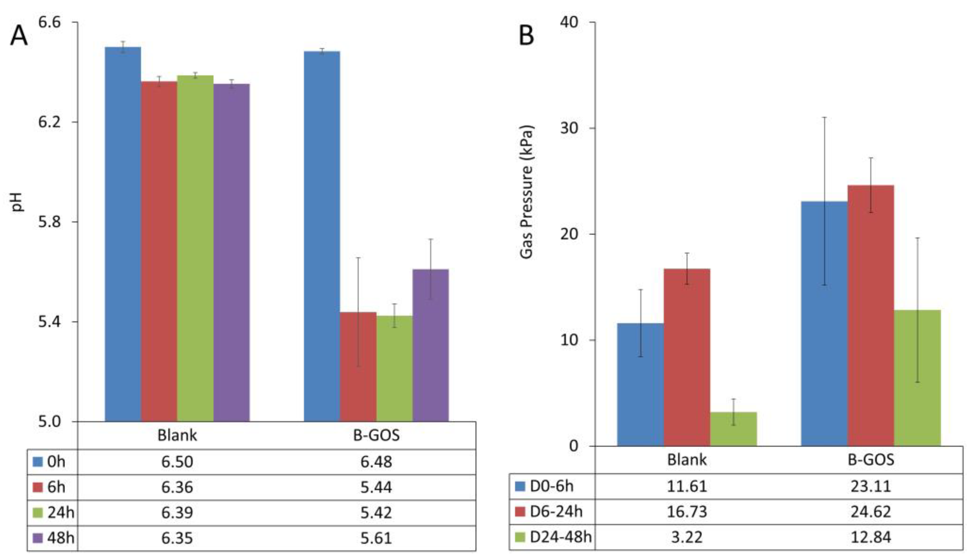
The pH remained relatively stable at all timepoints in the blank (untreated) reactors and was reduced relative to 0 h at 6 h, 24 h, and 48 h with GOS treatment (Figure 2A). For the blank samples, gas production was somewhat higher between 6 h and 24 h than between 0 h and 6 h, and decreased rather dramatically between 24 h and 48 h (Figure 2B). A similar pattern was observed for the GOS, though the gas production was greater overall. pH and gas production for the individual donors are shown in Figure S1a and Figure S1b, respectively.
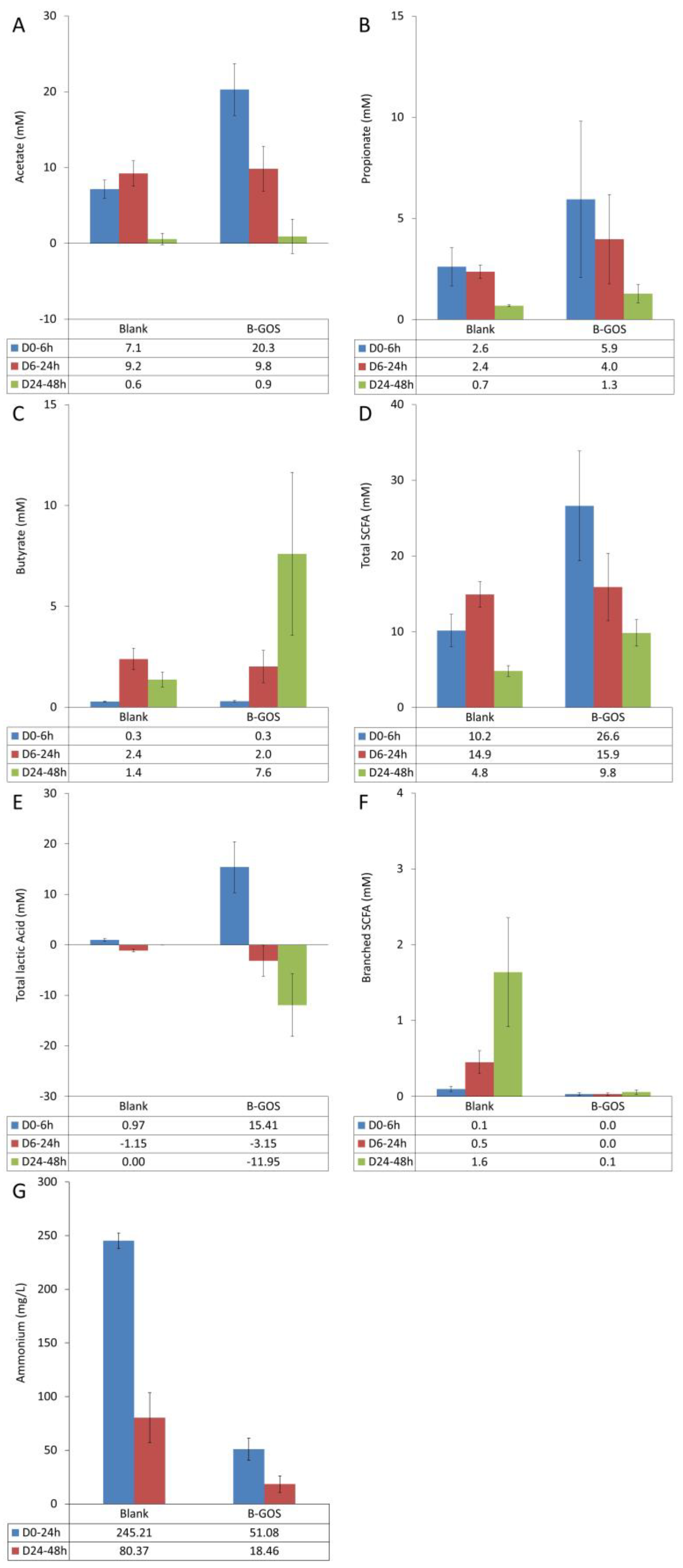
Average changes in SCFA levels are shown in Figure 3 and data for individual donors are shown in Figure S2. Acetate levels increased between 0 h and 6 h and between 6 h and 24 h, and then remained stable between 24 h and 48 h in the blank reactors (Figure 3A). In the reactors treated with GOS, the most dramatic production of acetate occurred between 0 h and 6 h, with a comparatively reduced level between 6 h and 24 h and the least production between 24 h and 48 h. Data for individual donors are shown in Figure S2a. The average propionate data showed that production was increased in the presence of GOS relative to the blank, and production was greatest between 0 h and 6 h, followed by between 6 h and 24 h, and was lowest between 24 h and 48 h (Figure 3B). Propionate production varied between donors (Figure S2b); little propionate was produced with Donor A and the levels were similar between treated and blank. Butyrate production showed a different pattern, with the lowest production between 0 h and 6 h and the highest production between 24 h and 48 h with GOS treatment (Figure 3C). While Donors B and C produced a similar total amount of butyrate with GOS (5.7 mM and 6.6 mM, respectively), the total amount produced by Donor A was comparatively much higher (17.4 mM in total) (Figure S2c). Total SCFA production is shown in Figure 3D. SCFA production was higher in the GOS-treated reactors compared with the blank reactors. In general, SCFA production was greatest between 0 h and 6 h, followed by between 6 h and 24 h; the least production was observed between 24 h and 48 h. Data for individual donors are shown in Figure S2d. Lactic acid levels were highest between 0 h and 6 h and the level was higher with GOS treatment versus blank (Figure 3E). Though there were some differences between donors (Figure S2e), lactic acid levels were reduced between 6 h and 24 h and between 24 h and 48 h. With the GOS treatment, the level of lactic acid decrease between 24 h and 48 h was greater than blank, though this was less pronounced for Donor C. The level of branched SCFAs was greatly reduced in the reactors treated with GOS compared with blank and the levels with GOS were extremely low (between 0.0 and 0.1 mM) (Figure 3F). The production of branched SCFA was low for Donor B relative to the others (Figure S2f). Treatment with GOS also reduced the production of ammonium for all donors (Figure 3G); there were no obvious differences among the donors (Figure S2g).

3.2. Microbial Community Composition

The microbial community composition at the start of the study is shown for each donor in Figure S3. While the number of cells/g fecal matter varied among the donors, Bacteroidetes were most abundant, followed by Firmicutes and Actinobacteria. The other phyla made up a very small proportion of the community composition. PCoA plots showing relative data at 6 h (Figure 4A) and absolute data at 24 h (Figure 4B) demonstrate clear shifts in microbial community composition between GOS and blank reactors for each of the donors as well as differences among donors.
Average relative abundances for the microbial community composition at the family level at 6 h are shown in Figure 5A. Treatment with GOS resulted in a significant decrease in the relative abundance of several families. LEfSe revealed a high LDA score of >4 for Bifidobacterium longum with GOS treatment at 6 h (relative abundance) (Figure 5B). The relative abundances of B. longum and Megamonas (unspecified) were significantly increased with GOS treatment compared with blank at 6 h (p = 0.006 and p = 0.03, respectively [ANOVA]) (Figure 5C). Changes in absolute abundances at the family level at 24 h are shown in Figure 6A. Compared with blank, treatment with GOS resulted in significant changes for several families; most notably, there was a significant increase in the absolute abundance of Bifidobacteriaceae (p < 0.001). Lactobacillaceae were also significantly increased (p < 0.05) and Clostridiales (p < 0.001), Erysipelotrichaceae (p < 0.05), Odoribacteraceae (p < 0.01), and Oscillospiraceae (p < 0.001) were significantly decreased with GOS treatment versus blank. Absolute abundances at the family level at 24 h are shown in Table S1 (according to donor and overall). The absolute abundance of Streptococcaceae increased significantly with GOS relative to blank for Donor A but was below the limit of quantification for the other two donors. LEfSe revealed a high LDA score of >4 for Bifidobacterium adolescentis, Collinsella spp., and Collinsella aerofaciens and of >3 (but <4) for Ruminococcus torques, Bifidobacterium kashiwanohense, and Bifidobacterium ruminantium with GOS treatment at 24 h (absolute abundance) (Figure 6B). The absolute abundances of B. adolescentis and B. ruminantium were significantly increased with GOS treatment compared with blank at 24 h (p < 0.001 and p = 0.01, respectively [ANOVA]) (Figure 6C).

3.3. GOS Utilization by the Gut Microbiota

After 6 h of fermentation, on average 18.79 +/− 0.11 GOS remained in the vessels. At the 24 h timepoint only 5.76% +/− 0.00 GOS was left, and at 48 h 5.41% +/− 0.02 GOS was present.

4. Discussion

Using short-term colonic incubations, we evaluated the effects of a specific GOS on the overall microbial fermentation, microbial metabolic activity, and microbial community composition of colonic bacteria isolated from healthy donors. The GOS was efficiently fermented (81% of GOS was consumed by the colonic microbiota during the first 6 h), resulting in notable effects on metabolite production, including increased SCFA production resulting from the enrichment of beneficial bacteria.
In our study, GOS increased the production of acetate, propionate, butyrate, and lactate. This result corresponds with previous in vitro studies using fecal batch cultures that reported an increased production of acetate with GOS supplementation [27], an increase in acetate and lactate with GOS supplementation [46], and an increased production of lactate and all SCFAs, particularly butyrate, with GOS supplementation [47]. The increased butyrate production was quite marked in our study and could be observed as early as 6 h to 24 h, demonstrating a maximum increase at 24 h to 48 h. The marked increase in butyrate production was likely largely driven by lactate-to-butyrate conversion [48], which would explain the consumption of lactic acid after 6 h. Acetate-to-butyrate conversion may also have contributed to the increase in butyrate [49]. Butyrate is preferentially consumed by colonocytes, making it an important component in gut membrane health [50]. Additionally, butyrate plays an important role in regulating the integrity of the epithelial barrier via coordinated regulation of tight junction proteins [51,52]. The importance of this function is highlighted by the fact that loss of barrier integrity is thought to contribute to metabolic disorders, inflammatory bowel disease, and obesity [53]. In obese adults, GOS supplementation was shown to improve intestinal barrier function [53]. In our study, GOS stimulated health-related microbial metabolites, confirming its usefulness as a prebiotic, and, for the first time, demonstrating its beneficial effects on unfavorable metabolites such as ammonium and branched SCFA.
With GOS supplementation, the production of ammonium was reduced relative to the blank, and little to no branched SCFAs were produced. This was in line with a previous report that GOS supplementation resulted in a decrease in the production of branched SCFAs in an in vitro model of the large intestine [46]. As these metabolites are markers of proteolytic fermentation, a reduction indicates that the level of proteolytic fermentation was low with this GOS, indicating a shift in the fermentation pattern to one more beneficial for the host. This may be considered beneficial, as some metabolic derivatives of proteolytic fermentation are implicated in disease, including colorectal cancer. Avoiding proteolytic fermentation is considered beneficial, as highly toxic compounds may be produced during this process [54].
A previous in vitro study reported that B. longum and other Bifidobacterium spp. along with several Lactobacillus spp. were directly involved in GOS fermentation [46,47]. In our study, the relative abundance of B. longum and Megamonas (unspecified) was significantly increased (versus blank) at the 6 h timepoint after GOS supplementation. In addition, at 24 h the absolute abundance of B. adolescentis and B. ruminantium was significantly increased (versus blank) as was the absolute abundance of Bifidobacteriaceae and Lactobacillaceae. Other in vitro fecal batch studies have shown that GOS supplementation (including the specific GOS tested in this study) increases the growth of bifidobacteria and lactobacilli [27,55] and studies in humans have demonstrated that GOS supplementation increased bifidobacteria and lactobacilli in healthy adults and the elderly [10,11,14]. Bifidobacteria and lactobacilli are well known for their role in human health. The increase in both was likely responsible for augmenting the observed increase in butyrate production via lactate production. Thus, the observed increase in bifidobacteria and Lactobacillaceae supports the prebiotic effects of this GOS. A role for Megamonas in health or disease has not been clearly defined, so it is difficult to speculate on the implications of this finding.
There were some donor-specific effects regarding propionate and butyrate production. Donor A produced very little propionate and there was no difference in propionate production between blank and GOS supplementation. However, this donor produced a much higher amount of butyrate with GOS supplementation than the other two donors. In addition, lactate production and consumption occurred to a greater degree in Donor A than the others. We observed an enrichment of Streptococcaceae for Donor A upon GOS supplementation. This may have contributed to the early strong increase in lactate production (0–6 h) and subsequent conversion into butyrate (24–48 h). Interestingly, Donor A and Donor B had an increase in the absolute abundance of both Bifidobacteriaceae and Lactobacillaceae while Donor C had an increase in Bifidobacteriaceae but not Lactobacillaceae. However, these differences in the effect of the GOS on the three donors do not completely explain the differences between donors in butyrate or lactate production.
As is the case with in vitro studies, our findings are limited in that they cannot directly translate to a biological response. However, these findings confirm the previously known prebiotic effects of the specific GOS tested in this study and provide insight into the mechanisms behind these observed effects. While we investigated the effects of this GOS on the gut microbiota of three donors, these effects need to be confirmed by performing testing on a greater variety of donors.

5. Conclusions

In conclusion, a specific GOS demonstrated prebiotic activity in short-term colonic incubations using the colonic microbiota of three healthy adult donors. A significant proportion of the GOS reached the colon and was not dialyzed during the in vitro simulation of intestinal absorption, a strong indication that the product is not absorbed at the level of the small intestine in vivo and as such is not bioavailable in itself. GOS thus reaches its target site, namely the colon, where 81% of the GOS was fermented during the initial 6 h timeframe, suggesting that the product is likely to be fermented in the proximal colon region in vivo. GOS fermentation was associated with production of bioactive metabolites, making GOS bioavailable through the action of the gut microbiota at the level of the proximal colon. The bioactive molecules produced through GOS fermentation include lactic acid and SCFAs, including butyrate. Furthermore, increased growth of the beneficial bacterial families Bifidobacteriaceae and Lactobacillaceae was observed, and for the first time it was demonstrated that this GOS reduced unfavorable metabolites such as ammonium and branched SCFAs. These results enable us to speculate on the effects the GOS will have in vivo, namely positive effects on gut membrane health resulting from a significant stimulation of butyrate production, as well as a reduction in proteolysis and thus toxic compounds. By being a substrate for fermentation for health-promoting gut members, the GOS resulted in the production of bioactive metabolites in the colon, making Bimuno® GOS an interesting candidate prebiotic. However, these effects need to be confirmed by performing tests on a greater number of donors.

Supplementary Materials

The following are available online at https://www.mdpi.com/article/10.3390/applmicrobiol3010008/s1, Table S1: Absolute abundances (log(cells/mL)) of bacterial families at 24 h of incubation, Figure S1: Overall microbial community activity (acidification and gas production) shown as (a) pH and (b) gas pressure for individual donors, Figure S2: Microbial metabolic activity (a) acetate, (b) propionate, (c) butyrate, (d) total SCFA, (e) lactate, (f) branched SCFA, and (g) ammonium for individual donors, Figure S3: Representation of major phyla in the fecal microbiota of each donor at 0 h.

Author Contributions

Conceptualization, A.M. and L.H.; methodology and formal analysis, J.G. and P.V.d.A.; Writing—original draft preparation, M.M.; Writing—review and editing, A.M., L.H. and J.G. All authors have read and agreed to the published version of the manuscript.

Funding

This research and the article processing charge were funded by Clasado Biosciences Ltd.

Institutional Review Board Statement

Fecal samples of the donors were collected according to the ethical approval of the University Hospital Ghent (reference number B670201836585).

Informed Consent Statement

Informed consent was obtained from all fecal donors.

Data Availability Statement

The data presented in this study are available on request from the corresponding author.

Acknowledgments

The authors thank Sarah Bubeck of Bubeck Scientific Communications for providing medical writing support. The authors would also like to thank Elena Conti, a former employee of Clasado Biosciences Ltd., for her contribution to early discussions of the results.

Conflicts of Interest

Lucien Harthoorn is currently an employee and Aleksandra Maruszak is a former employee of Clasado Biosciences Ltd.

References

  1. Gibson, G.R.; Hutkins, R.; Sanders, M.E.; Prescott, S.L.; Reimer, R.A.; Salminen, S.J.; Scott, K.; Stanton, C.; Swanson, K.S.; Cani, P.D.; et al. Expert consensus document: The International Scientific Association for Probiotics and Prebiotics (ISAPP) consensus statement on the definition and scope of prebiotics. Nat. Rev. Gastroenterol. Hepatol. 2017, 14, 491–502. [Google Scholar] [CrossRef] [PubMed] [Green Version]
  2. Bouhnik, Y.; Raskine, L.; Simoneau, G.; Vicaut, E.; Neut, C.; Flourie, B.; Brouns, F.; Bornet, F.R. The capacity of nondigestible carbohydrates to stimulate fecal bifidobacteria in healthy humans: A double-blind, randomized, placebo-controlled, parallel-group, dose-response relation study. Am. J. Clin. Nutr. 2004, 80, 1658–1664. [Google Scholar] [CrossRef] [PubMed] [Green Version]
  3. Walker, A.W.; Ince, J.; Duncan, S.H.; Webster, L.M.; Holtrop, G.; Ze, X.; Brown, D.; Stares, M.D.; Scott, P.; Bergerat, A.; et al. Dominant and diet-responsive groups of bacteria within the human colonic microbiota. ISME J. 2011, 5, 220–230. [Google Scholar] [CrossRef] [PubMed]
  4. Cotillard, A.; Kennedy, S.P.; Kong, L.C.; Prifti, E.; Pons, N.; Le Chatelier, E.; Almeida, M.; Quinquis, B.; Levenez, F.; Galleron, N.; et al. Dietary intervention impact on gut microbial gene richness. Nature 2013, 500, 585–588. [Google Scholar] [CrossRef]
  5. Halmos, E.P.; Christophersen, C.T.; Bird, A.R.; Shepherd, S.J.; Gibson, P.R.; Muir, J.G. Diets that differ in their FODMAP content alter the colonic luminal microenvironment. Gut 2015, 64, 93–100. [Google Scholar] [CrossRef]
  6. Costabile, A.; Klinder, A.; Fava, F.; Napolitano, A.; Fogliano, V.; Leonard, C.; Gibson, G.R.; Tuohy, K.M. Whole-grain wheat breakfast cereal has a prebiotic effect on the human gut microbiota: A double-blind, placebo-controlled, crossover study. Br. J. Nutr. 2008, 99, 110–120. [Google Scholar] [CrossRef]
  7. Carvalho-Wells, A.L.; Helmolz, K.; Nodet, C.; Molzer, C.; Leonard, C.; McKevith, B.; Thielecke, F.; Jackson, K.G.; Tuohy, K.M. Determination of the in vivo prebiotic potential of a maize-based whole grain breakfast cereal: A human feeding study. Br. J. Nutr. 2010, 104, 1353–1356. [Google Scholar] [CrossRef] [Green Version]
  8. Campana, R.; van Hemert, S.; Baffone, W. Strain-specific probiotic properties of lactic acid bacteria and their interference with human intestinal pathogens invasion. Gut Pathog. 2017, 9, 12. [Google Scholar] [CrossRef] [Green Version]
  9. Morrison, D.J.; Preston, T. Formation of short chain fatty acids by the gut microbiota and their impact on human metabolism. Gut Microbes 2016, 7, 189–200. [Google Scholar] [CrossRef] [Green Version]
  10. Depeint, F.; Tzortzis, G.; Vulevic, J.; I’Anson, K.; Gibson, G.R. Prebiotic evaluation of a novel galactooligosaccharide mixture produced by the enzymatic activity of Bifidobacterium bifidum NCIMB 41171, in healthy humans: A randomized, double-blind, crossover, placebo-controlled intervention study. Am. J. Clin. Nutr. 2008, 87, 785–791. [Google Scholar] [CrossRef]
  11. Vulevic, J.; Drakoularakou, A.; Yaqoob, P.; Tzortzis, G.; Gibson, G.R. Modulation of the fecal microflora profile and immune function by a novel trans-galactooligosaccharide mixture (GOS) in healthy elderly volunteers. Am. J. Clin. Nutr. 2008, 88, 1438–1446. [Google Scholar] [CrossRef] [PubMed]
  12. Davis, L.M.; Martinez, I.; Walter, J.; Goin, C.; Hutkins, R.W. Barcoded pyrosequencing reveals that consumption of galactooligosaccharides results in a highly specific bifidogenic response in humans. PLoS ONE 2011, 6, e25200. [Google Scholar] [CrossRef] [PubMed] [Green Version]
  13. Scalabrin, D.M.; Mitmesser, S.H.; Welling, G.W.; Harris, C.L.; Marunycz, J.D.; Walker, D.C.; Bos, N.A.; Tolkko, S.; Salminen, S.; Vanderhoof, J.A. New prebiotic blend of polydextrose and galacto-oligosaccharides has a bifidogenic effect in young infants. J. Pediatr. Gastroenterol. Nutr. 2012, 54, 343–352. [Google Scholar] [CrossRef] [PubMed]
  14. Vulevic, J.; Juric, A.; Walton, G.E.; Claus, S.P.; Tzortzis, G.; Toward, R.E.; Gibson, G.R. Influence of galacto-oligosaccharide mixture (GOS) on gut microbiota, immune parameters and metabonomics in elderly persons. Br. J. Nutr. 2015, 114, 586–595. [Google Scholar] [CrossRef] [PubMed] [Green Version]
  15. Ladirat, S.E.; Schuren, F.H.; Schoterman, M.H.; Nauta, A.; Gruppen, H.; Schols, H.A. Impact of galacto-oligosaccharides on the gut microbiota composition and metabolic activity upon antibiotic treatment during in vitro fermentation. FEMS Microbiol. Ecol. 2014, 87, 41–51. [Google Scholar] [CrossRef] [PubMed] [Green Version]
  16. Griffiths, E.A.; Duffy, L.C.; Schanbacher, F.L.; Qiao, H.; Dryja, D.; Leavens, A.; Rossman, J.; Rich, G.; Dirienzo, D.; Ogra, P.L. In vivo effects of bifidobacteria and lactoferrin on gut endotoxin concentration and mucosal immunity in Balb/c mice. Dig. Dis. Sci. 2004, 49, 579–589. [Google Scholar] [CrossRef]
  17. Wang, Z.; Xiao, G.; Yao, Y.; Guo, S.; Lu, K.; Sheng, Z. The role of bifidobacteria in gut barrier function after thermal injury in rats. J. Trauma 2006, 61, 650–657. [Google Scholar] [CrossRef]
  18. Wang, Z.T.; Yao, Y.M.; Xiao, G.X.; Sheng, Z.Y. Risk factors of development of gut-derived bacterial translocation in thermally injured rats. World J. Gastroenterol. 2004, 10, 1619–1624. [Google Scholar] [CrossRef]
  19. Ruiz, L.; Delgado, S.; Ruas-Madiedo, P.; Sanchez, B.; Margolles, A. Bifidobacteria and their molecular communication with the immune system. Front. Microbiol. 2017, 8, 2345. [Google Scholar] [CrossRef] [Green Version]
  20. Pompei, A.; Cordisco, L.; Amaretti, A.; Zanoni, S.; Matteuzzi, D.; Rossi, M. Folate production by bifidobacteria as a potential probiotic property. Appl. Environ. Microbiol. 2007, 73, 179–185. [Google Scholar] [CrossRef]
  21. Strozzi, G.P.; Mogna, L. Quantification of folic acid in human feces after administration of Bifidobacterium probiotic strains. J. Clin. Gastroenterol. 2008, 42 Pt 2 (Suppl. S3), S179–S184. [Google Scholar] [CrossRef] [PubMed]
  22. Sugahara, H.; Odamaki, T.; Hashikura, N.; Abe, F.; Xiao, J.Z. Differences in folate production by bifidobacteria of different origins. Biosci. Microbiota Food Health 2015, 34, 87–93. [Google Scholar] [CrossRef] [PubMed] [Green Version]
  23. Sivan, A.; Corrales, L.; Hubert, N.; Williams, J.B.; Aquino-Michaels, K.; Earley, Z.M.; Benyamin, F.W.; Lei, Y.M.; Jabri, B.; Alegre, M.L.; et al. Commensal Bifidobacterium promotes antitumor immunity and facilitates anti-PD-L1 efficacy. Science 2015, 350, 1084–1089. [Google Scholar] [CrossRef] [PubMed] [Green Version]
  24. Tzortzis, G.; Goulas, A.K.; Gibson, G.R. Synthesis of prebiotic galactooligosaccharides using whole cells of a novel strain, Bifidobacterium bifidum NCIMB 41171. Appl. Microbiol. Biotechnol. 2005, 68, 412–416. [Google Scholar] [CrossRef] [PubMed]
  25. Hasle, G.; Raastad, R.; Bjune, G.; Jenum, P.A.; Heier, L. Can a galacto-oligosaccharide reduce the risk of traveller’s diarrhoea? A placebo-controlled, randomized, double-blind study. J. Travel Med. 2017, 24. [Google Scholar] [CrossRef]
  26. Drakoularakou, A.; Tzortzis, G.; Rastall, R.A.; Gibson, G.R. A double-blind, placebo-controlled, randomized human study assessing the capacity of a novel galacto-oligosaccharide mixture in reducing travellers’ diarrhoea. Eur. J. Clin. Nutr. 2010, 64, 146–152. [Google Scholar] [CrossRef]
  27. Grimaldi, R.; Swann, J.R.; Vulevic, J.; Gibson, G.R.; Costabile, A. Fermentation properties and potential prebiotic activity of Bimuno(R) galacto-oligosaccharide (65 % galacto-oligosaccharide content) on in vitro gut microbiota parameters. Br. J. Nutr. 2016, 116, 480–486. [Google Scholar] [CrossRef] [Green Version]
  28. Silk, D.B.; Davis, A.; Vulevic, J.; Tzortzis, G.; Gibson, G.R. Clinical trial: The effects of a trans-galactooligosaccharide prebiotic on faecal microbiota and symptoms in irritable bowel syndrome. Aliment. Pharmacol. Ther. 2009, 29, 508–518. [Google Scholar] [CrossRef]
  29. Huaman, J.W.; Mego, M.; Manichanh, C.; Canellas, N.; Canueto, D.; Segurola, H.; Jansana, M.; Malagelada, C.; Accarino, A.; Vulevic, J.; et al. Effects of prebiotics vs a diet low in FODMAPs in patients with functional gut disorders. Gastroenterology 2018, 155, 1004–1007. [Google Scholar] [CrossRef]
  30. Vulevic, J.; Tzortzis, G.; Juric, A.; Gibson, G.R. Effect of a prebiotic galactooligosaccharide mixture (GOS(R)) on gastrointestinal symptoms in adults selected from a general population who suffer with bloating, abdominal pain, or flatulence. Neurogastroenterol. Motil. 2018, 30, e13440. [Google Scholar] [CrossRef]
  31. Kaur, A.; Rose, D.J.; Rumpagaporn, P.; Patterson, J.A.; Hamaker, B.R. In vitro batch fecal fermentation comparison of gas and short-chain fatty acid production using “slowly fermentable” dietary fibers. J. Food Sci. 2011, 76, H137–H142. [Google Scholar] [CrossRef] [PubMed]
  32. Hughes, S.A.; Shewry, P.R.; Gibson, G.R.; McCleary, B.V.; Rastall, R.A. In vitro fermentation of oat and barley derived beta-glucans by human faecal microbiota. FEMS Microbiol. Ecol. 2008, 64, 482–493. [Google Scholar] [CrossRef] [Green Version]
  33. Rycroft, C.E.; Jones, M.R.; Gibson, G.R.; Rastall, R.A. A comparative in vitro evaluation of the fermentation properties of prebiotic oligosaccharides. J. Appl. Microbiol. 2001, 91, 878–887. [Google Scholar] [CrossRef] [PubMed] [Green Version]
  34. Molly, K.; Vande Woestyne, M.; Verstraete, W. Development of a 5-step multi-chamber reactor as a simulation of the human intestinal microbial ecosystem. Appl. Microbiol. Biotechnol. 1993, 39, 254–258. [Google Scholar] [CrossRef] [PubMed]
  35. Eckburg, P.B.; Bik, E.M.; Bernstein, C.N.; Purdom, E.; Dethlefsen, L.; Sargent, M.; Gill, S.R.; Nelson, K.E.; Relman, D.A. Diversity of the human intestinal microbial flora. Science 2005, 308, 1635–1638. [Google Scholar] [CrossRef] [Green Version]
  36. Kerimi, A.; Kraut, N.U.; da Encarnacao, J.A.; Williamson, G. The gut microbiome drives inter- and intra-individual differences in metabolism of bioactive small molecules. Sci. Rep. 2020, 10, 19590. [Google Scholar] [CrossRef] [PubMed]
  37. Turnbaugh, P.J.; Ridaura, V.K.; Faith, J.J.; Rey, F.E.; Knight, R.; Gordon, J.I. The effect of diet on the human gut microbiome: A metagenomic analysis in humanized gnotobiotic mice. Sci. Transl. Med. 2009, 1, 6ra14. [Google Scholar] [CrossRef] [Green Version]
  38. Hoefman, S.; Pommerening-Röser, A.; Samyn, E.; De Vos, P.; Heylen, K. Efficient cryopreservation protocol enables accessibility of a broad range of ammonia-oxidizing bacteria for the scientific community. Res. Microbiol. 2013, 164, 288–292. [Google Scholar] [CrossRef]
  39. Van den Abbeele, P.; Kamil, A.; Fleige, L.; Chung, Y.; De Chavez, P.; Marzorati, M. Different oat ingredients stimulate specific microbial metabolites in the gut microbiome of three human individuals in vitro. ACS Omega 2018, 3, 12446–12456. [Google Scholar] [CrossRef] [Green Version]
  40. De Weirdt, R.; Possemiers, S.; Vermeulen, G.; Moerdijk-Poortvliet, T.C.; Boschker, H.T.; Verstraete, W.; Van de Wiele, T. Human faecal microbiota display variable patterns of glycerol metabolism. FEMS Microbiol. Ecol. 2010, 74, 601–611. [Google Scholar] [CrossRef]
  41. de Wiele, T.V.; Boon, N.; Possemiers, S.; Jacobs, H.; Verstraete, W. Prebiotic effects of chicory inulin in the simulator of the human intestinal microbial ecosystem. FEMS Microbiol. Ecol. 2004, 51, 143–153. [Google Scholar] [CrossRef] [PubMed]
  42. Ottesen, A.; Ramachandran, P.; Reed, E.; White, J.R.; Hasan, N.; Subramanian, P.; Ryan, G.; Jarvis, K.; Grim, C.; Daquiqan, N.; et al. Enrichment dynamics of Listeria monocytogenes and the associated microbiome from naturally contaminated ice cream linked to a listeriosis outbreak. BMC Microbiol. 2016, 16, 275. [Google Scholar] [CrossRef] [Green Version]
  43. Ponnusamy, D.; Kozlova, E.V.; Sha, J.; Erova, T.E.; Azar, S.R.; Fitts, E.C.; Kirtley, M.L.; Tiner, B.L.; Andersson, J.A.; Grim, C.J.; et al. Cross-talk among flesh-eating Aeromonas hydrophila strains in mixed infection leading to necrotizing fasciitis. Proc. Natl. Acad. Sci. USA 2016, 113, 722–727. [Google Scholar] [CrossRef] [PubMed] [Green Version]
  44. Hasan, N.A.; Young, B.A.; Minard-Smith, A.T.; Saeed, K.; Li, H.; Heizer, E.M.; McMillan, N.J.; Isom, R.; Abdullah, A.S.; Bornman, D.M.; et al. Microbial community profiling of human saliva using shotgun metagenomic sequencing. PLoS ONE 2014, 9, e97699. [Google Scholar] [CrossRef]
  45. Lax, S.; Smith, D.P.; Hampton-Marcell, J.; Owens, S.M.; Handley, K.M.; Scott, N.M.; Gibbons, S.M.; Larsen, P.; Shogan, B.D.; Weiss, S.; et al. Longitudinal analysis of microbial interaction between humans and the indoor environment. Science 2014, 345, 1048–1052. [Google Scholar] [CrossRef] [PubMed] [Green Version]
  46. Maathuis, A.J.; van den Heuvel, E.G.; Schoterman, M.H.; Venema, K. Galacto-oligosaccharides have prebiotic activity in a dynamic in vitro colon model using a (13)C-labeling technique. J. Nutr. 2012, 142, 1205–1212. [Google Scholar] [CrossRef] [Green Version]
  47. Perdijk, O.; van Baarlen, P.; Fernandez-Gutierrez, M.M.; van den Brink, E.; Schuren, F.H.J.; Brugman, S.; Savelkoul, H.F.J.; Kleerebezem, M.; van Neerven, R.J.J. Sialyllactose and galactooligosaccharides promote epithelial barrier functioning and distinctly modulate microbiota composition and short chain fatty acid production in vitro. Front. Immunol. 2019, 10, 94. [Google Scholar] [CrossRef] [Green Version]
  48. Bourriaud, C.; Robins, R.J.; Martin, L.; Kozlowski, F.; Tenailleau, E.; Cherbut, C.; Michel, C. Lactate is mainly fermented to butyrate by human intestinal microfloras but inter-individual variation is evident. J. Appl. Microbiol. 2005, 99, 201–212. [Google Scholar] [CrossRef]
  49. Duncan, S.H.; Holtrop, G.; Lobley, G.E.; Calder, A.G.; Stewart, C.S.; Flint, H.J. Contribution of acetate to butyrate formation by human faecal bacteria. Br. J. Nutr. 2004, 91, 915–923. [Google Scholar] [CrossRef] [Green Version]
  50. Clausen, M.R.; Mortensen, P.B. Kinetic studies on colonocyte metabolism of short chain fatty acids and glucose in ulcerative colitis. Gut 1995, 37, 684–689. [Google Scholar] [CrossRef]
  51. Geirnaert, A.; Calatayud, M.; Grootaert, C.; Laukens, D.; Devriese, S.; Smagghe, G.; De Vos, M.; Boon, N.; Van de Wiele, T. Butyrate-producing bacteria supplemented in vitro to Crohn’s disease patient microbiota increased butyrate production and enhanced intestinal epithelial barrier integrity. Sci. Rep. 2017, 7, 11450. [Google Scholar] [CrossRef] [PubMed] [Green Version]
  52. Peng, L.; Li, Z.R.; Green, R.S.; Holzman, I.R.; Lin, J. Butyrate enhances the intestinal barrier by facilitating tight junction assembly via activation of AMP-activated protein kinase in Caco-2 cell monolayers. J. Nutr. 2009, 139, 1619–1625. [Google Scholar] [CrossRef] [PubMed] [Green Version]
  53. Chelakkot, C.; Ghim, J.; Ryu, S.H. Mechanisms regulating intestinal barrier integrity and its pathological implications. Exp. Mol. Med. 2018, 50, 1–9. [Google Scholar] [CrossRef] [Green Version]
  54. Diether, N.E.; Willing, B.P. Microbial Fermentation of Dietary Protein: An Important Factor in Diet(-)Microbe(-)Host Interaction. Microorganisms 2019, 7, 19. [Google Scholar] [CrossRef] [PubMed] [Green Version]
  55. Rycroft, C.E.; Jones, M.R.; Gibson, G.R.; Rastall, R.A. Fermentation properties of gentio-oligosaccharides. Lett. Appl. Microbiol. 2001, 32, 156–161. [Google Scholar] [CrossRef] [PubMed]
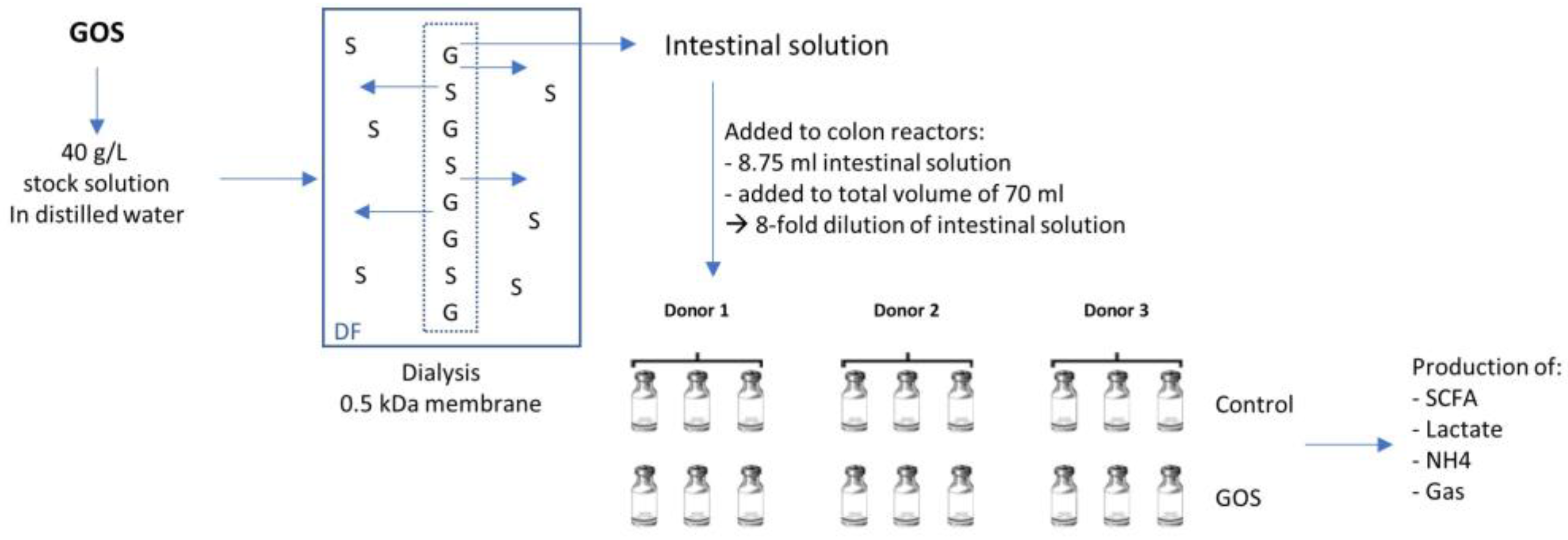
Figure 1. Experimental approach. GOS (provided by Clasado Biosciences Ltd., Reading, UK) was dialyzed prior to colonic incubation to simulate absorption during small intestinal passage. GOS stock solutions (40 g/L) were prepared in water and added inside dialysis membranes (0.5 kDa pore size). Membranes were sealed and the stock solution was dialyzed for 24 h in a dialysis fluid (DF) to allow for monosaccharides and disaccharides (S) to pass through the membrane, whereas larger molecules (G) were retained. The obtained intestinal solution was added to the reactors for the short-term colonic simulation, involving an 8-fold dilution.
Applmicrobiol 03 00008 g001
Figure 2. Overall microbial community activity (acidification and gas production) shown as (A) pH and (B) gas pressure. Measurements were collected in triplicate. Data for average values were derived using data from Donors A/B/C. Error bars represent standard deviation. GOS = Bimuno® galactooligosaccharide.
Applmicrobiol 03 00008 g002
Figure 3. Microbial metabolic activity (A) acetate, (B) propionate, (C) butyrate, (D) total SCFA, (E) lactate, (F) branched SCFA, and (G) ammonium. Measurements were collected in triplicate. Data for average values were derived using data from Donors A/B/C. Error bars represent standard deviation. GOS = Bimuno® galactooligosaccharide; SCFA = short-chain fatty acid.
Applmicrobiol 03 00008 g003
Figure 4. Principal Coordinate Analysis of species data using Bray–Curtis distance for each donor at (A) 6 h after treatment (relative data) and (B) 24 h after treatment (absolute data). Each dot represents one replicate. GOS = Bimuno® galactooligosaccharide.
Applmicrobiol 03 00008 g004
Figure 5. Changes in the microbial community composition (A) relative abundance (family level) 6 h after treatment, (B) linear discriminant analysis effect size of relative abundances (species level) 6 h after treatment, (C) boxplots for the most important enrichments (species level) 6 h after treatment. *: p < 0.05, **: p < 0.01, ***: p < 0.001. GOS = Bimuno® galactooligosaccharide; LDA = linear discriminant analysis.
Applmicrobiol 03 00008 g005
Figure 6. Changes in the microbial community composition (A) absolute abundances (family level) 24 h after treatment, (B) linear discriminant analysis effect size of absolute abundances (species level) 24 h after treatment, (C) boxplots for the most important enrichments (species level) 24 h after treatment. *: p < 0.05, **: p < 0.01, ***: p < 0.001. GOS = Bimuno® galactooligosaccharide; LDA = linear discriminant analysis.
Applmicrobiol 03 00008 g006
Disclaimer/Publisher’s Note: The statements, opinions and data contained in all publications are solely those of the individual author(s) and contributor(s) and not of MDPI and/or the editor(s). MDPI and/or the editor(s) disclaim responsibility for any injury to people or property resulting from any ideas, methods, instructions or products referred to in the content.