Article

Agrobacterium tumefaciens-Mediated Gene Transfer in a Major Human Skin Commensal Fungus, Malassezia globosa

1
Department of Microbiology, Meiji Pharmaceutical University, 2-522-1 Noshio, Kiyose, Tokyo 204-8588, Japan
2
Teikyo University Institute of Medical Mycology, 359 Otsuka, Hachioji, Tokyo 192-0395, Japan
*
Author to whom correspondence should be addressed.
Appl. Microbiol. 2022, 2(4), 827-836; https://doi.org/10.3390/applmicrobiol2040063
Submission received: 6 October 2022 / Revised: 19 October 2022 / Accepted: 21 October 2022 / Published: 25 October 2022

Abstract

:
Although the fungal microbiome in human skin mainly comprises lipophilic yeasts, including Malassezia species, these microorganisms can cause various dermatitis conditions, including pityriasis versicolor, seborrheic dermatitis, folliculitis, and atopic dermatitis, depending on the host condition. Both Malassezia globosa and Malassezia restricta are major species implicated in Malassezia-related dermatitis. However, the pathogenicity of these microorganisms has not been revealed at the genetic level owing to the lack of a genetic recombination system. Therefore, we developed a gene recombination system for M. globosa using Agrobacterium tumefaciens-mediated gene transfer of the target gene FKB1, which encodes the FKBP12 protein that binds the calcineurin inhibitor tacrolimus. The wild-type strain of M. globosa was sensitive to tacrolimus, whereas the FKB1 deletion mutant was resistant to tacrolimus. Reintroduction of FKB1 into the FKB1 deletion mutant restored wild-type levels of susceptibility to tacrolimus. Moreover, an FKB1-eGFP fusion gene was generated and expression of this fusion protein was observed in the cytoplasm. This newly developed gene recombination system for M. globosa will help further our understanding of the pathogenesis of M. globosa-related dermatitis at the genetic level.

1. Introduction

The human skin microbiome comprises bacteria, fungi, and viruses. In the bacterial microbiome, the genera Cutibacterium (formerly Propionibacterium), Corynebacterium, and Staphylococcus are predominant, whereas the skin fungal microbiota primarily comprises Malassezia species regardless of the skin area. Malassezia species commonly inhabit sebum-rich areas, such as the scalp, face, and neck, because they use sebum as a nutrient source [1,2]. The genus Malassezia includes 18 species, of which nine species (Malassezia dermatis, Malassezia furfur, Malassezia globosa, Malassezia japonica, Malassezia obtusa, Malassezia restricta, Malassezia slooffiae, Malassezia sympodialis, and Malassezia yamatoensis) inhabit the human skin. The remaining Malassezia species are commonly found on the skin of other animals. The fungal microbiome in humans primarily comprises M. globosa and M. restricta. Although Malassezia species are commensal microorganisms present on human skin, they also cause seborrheic dermatitis, including dandruff, pityriasis versicolor, and Malassezia folliculitis, and can exacerbate atopic dermatitis [3,4,5]. Although they inhabit human skin, the abundance of M. restricta and M. globosa differs depending on the skin disease. M. restricta is more abundant than M. globosa in the lesions of patients with seborrheic and atopic dermatitis, whereas M. globosa is more frequently detected in patients with pityriasis versicolor. Malassezia species produce many secretory lipases (thirteen in M. globosa and nine in M. restricta) that hydrolyze diglycerides or triglycerides in the sebum into glycerin and free fatty acids [6]. These metabolites are used as nutrients by many skin microbiota, including Malassezia. Oleic acid present in free fatty acids can induce seborrheic dermatitis; thus, lipases have been considered as virulence factors of Malassezia [7]. Anti-Malassezia manganese superoxide dismutase and cyclophilin-specific immunoglobulin E antibodies are produced in the sera of patients with atopic dermatitis, suggesting that the presence of Malassezia exacerbates this disease [8,9]. Pityriasis versicolor is characterized by scaly hypo- or hyperpigmented lesions usually affecting the trunk. Notably, a large amount of hyphae are observed in patient lesions, suggesting that dimorphic conversion may be responsible for the development of pityriasis versicolor [10]. Various factors have been suggested to be involved in Malassezia-related skin dermatitis. Deletion of the genes involved in virulence may help elucidate the functions of the relevant genes.
To date, gene deletion in Malassezia has only been conducted in M. furfur, M. sympodialis, and M. pachydermatis; gene deletion methods using clinically important species, M. globosa and M. restricta, have not yet been established [11,12,13]. However, we recently established a gene recombination system in M. restricta using Agrobacterium tumefaciens-mediated gene transfer (ATMT) [14]. Although Agrobacterium tumefaciens-mediated gene transfer (ATMT) was developed as a method for gene introduction into plant cells, it is possible to introduce genes into a wide range of hosts, including fungi. When Agrobacterium tumefaciens infects a fungus, it transfers exogenous genes into the host cell by using the property of integrating transferred DNA (T-DNA), part of the plasmid, into the chromosomal DNA of the host. The ATMT method has the advantage of simplicity compared to direct gene transfer methods such as the electroporation and particle gun methods [15]. To elucidate the pathogenicity or virulence of the causative agents of Malassezia-associated skin diseases, it will be important to establish a gene recombination system for M. globosa; targeting the genes of known function will be useful in establishing such a system for M. globosa.
The FKB1 gene, which encodes the 12-kDa FK506-binding protein (FKBP12), a protein that binds the calcineurin inhibitor tacrolimus, was previously deleted in M. sympodialis and M. restricta [12,14]. Since Malassezia species are sensitive to tacrolimus, deletion of the FKB1 gene results in resistance to tacrolimus. Therefore, it is easy to evaluate the function of the deleted gene. In the present study, we aimed to delete the FKB1 gene in M. globosa using ATMT and constructed an FKB1-enhanced green fluorescent protein (eGFP) fusion gene. The developed gene recombination system in this study will help further our understanding of the pathogenesis of M. globosa-related dermatitis at the genetic level.

2. Materials and Methods

2.1. Strains and Media

M. globosa CBS 7966 (type strain of the species) was obtained from the CBS-KNAW culture collection (https://wi.knaw.nl/ accessed on 1 July 2022) and maintained on modified Leeming and Notman agar (mLNA; 10 g/L polypeptone, 10 g/L glucose, 2 g/L yeast extract, 8 g/L ox bile, 10 mL/L glycerol, 0.5 g/L glycerol monostearate, 5 mL/L Tween-60, 20 mL/L olive oil, and 15 g/L agar) at 32 °C. The strains generated in this study are listed in Table 1.

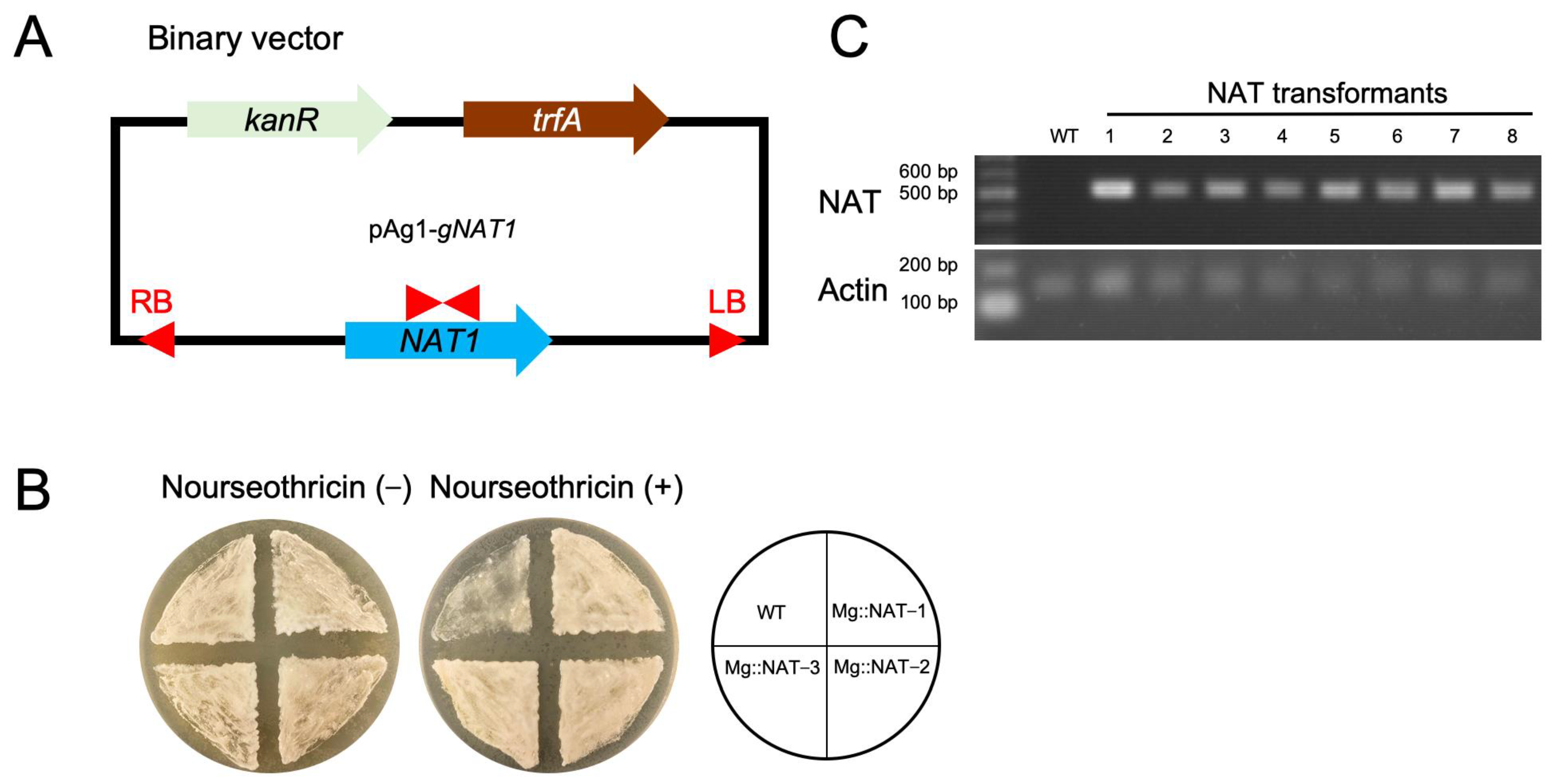
2.2. Construction of M. globosa Expressing the NAT1 Gene Using ATMT

ATMT was performed using a protocol previously developed by Cho et al. [14], Matsumoto et al. [16], and Yamada et al. [17], with minor modification. The binary vector pAg1-N-terminal acetyl transferase (gNAT1) was constructed using the primers FK5 and MgPactin1–3 (Table S1) (Figure 1A). The promoter region of the actin gene of M. globosa (MgPactin) was incorporated upstream of the NAT1 gene. pAg1-gNAT1 was introduced via electroporation into A. tumefaciens EHA105, which was then cultured in 2 × YT agar (16 g/L tryptone, 10 g/L yeast extract, 5 g/L NaCl, and 15 g/L agar) containing 50 μg/mL kanamycin (Fujifilm, Osaka, Japan) at 27 °C for 2 days. The cell concentration was adjusted to OD630 = 1 in Agrobacterium induction medium [AIM; 2.05 g/L K2HPO4, 1.45 g/L KH2PO4, 0.15 g/L NaCl, 0.5 g/L MgSO4·7H2O, 0.1 g/L CaCl2·6H2O, 0.0025 g/L FeSO4·7H2O, and 0.5 g/L (NH4)2SO4 supplemented with 40 mM 2-(N-morpholino)ethanesulfonic acid (pH 5.3), 10 mM glucose, and 0.5% (w/v) glycerol] containing 200 μg/mL acetosyringone (Fujifilm) and incubated at 27 °C for 6 h. M. globosa (100 μL; >2 × 10⁸ cells/mL) and Agrobacterium suspensions were spread onto sterilized nylon membranes (Hybond N+ membranes; Amersham Biosciences, Buckinghamshire, United Kingdom). The membranes were placed on AIM containing 200 μg/mL acetosyringone and 1.5% agar and incubated at 25 °C for 2 days. Transformants were obtained from mLNA supplemented with 200 μg/mL cefotaxime sodium and 100 μg/mL nourseothricin (Jena Bioscience, Jena, Germany) and further re-grown on mLNA containing 100 μg/mL nourseothricin. Polymerase chain reaction (PCR) was performed using the primers NAT-F/R and Actin-F/R (Table S1), which confirmed the introduction of the NAT1 gene into the M. globosa genome.

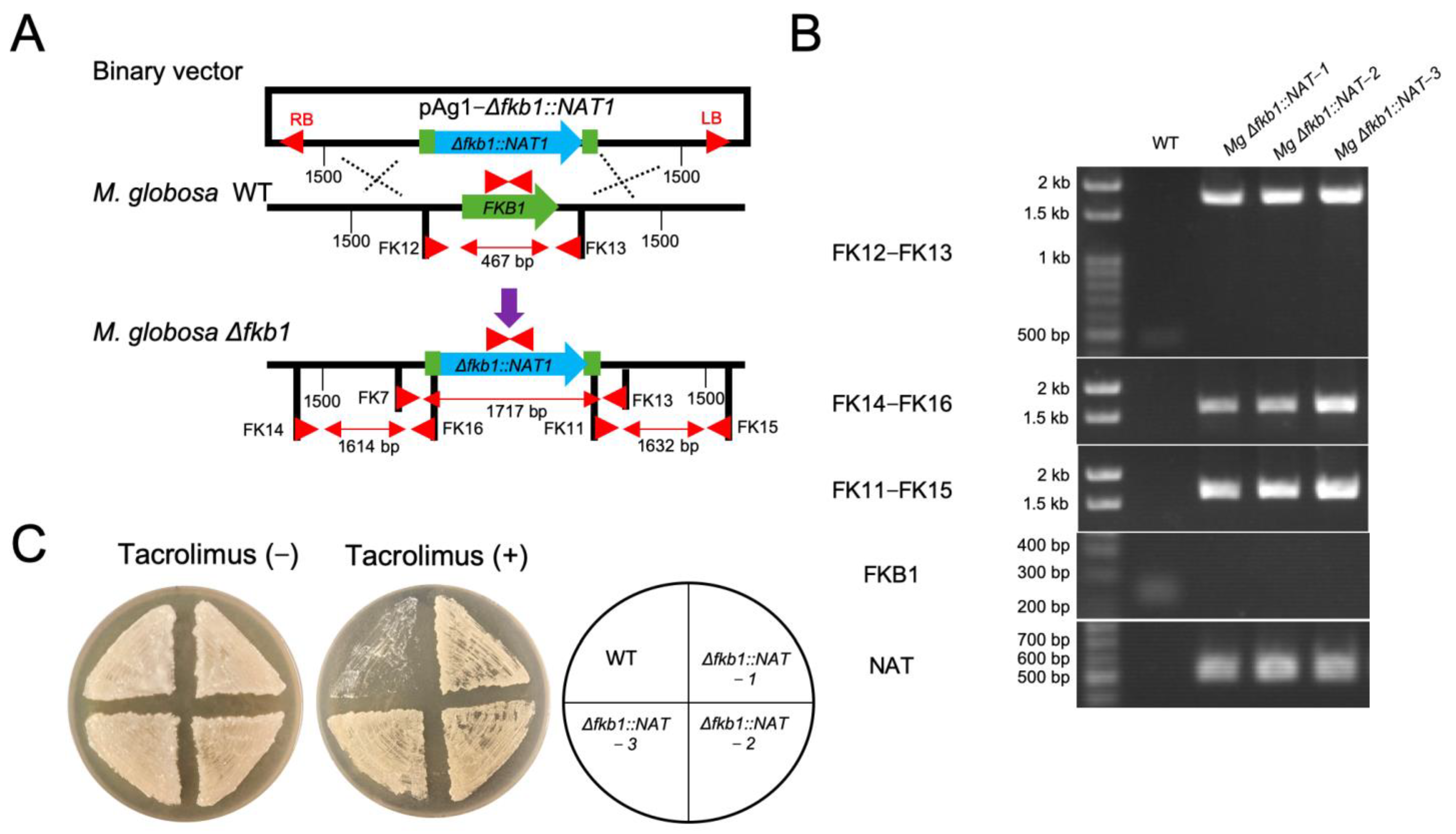
2.3. Targeted Gene Replacement in M. globosa via ATMT

The sequence of the target gene, FKB1, in the M. globosa CBS7966 (RPJNA27973) genome is similar to that of Candida albicans (AAA34367). Reciprocal basic local alignment search tool protein analysis of the C. albicans retinol-binding protein 1 gene revealed that FKBP12 was encoded by MGL_1262. Flanking regions of approximately 1.5 kb upstream and downstream of the FKB1 gene were introduced into the pAg1-gNAT1 vector; the vector and two flanking regions were amplified by PCR using the primers FK1–8 (Table S1) (Figure 2A). Amplicons were cloned into competent Escherichia coli (Thermo Fisher Scientific, Tokyo, Japan) using the In-FusionHDTM Cloning Kit (Takara, Shiga, Japan) according to the manufacturer’s instructions, and transformants were confirmed via PCR using the primers FK9 and FK10 (Table S1). The method of introduction of pAg1-Δfkb1::NAT1 into the M. globosa genome was similar to that of pAg1-gNAT1 described above. Deletion of FKB1 in the M. globosa genome was confirmed via PCR using the primers FK11–16, FKB1-F/R, and NAT-F/R (Table S1). M. globosa CBS 7966 and the FKB1 deletion mutant (Δfkb1) were grown on mLNA agar with or without 100 μg/mL tacrolimus at 32 °C for 4 days.

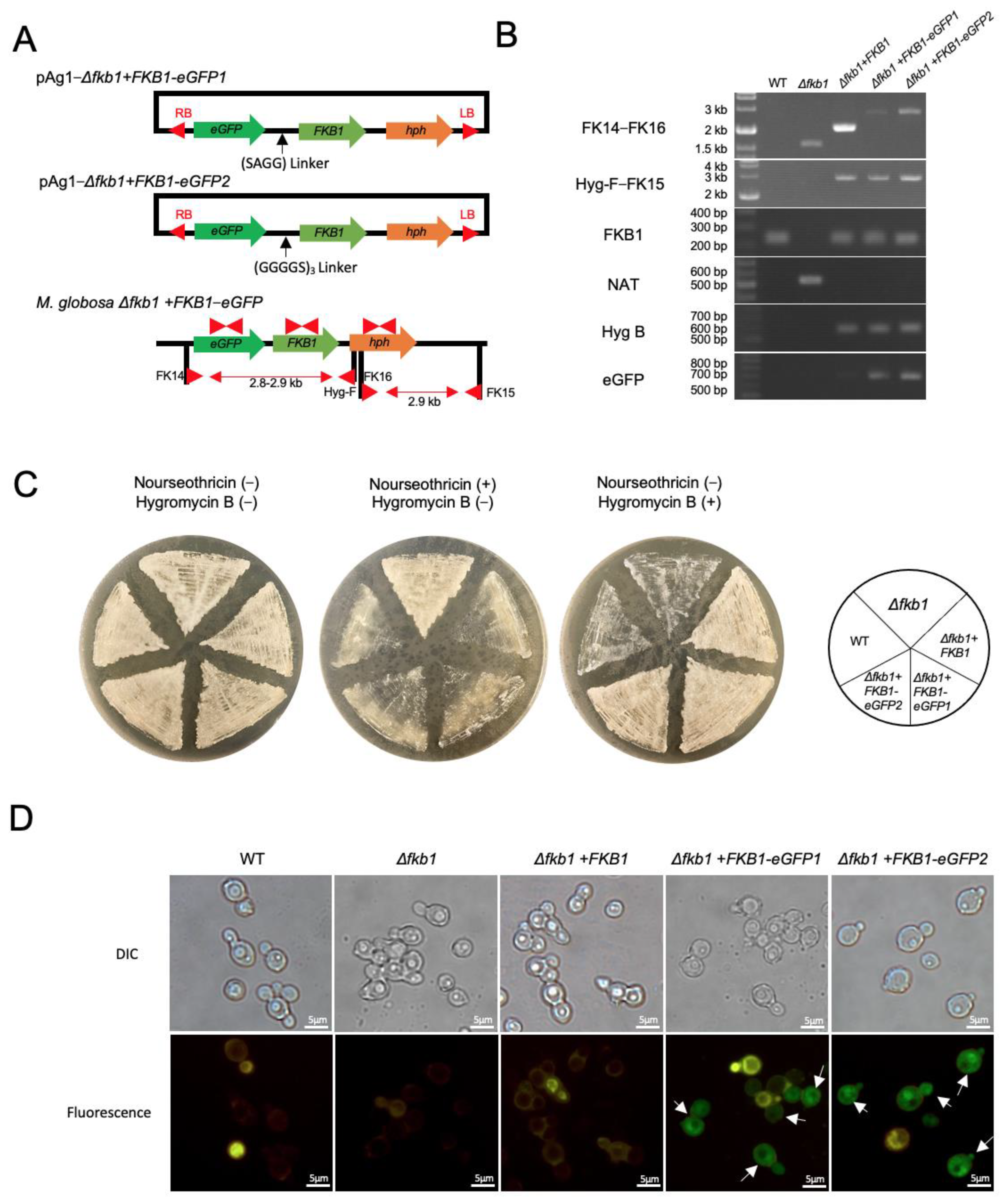
2.4. Reintroduction of the Target Gene into the M. globosa Δfkb1 Mutant

FKB1 was reintroduced into the M. globosa Δfkb1 mutant as previously described [14]. Briefly, flanking regions of approximately 200 bp downstream of FKB1 and hygromycin B phosphotransferase gene (hph) were introduced into the vector pAg1-Δfkb1::NAT1 (Figure 3A, and the binary vector pAg1-Δfkb1 + FKB1 was transformed into A. tumefaciens. Introduction of FKB1 into the genome of the transformants was confirmed via PCR using the primers FK14–16, FKB1-F/R, NAT-F/R, and Hyg-F/R (Table S1).

2.5. Construction of the FKB1-eGFP Fusion Gene in M. globosa

The binary vectors pAg1-Δfkb1 + FKB1-eGFP1 and pAg1-Δfkb1 + FKB1-eGFP2 were generated to reintroduce the target gene into the Δfkb1 mutant (Figure 4A), and the binary vector was transformed into A. tumefaciens as described above. The introduction of the FKB1-eGFP fusion genes into the genome of the transformants was confirmed via PCR using the primers FK14–16, FKB1-F/R, NAT-F/R, Hyg-F/R, and eGFP-F/R (Table S1). The selected transformants were grown on mLNA plates containing 100 μg/mL nourseothricin or 100 μg/mL hygromycin B (Nacalai Tesque, Kyoto, Japan). eGFP expression in Δfkb1, Δfkb1 + FKB1, and Δfkb1 + FKB1-eGFP M. globosa mutants was observed using a fluorescence microscope (BX61; Olympus Corporation, Tokyo, Japan).

2.6. Calcineurin Inhibitor Susceptibility

The minimum inhibitory concentrations (MICs) of the calcineurin inhibitors tacrolimus and cyclosporine A were determined using the broth microdilution method in accordance with CLSI M27-A3 as described by Rojas et al. [18]. M. globosa cells were incubated for 6 days at 32 °C in the presence of 0.03–100 μg/mL of tacrolimus (Fujifilm) or cyclosporine A (Fujifilm) in 96-well microtiter plates. The MIC was defined as the lowest concentration that completely inhibited growth.

3. Results

3.1. M. globosa Transformation Using ATMT

The binary vector pAg1-gNAT1 used in the ATMT system is presented in Figure 1A. Transformants were obtained from mLNA containing nourseothricin (Figure 1B), and NAT1 expression in the transformants was confirmed via PCR using NAT1-specific primers. Of the randomly selected eight colonies, all were positive; the wild-type strain was negative (Figure 1C).

3.2. FKB1 Gene Replacement in M. globosa

The binary plasmid pAg1-Δfkb1::NAT1, which contained regions of approximately 1.5 kb upstream and downstream of the FKB1 gene, was constructed for the FKB1 gene deletion (Figure 2A). Of the randomly selected 24 colonies, FKB1 was deleted in 23 transformants (95.8%) based on PCR confirmation (Figure 2B). The wild-type strain did not grow on tacrolimus-containing mLNA, whereas the Δfkb1 mutant showed growth in this medium (Figure 2C).

3.3. Reintroduction of the FKB1 Gene into the M. globosa Δfkb1 Mutant

The FKB1 gene was reintroduced into the M. globosa Δfkb1 mutant via ATMT using the pAg1-∆fkb1+FKB1 plasmid (Figure 3A). Of the 20 randomly selected colonies recovered from mLNA supplemented with 100 μg/mL hygromycin B, 11 transformants (55%) harbored the FKB1 gene in the genome based on PCR confirmation (Figure 3B).

3.4. Construction of the FKB1-eGFP Fusion Gene and Fluorescence Microscopy

To assess the localization of FKBP12, the M. globosa Δfkb1 + FKB1-eGFP mutant was generated using two binary vectors (pAg1-Δfkb1+FKB1-eGFP1 and pAg1-Δfkb1+FKB1-eGFP2) (Figure 4A). Reintroduction of FKB1-eGFP1 and FKB1-eGFP2 into the transformants was confirmed via PCR (Figure 4B). The M. globosa Δfkb1 + FKB1 and Δfkb1 + FKB1-eGFP mutants were sensitive to nourseothricin and resistant to hygromycin B (Figure 4C). eGFP expression was observed in only Δfkb1 + FKB1-eGFP mutants; yellow autofluorescence was observed in each strain (Figure 4D). Fluorescence microscopy revealed that the FKB1-eGPF fusion protein was expressed in the cytoplasm.

3.5. Drug Susceptibility to Calcineurin Inhibitors

The MICs of tacrolimus ranged from 0.06 to 0.12 μg/mL for the wild-type strain of M. globosa and were >100 μg/mL for the Δfkb1 mutant. The gene-complemented mutant, Δfkb1 + FKB1, had the same MIC as that of the wild-type strain. The susceptibility of the Δfkb1 + FKB1-eGFP mutant to tacrolimus was similar to that of the wild type and Δfkb1 + FKB1 mutant (Table 2). However, the wild type and all mutants (Δfkb1, Δfkb1 + FKB1, and Δfkb1 + FKB1-eGFP) showed no difference in susceptibility to cyclosporine A, which does not bind FKBP12 (Table 2).

4. Discussion

Genetic studies on Malassezia have not progressed as rapidly as those on Candida and Cryptococcus. In recent years, gene recombination has been successfully reported for M. furfur, M. sympodialis, and M. pachydermatis using ATMT [11,12,13]. The main causative or exacerbating agents of Malassezia-related skin dermatitis are M. restricta and M. globosa [5,19,20,21]. Following our recently established gene recombination method for M. restricta [14], in the present study, we developed a similar method for M. globosa targeting the FKB1 gene. The FKB1 gene encodes FKBP12, a protein that binds the calcineurin inhibitor tacrolimus; therefore, the Δfkb1 mutant was no longer susceptible to tacrolimus [22,23].
The gene recombination method for M. globosa was essentially the same as that for M. restricta, with the following modifications. First, the promoter of the binary vector was changed from the Cryptococcus neoformans actin promoter to the M. globosa actin promoter. Although the C. neoformans actin promoter may function in Malassezia cells, a promoter derived from the same species was expected to function better. The M. globosa actin promoter was functional in M. globosa mutants in this study. Second, the concentration of Malassezia cells was critical for transformation. We optimized the ATMT protocol for M. globosa to use a cell concentration of 2 × 10⁸ cells/mL, whereas the protocol for M. restricta uses 6 × 10⁸ cells/mL. With cell concentrations of <2 × 10⁸ cells/mL, few transformants were detected. Finally, the concentration of acetosyringone (200 μg/mL) in the AIM and the incubation temperature of 25 °C were optimized for M. globosa to increase transformation efficiency (for M. restricta, 40 μg/mL of acetosyringone and 27 °C of incubation temperature were applied).
GFP fusion proteins are widely used to investigate protein localization within cells, and the expression of GFP fusion genes has been successfully observed in Malassezia species [24,25]. We used two linker sequences, SAGG and (GGGGS)3, to create eGFP fusion genes [26]. We found that eGFP fusion genes could be created using either linker sequence in M. globosa.
Although M. globosa is implicated in all Malassezia-related dermatitis conditions, it is thought to be more involved in the development of pityriasis versicolor [19,27,28]. M. globosa is relatively more abundant in lesions of patients with pityriasis versicolor, and numerous hyphae formed by the microorganism is observed, suggesting that dimorphic conversion of M. globosa may be involved in the pathogenesis of dermatitis [5,20]. However, a genetic understanding of the dimorphism pathway in M. globosa is lacking. M. globosa is also responsible for exacerbating atopic dermatitis. MGL_1304 in M. globosa has been identified as a causative antigen of sweat allergies observed in patients with atopic dermatitis and cholinergic urticaria; the present study suggests that the skin microbiota may represent a potential allergen [29,30]. Further elucidation of the functions of dimorphism-related and allergen-encoding genes of M. globosa will be possible using the gene recombination method established in this study.

Supplementary Materials

The following supporting information can be downloaded at: https://www.mdpi.com/article/10.3390/applmicrobiol2040063/s1, Table S1. Primers used in this study.

Author Contributions

Conceptualization, O.C. and T.S.; methodology, O.C., Y.M. and T.Y.; investigation, O.C., Y.M. and T.Y.; data curation, O.C., Y.M., T.Y. and T.S.; writing—original draft, O.C.; writing—review and editing, Y.M., T.Y. and T.S.; visualization, O.C., Y.M. and T.Y.; project administration, T.S. All authors have read and agreed to the final version of the manuscript.

Funding

This work was partially supported by the Japan Society for the Promotion of Science KAKENHI (grant number JP20K07208 to O.C.).

Institutional Review Board Statement

Not applicable.

Informed Consent Statement

Not applicable.

Data Availability Statement

Essential data supporting the reported results are provided in the article.

Conflicts of Interest

The authors have no financial, consultant, institutional, and other relationships that might lead to bias or conflict of interest.

References

  1. Findley, K.; Oh, J.; Yang, J.; Conlan, S.; Deming, C.; Meyer, J.A.; Schoenfeld, D.; Nomicos, E.; Park, M.; NIH Intramural Sequencing Center Comparative Sequencing Program; et al. Topographic diversity of fungal and bacterial communities in human skin. Nature 2013, 498, 367–370. [Google Scholar] [CrossRef] [PubMed] [Green Version]
  2. Byrd, A.L.; Belkaid, Y.; Segre, J.A. The human skin microbiome. Nat. Rev. Microbiol. 2018, 16, 143–155. [Google Scholar] [CrossRef]
  3. Harada, K.; Saito, M.; Sugita, T.; Tsuboi, R. Malassezia species and their associated skin diseases. J. Dermatol. 2015, 42, 250–257. [Google Scholar] [CrossRef]
  4. Theelen, B.; Cafarchia, C.; Gaitanis, G.; Bassukas, I.D.; Boekhout, T.; Dawson, T.L., Jr. Malassezia ecology, pathophysiology, and treatment. Med. Mycol. 2018, 56, S10–S25. [Google Scholar] [CrossRef] [Green Version]
  5. Sugita, T.; Boekhout, T.; Velegraki, A.; Guillot, J.; Hađina, S.; Cabañes, F.J. Epidemiology of Malassezia-related skin diseases. In Malassezia and the Skin: Science and Clinical Practice; Velegraki, A., Boekhout, T., Mayser, P., Guého-Kellermann, E., Eds.; Springer: Heidelberg/Berlin, Germany, 2010; pp. 65–119. [Google Scholar]
  6. Dawson, T.L., Jr. Malassezia globosa and restricta: Breakthrough understanding of the etiology and treatment of dandruff and seborrheic dermatitis through whole-genome analysis. J. Investig. Dermatol. Symp. Proc. 2007, 12, 15–19. [Google Scholar] [CrossRef] [PubMed] [Green Version]
  7. DeAngelis, Y.M.; Gemmer, C.M.; Kaczvinsky, J.R.; Kenneally, D.C.; Schwartz, J.R.; Dawson, T.L., Jr. Three etiologic facets of dandruff and seborrheic dermatitis: Malassezia fungi, sebaceous lipids, and individual sensitivity. J. Investig. Dermatol. Symp. Proc. 2005, 10, 295–297. [Google Scholar] [CrossRef] [PubMed] [Green Version]
  8. Schmid-Grendelmeier, P.; Flückiger, S.; Disch, R.; Trautmann, A.; Wüthrich, B.; Blaser, K.; Scheynius, A.; Crameri, R. IgE-mediated and T cell-mediated autoimmunity against manganese superoxide dismutase in atopic dermatitis. J. Allergy Clin. Immunol. 2005, 115, 1068–1075. [Google Scholar] [CrossRef]
  9. Flückiger, S.; Fijten, H.; Whitley, P.; Blaser, K.; Crameri, R. Cyclophilins, a new family of cross-reactive allergens. Eur. J. Immunol. 2002, 32, 10–17. [Google Scholar] [CrossRef]
  10. Civila, E.S.; Vignale, R.; Sanjinés, A.; Conti-Diaz, I.A. Hyphal production by Pityrosporum ovale. Int. J. Dermatol. 1978, 17, 74–77. [Google Scholar] [CrossRef]
  11. Ianiri, G.; Averette, A.F.; Kingsbury, J.M.; Heitman, J.; Idnurm, A. Gene function analysis in the ubiquitous human commensal and pathogen Malassezia genus. mBio 2016, 7, e01853-16. [Google Scholar] [CrossRef]
  12. Ianiri, G.; Applen Clancey, S.; Lee, S.C.; Heitman, J. FKBP12-Dependent inhibition of calcineurin mediates immunosuppressive antifungal drug action in Malassezia. mBio 2017, 8, e01752-17. [Google Scholar] [CrossRef] [PubMed] [Green Version]
  13. Celis, A.M.; Vos, A.M.; Triana, S.; Medina, C.A.; Escobar, N.; Restrepo, S.; Wösten, H.A.; de Cock, H. Highly efficient transformation system for Malassezia furfur and Malassezia pachydermatis using Agrobacterium tumefaciens-mediated transformation. J. Microbiol. Methods. 2017, 134, 1–6. [Google Scholar] [CrossRef] [PubMed]
  14. Cho, O.; Matsumoto, Y.; Yamada, T.; Sugita, T. Establishment of a gene recombination method for a major human skin commensal fungus, Malassezia restricta, using Agrobacterium tumefaciens-mediated gene transfer system. Med. Mycol. in press.
  15. Idnurm, A.; Bailey, A.M.; Cairns, T.C.; Elliott, C.E.; Foster, G.D.; Ianiri, G.; Jeon, J. A silver bullet in a golden age of functional genomics: The impact of Agrobacterium-mediated transformation of fungi. Fungal Biol. Biotechnol. 2017, 4, 6. [Google Scholar] [CrossRef] [PubMed] [Green Version]
  16. Matsumoto, Y.; Yamazaki, H.; Yamasaki, Y.; Tateyama, Y.; Yamada, T.; Sugita, T. A novel silkworm infection model with fluorescence imaging using transgenic Trichosporon asahii expressing eGFP. Sci. Rep. 2020, 10, 10991. [Google Scholar] [CrossRef]
  17. Yamada, T.; Makimura, K.; Satoh, K.; Umeda, Y.; Ishihara, Y.; Abe, S. Agrobacterium tumefaciens-mediated transformation of the dermatophyte, Trichophyton mentagrophytes: An efficient tool for gene transfer. Med. Mycol. 2009, 47, 485–494. [Google Scholar] [CrossRef] [Green Version]
  18. Rojas, F.D.; Sosa, M.d.L.A.; Fernández, M.S.; Cattana, M.E.; Córdoba, S.B.; Giusiano, G.E. Antifungal susceptibility of Malassezia furfur, Malassezia sympodialis, and Malassezia globosa to azole drugs and amphotericin B evaluated using a broth microdilution method. Med. Mycol. 2014, 52, 641–646. [Google Scholar] [CrossRef] [Green Version]
  19. Sugita, T.; Suto, H.; Unno, T.; Tsuboi, R.; Ogawa, H.; Shinoda, T.; Nishikawa, A. Molecular analysis of Malassezia microflora on the skin of atopic dermatitis patients and healthy subjects. J. Clin. Microbiol. 2001, 39, 3486–3490. [Google Scholar] [CrossRef] [Green Version]
  20. Morishita, N.; Sei, Y.; Sugita, T. Molecular analysis of Malassezia microflora from patients with pityriasis versicolor. Mycopathologia 2006, 161, 61–65. [Google Scholar] [CrossRef]
  21. Tajima, M.; Sugita, T.; Nishikawa, A.; Tsuboi, R. Molecular analysis of Malassezia microflora in seborrheic dermatitis patients: Comparison with other diseases and healthy subjects. J. Invest. Dermatol. 2008, 128, 345–351. [Google Scholar] [CrossRef] [Green Version]
  22. Park, H.S.; Lee, S.C.; Cardenas, M.E.; Heitman, J. Calcium-calmodulin-calcineurin signaling: A globally conserved virulence cascade in eukaryotic microbial pathogens. Cell Host Microbe. 2019, 26, 453–462. [Google Scholar] [CrossRef] [PubMed]
  23. Vellanki, S.; Garcia, A.E.; Lee, S.C. Interactions of FK506 and rapamycin with FK506 binding protein 12 in opportunistic human fungal pathogens. Front. Mol. Biosci. 2020, 7, 588913. [Google Scholar] [CrossRef] [PubMed]
  24. Ianiri, G.; Coelho, M.A.; Ruchti, F.; Sparber, F.; McMahon, T.J.; Fu, C.; Bolejack, M.; Donovan, O.; Smutney, H.; Myler, P.; et al. HGT in the human and skin commensal Malassezia: A bacterially derived flavohemoglobin is required for NO resistance and host interaction. Proc. Natl Acad. Sci. USA 2020, 117, 15884–15894. [Google Scholar] [CrossRef] [PubMed]
  25. Sankaranarayanan, S.R.; Ianiri, G.; Coelho, M.A.; Reza, M.H.; Thimmappa, B.C.; Ganguly, P.; Vadnala, R.N.; Sun, S.; Siddharthan, R.; Tellgren-Roth, C.; et al. Loss of centromere function drives karyotype evolution in closely related Malassezia species. eLife 2020, 9, e53944. [Google Scholar] [CrossRef] [PubMed]
  26. Chen, X.; Zaro, J.L.; Shen, W.C. Fusion protein linkers: Property, design and functionality. Adv. Drug Deliv. Rev. 2013, 65, 1357–1369. [Google Scholar] [CrossRef] [Green Version]
  27. Aghaei Gharehbolagh, S.; Mafakher, L.; Salehi, Z.; Asgari, Y.; Hashemi, S.J.; Mahmoudi, S.; Nasimi, M.; Rezaie, S. Unveiling the structure of GPI-anchored protein of Malassezia globosa and its pathogenic role in pityriasis versicolor. J. Mol. Model. 2021, 27, 246. [Google Scholar] [CrossRef]
  28. Aghaei Gharehbolagh, S.; Kordbacheh, P.; Hashemi, S.J.; Daie Ghazvini, R.; Asgari, Y.; Agha Kuchak Afshari, S.; Seyedmousavi, S.; Rezaie, S. MGL_3741 gene contributes to pathogenicity of Malassezia globosa in pityriasis versicolor. Mycoses 2018, 61, 938–944. [Google Scholar] [CrossRef]
  29. Hiragun, T.; Ishii, K.; Hiragun, M.; Suzuki, H.; Kan, T.; Mihara, S.; Yanase, Y.; Bartels, J.; Schröder, J.M.; Hide, M. Fungal protein MGL_1304 in sweat is an allergen for atopic dermatitis patients. J. Allergy Clin. Immunol. 2013, 132, 608–615.e4. [Google Scholar] [CrossRef]
  30. Takahagi, S.; Tanaka, A.; Hide, M. Sweat allergy. Allergol. Int. 2018, 67, 435–441. [Google Scholar] [CrossRef]
Figure 1. Agrobacterium tumefaciens-mediated transformation. (A) Schematic representation of the binary vector containing NAT1 introduced into Malassezia globosa. The promoter sequence of the actin gene of M. globosa was incorporated upstream of the NAT1 gene, and the termination sequence of the TRP1 gene of Cryptococcus neoformans was incorporated downstream of the NAT1 gene. Red arrowheads indicate primer-binding sites. (B) Sensitivity of the WT strain and Δfkb1 M. globosa mutant on modified Leeming and Notman agar containing nourseothricin. (C) Polymerase chain reaction confirmation of the introduction of the NAT1 gene into the M. globosa genome. NAT1, nourseothricin resistance gene; WT, wild-type strain; Δfkb1, FKB1-knockout mutant.
Applmicrobiol 02 00063 g001
Figure 2. FKB1 gene replacement in Malassezia globosa. FKB1 in M. globosa. (A) Schematic representation of targeted gene replacement of the FKB1 gene in M. globosa. Red arrowheads indicate primer-binding sites. (B) Confirmation of the M. globosa Δfkb1 strain by PCR. (C) Sensitivity of the WT strain and the Δfkb1 mutant to tacrolimus. The medium contained 100 μg/mL of tacrolimus. NAT1, nourseothricin resistance gene; WT, wild-type strain; Δfkb1, FKB1-deletion mutant.
Applmicrobiol 02 00063 g002
Figure 3. Reintroduction of the FKB1 gene into the Malassezia globosa Δfkb1 mutant. (A) Schematic representation of reintroduction of FKB1 into the M. globosa Δfkb1 mutant. Red arrowheads indicate primer-binding sites. (B) Polymerase chain reaction confirmation of reintroduction of the FKB1 gene into the M. globosa Δfkb1 mutant. NAT1, nourseothricin resistance gene; WT, wild-type strain; Δfkb1, FKB1-knockout mutant; hph, hygromycin B phosphotransferase.
Applmicrobiol 02 00063 g003
Figure 4. Construction of the FKB1-eGFP fusion gene and fluorescence microscopy. (A) Schematic representation of the FKB1-eGFP fusion gene. Two types of linker, SAGG or (GGGGS)3, were used for construction. Red arrowheads indicate primer-binding sites. (B) PCR confirmation of reintroduction of the FKB1-eGFP fusion gene into the Malassezia globosa Δfkb1 mutant. (C) Sensitivity of WT strain and Δfkb1, Δfkb1 + FKB1, and Δfkb1 + FKB1-eGFP M. globosa mutants to modified Leeming and Notman agar containing nourseothricin or hygromycin B. (D) Fluorescence microscopy of WT M. globosa and mutants (Δfkb1, Δfkb1 + FKB1, and Δfkb1 + FKB1-eGFP). Images were compiled using Adobe Photoshop. Arrows indicate eGFP expression in cells (green fluorescence). Yellow fluorescence represents autofluorescence of M. globosa strains. Scale bars, 5 µm. NAT1, nourseothricin resistance gene; eGFP, enhanced green fluorescence protein; WT, wild-type strain; Δfkb1, FKB1-knockout mutant.
Applmicrobiol 02 00063 g004
Table 1. Malassezia globosa strains used in this study.
M. globosa StrainsRelevant GenotypeBackgroundReference
CBS 7966Wild-type
Mg::NAT-1NAT1CBS 7966This study
Mg::NAT-2NAT1CBS 7966This study
Mg::NAT-3NAT1CBS 7966This study
Mg ∆fkb1::NAT-1∆fkb1::NAT1CBS 7966This study
Mg ∆fkb1::NAT-2∆fkb1::NAT1CBS 7966This study
Mg ∆fkb1::NAT-3∆fkb1::NAT1CBS 7966This study
Δfkb1 + FKB1∆fkb1::NAT1 FKB1::hphCBS 7966 ∆fkb1This study
Δfkb1 + FKB1-eGFP1∆fkb1::NAT1 FKB1::eGFP::hphCBS 7966 ∆fkb1This study
Δfkb1 + FKB1-eGFP2∆fkb1::NAT1 FKB1::eGFP::hphCBS 7966 ∆fkb1This study
Table 2. Drug sensitivity of Malassezia globosa strains to tacrolimus and cyclosporine A.
M. globosa StrainMIC (μg/mL)
TacrolimusCyclosporin A
Wild-type0.06–0.128–16
Δfkb1>1008–16
Δfkb1 + FKB10.06–0.128–16
Δfkb1 + FKB1-eGFP10.06–0.128–16
Δfkb1 + FKB1-eGFP20.06–0.128–16
Publisher’s Note: MDPI stays neutral with regard to jurisdictional claims in published maps and institutional affiliations.

