Article

Synthesis, Identification and Antibacterial Activities of Amino Acid Schiff Base Cu(II) Complexes with Chlorinated Aromatic Moieties

1
Department of Chemistry, Faculty of Science, Tokyo University of Science, 1-3 Kagurazaka, Shinjuku-ku, Tokyo 162-8601, Japan
2
ESCOM, TIMR (Integrated Transformations of Renewable Matter), Centre de Recherche Royallieu, University of Technology of Compiegne, CS 60 319, CEDEX, 60203 Compiegne, France
*
Authors to whom correspondence should be addressed.
Appl. Microbiol. 2022, 2(2), 438-448; https://doi.org/10.3390/applmicrobiol2020032
Submission received: 10 May 2022 / Revised: 13 June 2022 / Accepted: 15 June 2022 / Published: 18 June 2022

Abstract

:
Amino acid Schiff base Cu(II) complexes were synthesized under microwave irradiation using methanol as a solvent, to maximize the best conditions to obtain the attained compounds, containing aromatics possessing no, one or two chlorine atoms. The compounds’ antibacterial activities were tested against Gram-positive and Gram-negative bacteria, and the most active were tested for their antioxidant activities, and as E. coli, in particular, was found to be sensitive to these compounds, their interaction with this bacterium was investigated. It was found that, depending on the amino acid used for the formation of the Schiff base ligand, its LogPo/w mono-chlorinated or bis-chlorinated compounds are the most efficient against the tested bacteria.

1. Introduction

Schiff bases were discovered by Hugo Schiff, and their metal complex derivatives were largely synthesized, for example, incorporating amino acids [1]. Their fields of application are varied and concern, for example, environmental sensors [2], catalysis [3,4], and anticancer [5,6] or antioxidant agents, but they also serve as antimicrobial agents, particularly against bacterial and fungal pathogens, showing that, compared to Co(II), Ni(II) or Zn(II), Cu(II) complexes show lower MIC values, revealing that these complexes present a better growth inhibitory activity [7]. Other Schiff bases’ copper complexes were described from salicylaldehyde derivatives and tested as antimicrobials [8,9], but in all the previous literature on amino acid copper-complex Schiff base syntheses [10], it is noticeable that the reaction time is somehow long, lasting from several hours to days.
Since the use of microwave heating to accelerate organic chemical transformations was first reported by the group of Gedye and Giguere/Majetich in 1986 [11,12], many studies using microwaves have been published [13] about the wavelengths, frequencies and applications of various radio waves used in our daily lives. Controlled microwave heating under closed vessel conditions greatly accelerates reactions compared to conventional synthesis methods using external heating, resulting in higher yields of products, fewer unwanted side reactions and the ability to synthesize high-purity compounds with fewer raw materials [13,14]. Microwave-assisted synthesis typically utilizes “microwave dielectric heating” phenomena, such as dipolar polarization and ionic conduction mechanisms, and relies on the ability of the reacting mixture to efficiently absorb microwave energy [15]. The ability of a specific solvent to convert microwave energy into heat at a given frequency and temperature is determined by the so-called loss tangent (tan δ), expressed as the quotient, tan δ = ε″/ε′, where ε″ is the dielectric loss, indicative of the efficiency with which electromagnetic radiation is converted into heat, and ε’ is the dielectric constant, describing the ability of molecules to be polarized by the electric field. A reaction medium with a high tan δ at the standard operating frequency of a microwave synthesis reactor (2.45 GHz) is required for good absorption and, consequently, efficient heating [15]. In general, solvents used for microwave synthesis are classified as having high (tan δ > 0.5), medium (tan δ 0.1–0.5) or low (tan δ < 0.1) microwave absorption. Microwave synthesis in low-absorbing or microwave-permeable solvents are often not feasible. Among the solvents possessing a high tan δ, methanol is one of the most suitable solvents for microwave synthesis because of its high microwave absorption with a loss tan δ of 0.659 [16]. That is why this method was previously employed for the synthesis of copper Schiff base complexes by microwave from a 2-step procedure, lowering the global reaction time to 10 min [17], leading, for example, to di-chlorinated compounds and, thanks to the synthetic optimized method, new mono-chlorinated Schiff base copper complexes were synthesized in this work, and all these compounds were tested against model bacteria to investigate how they inhibit the growth of these bacteria.

2. Materials and Methods

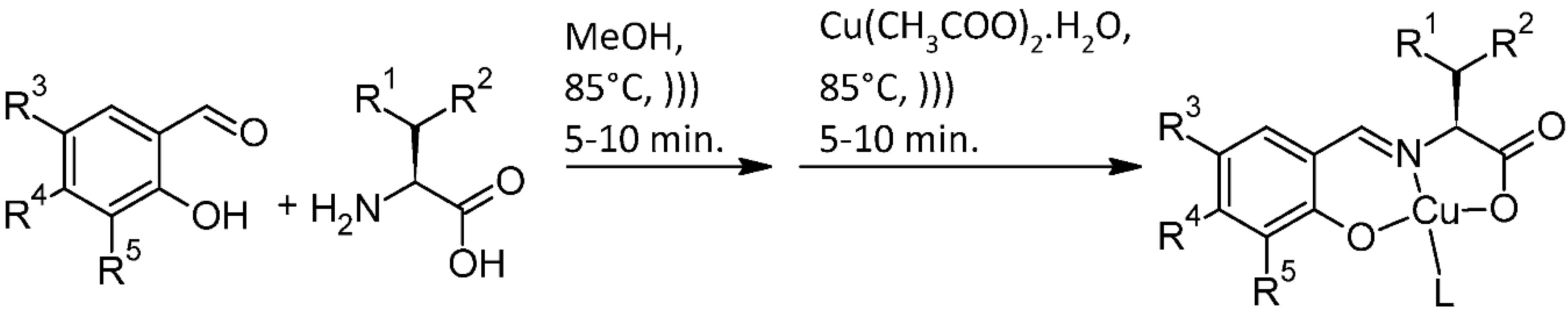
2.1. Compound Syntheses

Known compounds C1.1–C8.1 were prepared in the previous paper and used as described in [17]. New compounds entries C1.2–C12.2 were prepared and characterized as follows.
Cu(CH3COO)2·H2O, L-alanine, L-leucine, L-serine and L-threonine used in this study were purchased from Wako Fujifilm (Osaka, Japan), and salicylaldehyde, 3-chlorosalicylaldehyde, 4-chlorosalicylaldehyde and 5-chlorosalicylaldehyde were purchased from TCI (Tokyo, Japan). The synthesis of the compounds was conducted at 358 K using an Initiator+ microwave apparatus (Biotage, Tokyo, Japan).

2.1.1. General Procedures

To a methanol solution (20 mL) of L-amino acid (0.2 mmol) was added the proper chlorosalicylaldehyde (0.2 mmol), and the mixture was stirred under microwave irradiation at 358 K for 10 min. Then, a methanol solution (20 mL) of Cu(CH3COO)2·H2O (0.0399 g, 0.2 mmol) was added and the mixture was stirred under microwave irradiation for 10 min at 358 K to produce the final product. For additional spectra, see Supplementary Materials.

2.1.2. Physical Measurements

Elemental analyses were conducted with a Perkin-Elmer 2400II CHNS/O analyzer, (Waltham, MA, USA) at the Tokyo University of Science. Infrared (IR) spectra were recorded on a JASCO FT-IR 4200 spectrophotometer (JASCO, Tokyo, Japan) in the range of 4000–400 cm−1 at 298 K. Absorption electronic (UV-Vis) spectra were measured on a JASCO V-570 spectrophotometer in the range of 800−250 nm at 298 K.

2.1.3. Preparation of C1.2

Using the general procedure with L-leucine (0.02623 g, 0.2 mmol) and 3-chlorosalicylaldehyde (0.03131 g, 0.2 mmol) produced a green compound (yield 0.06985 g, 54.73%). The resulting crude compound was filtered and the precipitate was washed with diethyl ether and ethanol, and dried in a desiccator for several days. This product was then filtered. Anal. Calcd. for C13H16ClCuNO4: C, 44.71; H, 4.62; N, 4.01%; found: C, 45.69; H, 3.5; N, 3.84%.

2.1.4. Preparation of C2.2

Using the general procedure with L-leucine (0.02623 g, 0.2 mmol) and 4-chlorosalicylaldehyde (0.03131 g, 0.2 mmol) produced a green compound (yield 0.06985 g, 32.85%). The resulting crude compound was filtered and the precipitate was washed with diethyl ether, acetone and ethanol, then dried in a desiccator for several days. This product was filtered. Anal. Calcd. for C13H16ClCuNO4: C, 44.71; H, 4.62; N, 4.01%; found: C, 43.39; H, 2.51; N, 1.20%.

2.1.5. Preparation of C3.2

Using the general procedure with L-leucine (0.02623 g, 0.2 mmol) and 5-chlorosalicylaldehyde (0.03131 g, 0.2 mmol) produced a green compound (yield 0.03918 g, 56.09%). The resulting crude compound was filtered and the precipitate was washed with diethyl ether, acetone and ethanol, then dried in a desiccator for several days. This product was filtered. Anal. Calcd. for C13H16ClCuNO4: C, 44.71; H, 4.62; N, 4.01%; found: C, 45.1; H, 3.71; N, 3.82%.

2.1.6. Preparation of C4.2

Using the general procedure with L-alanine (0.01782 g, 0.2 mmol) and 3-chlorosalicylaldehyde (0.03131 g, 0.2 mmol) produced a green compound (yield 0.40000 g, 65.11%). The resulting crude compound was filtered and the precipitate was washed with diethyl ether, acetone and ethanol, then dried in a desiccator for several days. This product was filtered. Anal. Calcd. for C10H10ClCuNO4: C, 39.10; H, 3.28; N, 4.56%; found: C, 40.78; H, 2.45; N, 4.43%.

2.1.7. Preparation of C5.2

Using the general procedure with L-alanine (0.01782 g, 0.2 mmol) and 4-chlorosalicylaldehyde (0.03131 g, 0.2 mmol) produced a green compound (yield 0.04146 g, 67.48%). The resulting crude compound was filtered and the precipitate was washed with diethyl ether, acetone and ethanol, then dried in a desiccator for several days. This product was filtered. Anal. Calcd. for C10H10ClCuNO4: C, 39.10; H, 3.28; N, 4.56%; found: C, 41.39; H, 2.32; N, 4.54%.

2.1.8. Preparation of C6.2

To a methanol solution (20 mL) of alanine (0.01782 g, 0.2 mmol) was added 4-chlorosalicylaldehyde (0.03131 g, 0.2 mmol) and stirred at 358 K for 10 min to obtain a pale yellow color. Then, a methanol solution (20 mL) of Cu(CH3COO)2·H2O (0.03990 g, 0.2 mmol) was added, and the mixture was stirred for 10 min at 358 K to produce a green compound (yield 0.01060 g, 17.25%). The resulting crude compound was filtered and the precipitate was washed with diethyl ether, acetone and ethanol, then dried in a desiccator for several days. This product was filtered. Anal. Calcd. for C10H10ClCuNO4: C, 39.10; H, 3.28; N, 4.56%; found: C, 36.6; H, 3.13; N, 4.08%.

2.1.9. Preparation of C7.2

To a methanol solution (20 mL) of serine (0.02102 g, 0.2 mmol) was added 3-chlorosalicylaldehyde (0.03131 g, 0.2 mmol) and stirred at 358 K for 10 min to obtain a pale yellow color. Then, a methanol solution (20 mL) of Cu(CH3COO)2·H2O (0.0399 g, 0.2 mmol) was added, and the mixture was stirred for 10 min at 358 K to produce a green compound (yield 0.03737 g, 58.04%). The resulting crude compound was filtered and the precipitate was washed with diethyl ether, acetone and ethanol, then dried in a desiccator for several days. This product was filtered. Anal. Calcd. for C10H10ClCuNO5: C, 37.16; H, 3.12; N, 4.33%; found: C, 33.17; H, 3.27; N, 7.17%.

2.1.10. Preparation of C8.2

Using the general procedure with L-serine (0.02102 g, 0.2 mmol) and 4-chlorosalicylaldehyde (0.03131 g, 0.2 mmol) produced a green compound (yield 0.04404 g, 68.40%). The resulting crude compound was filtered and the precipitate was washed with diethyl ether, acetone and ethanol, then dried in a desiccator for several days. This product was filtered. Anal. Calcd. for C10H10ClCuNO5: C, 37.16; H, 3.12; N, 4.33%; found: C, 35.31; H, 2.88; N, 4.95%.

2.1.11. Preparation of C9.2

Using the general procedure with L-serine (0.02102 g, 0.2 mmol) and 5-chlorosalicylaldehyde (0.03131 g, 0.2 mmol) produced a green compound (yield 0.04678 g, 72.65%). The resulting crude compound was filtered and the precipitate was washed with diethyl ether, acetone and ethanol, then dried in a desiccator for several days. This product was filtered. Anal. Calcd. for C10H10ClCuNO5: C, 37.16; H, 3.12; N, 4.33%; found: C, 34.34; H, 2.42; N, 3.17%.

2.1.12. Preparation of C10.2

Using the general procedure with L-threonine (0.02380 g, 0.2 mmol) and 4-chlorosalicylaldehyde (0.03131 g, 0.2 mmol) produced a green compound (yield 0.03446 g, 51.09%). The resulting crude compound was filtered and the precipitate was washed with diethyl ether, acetone and ethanol, then dried in a desiccator for several days. This product was filtered. Anal. Calcd. for C11H12ClCuNO5: C, 39.18; H, 3.59; N, 4.15%; found: C, 34.34; H, 3.28; N, 6.37%.

2.1.13. Preparation of C11.2

Using the general procedure with L-threonine (0.02382 g, 0.2 mmol) and 4-chlorosalicylaldehyde (0.03135 g, 0.2 mmol) produced a green compound (yield 0.02844 g, 42.17%). The resulting crude compound was filtered and the precipitate was washed with diethyl ether, acetone and ethanol, then dried in a desiccator for several days. This product was filtered. Anal. Calcd. for C11H12ClCuNO5: C, 39.18; H, 3.59; N, 4.15%; found: C, 44.48; H, 1.62; N, 6.71%.

2.1.14. Preparation of C12.2

Using the general procedure, L-threonine (0.02382 g, 0.2 mmol) and 4-chlorosalicylaldehyde (0.03135 g, 0.2 mmol) produced a green compound (yield 0.13912 g, 20.63%). The resulting crude compound was filtered and the precipitate was washed with diethyl ether, acetone and ethanol, then dried in a desiccator for several days. This product was filtered. Anal. Calcd. for C11H12ClCuNO5: C, 39.18; H, 3.59; N, 4.15%; found: C, 44.62; H, 1.53; N, 14.03%.

2.2. Antimicrobial Assays

The bacterial strains used were E. coli ATCC 25922TM (American Type Culture Collection, Manassas, VA, USA), M. luteus CRBIP 107660 (Institut Pasteur, Lille, France), S. saprophyticus (isolated by TIMR Laboratory) and B. subtilis ATCC 6051 (American Type Culture Collection, Manassas, VA, USA). They were grown on trypto-casein soy solid medium (TSA, Conda, Madrid, Spain) and incubated at 30 °C in the dark (SANYO, incubator, MIR-253) for 24 h. The solid cultures were then stored in a cold room at 4 °C or used to inoculate 5 mL of liquid mineral medium 24 h before the microplate test (glucose 10 g/L, KCl 0.250 g/L, NaH2PO4 6.464 g/L, Na2HPO4.2H2O 10.408 g/L, MgSO4 0.244g/L, NO3NH4 1 g/L, MgCl2 0.05 g/L; pH = 7). A suspension of bacterial inoculum with an optical density (OD) between 0.1 and 0.2 (Ultrospec10 Amersham Bioscience) was prepared from a pre-culture for 24 h. The tests were performed on 96-well microplates (Thermo Scientific, Nunc™ Edge) in triplicate. The tested products were dissolved in DMSO as 100-times-concentrated stock solutions. Blanks were composed of non-inoculated mineral medium with or without the tested compounds to withdraw their eventual variation of absorbance along with time; positive controls were conducted with inoculated mineral medium without the compound and tests were inoculated media with the tested compounds. Growth monitoring was performed with a spectrophotometer Thermo Fisher Scientific Multiskan GO (type 1510). It was set to perform OD measurements at 600 nm of each well every 15 min for 24 h to follow the growth kinetics.
The percentage of inhibition was calculated as follows:
% I n h i b i t i o n = ( 1 O D v a r   g r o w t h O D v a r   c o n t r o l ) 100
ODvar growth was the difference in OD between the highest and lowest points on the growth curve in the tests. ODvar control was the difference in OD between the highest and lowest points on the positive control curve.
The MIC95 value was considered to be the lowest tested concentration value at which at least 95% inhibition was achieved.

2.3. Antioxidant Assays

The tests were performed on 96-well microplates (Thermo Scientific, Nunc™ Edge) in triplicate. The tested products were dissolved in DMSO. Oxidation monitoring was performed with a spectrophotometer Thermo Fisher Scientific Multiskan GO (Type 1510). It was set to perform OD measurements at 450 nm of each well every 1 min for 6 h to follow the oxidation kinetics (2 µL of sodium persulfate at 5 mg/mL; 2 µL of the tested compound of 50 µL ABTS at 0.08%) in a citrate-phosphate buffer (0.1 M) for a total volume of 200 µL. The maximum oxidation curve for the positive control reached the absorbance of 0.7. The percentage of inhibition was calculated as follows:
% I n h i b i t i o n = ( 1 ( O D v a r   t e s t ) / ( O D v a r   c o n t r o l ) ) 100
ODvar test was the difference in OD between the highest and the lowest points on the test curve. ODvar control was the difference in OD between the highest and lowest points on the control curve.

2.4. Interaction with E. coli

The tested compounds in DMSO (200 µg/mL as the final concentration) were mixed in culture media with E. coli. Their UV-Vis spectra were recorded after filtration on a 0.2 µm PTFE syringe filter at different times of incubation at 30 °C for up to 24 h. The values were recorded depending on the complex λmax values (384 nm for C4.1, 372 nm for C6.1, 369 nm for C2.2, 357 nm for C8.2, 366 nm for C9.2 and 378 nm for C11.2) and compared to the solutions without culture media.

2.5. Log Po/w Calculation

The theoretical Log Po/w were calculated thanks to SwissADME from the Swiss Institute of Bioinformatics under the CC-BY 4.0 Creative Commons 4.0 International License.

2.6. Statistical Analysis

Data are expressed as means ± standard deviation, and the statistical significance (p  <  0.05) was determined by one-way ANOVA with Tukey’s post hoc analysis.

3. Results and Discussions

3.1. Chemistry

For the preparation of L-amino acid derivative Schiff base Cu(II) complexes, two-step reactions were employed, namely, (1) imine condensation of primary amine (L-amino acid) and aldehyde, and (2) coordination of the Cu(II) ion from an acetate source (Scheme 1). In previous work, microwave syntheses, conventional heating and mechanochemistry for the corresponding L-amino acid derivative Schiff base Cu (II) complexes (series 1) [17] were compared, and this compound library was enriched with various mono-chloride compounds (series 2) to better understand the structure–activity link between the amino acid functional group/number and place of the chlorine moiety, as well as the antibacterial activity.
From the previous work (series 1), it is noteworthy that a two-step 5 + 5 (min) microwave synthesis at 85 °C is really efficient for the reaction between valine or threonine and salicylaldehyde (entries 1 and 7, 86% yield), while dichloro salicylaldehyde with threonine or alanine produced lower results (entries 6 and 8, 75% and 78% yield). The new series was then synthesized thanks to the screening antimicrobial results (see 3.2 Antibacterial Effect), and L-leucine (non-polar amino acid, entries 9–11) showed that the chlorine position in R3 or R5 produced a yield of 55% and 56%; whereas, with R4 = Cl, only a 33% yield was obtained. With L-alanine (non-polar amino acid, entries 12–14), the best result was obtained for R4 = Cl (67% yield); whereas, for L-threonine (non-charged polar amino acid, entries 18–20), the best result was obtained with R5 = Cl (51% yield, respectively) lowered to R3 = Cl or both amino acids (17% and 21% yields, respectively). As for the L-serine derivatives (non-charged polar amino acid entries 15–17), the best result was obtained with R3 = Cl (73% yield) and the lower yield was obtained for R5 = Cl (58%). All these results show that there is clearly an impact from the chlorine position and the amino acid functional group on the yield obtained, not really correlated to their polarity or their charge.

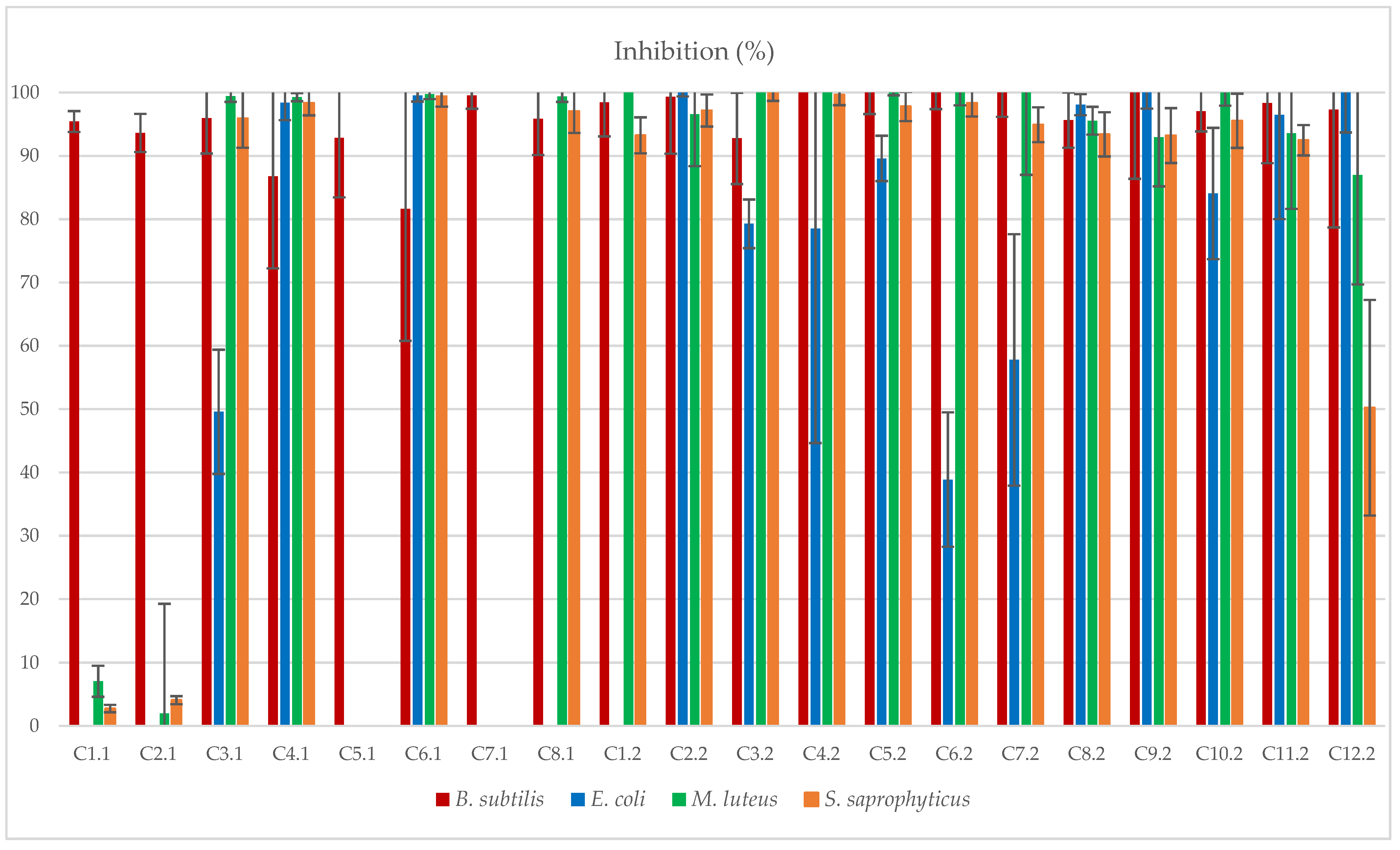
3.2. Antibacterial Effect

The synthesized 20 compounds were tested at a concentration of 50 µg/mL against four bacteria Bacillus subtilis (Gram (+), rod-shaped), Staphylococcus saprophyticus (Gram (+), coccus), Micrococcus luteus (Gram (+), coccus) and Escherichia coli (Gram (−), rod-shaped). These bacterial strains were chosen in order to have a good representativity of bacterial morphologies, cell grouping and cell wall structures, which are important factors influencing resistance to antibacterial molecules. Moreover, the species studied are described as human pathogens regarding E. coli [18] and S. saprophyticus [19], or in food poisoning with B. subtilis [20] and M. luteus [21]. The first group (Table 1, Cn.1, entries 1–8) was chosen for its various functions led by the amino acids (R1 and R2), as well as the presence of 2 or no chlorides (R3 and R5). Then, the second group (Table 1, Cn.2, entries 9–20) was chosen according to the best antibacterial results obtained with group 1, and the mono-chlorinated positions varied (R3, R4 and R5). It was clear from the antibacterial tests of the first group that compounds C3.1, C4.1 and C6.1 (entries 3, 4 and 6) had the best antibacterial effect (Figure 1). Indeed, C3.1 was active against all the Gram (+) bacteria, while C4.1 and C6.1 were active against both Gram (+) and Gram (−) bacteria. The last compound, C8.1, was found to be active against all the Gram (+) bacteria, but totally inactive against E. coli. The common factor of these three compounds compared to the others (C1.1, C2.1, C5.1 and C7.1) was the presence of the bis-chlorine atoms. Moreover, focusing on the Gram (−) bacteria, C4.1 and C6.1, which were active against this strain, were exempted from oxygen moiety afforded by the amino acid part, whereas both C3.1 and C8.1 possessed a hydroxyl group. Lastly, between the two hydroxylated compounds, the methyl moiety seemed to have a deleterious effect on the activity compared to the H atom. Based on these first excellent results by C3.1, C4.1, C6.1 and C8.1, the second group (Cn.2) was synthesized, but with moderating the place of the mono-chlorine atom (R3, R4 and R5) and then tested against the four same bacteria. First, it was really interesting to observe that C6.1 mono-chlorinated isosteres (C4.2, C5.2, C6.2) were less interesting than the bis-chlorinated compound. Then, comparing C1.2, C2.2 and C3.2 led to the observation that the most active compounds against all the strains possessed chlorine in the R4 position. In a similar manner, comparing C7.2, C8.2 and C9.2 found that C8.2 and C9.2 were the most active compounds and, most importantly, even better than their mono-chlorinated isostere C3.1. As expected, C10.2, C11.2 and C12.2 compounds were less active as their isostere C8.1, even though the mono-chlorinated compound C11.2 was the best of them. According to this antibacterial screening result, MIC (minimum inhibition concentration) values were tested and produced the following results.

3.3. MIC95 and MIC50 Values

MIC95 values are the minimal concentration where the inhibition reaches at least 95% and MIC50 values are minimal concentration where the inhibition reaches at least 50%. The results for the tested concentrations are 50, 25, 12.5, 6.25, 3.12, 1.56 and 0.78 µg/mL. From Table 2, it can be observed that, for the bacteria S. saprophyticus, the bis-chlorinated molecules exhibited the lowest MIC value. This was totally inverted as for one of the other Gram (+) strains, B. subtilis, since the mono-chlorinated compounds possessed the lowest MIC values, maybe due to the difference of cell morphology between the two bacteria. Surprisingly, for E. coli, which is a Gram (−) bacteria, the MIC values were the lowest, whatever the molecules, with the best values for C6.1, C2.2 and C11.2. Indeed Gram (−) bacteria are generally described as more resistant against antibacterial molecules due to their more selective cell wall and other more specific mechanisms of resistance [22]. By comparison, between all the tested molecules, on S. saprophyticus, E. coli and M. luteus, the compounds C8.2 and C9.2 exhibited the lowest activity. Moreover, C2.2 and C11.2 were more active against the rod-shaped molecules than the coccus.

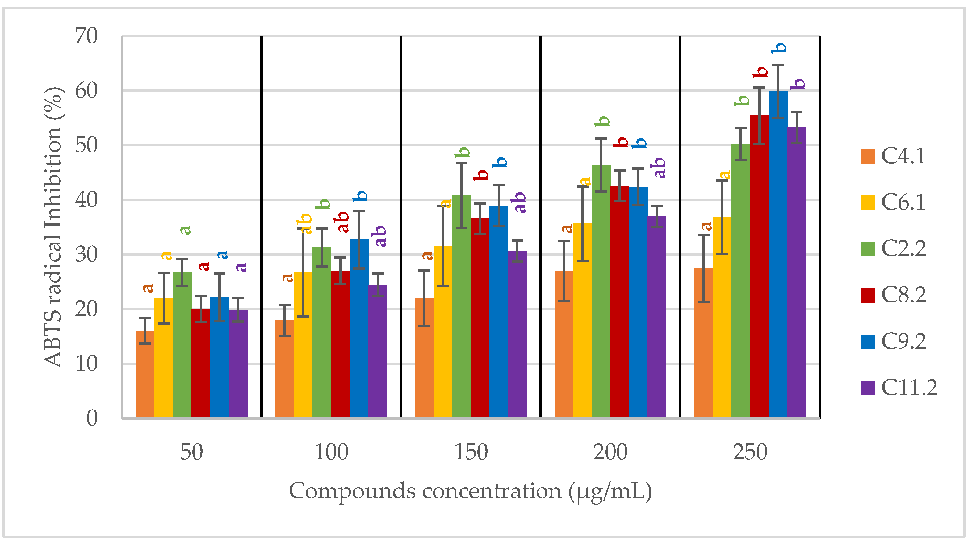
3.4. Antioxidant Activity

ROS (reactive oxygen species) were suspected to be the cause of the bacterial inhibition, so oxidation tests were performed with ABTS (2,2′-azinobis-3-ethylbenzothiazoline-6-sulfonic acid) and copper compounds (C4.1, C6.1, C2.2, C8.2, C9.2 and C11.2), and compared to sodium persulfate, in the dark or under a white lamp (60 watt). The result produced absolutely no oxidized ABTS species, And, as mentioned in the literature, copper Schiff-based compounds can exhibit antioxidant activity [23,24,25]. The antioxidant activity of the best antibacterial compounds C4.1, C6.1, C2.2, C8.2, C9.2 and C11.2 was kinetically tested against the oxidation of ABTS to the ABTS+ radical [26] by sodium persulfate (Figure 2).
Two groups of antiradical compounds were identified, as it was clear that the first group (C4.1 and C6.1) did not reach a 50% inhibition with a plateau of antioxidant capability from 200–250 µg/mL and over. However, the second group of compounds (C2.2, C8.2, C9.2 and C11.2) was able to reach 50% inhibition at a 250 µg/mL concentration and in the same statistical group. The conclusion may be that the mono-chlorinated compounds of these series of Schiff bases were more active against the radical oxidation of ABTS than the bis-chlorinated ones. IC50 was thus calculated from this second group of compounds and compared to the standard L-ascorbic acid (AA) (Table 3).

3.5. Interaction of Copper Complexes with E. coli

Copper ions and complexes are known to be biologically active due to the redox processes involved [27]. However, if E. coli is equipped with multiple systems to ensure safe copper handling under varying environmental conditions [28,29], in our case, this bacteria was the most sensitive to the copper Schiff base compounds. Therefore, the method of Joseph et al. [30] was adapted to evaluate the capability of the most active compounds to interact with E. coli. The absorption band of the complex in the culture media at their respective λmax wavelengths lowered after interacting with E. coli (Table 4).
This absorbance reduction after 24 h of incubation at 30 °C can be correlated to the compound activity. Indeed, the lowest absorbance reductions (2% and 12%) were observed for C9.2 and C8.2, respectively, and for these two compounds, the MIC95 values of 50 µg/mL were the highest. As for the other compounds, good absorbance reduction greater than 34% led to lower MIC95 from 6.25 to 25 µg/mL. In this group of active compounds, it is important to notice that, even if C2.2 was less absorbed than C11.2 or C4.1 by E. coli, its better activity might be attributed to its functional groups and chlorine placement. Additionally, the theoretical LogPo/w is interesting while comparing the uncomplexed ligands. Indeed, while this Log Po/w for L4.1 led to C4.1 when complexed copper was high (3.31), the values for L8.2 and L9.2 were low (1.07 and 1.06, respectively). This could also explain why if C4.1 is highly absorbed by E. coli, its activity is not as good as the activities of C2.2, C6.1 and C11.2 (Log Po/w for L2.2 = 2.69, for L6.1 = 2.37 and for L11.2 = 1.39).

4. Conclusions

Amino acid Schiff base Cu(II) complexes were synthesized under microwave irradiation using methanol as a solvent, to maximize the best conditions to obtain the obtained compounds. First, it was found that some compounds possessing bis-chlorinated moieties had better antibacterial activity compared to non-chlorinated aromatic. The place and number of chlorine atoms on the salicylaldehyde was then investigated and, depending on the amino acid used for the formation of the Schiff base ligand, its LogPo/w mono-chlorinated or bis-chlorinated compounds were the most efficient against the tested bacteria. Finally, the light antioxidant effect and the sensitivity of E. coli towards the most active compounds led us to test their interaction capability to be absorbed by E. coli. Subsequently, we plan to verify the toxicity of the molecules to guide the application, depending on the results obtained for skin, kidney, liver cells.

Supplementary Materials

The following supporting information can be downloaded at: https://www.mdpi.com/article/10.3390/applmicrobiol2020032/s1: Figures S1–S6: IR spectra, Figures S7–S12: UV-Vis spectra, Figure S13: Tentative crystal structures; Table S1: Compounds.

Author Contributions

E.L. (PA, INV, MET, WOD); A.F. (MET, VAL, WRE); N.O. (INV), D.N. (VAL), T.A. (CO, PA, SUP, WOD). (CO) Conceptualization; (DC) Data curation; (FA) Formal Analysis; (FUA) Funding acquisition; (INV) Investigation; (MET) Methodology; (PA) Project administration; (RES) Resources; (SOF) Software; (SUP) Supervision; (VAL) Validation; (VIS) Visualization; (WOD) Writing—original draft; (WRE) Writing—review and editing. All authors have read and agreed to the published version of the manuscript.

Funding

This research was funded by TIMR UTC-ESCOM, and this work was supported by a Grant-in-Aid for Scientific Research (A) KAKENHI (20H00336).

Data Availability Statement

Not applicable.

Acknowledgments

The authors thank Yuta Mitani for his help with the X-ray crystallography.

Conflicts of Interest

There are no conflicts to declare.

References

  1. Arunadevi, A.; Raman, N. Biological Response of Schiff Base Metal Complexes Incorporating Amino Acids—A Short Review. J. Coord. Chem. 2020, 73, 2095–2116. [Google Scholar] [CrossRef]
  2. Cimerman, Z.; Galic, N.; Bosner, B. The Schiff Bases of Salicylaldehyde and Aminopyridines as Highly Sensitive Analytical Reagents. Anal. Chim. Acta 1997, 343, 145–153. [Google Scholar] [CrossRef]
  3. Al-Hussein, M.F.I.; Adam, M.S.S. Catalytic Evaluation of Copper (II) N-Salicylidene-Amino Acid Schiff Base in the Various Catalytic Processes. Appl. Organomet. Chem. 2020, 34, e5598. [Google Scholar] [CrossRef]
  4. Chakraborty, H.; Paul, N.; Rahman, M.L. Catalytic Activities of Schiff Base Aquocomplexes of Copper(II) towards Hydrolysis of Amino Acid Esters. Transit. Met. Chem. 1994, 19, 524–526. [Google Scholar] [CrossRef]
  5. Zehra, S.; Roisnel, T.; Arjmand, F. Enantiomeric Amino Acid Schiff Base Copper(II) Complexes as a New Class of RNA-Targeted Metallo-Intercalators: Single X-Ray Crystal Structural Details, Comparative in Vitro DNA/RNA Binding Profile, Cleavage, and Cytotoxicity. ACS Omega 2019, 4, 7691–7705. [Google Scholar] [CrossRef]
  6. Zuo, J.; Bi, C.; Fan, Y.; Buac, D.; Nardon, C.; Daniel, K.G.; Dou, Q.P. Cellular and Computational Studies of Proteasome Inhibition and Apoptosis Induction in Human Cancer Cells by Amino Acid Schiff Base–Copper Complexes. J. Inorg. Biochem. 2013, 118, 83–93. [Google Scholar] [CrossRef] [Green Version]
  7. Kumaravel, G.; Ponya Utthra, P.; Raman, N. Exploiting the Biological Efficacy of Benzimidazole Based Schiff Base Complexes with L-Histidine as a Co-Ligand: Combined Molecular Docking, DNA Interaction, Antimicrobial and Cytotoxic Studies. Bioorganic Chem. 2018, 77, 269–279. [Google Scholar] [CrossRef]
  8. Saikumari, N. Synthesis and Characterization of Amino Acid Schiff Base and Its Copper (II) Complex and Its Antimicrobial Studies. Mater. Today Proc. 2021, 47, 1777–1781. [Google Scholar] [CrossRef]
  9. Costamagna, J.; Vargas, J.; Latorre, R.; Alvarado, A.; Mena, G. Coordination Compounds of Copper, Nickel and Iron with Schiff Bases Derived from Hydroxynaphthaldehydes and Salicylaldehydes. Coord. Chem. Rev. 1992, 119, 67–88. [Google Scholar] [CrossRef]
  10. Nakao, Y.; Sakurai, K.; Nakahara, A. Copper (II) Chelates of Schiff Bases Derived from Salicylaldehyde and Various α-Amino Acids. Bull. Chem. Soc. Jpn. 1967, 40, 1536–1538. [Google Scholar] [CrossRef]
  11. Gedye, R.; Smith, F.; Westaway, K.; Ali, H.; Baldisera, L.; Laberge, L.; Rousell, J. The Use of Microwave Ovens for Rapid Organic Synthesis. Tetrahedron Lett. 1986, 27, 279–282. [Google Scholar] [CrossRef]
  12. Giguere, R.J.; Bray, T.L.; Duncan, S.M.; Majetich, G. Application of Commercial Microwave Ovens to Organic Synthesis. Tetrahedron Lett. 1986, 27, 4945–4948. [Google Scholar] [CrossRef]
  13. Gedye, R. Microwave-Enhanced Chemistry. Fundamentals, Sample Preparation and Applications Edited by H.M. Kingston (Duquesne University) and Stephen J. Haswell (University of Hull); American Chemical Society: Washington, DC, USA, 1997; p. 772. ISBN 0-8412-3375-6. [Google Scholar]
  14. Strauss, C.R. Microwave-Assisted Reactions in Organic Synthesis—Are There Any Nonthermal Microwave Effects? Response to the Highlight by N. Kuhnert. Angew. Chem. Int. Ed. 2002, 41, 3589–3591. [Google Scholar] [CrossRef]
  15. Gabriel, C.; Gabriel, S.; Grant, E.H.; Grant, E.H.; Halstead, B.S.J.; Mingos, D.M.P. Dielectric Parameters Relevant to Microwave Dielectric Heating. Chem. Soc. Rev. 1998, 27, 213–224. [Google Scholar] [CrossRef]
  16. Dallinger, D.; Kappe, C.O. Microwave-Assisted Synthesis in Water as Solvent. Chem. Rev. 2007, 107, 2563–2591. [Google Scholar] [CrossRef]
  17. Otani, N.; Furuya, T.; Katsuumi, N.; Haraguchi, T.; Akitsu, T. Synthesis of Amino Acid Derivative Schiff Base Copper(II) Complexes by Microwave and Wet Mechanochemical Methods. J. Indian Chem. Soc. 2021, 98, 100004. [Google Scholar] [CrossRef]
  18. Kaper, J.B.; Nataro, J.P.; Mobley, H.L.T. Pathogenic Escherichia Coli. Nat. Rev. Microbiol. 2004, 2, 123–140. [Google Scholar] [CrossRef]
  19. Kuroda, M.; Yamashita, A.; Hirakawa, H.; Kumano, M.; Morikawa, K.; Higashide, M.; Maruyama, A.; Inose, Y.; Matoba, K.; Toh, H.; et al. Whole Genome Sequence of Staphylococcus Saprophyticus Reveals the Pathogenesis of Uncomplicated Urinary Tract Infection. Proc. Natl. Acad. Sci. USA 2005, 102, 13272–13277. [Google Scholar] [CrossRef] [Green Version]
  20. Turnbull, P.C.B. Bacillus. In Medical Microbiology; Baron, S., Ed.; University of Texas Medical Branch at Galveston: Galveston, TX, USA, 1996; ISBN 978-0-9631172-1-2. [Google Scholar]
  21. Zhang, R.; Zhang, Y.; Zhang, T.; Xu, M.; Wang, H.; Zhang, S.; Zhang, T.; Zhou, W.; Shi, G. Establishing a MALDI-TOF-TOF-MS Method for Rapid Identification of Three Common Gram-Positive Bacteria (Bacillus Cereus, Listeria Monocytogenes, and Micrococcus Luteus) Associated with Foodborne Diseases. Food Sci. Technol 2022, 42. [Google Scholar] [CrossRef]
  22. Breijyeh, Z.; Jubeh, B.; Karaman, R. Resistance of Gram-Negative Bacteria to Current Antibacterial Agents and Approaches to Resolve It. Molecules 2020, 25, 1340. [Google Scholar] [CrossRef] [Green Version]
  23. Shah, S.S.; Shah, D.; Khan, I.; Ahmad, S.; Ali, U.; ur Rahman, A. Synthesis and Antioxidant Activities of Schiff Bases and Their Complexes: An Updated Review. Biointerf. Res. Appl. Chem. 2020, 10, 6936–6963. [Google Scholar]
  24. Ganji, N.; Rambabu, A.; Vamsikrishna, N.; Daravath, S. Copper (II) Complexes with Isoxazole Schiff Bases: Synthesis, Spectroscopic Investigation, DNA Binding and Nuclease Activities, Antioxidant and Antimicrobial Studies. J. Mol. Struct. 2018, 1173, 173–182. [Google Scholar] [CrossRef]
  25. Soberanes, Y.; López-Gastélum, K.-A.; Moreno-Urbalejo, J.; Salazar-Medina, A.J.; del Carmen Estrada-Montoya, M.; Sugich-Miranda, R.; Hernandez-Paredes, J.; Gonzalez-Córdova, A.F.; Vallejo-Cordoba, B.; Sotelo-Mundo, R.R.; et al. Tetrameric Copper(II) Metallocyclic Complex Bearing an Amino Acid Derived Schiff Base Ligand: Structure, Catalytic and Antioxidant Activities. Inorg. Chem. Commun. 2018, 94, 139–141. [Google Scholar] [CrossRef]
  26. Ilyasov, I.R.; Beloborodov, V.L.; Selivanova, I.A. Three ABTS•+ Radical Cation-Based Approaches for the Evaluation of Antioxidant Activity: Fast-and Slow-Reacting Antioxidant Behavior. Chem. Pap. 2018, 72, 1917–1925. [Google Scholar] [CrossRef]
  27. Mitra, D.; Kang, E.-T.; Neoh, K.G. Antimicrobial Copper-Based Materials and Coatings: Potential Multifaceted Biomedical Applications. ACS Appl. Mater. Interfaces 2020, 12, 21159–21182. [Google Scholar] [CrossRef]
  28. Rensing, C.; Grass, G. Escherichia Coli Mechanisms of Copper Homeostasis in a Changing Environment. FEMS Microbiol. Rev. 2003, 27, 197–213. [Google Scholar] [CrossRef] [Green Version]
  29. Vincent, M.; Duval, R.; Hartemann, P.; Engels-Deutsch, M. Contact Killing and Antimicrobial Properties of Copper. J. Appl. Microbiol. 2018, 124, 1032–1046. [Google Scholar] [CrossRef] [Green Version]
  30. Joseph, J.; Nagashri, K.; Rani, G.A.B. Synthesis, Characterization and Antimicrobial Activities of Copper Complexes Derived from 4-Aminoantipyrine Derivatives. J. Saudi Chem. Soc. 2013, 17, 285–294. [Google Scholar] [CrossRef] [Green Version]
Scheme 1. Typical reaction scheme in a solution for the L-amino acid derivative Schiff base Cu(II) complexes.
Applmicrobiol 02 00032 sch001
Figure 1. Inhibition rate of Cn.1 and Cn.2 against B. subtilis, E. coli, M. luteus and S. saprophyticus.
Applmicrobiol 02 00032 g001
Figure 2. ABTS radical inhibition by C4.1, C6.1, C8.2, C9.2 and C11.2 compounds. Bars with different letters indicate statistically significant differences among the groups.
Applmicrobiol 02 00032 g002
Table 1. Summary of the results for the obtained 2-step compounds.
EntrySeriesCompoundR 1R 2R 3R 4R 5Yield a
11C1.1CH3CH3HHH86% b
2C2.1OHHHHH65% b
3C3.1OHHClHCl49% b
4C4.1iPrHClHCl31% b
5C5.1HHHHH67% b
6C6.1HHClHCl75% b
7C7.1OHCH3HHH86% b
8C8.1OHCH3ClHCl78% b
92C1.2iPrHHHCl55%
10C2.2iPrHHClH33%
11C3.2iPrHClHH56%
12C4.2HHHHCl65%
13C5.2HHHClH67%
14C6.2HHClHH17%
15C7.2OHHHHCl58%
16C8.2OHHHClH68%
17C9.2OHHClHH73%
18C10.2OHCH3HHCl51%
19C11.2OHCH3HClH42%
20C12.2OHCH3ClHH21%
a Isolated yields; b from previous work [17].
Table 2. MIC95/MIC50 values (µg/mL).
CompoundsS. saprophyticusE. coliM. luteusB. subtilis
C4.125/2525/2550/12.5>50/50
C6.112.5/12.512.5/12.525/6.25>50/50
C2.250/256.25/6.2550/12.56.25/3.12
C8.250/5050/50>50/12.525/12.5
C9.250/5050/50>50/5025/12.5
C11.250/5012.5/12.5>50/5012.5/12.5
Streptomycin25/6.25---
Daptomycin--7.75/230/15
Polymyxin B-3.75/1--
Table 3. IC50 (mM) compared to standard L-ascorbic acid.
C2.2C8.2C9.2C11.2AA
IC50 (mM)0.720.770.770.740.14
Table 4. Absorbance reduction (%) of copper Schiff base compounds in solution after E. coli incubation.
C4.1C6.1C2.2C8.2C9.2C11.2
Absorbance reduction (%)60.334.334.411.92.382.3
Publisher’s Note: MDPI stays neutral with regard to jurisdictional claims in published maps and institutional affiliations.

