1. Introduction
Antipsychotic drugs are used primarily for the treatment of psychiatric disorders, including schizophrenia, bipolar disorder, and severe depression, and are traditionally classified into two main categories: first-generation antipsychotics (typical antipsychotics) and second-generation antipsychotics (atypical antipsychotics) [
1]. Haloperidol and chlorpromazine are examples of first-generation antipsychotics, which were the first class of antipsychotics to be launched in the 1950s. They mostly act as dopamine D2 and D3 receptor antagonists, which decrease psychotic symptoms, but they are associated with serious adverse effects such as extrapyramidal symptoms (EPS) [
2] and tardive dyskinesia [
3].
Second-generation antipsychotics, such as olanzapine, quetiapine, and risperidone, have a wider range of action interacting with a wide range of receptors, including serotonin (5-HT2A, 5-HT2C), histamine (H1), and adrenergic receptors in addition to blocking dopamine receptors. Compared to typical antipsychotics, atypical antipsychotics often have fewer side effects, and several have demonstrated efficacy in treating mood disorders in addition to psychotic symptoms. Still, their use is associated with significant metabolic disturbances including weight gain, dyslipidemia, and insulin resistance [
4].
Emerging evidence suggests that antipsychotic drugs may exert anticancer effects through multiple cellular processes involved in tumor growth and survival. Since the late 2010s, there has been growing scientific interest in repurposing antipsychotic medicines as anticancer agents, driven by accumulating preclinical and epidemiological evidence of their tumor-inhibitory effects [
5].
Dopamine D2 receptor (DRD2) antagonism represents one of the principal pharmacological mechanisms shared by many antipsychotic agents and provides an important biological rationale for their investigation in oncology. DRD2 has been reported to be expressed in several malignancies, where it has been implicated in regulating tumor cell proliferation, survival, angiogenesis, and therapeutic resistance. The receptor exists as two alternatively spliced isoforms, the long (D2L) and short (D2S) variants, which differ in their signaling properties and tissue distribution. In addition to classical Gi/o protein-mediated signaling, DRD2 can also signal through β-arrestin-dependent pathways that regulate downstream effectors including Akt and glycogen synthase kinase-3β (GSK-3β). Nevertheless, the anticancer activity of antipsychotic drugs extends beyond DRD2 antagonism and involves modulation of multiple oncogenic pathways, including PI3K/Akt/mTOR, STAT3, NF-κB, Wnt/β-catenin, and mitochondrial apoptotic signaling, thereby influencing cancer cell proliferation, survival, invasion, and therapeutic response [
6,
7,
8,
9,
10].
Beyond their impact on intracellular signaling, recent studies have shown that antipsychotics also influence the tumor microenvironment (TME), a critical player in cancer progression and therapy resistance. These drugs have been reported to inhibit angiogenesis, reprogram immune signaling, and suppress inflammatory pathways such as NF-κB, all of which are essential for tumor growth and metastatic potential [
11,
12].
As these drugs impact both cancer cells and the surrounding microenvironment, their potential to serve as adjuncts to conventional therapies, such as chemotherapy and radiotherapy, is becoming an area of significant interest. In addition, novel drug delivery systems, including nanoparticles, may further enhance tumor targeting while minimizing systemic toxicity. This review examines the clinical relevance of repurposing antipsychotic drugs for cancer treatment, assessing their therapeutic potential, safety, and limitations in oncology. By highlighting promising candidates and identifying gaps in translational research, it aims to guide future studies toward realistic and clinically viable applications.
The following sections summarize representative typical and atypical antipsychotic agents for which the strongest preclinical and emerging clinical evidence of anticancer activity is currently available and are not intended to provide an exhaustive review of all antipsychotic compounds investigated for oncological applications.
2. Literature Selection—Methods
This review examines the anticancer potential of repurposed typical and atypical antipsychotic drugs, focusing on preclinical evidence, combination strategies with chemotherapy and radiotherapy, and novel delivery systems such as nanoparticles. Key mechanisms of action and tumor microenvironment effects are highlighted, along with critical discussion of translational challenges and future research priorities, including dosing, toxicity, and clinical applicability.
A structured literature search was conducted to identify studies investigating the anticancer effects of antipsychotic drugs. The main search was performed in PubMed/MEDLINE, Scopus, and Google Scholar from January 2019 to December 2024.
To ensure a comprehensive review, important studies published before 2019 were also included when they provided key evidence on the anticancer effects of the antipsychotic drugs. These studies were identified through screening the reference lists of relevant articles.
The search strategy was guided by the Population–Concept–Context (PCC) framework. The population included cancer-related models and tumor types (e.g., glioblastoma, breast cancer, colorectal cancer, pancreatic cancer, melanoma, leukemia, and other malignancies). The concept focused on antipsychotic drugs and dopamine D2 receptor antagonists investigated for potential anticancer or tumor-modulating effects. The context included experimental, translational, and clinical oncology settings.
Search terms combined antipsychotic drug names (haloperidol, chlorpromazine, penfluridol, trifluoperazine, olanzapine, quetiapine, aripiprazole, risperidone, pimozide, sertindole, ziprasidone, and lurasidone) with oncology-related keywords such as “cancer”, “tumor”, “neoplasm”, “glioblastoma”, “breast cancer”, “colorectal cancer”, “pancreatic cancer”, “melanoma”, and “leukemia”, as well as mechanistic terms including “apoptosis”, “autophagy”, “cell cycle arrest”, “NF-κB”, “STAT3”, and “PI3K/Akt signaling”.
Studies were included if they: (1) investigated antipsychotic drugs in cancer-related experimental or clinical contexts; (2) reported relevant anticancer outcomes such as tumor growth, survival, metastasis, or molecular signaling effects; and (3) included preclinical (in vitro or in vivo), translational, or clinical evidence. Only peer-reviewed English-language articles were considered. Studies were excluded if they were unrelated to oncology, lacked relevant tumor-related outcomes, or were duplicate/non-peer-reviewed reports.
Data were extracted and organized according to antipsychotic drug class, cancer type, and proposed molecular mechanisms. Studies were further categorized based on evidence level, including in vitro studies, in vivo animal models, xenograft studies, and clinical or translational evidence. Due to heterogeneity in study design and outcomes, a narrative synthesis approach was used.
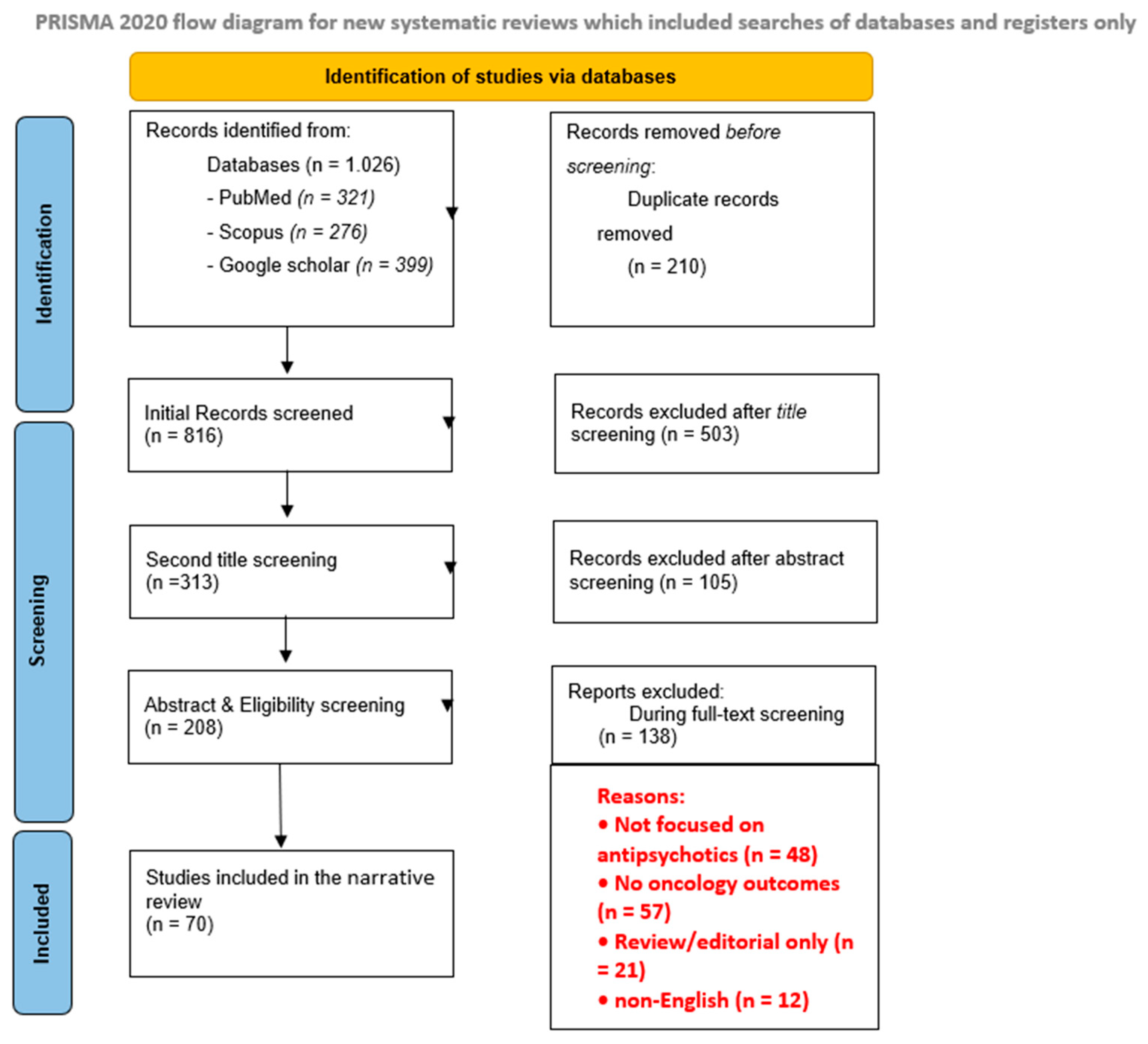
Study selection followed a structured screening process. Records identified through database searches were screened by title and abstract, followed by full-text assessment for eligibility. This structured approach was used to enhance transparency and reproducibility while maintaining the narrative nature of the review. The study selection process is summarized in the PRISMA 2020 flow diagram (
Figure 1).
2.1. Typical Antipsychotics
2.1.1. Haloperidol
Melanoma, Colon, and Breast Cancer
Haloperidol (HAL), a traditional antipsychotic, has been found to exhibit anticancer effects in solid cancers including melanoma, colon, and breast primarily by blocking cell division and promoting apoptosis in preclinical studies [
13,
14,
15]. These findings suggest broad cytotoxic potential beyond its neurological targets.
Pancreatic Cancer
In contrast to conventional cancer-suppressing mechanisms, HAL demonstrates a unique action in pancreatic cancer by inducing endoplasmic reticulum stress and apoptosis. This effect is achieved through the inhibition of DRD2’s coupling with the G protein subunit αi2, leading to increased cAMP and protein kinase A activity, both of which promote cancer cell death [
16,
17].
Glioblastoma
In glioblastoma cell models (in vitro), Liu et al. [
18] demonstrated that haloperidol synergizes with typical anti-glioma chemotherapy, temozolomide (TMZ), in glioblastoma cells (in vitro) by enhancing DNA damage, evidenced by γH2AX markers and by inhibiting autophagy. HAL’s inhibition of DR2 reduces ERK activation and enhanced TMZ’s cytotoxicity thus providing a novel therapeutic strategy for GMB treatment.
Further studies have revealed a molecular interplay between EGFR and DRD2 in glioblastoma cells, highlighting EGFR as one of the factors that affect GBM’S response to DRD2 inhibitors. This suggests that GBM may be treated with antipsychotic medications such as HAL, which function as DRD2 inhibitors, particularly in patients with elevated EGFR expression. Combining EGFR inhibitors with HAL could potentially amplify anticancer effects, possibly overcome resistance and improving tumor growth inhibition [
19].
These findings may have potential relevance for future clinical translation, especially for patients with TMZ-resistant glioblastoma or EGFR-overexpressing tumors, where haloperidol could serve as an adjunct to enhance treatment efficacy.
Moreover, HAL exhibits anti-glioblastoma activity by stabilizing PTEN, a key tumor suppressor protein, and disrupting lysosomal function. This interference hampers glioblastoma cells’ ability to recycle intracellular components necessary for survival, leading to tumor growth inhibition or apoptosis [
20]. In U87 and T98 glioblastoma cells haloperidol when combined with TMZ and radiation therapy increased cell death. Furthermore, haloperidol caused apoptosis in a dose-dependent manner, inhibited cell migration and altered the expression of CD24/CD44 [
21].
Breast Cancer
Lee et al. [
22] further support HAL’s efficacy in breast cancer. In MDA-MB-231 and MDA-MB-468 (in vitro) HAL inhibits IQGAP1 (IQ motif containing GTPase activating protein 1), a scaffolding protein involved in cancer-promoting Ras/ERK and PI3K/Akt pathways. By targeting IQGAP1, HAL disrupts key drivers of proliferation and survival in triple-negative breast cancer [
23].
Given the lack of targeted therapies in triple-negative breast cancer, these findings suggest HAL’s repurposing potential in this aggressive cancer subtype, warranting further translational research and clinical investigation.
Other tumor models (neuroblastoma and hepatocellular carcinoma).
In addition, haloperidol analogs have also shown cytotoxic effects in neuroblastoma (SH-SY5Y) and hepatocellular carcinoma (HUH-7) (in vitro) cells by targeting sigma receptors (SR) which regulate apoptosis, calcium signaling, and the cell cycle [
24]. Unlike HAL, which mainly targets SR1, these analogues target both SR1 and SR2, potentially offering a broader mechanism of anticancer activity and suggesting a new avenue for drug development [
25].
Taken together, these studies underscore haloperidol’s potential relevance as both a monotherapy and a sensitizer in combination regimens, particularly for difficult-to-treat cancers like glioblastoma and triple-negative breast cancer. However, most findings remain at the experimental stage, and further translational and clinical validation is required before therapeutic application.
2.1.2. Chlorpromazine
Chlorpromazine (CPZ) is another typical antipsychotic that has been found to induce cell cycle arrest, autophagy, and apoptosis while also disrupting cellular responses to stress in malignancies such as colorectal cancer and glioblastoma [
26]. Its ability to cross the blood-brain barrier makes it especially promising for treating brain tumors [
27].
Acute Myeloid Leukemia (AML)
In AML models, CPZ has demonstrated enhanced anticancer efficacy when formulated in PEGylated PLGA nanoparticles. This delivery system improves cellular uptake in AML cells and enables sustained drug release, with approximately 80% release over 24 h. Importantly, in vivo zebrafish larva models showed no brain accumulation of the nanoparticles, suggesting reduced central nervous system toxicity.
In the same experimental context, CPZ demonstrated synergistic effects with bortezomib and daunorubicin in MOLM-13 cells, as confirmed by Combination Drug Index (CDI) analysis [
28]. These findings suggest improved therapeutic potential when CPZ is used in combination regimens in preclinical AML models.
Encapsulating CPZ in nanoparticles not only improves its delivery to AML cells but also enhances its effectiveness when combined with chemotherapy. This strategy addresses the challenges of chemotherapy resistance while minimizing neurotoxic side effects.
Rai et al. further showed that CPZ targets AML cells carrying the KIT D816V mutation, a high-risk variant associated with poor prognosis. Mechanistically, CPZ disrupts KIT-D816V intracellular localization, leading to inhibition of Akt signaling and induction of apoptosis in mutated AML cells [
29].
Glioblastoma
In glioblastoma models, CPZ has been shown to enhance the cytotoxic effects of temozolomide (TMZ) by inhibiting multiple DNA repair pathways, including base excision repair (BER), nucleotide excision repair (NER), homologous recombination (HR), non-homologous end joining (NHEJ), and potentially mismatch repair (MMR). CPZ has also been linked to modulation of connexin 43 (Cx43), a protein associated with chemotherapy resistance [
30].
Clinically relevant translational evidence comes from the Phase II RACTAC trial, which demonstrated that CPZ improved TMZ efficacy in glioblastoma patients with an unmethylated MGMT promoter, a subgroup typically resistant to standard therapy [
31]. This supports potential clinical applicability, although further validation is required.
Breast Cancer
In breast cancer, CPZ has been shown to enhance the cytotoxic effects of 266 nm laser radiation by increasing ROS production and inducing cytoskeletal disruption, leading to increased tumor cell death [
32]. These findings suggest a potential role for CPZ in combination with physical or photodynamic therapeutic strategies.
Hepatocellular Carcinoma
CPZ can demonstrate direct anticancer properties such as inducing cellular stress and non-apoptotic cell death in hepatocellular carcinoma [
33].
Endometrial Cancer
Additionally, its derivative, JX57, inhibits endometrial cancer progression by activating AMP-activated protein kinase (AMPK) and disturbing calcium homeostasis, highlighting the therapeutic potential of structural analogues [
34].
Further translational studies in animal models are needed to confirm tumor response, toxicity, and dosing strategies for CPZ. The ongoing Phase II trial (NCT04224441) will provide important evidence on the safety and efficacy of CPZ combined with temozolomide in glioblastoma.
Given its established clinical use in psychiatry, CPZ remains a promising repurposing candidate in oncology, particularly in combination-based strategies. However, current evidence from preclinical and early translational studies requires further validation before clinical application, including careful assessment of toxicity profiles across neurological, hematological and cardiovascular systems.
2.1.3. Penfluridol
Penfluridol (PNF), a long-acting antipsychotic, has gained attention for its broad anticancer activity, particularly its anti-metastatic and radiosensitizing properties. It has been shown to suppress angiogenesis, thereby limiting tumor vascularization and metastatic spread [
35].
DNA Damage Repair/Radiosensitization
PNF inhibits the non-homologous end joining (NHEJ) DNA repair pathway, reducing the ability of cancer cells to repair double-strand DNA breaks. This increases cellular sensitivity to radiation, supporting its role as a potential radiosensitizer for enhancing radiotherapy efficacy [
36].
Triple-Negative Breast Cancer
In triple-negative breast cancer (TNBC), PNF has been shown to inhibit integrin and Akt signaling pathways, which are critical for tumor invasion and metastatic progression, particularly to the brain. Preclinical in situ metastatic mouse models demonstrated strong anti-metastatic effects, including approximately 90% reduction in brain metastases and 49% reduction in primary tumor growth [
37].
Gupta et al. further reported that PNF can overcome paclitaxel resistance in breast cancer by inhibiting the HER2/β-catenin signaling axis, which contributes to chemoresistance. In vivo models confirmed significant suppression of tumor growth and metastasis, supporting its potential use as an adjuvant to improve chemotherapy response [
38].
Glioblastoma
In glioblastoma models, PNF has been shown to enhance the efficacy of temozolomide (TMZ). It reverses epithelial–mesenchymal transition (EMT), suppresses integrin signaling, and reduces stemness markers such as CD133 and Nestin. In vivo studies demonstrated synergistic tumor suppression and improved survival when combined with TMZ, highlighting its potential in treatment-resistant glioblastoma [
39].
Other solid tumors (lung, renal, esophageal, ovarian, liver, and melanoma models;)
PNF also exhibits anticancer effects across multiple tumor types. In lung cancer [
40] and renal cell carcinoma [
41], it induces apoptosis and inhibits tumor cell growth. In esophageal cancer, PNF suppresses tumorigenesis by targeting PFKL, thereby inhibiting glycolysis in xenograft models [
42]. In epithelial ovarian cancer, PNF inhibits proliferation and migration via the MAPK pathway and shows synergy with paclitaxel in chemoresistant settings [
43]. In liver cancer, it disrupts KEAP1–NRF2 signaling, inhibiting MYC-driven ANLN expression and reducing tumor growth in both in vitro and syngeneic mouse models [
44]. In melanoma, PNF promotes degradation of CIP2A via the von Hippel–Lindau pathway, contributing to reduced tumor progression and metastasis [
45].
Overall, preclinical evidence indicates that penfluridol exerts broad anticancer effects across multiple tumor types, primarily through inhibition of metastasis, disruption of survival signaling pathways, and impairment of DNA repair mechanisms. Most findings remain at the in vitro and in vivo preclinical stage, with strong evidence of synergy with chemotherapy and radiotherapy. However, further optimization of dosing, toxicity profiling, and clinical validation are required before translational application.
PNF shares side effects with other treatments like paclitaxel, which is associated with neuropathy and hematological toxicity [
46,
47].
2.1.4. Trifluoperazine
Trifluoperazine (TFP), a well-known calmodulin inhibitor, has been studied for its ts anticancer and chemosensitizing properties across multiple tumor types. In various cancer types, such as triple-negative breast cancer (TNBC), lung cancer, and brain metastases, TFP causes apoptosis [
48,
49].
Urothelial Carcinoma
In urothelial carcinoma cells, TFP has been reported to overcome cisplatin resistance by suppressing the anti-apoptotic protein Bcl-xL. This enhances cisplatin-induced apoptosis, indicating a potential role for TFP in improving responses to conventional chemotherapy in resistant cancers [
50].
Osteosarcoma
Recent studies have shown that TFP induces mitophagy in osteosarcoma through activation of the AMPK/mTOR/ULK1 signaling pathway. This leads to the removal of damaged mitochondria, reduced oxidative stress, and inhibition of tumor cell proliferation, highlighting mitochondrial regulation as a key anticancer mechanism [
51].
Oral Cancer
In oral cancer models, TFP has been shown to induce ferroptosis by targeting the SLC7A11/GPX4 axis. This results in reactive oxygen species (ROS) accumulation and activation of autophagy-related cell death pathways [
52]. These findings suggest that TFP can trigger non-apoptotic cell death mechanisms, expanding its therapeutic potential in apoptosis-resistant cancers.
Lung Cancer
Nanoparticle-based delivery systems have been developed to enhance the anticancer efficacy of TFP. In lung cancer models, mesoporous silica nanoparticles (MSNs) loaded with TFP and modified with PEG and anisamide demonstrated targeted delivery and strong calmodulin inhibition, resulting in apoptosis induction and cell cycle arrest.
In vivo studies confirmed significant tumor suppression with reduced systemic toxicity. Additionally, lower brain tissue accumulation of TFP was observed using HPLC analysis, suggesting reduced central nervous system exposure and improved targeting efficiency [
53].
Overall, preclinical evidence indicates that trifluoperazine exerts multi-modal anticancer effects across diverse tumor types, including apoptosis induction, chemosensitization, metabolic disruption, and ferroptosis activation. Most findings remain at the in vitro and in vivo experimental stage, with emerging evidence supporting nanoparticle-based delivery strategies. However, further translational and clinical studies are required to establish safety, optimal dosing, and therapeutic efficacy in oncology.
2.1.5. Zuclopenthixol
Zuclopenthixol (ZPX), a typical antipsychotic, has been less extensively studied in oncology; however, emerging evidence suggests potential anticancer and anti-metastatic effects across several tumor types.
Glioblastoma
In glioblastoma models, ZPX has been shown to inhibit dopamine receptor signaling, reducing neuronal hyperexcitability associated with tumor progression. This may contribute not only to anticancer effects but also to modulation of tumor-associated neurological dysfunction [
55].
Melanoma
In melanoma models, ZPX has been reported to induce apoptosis and cell cycle arrest at the G1 phase, leading to reduced tumor cell proliferation. It has also been suggested to play a role in limiting metastatic spread, including potential prevention of brain metastases [
56].
Overall, available preclinical evidence suggests that zuclopenthixol may exert anticancer effects through HER2 modulation, dopaminergic signaling inhibition, and induction of cell cycle arrest and apoptosis. However, current findings remain limited and primarily preclinical, and further studies are required to clarify its mechanisms in non-HER2 tumors and to evaluate its safety, particularly regarding long-term central nervous system effects.
2.1.6. Pimozide
Pimozide (PMZ), a well-known antipsychotic, has demonstrated broad anticancer activity across multiple tumor types, including leukemia, breast cancer, non-small cell lung cancer, hepatocellular carcinoma, pancreatic cancer, and osteosarcoma. Its anticancer effects are primarily associated with disruption of oncogenic signaling pathways and induction of programmed cell death [
57].
Leukemia and STAT Signaling Inhibition (In Vitro Mechanistic Studies)
In leukemia models, PMZ has been shown to inhibit STAT5 signaling, while in other cancer types it suppresses STAT3 phosphorylation at Tyr705. This leads to reduced cell viability and impaired proliferative signaling, highlighting STAT pathway inhibition as a central mechanism of action [
57].
Breast Cancer
In breast cancer cells, PMZ has been reported to induce apoptosis and autophagy through activation of RAF/ERK signaling, with downstream caspase activation contributing to programmed cell death. These findings suggest a potential role for PMZ in enhancing the effectiveness of existing breast cancer therapies through modulation of cell death pathways [
58].
Liver Cancer and Osteosarcoma (In Vitro Mechanistic Effects)
In hepatocellular carcinoma, PMZ has been shown to reverse cancer stem-like properties, while in osteosarcoma it induces apoptosis and increases reactive oxygen species (ROS), contributing to suppression of tumor growth and aggressive phenotypes [
41].
Prostate Cancer (Drug Resistance and In Silico Evidence)
Hongo et al. identified PMZ as a potential agent against cabazitaxel-resistant castration-resistant prostate cancer in DU145CR and PC3CR cell lines. In silico screening suggested that PMZ may target resistance-associated genes such as AURKB and KIF20A, supporting its potential role as a chemosensitizing agent in resistant prostate cancer [
59].
Nanoparticle-Based Delivery Systems (In Vitro + Formulation Studies)
To improve delivery and therapeutic efficacy, nanostructured lipid carriers (NLCs) loaded with PMZ have been developed. These formulations demonstrated enhanced anticancer activity compared to free PMZ, with reduced IC
50 values and pH-sensitive drug release profiles that may improve tumor-targeted delivery while reducing systemic exposure [
60].
Overall, preclinical evidence indicates that pimozide exerts broad anticancer effects across multiple tumor types through inhibition of STAT signaling, induction of apoptosis and autophagy, and modulation of oxidative stress and cancer stem-like properties. Emerging nanocarrier-based delivery systems further enhance its therapeutic potential. However, most findings remain at the preclinical stage, and further in vivo and clinical studies are required to validate safety, efficacy, and translational applicability.
To provide an integrated overview of the findings discussed above, the anticancer properties of typical antipsychotics are summarized in
Table 1 and
Table 2.
Table 1 compares the available evidence, tumor types, molecular mechanisms, and key outcomes for each drug, whereas
Table 2 categorizes the shared anticancer mechanisms and their associated biological effects across this drug class.
2.2. Atypical Antipsychotics
Atypical antipsychotics are second-generation drugs that target a broader range of receptors, including dopamine, serotonin, and other neurotransmitter systems. Their anticancer properties are particularly intriguing because they act on serotonin and dopamine receptors simultaneously. The following section highlights atypical antipsychotic drugs investigated for their anti-cancer potential in both earlier and ongoing research.
2.2.1. Olanzapine
Olanzapine (OZP), an atypical antipsychotic, has demonstrated anticancer effects in several malignancies, including pancreatic, lung, breast, and glioblastoma cancers. One of its primary mechanisms involves suppression of NF-κB signaling through inhibition of p65 nuclear translocation, resulting in reduced cancer cell proliferation and induction of autophagy [
61].
Glioblastoma
In glioblastoma models, OZP has been shown to induce reactive oxygen species (ROS) production, a potent mediator of cancer cell death [
62]. Increased ROS levels enhanced the cytotoxic effects of temozolomide (TMZ), suggesting a synergistic interaction between OZP and standard glioblastoma therapy. These findings indicate that OZP may have potential as a chemosensitizing agent in glioblastoma treatment.
Breast Cancer
In breast cancer models, OZP has demonstrated radiosensitizing properties when combined with radiation therapy [
63]. This effect is associated with increased apoptosis through downregulation of the anti-apoptotic protein Bcl-2 and upregulation of the pro-apoptotic protein Bax. Additionally, OZP enhanced DNA damage, contributing to improved therapeutic efficacy.
Lung Cancer
More recently, Lu et al. [
64] demonstrated that OZP inhibits norepinephrine release and subsequently blocks the ADRB2-cAMP-PKA-CREB signaling pathway, which promotes cancer cell proliferation and survival. This inhibition altered cancer stem cell characteristics and sensitized lung cancer cells to gemcitabine under chronic stress conditions.
These findings suggest that OZP may help overcome chemotherapy resistance in lung cancer. Overall, preclinical evidence indicates that olanzapine exerts anticancer activity through multiple mechanisms, including NF-κB suppression, ROS generation, apoptosis induction, and modulation of stress-related signaling pathways [
61,
62,
63,
64]. In addition to its direct anticancer effects, OZP has shown potential to enhance responses to chemotherapy and radiotherapy. However, further in vivo and clinical studies are required to establish its translational value in oncology.
2.2.2. Quetiapine
Quetiapine (QTP), an atypical antipsychotic, has demonstrated anticancer activity in several malignancies, particularly hepatocellular carcinoma and glioma models. Its reported effects include induction of apoptosis, inhibition of invasion and angiogenesis, modulation of the tumor microenvironment, enhancement of radiotherapy efficacy, and reversal of multidrug resistance [
65,
66,
67,
68].
Hepatocellular Carcinoma
QTP has shown anticancer activity in hepatocellular carcinoma (HCC) models by inducing apoptosis, suppressing tumor invasion, and inhibiting angiogenesis [
65,
66,
67]. Mechanistically, QTP downregulates the PI3K/Akt/mTOR signaling pathway and reduces the expression of the pro-inflammatory cytokines IL-6 and TNF-α, potentially limiting the inflammatory microenvironment that supports tumor progression [
65]. These findings suggest that QTP may exert both direct antitumor effects and indirect effects through modulation of the tumor microenvironment.
Recent studies have further demonstrated that QTP can enhance the efficacy of radiotherapy in HCC models. Wang et al. reported that QTP improved radiosensitivity by targeting HIF-1α-mediated radioresistance while simultaneously amplifying radiation-induced oxidative stress. These findings support the potential use of QTP as a radiosensitizing agent in liver cancer treatment [
67].
Glioma
In glioma models, QTP has been shown to reduce the viability of glioma stem-like cells by inducing G2/M cell cycle arrest and promoting their differentiation into less aggressive oligodendrocyte-like cells [
66]. Given the role of cancer stem cells in tumor recurrence and treatment resistance, these findings highlight a potentially important therapeutic application of QTP in glioma management.
Multidrug-Resistant Cancers
Beyond its direct anticancer effects, QTP has demonstrated the ability to overcome multidrug resistance in ABCB1-overexpressing cancers through inhibition of P-glycoprotein activity [
68]. Since P-glycoprotein-mediated drug efflux is a major mechanism of chemotherapy resistance, QTP may enhance the effectiveness of conventional anticancer agents when used in combination therapies.
Despite these promising findings, several challenges remain for the clinical translation of QTP in oncology. Effective doses reported in preclinical studies may approach levels associated with QTc prolongation and metabolic adverse effects, particularly in patients with impaired hepatic function [
67]. Furthermore, tumor penetration and pharmacokinetic behavior in cancer patients remain insufficiently characterized. Future studies should focus on defining safe dosing strategies, evaluating tumor drug accumulation, and validating predictive biomarkers such as HIF-1α and ABCB1 to support clinical development [
67,
68].
Overall, preclinical evidence suggests that quetiapine exerts anticancer activity through multiple mechanisms, including apoptosis induction, inhibition of angiogenesis and invasion, modulation of inflammatory signaling, radiosensitization, and reversal of multidrug resistance [
65,
66,
67,
68]. While these findings support its potential repurposing in oncology, further translational and clinical studies are required to establish its safety, efficacy, and therapeutic applicability in cancer patients.
2.2.3. Aripiprazole
Aripiprazole (ARP) has demonstrated promising anticancer effects across several tumor types, mainly through modulation of apoptosis, autophagy, oxidative stress, and drug resistance pathways [
69,
70,
71,
72,
73,
74].
Breast Cancer
In breast cancer, particularly in the MCF-7 cell line, ARP induces apoptosis and causes cell cycle arrest, leading to reduced tumorigenic potential [
69]. However, some reports also indicate suppression of apoptosis depending on experimental context, suggesting that its effects may be dose- and pathway-dependent [
69].
In drug-resistant breast cancer models (MCF-7/ADR), low-dose ARP enhances the efficacy of vincristine by modulating P-glycoprotein (P-gp) expression and altering cellular stress response pathways. This results in increased apoptosis and reduced viability specifically in P-gp–overexpressing cells, while showing limited toxicity in drug-sensitive MDA-MB-231 cells, indicating a degree of tumor-selective action [
72].
Additionally, ARP has been incorporated into nanotechnology-based delivery systems. A niosome/chitosan–gold nanoparticle formulation loaded with ARP significantly enhances cytotoxicity in MCF-7 cells, with improved pH-sensitive release and drug-loading efficiency, suggesting improved bioavailability and targeted delivery potential [
73].
Head and Neck Cancer
In head and neck cancer, ARP increases reactive oxygen species (ROS) production, thereby enhancing tumor cell sensitivity to radiation therapy and promoting selective tumor cell death. This positions ARP as a potential radiosensitizing agent.
In vivo xenograft studies further support this effect, showing that ARP alone does not significantly inhibit tumor growth, but when combined with ionizing radiation (IR), it markedly enhances tumor suppression without causing significant weight loss or systemic toxicity in mice [
70]. This indicates a favorable safety profile for combination therapy.
Pancreatic Cancer
In pancreatic cancer, ARP enhances chemosensitivity to cisplatin by inducing both apoptosis and autophagy. Mechanistically, this effect is mediated through inhibition of STAT3 signaling, activation of caspase-3, and downregulation of anti-apoptotic proteins such as XIAP and MCL-1 [
71].
In vivo xenograft models confirm that ARP combined with cisplatin significantly reduces tumor growth without notable toxicity or weight loss, suggesting strong translational potential as a chemosensitizing agent [
71].
Colorectal Cancer
In colorectal cancer, ARP has been investigated primarily as a monotherapy targeting key survival pathways. It modulates mTOR signaling, the RNH1/miR-99a axis, and lysosome-associated membrane protein LAMP2a pathways, ultimately inducing autophagy-associated cell death in cancer cells [
74].
These findings suggest that ARP may act as a pathway-specific regulator of tumor survival mechanisms, supporting its potential as a standalone therapeutic candidate in colorectal cancer [
74].
Across cancer types, ARP consistently exerts anticancer effects through induction of apoptosis, autophagy modulation, ROS generation, and suppression of pro-survival signaling pathways such as STAT3 and mTOR. In addition, it demonstrates a notable ability to overcome chemoresistance via P-gp modulation and to enhance the efficacy of radiotherapy and chemotherapy.
Most evidence for ARP remains preclinical, with strong representation from in vitro and xenograft models. However, the consistency of its effects across multiple cancer types and treatment modalities (chemotherapy, radiotherapy, and nanodelivery systems) supports its potential as a multifunctional anticancer adjunct, though clinical validation is still lacking.
2.2.4. Sertindole
Sertindole (STR) has shown broad anticancer potential across multiple tumor types, mainly through regulation of apoptosis, cell cycle progression, STAT3 signaling, and autophagy-related pathways.
Breast Cancer
In breast cancer cell lines including MDA-MB-231, MDA-MB-468, and SKBR3, STR suppresses cell proliferation by inducing G1-phase cell cycle arrest and promoting apoptosis. These effects are associated with downregulation of the anti-apoptotic protein Bcl-2 and activation of caspase-dependent pathways [
75]. Collectively, these findings suggest that STR interferes with key survival mechanisms in breast cancer cells and may function as a potential therapeutic agent targeting tumor viability [
75].
Gastric Cancer
In gastric cancer models, STR inhibits the STAT3 signaling pathway and its downstream targets, including Bcl-2 and Cyclin D1. This inhibition leads to reduced cellular proliferation and increased apoptosis, indicating that disruption of STAT3-mediated survival signaling is a key mechanism of STR action in gastric cancer [
76].
Glioma
In glioma, STR suppresses tumor growth by inhibiting autophagic flux. This effect is associated with downregulation of key autophagy-related proteins, including Beclin-1 and LC3, resulting in reduced cell viability and enhanced apoptosis [
77]. These findings suggest that STR may exert anticancer effects in glioma by disrupting autophagy-dependent tumor survival mechanisms [
77].
Bladder Cancer
In bladder cancer, STR promotes apoptosis through inhibition of the STAT3/Bcl-XL signaling axis. This disruption of STAT3-dependent survival pathways leads to increased tumor cell death, further supporting the role of STR as a modulator of key oncogenic signaling networks [
78].
Melanoma/Uveal Melanoma
In melanoma, particularly tumors driven by GNAQ mutations, STR demonstrates selective anticancer activity. It shows potential therapeutic relevance in genetically defined uveal melanoma by targeting mutation-associated signaling dependencies, suggesting a possible role in personalized cancer therapy approaches [
79].
Across cancer types, STR consistently targets key oncogenic pathways including STAT3 signaling, apoptosis regulation (Bcl-2/Bcl-XL), cell cycle progression (Cyclin D1), and autophagy-related mechanisms (Beclin-1, LC3). These converging effects lead to reduced proliferation and enhanced apoptosis across multiple tumor models.
Current evidence for STR is primarily preclinical, with consistent in vitro findings across several cancer types and mechanistic pathways. However, further in vivo validation and translational studies are needed to confirm its therapeutic potential and clinical applicability.
2.2.5. Risperidone
Risperidone (RSD) has been investigated for its anticancer potential across multiple tumor types, with its effects mainly linked to oxidative stress induction, apoptosis activation, DNA damage, and inhibition of cell migration.
Gastric Cancer
In gastric cancer models, RSD induces reactive oxygen species (ROS) production, which leads to inhibition of cell proliferation and activation of apoptosis [
80]. These findings suggest that oxidative stress–mediated cytotoxicity may represent a key mechanism underlying its anticancer effects in gastric tumor cells [
80].
Colorectal Cancer
Similarly, in colorectal cancer, RSD increases ROS generation, resulting in suppressed proliferation and enhanced apoptotic cell death [
81]. This further supports the role of ROS-mediated signaling disruption as a shared mechanism of RSD activity across gastrointestinal malignancies [
81].
Breast Cancer
In breast cancer, particularly MCF-7 cells, RSD has been shown to induce apoptosis, inhibit cell migration, and slow tumor progression. These effects are associated with DNA damage induction, suggesting that RSD interferes with genomic stability and tumor cell survival pathways [
82].
Despite earlier reports suggesting a potential association between RSD and increased breast cancer risk, more recent evidence has begun to challenge this concern. This evolving perspective has renewed interest in its anticancer properties and supports further investigation into its therapeutic potential in oncology [
83].
Across cancer types, RSD primarily exerts anticancer effects through ROS generation, apoptosis induction, DNA damage, and inhibition of migration. These mechanisms converge on disruption of cellular redox balance and genomic integrity, ultimately leading to reduced tumor cell viability.
The current evidence for RSD remains largely preclinical and somewhat fragmented, with most findings derived from in vitro studies. However, consistent mechanistic patterns across gastric, colorectal, and breast cancer models suggest potential anticancer activity that warrants renewed and more systematic investigation.
Similarly,
Table 3 and
Table 4 summarize the available evidence and major anticancer mechanisms of atypical antipsychotics.
2.2.6. Emerging Atypical Antipsychotics
While the focus has traditionally been on widely studied atypical antipsychotics, emerging evidence suggests potential anticancer activity for agents like Lurasidone, Ziprasidone, and Asenapine. These drugs, although less studied, merit further investigation due to their unique receptor profiles and tolerability [
84,
85,
86].
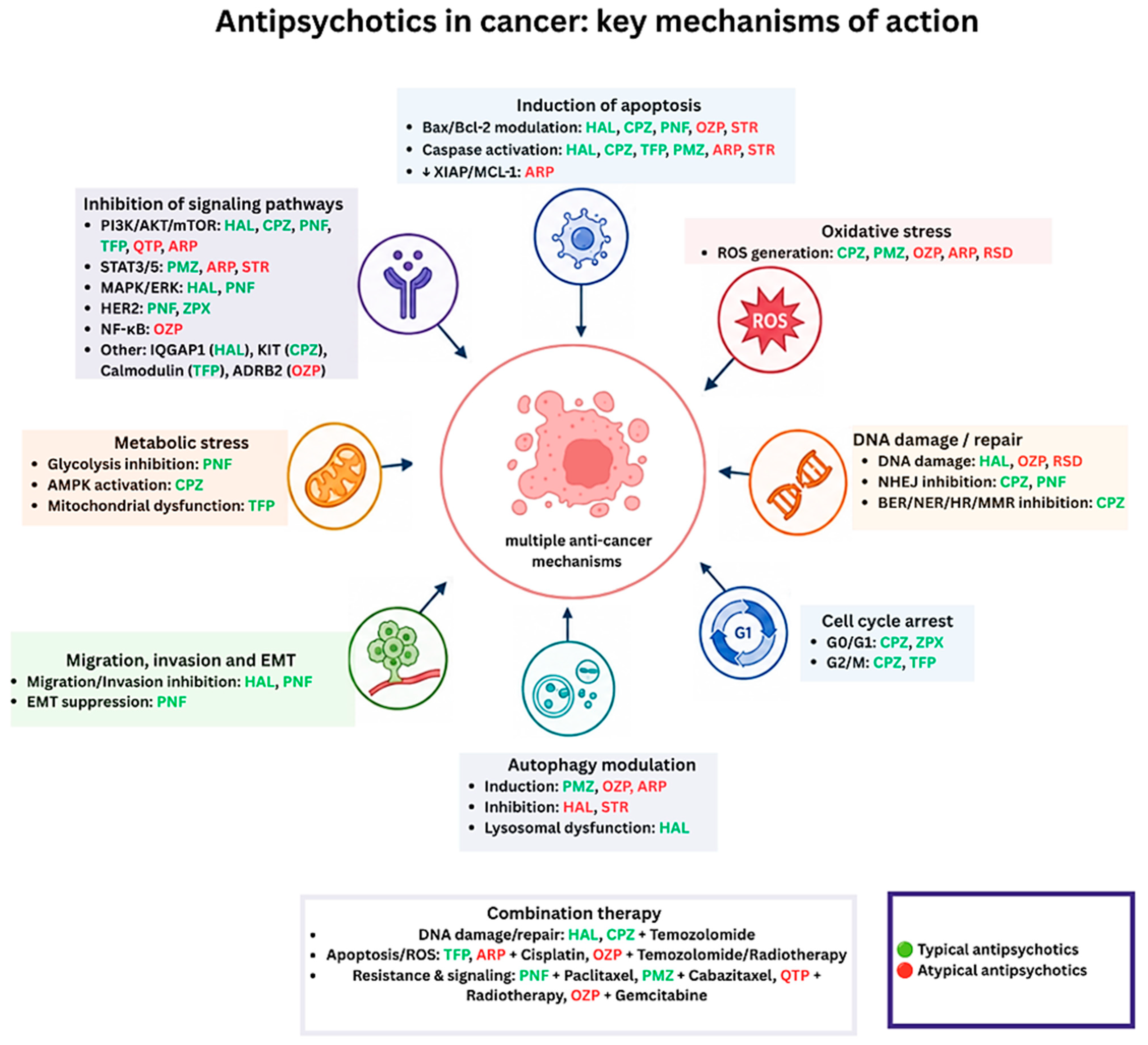
To provide a visual overview of the shared and distinct anticancer mechanisms of typical and atypical antipsychotics, the principal molecular pathways and biological processes targeted by these agents are summarized in
Figure 2.
To integrate the findings presented for both typical and atypical antipsychotics
Table 5,
Table 6 and
Table 7 provide a comparative overview of their molecular mechanisms, therapeutic profiles, combination strategies with conventional anticancer therapies, and emerging nanotechnology-based delivery systems.
3. Risks of Using Antipsychotic Drugs Against Cancer
While these preclinical findings on the anticancer effects of antipsychotic drugs are promising, clinical evidence remains limited. Future research should focus on translating these results into clinical settings while carefully evaluating the pharmacokinetic and toxicity profiles of these agents in cancer patients.
One significant concern is the use of prolactin-elevation antipsychotics, such haloperidol, and risperidone, which may raise the risk of breast cancer. These drugs can induce hyperprolactinemia, which activates the JAK-STAT signaling pathway, potentially promoting breast tissue cell survival and proliferation [
87]. In addition, vascular complications represent another potential risk. Antipsychotics including haloperidol, quetiapine, and olanzapine have been associated with an increased risk of pulmonary embolism, possibly through effects on endothelial function and platelet aggregation, which may contribute to increased mortality in cancer patients [
88].
Atypical antipsychotics such as olanzapine and clozapine may also cause metabolic disturbances, including obesity and insulin resistance. These conditions increase the risk of hormone-sensitive malignancies such breast and endometrial cancers [
89]. Clozapine, commonly prescribed drug for treatment- resistant schizophrenia, has been linked to an increased risk of both non-hematologic cancers and hematologic cancers, including leukemia and lymphoma although the clinical significance of these associations remains uncertain [
90,
91].
Beyond pharmacological risks, the incorporation of antipsychotic drugs in advanced delivery systems or combination therapies introduces additional considerations. Nanoparticle-based delivery systems, may improve tumor targeting and reduce systemic exposure but may also present challenges related to long-term toxicity, bioaccumulation, and immune responses [
92]. Likewise, combining antipsychotics with chemotherapy or radiotherapy may enhance anticancer efficacy while simultaneously increasing the risk of organ-specific toxicities, drug–drug interactions, and cumulative cardiotoxic or hepatotoxic effects [
93].
These findings highlight the necessity careful patient selection, individualized dosing strategies and close monitoring when considering antipsychotic drugs for oncology applications. Future studies should also investigate pharmacogenomic predictors of efficacy and toxicity to support safer and more personalized treatment approaches.
4. Conclusions
The repurposing of antipsychotic drugs in cancer treatment represents a promising area of oncology research. Evidence from preclinical studies demonstrates that both typical and atypical antipsychotics can inhibit tumor growth through multiple mechanisms, including apoptosis induction, autophagy modulation, ferroptosis, cell cycle arrest, and disruption of oncogenic signaling pathways. Several agents have also shown the ability to enhance the efficacy of chemotherapy and radiotherapy, highlighting their potential as adjunctive therapeutic strategies.
To minimize these risks, it is crucial to implement patient-tailored strategies. Patients who are likely to benefit from antipsychotic repurposing should be selected, while those with pre-existing risk factors, such as metabolic syndrome or hormone-sensitive malignancies, should be excluded. For example, atypical antipsychotics like olanzapine, known for inducing metabolic disturbances, should be avoided in patients with diabetes or obesity.
A promising strategy involves using lower, tailored doses of antipsychotics to preserve their anticancer efficacy while reducing off-target effects. For instance, low dose trifluoperazine has been shown to induce ferroptosis in oral cancer cells while minimizing adverse effects [
36]. Additionally, combination therapy with other cancer medications could prove essential; combining antipsychotics with chemotherapy or radiotherapy allows for the lowering of antipsychotic doses, thus reducing the risk of toxicity. Quetiapine, for example, has been shown to increase radiation efficacy in hepatocellular cancer, allowing for reduced chemotherapy doses [
12].
The development of innovative drug delivery systems, such as nanoparticle-based carriers (e.g., pimozide-loaded nanoparticles), holds significant promise for improving the specificity and reducing the systemic toxicity of antipsychotic drugs in cancer therapy [
43]. However, the potential risks associated with nanoparticles, including long-term toxicity, bioaccumulation, and unpredictable immune responses, need further investigation.
Despite these promising strategies, most of the research conducted to date is based on in vitro and in vivo experiments. As a result, further studies are essential to evaluate the pharmacokinetics, toxicity profiles, and clinical relevance of antipsychotic drugs in human cancer patients. This underscores the importance of careful patient monitoring—including regular imaging, hormone testing, and metabolic profiling—to identify and manage side effects early.
In conclusion, antipsychotic drugs represent promising candidates for drug repurposing in oncology, but further translational and clinical research is essential before their integration into routine cancer care.
5. Expert Opinion
Preliminary studies suggest that antipsychotic drugs possess meaningful anticancer potential across a wide range of malignancies. However, current data are not sufficiently robust to support their use as standalone anticancer therapies, as most findings originate from preclinical studies and the underlying mechanisms remain incompletely understood.
In our view, the greatest potential of antipsychotic drugs lies in their use as adjunctive agents capable of enhancing existing treatment modalities, overcoming therapeutic resistance, and targeting multiple cancer-related pathways simultaneously. Particularly encouraging results have been reported in glioblastoma, triple-negative breast cancer, pancreatic cancer, and other treatment-resistant malignancies.
The next phase of research should prioritize well-designed clinical trials aimed at evaluating efficacy, pharmacokinetics, safety, and optimal dosing strategies. Equally important will be the identification of predictive biomarkers and patient populations most likely to benefit from these therapies. Advances in targeted drug delivery systems and combination treatment approaches may further improve the therapeutic index of these agents.
Although substantial challenges remain, the repurposing of antipsychotic drugs represents a scientifically compelling strategy with the potential to expand the current oncology treatment landscape. Continued translational and clinical research will be critical to determining whether these promising preclinical findings can ultimately be translated into meaningful patient benefit.
Expert Opinion Summary
Antipsychotic drugs demonstrate promising anticancer activity in preclinical models and may be particularly valuable as adjuncts to existing cancer therapies. Future research should focus on clinical validation, biomarker-guided patient selection, dose optimization, and combination strategies to establish their role in oncology.




 ,
,