Next Article in Journal
Intercellular Mitochondrial Trafficking as a Master Regulator of Tumor Progression and Cancer Stem Cell Plasticity
Previous Article in Journal
Macrophage Activation Syndrome Following Atezolizumab in Advanced Non-Small-Cell Lung Cancer: A Case Report
 
 
Correction published on 22 June 2026, see Onco 2026, 6(2), 30.
Article

Development of a Murine Intracranial Surgical Resection Glioblastoma Model to Facilitate Preclinical In Vivo Drug Screening


1
Department of Neurosurgery, Hollings Cancer Center, Medical University of South Carolina, Charleston, SC 29425, USA
2
NICO Corporation, Indianapolis, IN 46240, USA
3
Terason, Burlington, MA 01803, USA
4
Department of Neurosurgery, Endeavor Health, Evanston, IL 60201, USA
5
Pritzker School of Medicine, University of Chicago, Chicago, IL 60637, USA
6
Intent Medical Group, Endeavor Health Advanced Neurosciences Institute, Arlington Heights, IL 60005, USA
7
Department of Medicine/Hematology-Oncology, UMass Memorial Health, Worcester, MA 01655, USA
*
Author to whom correspondence should be addressed.
Current address: Endeavor Health Neurosciences Institute, Evanston, IL 60201, USA.
Submission received: 27 March 2026 / Revised: 30 April 2026 / Accepted: 13 May 2026 / Published: 17 May 2026 / Corrected: 22 June 2026

Simple Summary

Glioblastoma (GBM) is the most common type of brain cancer. Treatment involves surgical removal followed by radiation and chemotherapy. While tumor removal increases survival time, it also changes the brain environment in ways that may promote tumor regrowth. Mouse tumor models are used to develop and test new treatments for GBM; however, because the small size of the mouse brain makes tumor removal difficult, it is not usually done. The goal of this study was to develop a surgical murine GBM resection model (Sur-rGBM) that included a reliable, standardized method for tumor removal, to more closely mimic human GBM treatment. Our mouse model uses a specific tool to both create a cavity within the brain for tumor cell implantation and to remove the tumor after it grows. The model allowed us to examine how surgical removal might promote tumor growth and drug response, and to assess its effect on overall survival.

Abstract

Background: Current murine glioblastoma (GBM) models do not incorporate tumor resection and thus do not allow study of recurrent GBM after surgery, including postsurgical changes in the tumor microenvironment (TME), thereby limiting translational relevance. Methods: In phase 1 of a three-phase study, we compared tumor cell implantation into a cavity created using conventional microdissection techniques or the Myriad Research Laboratory System (MRLS) versus direct implantation into the brain without a cavity, and assessed morbidity using the neurological severity score (NSS). In phase 2, we developed a new surgical resection model, the Surgical murine GBM resection model (Sur-rGBM), and examined the effects of tumor resection on the tumor microenvironment (TME) and on overall survival. In phase 3, we compared the therapeutic response to temozolomide (TMZ) with or without anti-VEGF antibody, after resection (Sur-rGBM) or no resection. Tumor growth was confirmed before and after resection by ultrasound. Animals were euthanized for immunohistochemical assessment at maximal tumor growth. Results: Creating a cavity for tumor cell implantation using MRLS improved survival compared to direct cell injection with no cavity. Tumor resection increased survival, and TMZ combined with an anti-VEGF antibody after tumor resection improved survival compared with surgery or TMZ alone. Resection induced significant changes in biomarker expression within the TME. Conclusions: Our novel murine GBM surgical resection model (Sur-rGBM) provides reliable, controlled tumor growth and a standardized resection technique to facilitate studies on TME changes and therapeutic response after tumor resection.

1. Introduction

Glioblastoma (GBM), an isocitrate dehydrogenase (IDH) wild-type WHO Grade IV astrocytoma, is fast-growing, aggressive, and the most prevalent malignant primary brain tumor in adults [1,2]. Despite a variety of modern therapies developed against GBM, it remains a deadly disease with an extremely poor prognosis and a median survival of approximately 14–15 months from diagnosis [1,2]. The current standard of care for GBM involves maximal surgical resection followed by temozolomide (TMZ)-based chemoradiation and adjuvant TMZ therapy [3]. The extent of surgical resection provides the single greatest survival benefit for patients, with evidence suggesting that surgery may improve the efficacy of adjuvant therapy [4]. Recent studies have shown that combining advanced microsurgical techniques with diffusion tensor imaging (DTI)-fiber mapping, functional MRI, and fluorescence agents, e.g., fluorescein and 5-aminolevulinic acid (5-ALA), is beneficial for achieving maximal gross tumor resection. However, GBM cells are diffusely infiltrative [5], making complete curative resection impossible, and most GBM patients experience tumor recurrence around the resection margin [6]. While many potential therapies, including immunotherapy and oncolytic virotherapy [7,8,9], have shown some promise in preclinical studies, none have significantly improved survival in clinical trials to date.
Perhaps one reason why GBM-eradicating treatments remain elusive is that current preclinical GBM models do not faithfully recapitulate the key pathological processes that occur after tumor resection and during recurrence [10]. The majority of pre-clinical studies employing GBM rodent models focus solely on pharmacologic regimens and do not consider the therapeutic advantage of surgery, which may significantly limit their translational relevance [5,11,12,13,14]. As surgical resection may promote an immunosuppressive tumor microenvironment [4,5,15,16,17,18,19,20,21,22,23], a murine GBM surgical resection model could be beneficial for both developing potential therapies and increasing their translatability to human patients. Thus, the goals of this study were to demonstrate the feasibility of tumor resection in a live-mouse GBM model and further to use this model to evaluate changes in the TME and treatment response over time.
We describe the development of a stable surgical orthotopic mouse GL261 glioma resection model that recapitulates the characteristics of slow GBM recurrence following tumor resection. This model enabled us to examine the effects of resection on treatment response, to explore the molecular and environmental mechanisms underlying the immunosuppressive effects of surgical stress on the TME, and to evaluate immunotherapy as part of a combined treatment strategy.

2. Materials and Methods

2.1. Animals

C57BL/6J male mice were purchased from the Jackson Laboratory (Bar Harbor, ME, USA) for all phases of this study. This study was approved by the Institutional Animal Care and Use Committee (IACUC) at the Medical University of South Carolina.

2.2. Glioma Cell Line

The GL261 line was established in 1970 via chemical induction—methylcholanthrene pellets implanted into the brains of C57BL/6 mice [24]—and subsequently maintained by direct tumor transfer, with stable cell lines cultured in the 1990s. Currently, the GL261 line in C57BL/6 mice remains the gold standard for orthotopic glioma studies due to its predictable growth kinetics and immunocompetence. GL261 is a robust model for neuro-oncology; it exhibits a high mutational burden, which confers a maximal GBM phenotype in an immunocompetent host, and is widely used and extensively characterized [25]. In addition, GL261 cells are readily maintained in culture for in vitro studies and can be expanded for in vivo implantation.
GL261 cells were purchased from the National Cancer Institute Division of Cancer Treatment and Diagnosis Repository (Frederick, MD, USA) and tested negative for mycoplasma. Cells were cultured in Dulbecco’s Modified Eagle’s Medium (DMEM; 10-013-CV, Corning Inc. New York, NY, USA) containing 5 mmol/L HEPES, 1.3 mmol/L L-glutamine, 50 µmol/L 2-ME, penicillin, streptomycin, and 10% fetal bovine serum (FBS) at 37 °C and 5% CO2.

2.3. Phase 1: Optimizing Tumor Cell Implantation Method for Murine GBM Tumor Model

The mouse cortex is proportionally a smaller fraction of the brain than the human cortex. Thus, traditional methods of cell implantation through needle injection can result in a variety of challenges that limit tumor growth and lead to unintentional complications for the animal. Creating a cavity for tumor cell implantation may prevent cell efflux through the craniotomy and subsequent superficial tumor growth. Following the methods described by Safwan et al. [26], we examined two approaches to create a cavity for tumor cell implantation without causing neurological damage in the animal. One method used the traditional microdissection method and the other used the Myriad Research Laboratory System (MRLS) (NICO Corporation, IN, USA) (Figure 1A). The NICO Myriad™ is a multifunctional clinical device that enables automated, user-controlled tissue removal via suction and cutting in both open and narrow corridors [27]. It is non-ablative and does not generate heat, allowing for precise tissue removal without injury to adjacent critical structures [28]. The MRLS, adapted for use in the pre-clinical setting, applies the functionality and benefits of the clinical Myriad design to provide the same controlled automated tissue resection and collection as in human glioma surgery.

2.3.1. Cavity Creation and Tumor Implantation

We prepared all mice for cavity creation in the same manner, regardless of whether tumor cells were subsequently implanted [28]. This allowed us to distinguish between tumor progression and cavity creation surgery as the cause of any neurologic changes.
Mice (6–10-week-old, male) were anesthetized via intraperitoneal injection with 10 mg/kg xylazine plus 80 mg/kg ketamine at 0.1 mL/10 g body weight to produce a deep stage of anesthesia (i.e., 0.25 mL anesthesia for a 25 g mouse) and fixed in a stereotactic frame. Toe-pinch response was used to confirm appropriate anesthesia. The skin was then prepped with betadine, and surgery was performed under aseptic conditions, with body temperature maintained at 37 °C by a heating pad. The scalp was retracted, and the skull base region was located 3 mm to the right of the midline, just anterior to the coronal suture. A small (3 mm) burr hole was made in the skull using a micro-handheld drill (RWD Life Science Inc., Dover, DE, USA).
In Groups 1 and 2, we then created a cavity using the Myriad Research Laboratory System (MRLS) (NICO Corporation, Indianapolis, IN, USA). Using a specific rotation technique (per the manufacturer’s protocol), the MRLS enabled us to standardize cavity creation. Cavities of two different volumes were created: In Group 1, a single full rotation of the MRLS cannula was performed, opening a 9.4 mm3 cavity. Two full rotations of the MRLS cannula (Group 2) created a 23.2 mm3 cavity. Cavity creation in Group 3 was performed by gentle microdissection using microscissors.
For mice undergoing tumor implantation (tumor groups), exponentially growing GL261 cells were orthotopically allografted into the cortex, either into a cavity or directly into brain tissue. Approximately 1–2 × 10⁶ cells suspended in 4 µL of media + Matrigel were steadily implanted over 70 s; the needle remained in place for 1 min before being slowly withdrawn. The burr hole was sealed with bone wax, and the skin incision was closed with staples. In our sham surgery controls (non-tumor bearing), the same cranial surgery was performed: a 3- to 5-mm midline incision, craniotomy over the right hemisphere, and dura mater incision. A 2-mm hemispherical cavity was created via vacuum aspiration to mimic the implantation process. To ensure the cavity was simply a void, 1 µL of sterile Matrigel was added, and the site was sealed with glue. These mice underwent the same intensive monitoring as the experimental groups.
After surgery, mice were placed in individual cages with fresh bedding, with every effort made to reduce discomfort, and monitored twice daily. Staples were removed 12 days after surgery.

2.3.2. Neurological Evaluation

Each mouse was evaluated daily for 14 days after surgery using a ten-point neurological severity score (NSS) (Table 1), modified from the system described by Rogers et al. [29]. Animals were individually placed in a purpose-built obstacle course and assessed by three independent observers. The mouse received one point for each failed task, with the maximum NSS of 9 if all tasks were failed. Both tumor and non-tumor mice, with and without cavities, were evaluated to avoid confounding NSS measures by neurological deterioration related to tumor growth.

2.4. Phase 2: Evaluating the Impact of Surgical Resection on Tumor Microenvironment (TME)

In two cohorts of mice with implanted tumor cells—the no-cavity group and the group with a 23.2 mm3 cavity created by two MRLS cannula rotations—tumor growth was monitored by ultrasound imaging using the Terason model 3200T ultrasound system (Terason, Burlington, MA, USA) (Figure 1B) to confirm and monitor tumor growth. When sufficient growth was visible (typically around days 12–14 post-implantation), each group was divided into non-resection and resection cohorts, with the latter cohorts prepared for surgery.

2.4.1. Tumor Resection—Surgical Murine GBM Resection Model (Sur-rGBM)

Mice were given pre-emptive analgesia (subcutaneous buprenorphine; 0.01 mg/kg) 30 min prior to the planned resection. Alcohol and iodine swabs were used to sterilize the scalp, and the eyes were protected with petrolatum ophthalmic ointment. The animals were anesthetized with an isoflurane-O2 gas mixture in an induction chamber or with an intraperitoneal injection of xylazine/ketamine solution, and then placed on a stereotactic stage. For induction of gas anesthesia, which typically requires 3–5 min in the chamber, we set the gas flow rate to 1.0 mL/min and the vaporizer to 2.0%. During the surgical procedure, the mouse continued to receive an isoflurane-O2 mixture (1.5%) through a nose cone.
Surgery was performed using an operating microscope with a 200-mm working distance and 10–25× magnification. Microsurgical instruments were obtained from Accurate Surgical and Scientific Instruments (Westbury, NY, USA). Previous staples from the GL261 glioma cell implantation site were removed. Hair was removed with either a depilatory cream or by shaving, and the skin was disinfected with alternating cycles of a chlorhexidine/betadine-based scrub and alcohol. A sterile scalpel was used to create a 1 cm longitudinal midline incision along the previous surgical scar. We used the same burr hole created for the initial implantation of GL261 tumor cells to minimize alterations to regional anatomy and reduce the risk of significant blood loss. The MRLS handpiece was attached to the stereotactic arm via the stage adapter/handpiece holder to enhance stability and precision (Figure 1A). For resection of the GL261 tumor, the MRLS cannula was inserted to a depth of 2.5 mm and turned 1 full rotation to obtain a tumor volume of up to 9.4 mm3.
After resection was complete, each mouse was placed on a heating pad or under a warming light during recovery from anesthesia, before being returned to its cage. Mice were monitored for any neurological changes (abnormal erratic movements or seizures) following tumor resection. Animals displaying severe neurological impairments (lethargy, gaunt appearance, hunched back, or erratic movements) were euthanized.

2.4.2. Monitoring Tumor Regrowth After Resection

A Terason ultrasound device with a high-frequency, small-footprint linear transducer was used to assess tumor size during tumor recurrence. An ultrasound gel-based stand-off pad (Hill Laboratories, Frazer, PA, USA) was used to enhance imaging performance. Each mouse was positioned appropriately, with its skull exposed. A small section of the gel pad was gently placed over the exposed skull at the burr hole site, and the ultrasound transducer was placed on top of the pad and slowly manipulated to visualize any tumor growth. Individual still images and short real-time video clips were recorded and stored on the ultrasound system.
Once maximal tumor growth was reached or the animal became immobile, the animal was sacrificed, and the survival time (days) was recorded. Tumor sections from resected and non-resected groups were subjected to histological evaluation by hematoxylin and eosin (H&E) staining at magnification under a BX40 light microscope (Olympus Corp., Sanford, NC, USA).

2.5. Phase 3: Therapeutic Intervention with Unresected Tumor and Sur-rGBM Model

In Phase 3, we evaluated the effects of therapeutic intervention with or without surgical resection. Mice implanted with tumor cells into 23.2 mm3 cavities created by the MRLS were divided into two groups. Tumors were either resected using the MRLS (Sur-rGBM) or not resected. Mice in each group received either (1) no therapeutics, (2) TMZ alone (dissolved in PBS + 1% BSA at pH 5), or (3) TMZ and anti-VEGF monoclonal antibody (mAb) (3500 ng/kg for 14 days in 84 μL, corresponding to 250 ng/kg per day) for 14 days via Alzet osmotic pumps (Alset LLC, Campbell, CA, USA). After receiving the specified treatment, animals were monitored by ultrasound for maximal tumor growth or until they became immobile. At this final endpoint, animals were sacrificed, and survival time (days) was recorded. Tumor sections from all treatment groups were subjected to histological evaluation by H&E staining at magnification under a light microscope.

2.6. Immunohistochemistry

Brain sections (10 μm) were obtained by cryosectioning, transferred to Superfrost Menzel-Glaser glass slides (Thermo-Fisher, Waltham, MA, USA), and washed 3 times in PBS to remove excess optimal cutting temperature compound. Slides were incubated for 5 min at room temperature. Antigen retrieval was performed using 1% sodium dodecyl sulphate (SDS) in PBS as previously described [30]. Residual SDS was removed by an additional three washes in PBS before the slides were transferred to Tris-buffered saline (TBS). Slides were incubated for 1 h at room temperature in TBS containing 10% normal goat serum, 0.01% Thimerosal, and 0.01% Tween-20 for initial antigen blocking, then incubated overnight at 4 °C with primary antibody. Antibodies for GFAP (Catalog Number: G3893, Sigma-Aldrich, St. Louis, MO, USA), CD11b+ (Catalog Number: MA1-80091, Thermo Fisher Scientific, San Diego, CA, USA), IBA-1 (Catalog Number: HL22, MA5-36257, Thermo Fisher Scientific, San Diego, CA, USA), TGM2 (Catalog Number: TA809245, Thermo Fisher Scientific, San Diego, CA, USA), CCL22 (Catalog Number: MAB279, R&D Systems, Minneapolis, MN, USA), CDC26 (Catalog Number: HPA044130, Sigma-Aldrich,St. Louis, MO, USA), VEGF (Catalog Number: MA5-13182, Thermo Fisher Scientific, San Diego, CA, USA), Ki-67 (Catalog Number: MA5-14520, Thermo Fisher Scientific, San Diego, San Diego, CA, USA), CD133 (Catalog Number: 14-1331-82, Thermo Fisher Scientific, San Diego, CA, USA), Nestin (Catalog Number: E5C7I, Cell Signaling Technology, Danvers, MA, USA), Olig2 (Catalog Number: MA5-42372, Thermo Fisher Scientific, San Diego, CA, USA), and CD8 (Catalog Number: MA1-80231, Thermo Fisher Scientific, CA) were diluted 1:200 in TBS containing 2% normal goat serum, 0.01% thimerosal, and 0.01% Tween-20. After three washes in 1xTBS/0.01% Tween-20, antibody binding was detected according to the manufacturer’s instructions (Vector Laboratories, Newark, CA, USA). Antibody specificity was confirmed by the presence of a single band of the predicted size in Western blots and by the absence of signal in negative controls where the primary antibody was omitted. Images were captured and evaluated using an IX73 microscope (Olympus Corp., Sanford, NC, USA). ImageJ software (version 1.45) of the National Institutes of Health (NIH), Bethesda, MA, USA, was used to quantify fluorescence images. Three to five sections from each sample, approximately 150–250 µm apart, were used for quantification and reported as arbitrary units.

2.7. Statistical Analysis

Results were assessed in Stat View software (version 5.0, Abacus Concepts, Piscataway, NJ, USA) and compared using one-way analysis of variance (ANOVA) with Fisher’s protected least significant difference post hoc test at 95% confidence interval. The statistical significance of Kaplan–Meier survival curves was assessed using a post hoc log-rank test. Data were expressed as mean ± SEM (n = 3–7). A * p < 0.05 and ** p < 0.01 was considered statistically significant.

3. Results

3.1. Creation of Cavities for Tumor Cell Implantation Was Associated with Minimal Neurological Morbidity

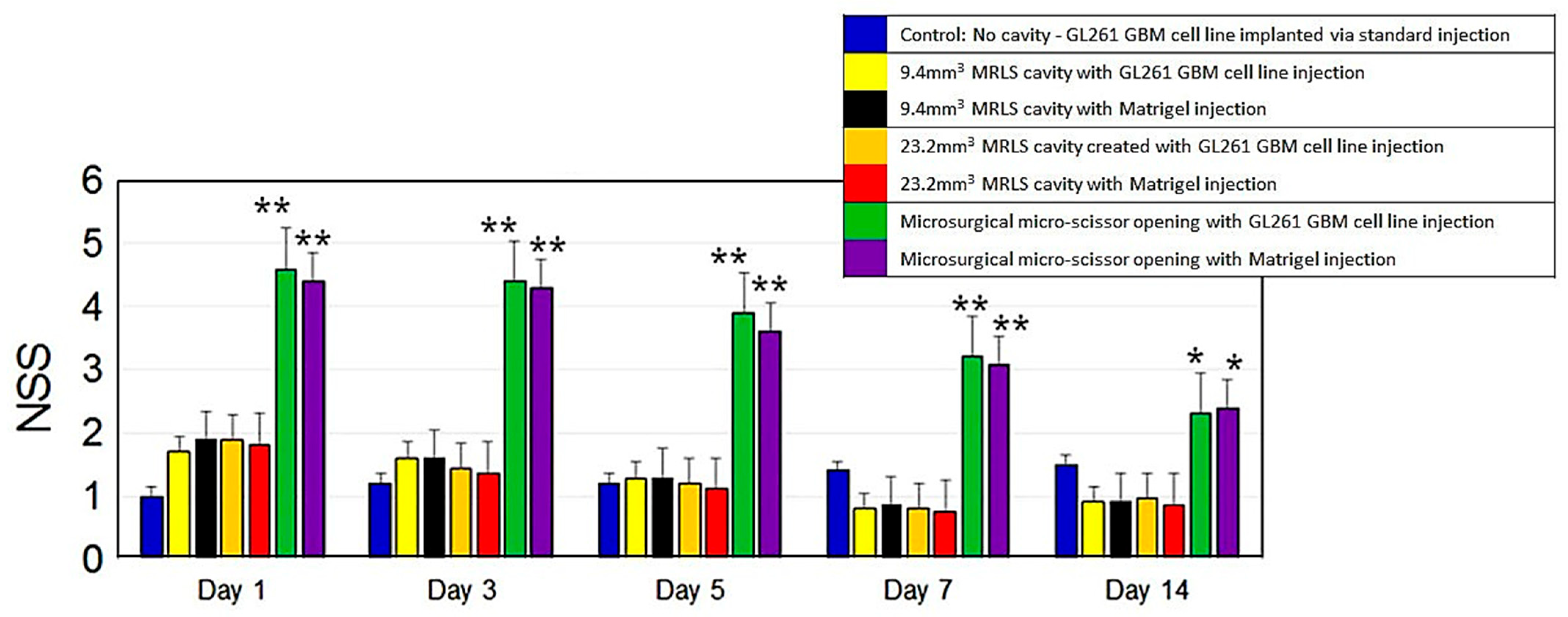
Our methods for creating a cavity in the mouse brain for glioma cell injection did not cause significant blood loss or disruption of surrounding brain architecture, though fluid accumulated in the cavity in some cases. Significant differences in NSS were observed between groups undergoing cavity creation via the MRLS and those undergoing cavity creation with traditional microsurgical techniques and microscissors. The microdissection group (Group 3), with or without tumor cell implantation, showed higher NSS (i.e., more neurological effects) throughout the observation period, with the mean NSS on day 14 exceeding the highest NSS recorded on day 1 for all other groups (Figure 2). Notably, when the MRLS was used to create a space for tumor implantation, neurological morbidity returned to baseline within 3–5 days post-procedure, despite the relatively large volumes of tissue removed in this process.
No statistically significant morbidity differences in NSS between tumor and non-tumor groups were detected following implantation of GL261 glioma cells (or Matrigel in non-tumor groups), despite subsequent tumor growth (Mean NSS < 2, range 0–3). The similarity in NSSs between tumor and non-tumor (sham) groups indicates that the surgical cavity itself did not induce severe neurological deficits, and that the behavioral functional recovery observed in later phases was attributable to the intervention rather than the healing process alone.
Independent of tumor progression, these results demonstrated that creating a cavity of up to 23.2 mm3 prior to cell implantation with the MRLS did not increase morbidity compared with injecting cells without a cavity and provided a reproducible method for achieving tumor growth at a specified location. Thus, in Phase 2 of our study, to evaluate the impact of surgical resection on the tumor microenvironment, cavities (23.2 mm3) were created with the MRLS prior to GL261 implantation.

3.2. Ultrasound and H&E Validate the Benefits of Cavity vs. No Cavity for Tumor Growth

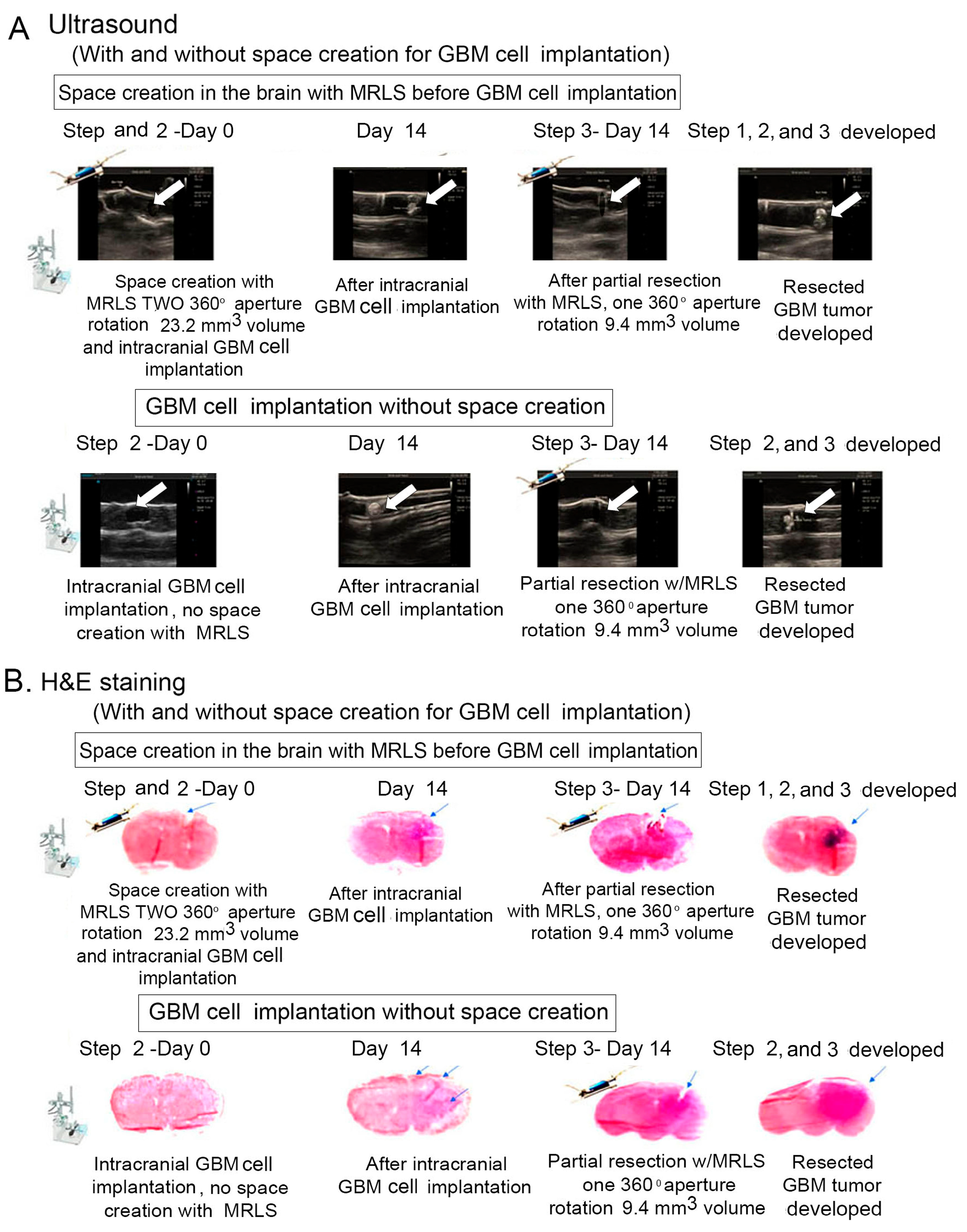
Glioma allograft models, generated either by direct implantation of GL261 cells into the brain (no cavity) or into a 23.2 mm3 cavity created by MRLS, were evaluated using in vivo ultrasound and post-mortem H&E staining (Figure 3). The Terason model 3200T ultrasound system was used to calculate tumor volumes at various time points throughout tumor progression. Images and/or short, real-time video clips were recorded at tumor implantation, at surgical resection, and at the prespecified point of maximal tumor growth or full loss of mobility, at which point the animal was sacrificed. Our results show that creating a cavity with the MRLS to implant GL261 GBM cells in mice minimized or prevented GBM cell migration to other locations and enabled greater accuracy in surgical resection and/or therapeutic delivery (Figure 3A). Histological examination indicated greater tumor localization in mice with a cavity than in those without (Figure 3B). H&E staining in these mice showed a clear, circular cavity with residual tumor cells (dark purple), a finding further confirmed by ultrasound imaging.

3.3. MRLS Resection of GBM Following Tumor Growth Prolonged Median Survival

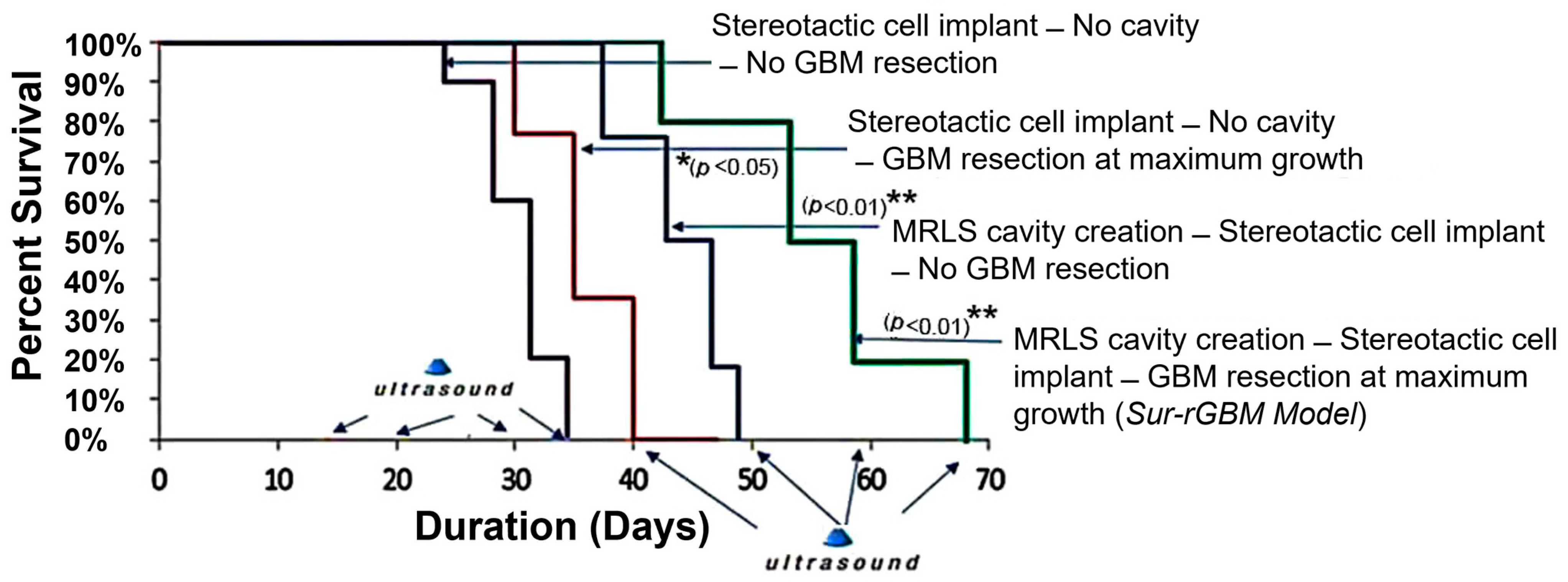
Ultrasound imaging confirmed that gross total resection (i.e., all visible tumor removed) was achieved in 65% of mice undergoing surgical resection, and tumor recurrence was noted in 65% of animals at four weeks post-resection [31]. As shown in Figure 4, total surgical resection improved survival in animals with no cavity and animals with MRLS-created cavities, compared to no resection.
All mice undergoing tumor resection showed a significant change in mobility immediately after surgery (mean NSS > 4). However, the overall increase in survival after surgical resection of GBM was consistent with prior data supporting surgical resection as a means of potentially prolonging survival. Remarkably, animals with MRLS-created cavities prior to implantation of GL261 GBM cells survived longer than animals with direct cell implantation and no cavity, regardless of subsequent resection (Figure 4) [32].

3.4. Phase 3: Tumor Resection Leads to Significant Changes in TME

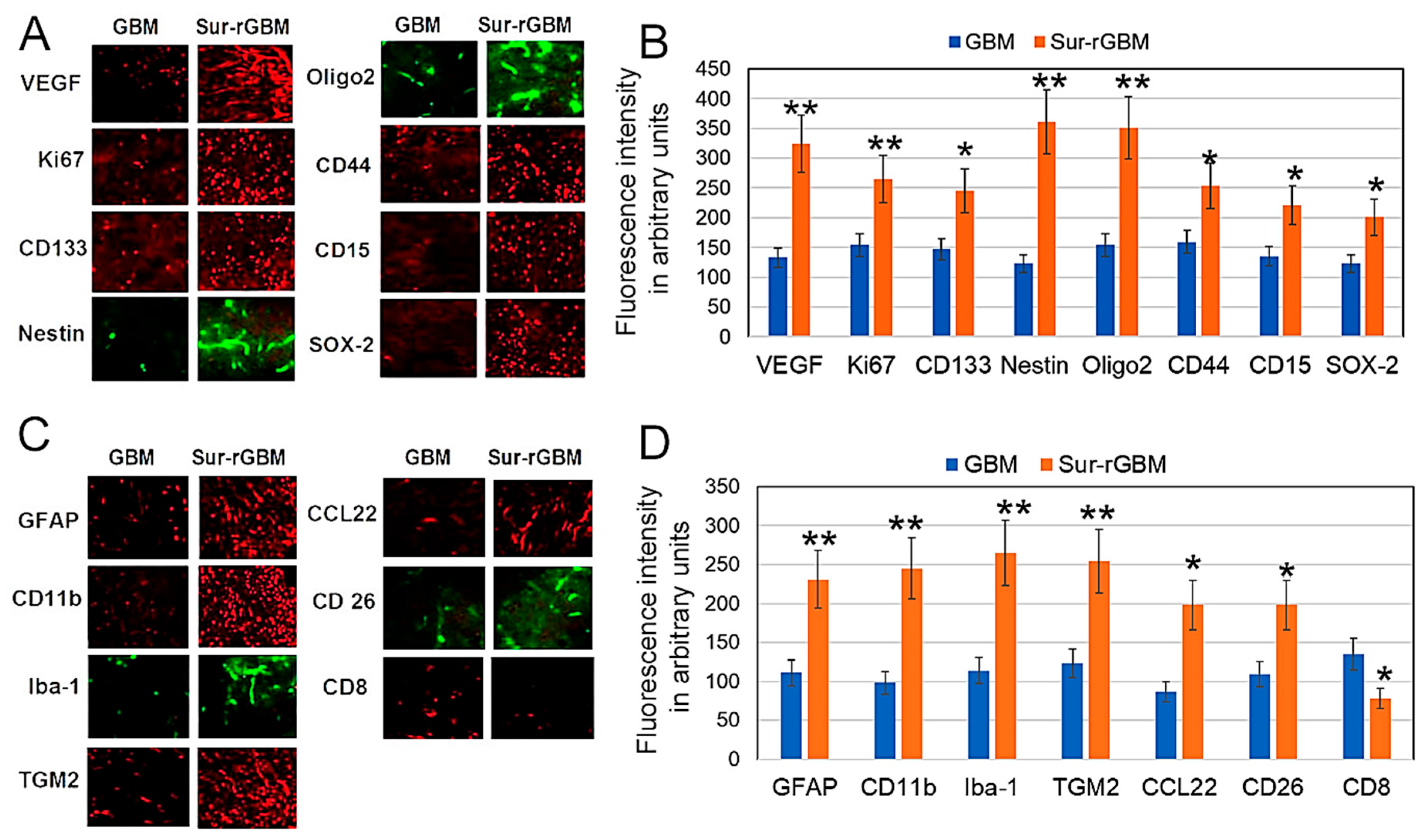
To identify surgical resection markers that may affect tumor regrowth in mice, we assessed the expression of a panel of cellular biomarkers in brain slices using immunohistochemistry. Our results demonstrated significant (p < 0.01) increases in astrocytic activation (GFAP+), increased microglia/macrophage (CD11b+, and IBA-+1) infiltration to surrounding tissue, and increases in M2 macrophages (TGM2+ and CCL22+) and NK regulatory cells (CDC 26+) in resected compared to unresected tumors (Figure 5A,B). We also observed significantly increased markers of angiogenesis (VEGF), increased proliferation (Ki-67), and upregulated expression of stem cell markers CD133, Nestin, and Olig2 after tumor resection (Figure 5C,D). Negligible or focal expression of these markers was observed in tumor tissue from non-resected groups. These results suggest that changes in astrocyte, microglia/macrophage, NK regulatory cell, tumor proliferation, and stem cell markers occur after tumor resection. These changes may contribute to tumorigenesis and serve as biomarkers of tumor recurrence or therapeutic targets for recurrent tumors.

3.5. Resection Enhances Survival After TMZ Alone or Combined with VEGF mAb

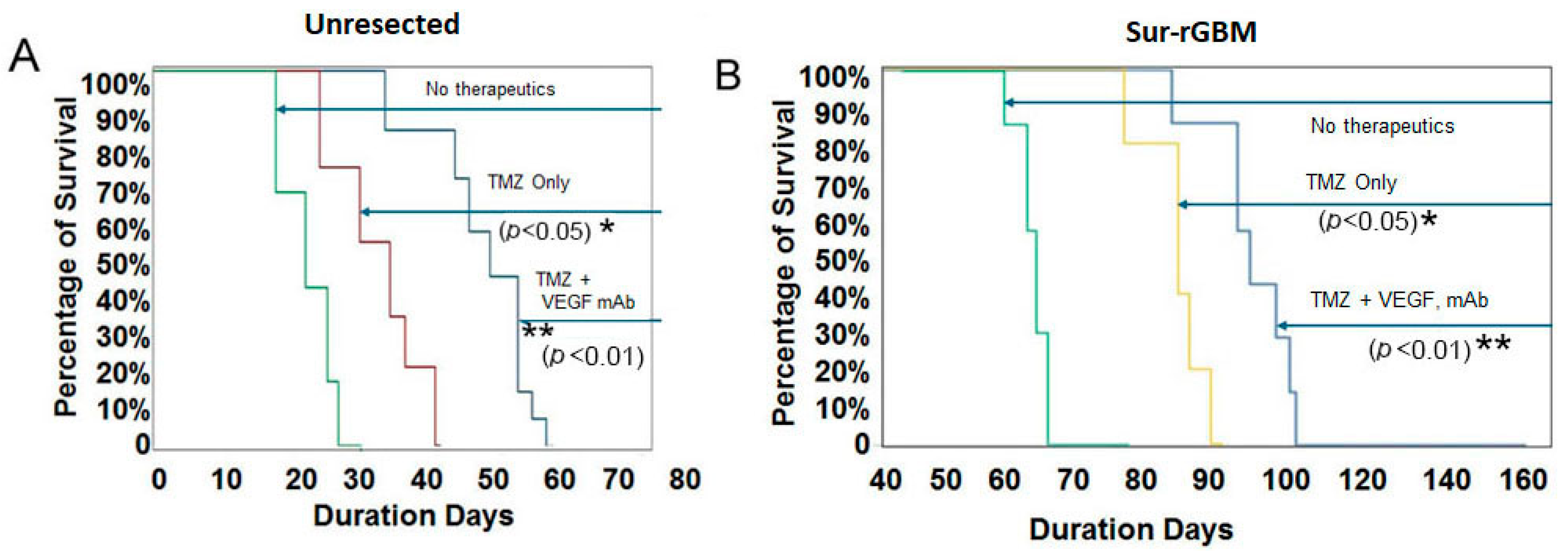
While TMZ and anti-VEGF are standard in recurrent or adjuvant settings for GBM, their use post-resection is critical because surgery alone cannot eliminate the infiltrative ‘rim’ of glioma cells. The goal is to stabilize the disrupted blood–brain barrier post-surgery and to sensitize residual, hypoxic niches to alkylating agents during the window of maximal cytoreduction. Thus, here, unresected and resected (Sur-rGBM) GL261 tumor mice were treated with TMZ alone or TMZ in combination with anti-VEGF mAb. Mice with unresected GL261 GBM tumors, with and without therapeutic intervention, exhibited lower median survival than all Sur-rGBM groups (Figure 6). Among unresected tumor groups, therapeutic intervention increased survival compared to no intervention; the group receiving TMZ in combination with VEGF mAb had the longest survival.
Similarly, after tumor resection (Sur-rGBM), mice showed improved survival with therapeutic intervention, with the greatest survival observed in animals receiving combined TMZ and VEGF mAb. Thus, in both Sur-rGBM groups and no-resection groups, survival was improved with the addition of VEGF mAb over TMZ alone. However, across all groups, survival after resection was considerably longer than without resection. We suggest that this may be due to the TME changes observed in Phase 2, resulting from surgical tumor resection.

4. Discussion

To the best of our knowledge, no prior studies have reported using a murine surgical resection model to investigate the effects of glioma resection on disease course or therapeutic interventions over time. This may be due to the challenges created by the small size of the mouse brain and the relatively large size of surgical instrumentation used clinically in human patients. Standard preclinical glioma models rely on histological examination after animal sacrifice to monitor molecular characteristics. Not only does this process require the use of large numbers of animals, but models that do not include resection may not fully recapitulate the clinical experience, limiting their translational value. In the present study, we established an ultrasound-imageable murine model of orthotopic GL261 glioma (Sur-rGBM) that allowed for tumor implantation within a cavity, tumor resection, monitoring of tumor recurrence, detection of TME changes, and evaluation of therapeutic effects and survival over time.
Our goal was to recapitulate the features of recurrence in human GBM following surgery, thereby enabling investigators to study changes in the tumor and TME after resection. Several factors contributed to the success of the Sur-rGBM model: by first creating a standardized open cavity for GL261 GBM cell delivery, we controlled the tumor location, making it a more accessible target for resection and/or therapeutic delivery at later time points. Using the MRLS device enabled us to minimize challenges encountered with standard cell implantation through needle injection, including cell efflux through the cranial opening. Using the MRLS for resection, we achieved a gross tumor resection (GTR) rate of 65% in our Sur-rGBM model, which mirrors clinical data reporting GTR rates of 65–94% in human patients, depending on the neurosurgical center and the methods employed to maximize resection. The standardization of the implantation process and resection presents a compelling opportunity for other preclinical labs investigating GBM to replicate this model.
Despite the lack of surgical adjuncts such as 5ALA to increase visualization of the infiltrative tumor margin, we demonstrated that surgery alone provided a survival benefit, as noted by Kaplan–Meier analysis (p = 0.01), and that early surgery appeared more effective, reflecting clinically observed survival benefits and reinforcing the long-standing view that early detection and treatment of GBM is associated with a better prognosis. Mice that underwent tumor resection demonstrated prolonged survival compared to mice with unresected tumors (Figure 3). This was expected, given that tumor burden was significantly reduced in the resected group compared with the unresected control group. Tumor recurrence was consistent in the days following resection due to the infiltrative nature of GL261, which inevitably resulted in residual tumor cells remaining.
In Sur-rGBM mice, 50% achieved long-term survival (defined as >70 days), the equivalent of just over 6 human years [33], and resection resulted in additional survival time. However, remarkable changes were observed in the tumor microenvironment following resection, including increased activity of markers implicated in tumorigenesis. Therefore, surgical resection must be considered as a double-edged sword. Resection is a necessity to relieve symptoms associated with tumor burden and to improve patient survival, yet, as our results showed, it simultaneously induces tumor-promoting changes in the TME and directly promotes glioblastoma stem cell propagation. Our results demonstrated a significant increase in astrocytic activation (GFAP+), increased microglia/macrophage (CD11b+, and IBA-+1) infiltration to surrounding tissue, and increases in M2 macrophages (TGM2+ and CCL22+) and NK regulatory cells (CDC 26+) in Sur-rGBM compared to unresected GBM (Figure 5). Populations of KI67+ proliferative cells, high levels of the angiogenic (VEGF) biomarker, and CD133+, Nestin+, and Olig2+ stem cell biomarkers were found in Sur-rGBM at both the resection site and distant infiltrated areas (Figure 5).
To evaluate the response of the model to standard GBM treatment strategies, we evaluated the effects of TMZ alone or with anti-VEGF monotherapy (Bevacizumab, an approved agent for recurrent GBM). We observed differential effects of TMZ plus VEGF mAb compared with TMZ alone in both unresected GL261 GBM tumors and in the Sur-rGBM model; however, increased survival after resection (Sur-rGBM) was observed in all groups, irrespective of subsequent treatment, which may reflect differences in the tumor microenvironment between resected and unresected GBM. Parallels may be drawn between our model and GBM patients regarding the effects of resection on tumor biology during the time window between resection and the onset of adjuvant therapy. These results indicate that our GL261 model recapitulates key histological and immunological aspects of GBM, a necessary step before using it to evaluate novel treatment strategies.
Taken together, these findings suggest that both non-resection GBM models and our Sur-rGBM model are necessary for preclinical drug testing, as therapeutic responses may differ as the TME evolves following resection. Our resection model is an important step toward more advanced preclinical GBM models.
This study had some limitations: (i) A limitation of the GL261 surgical model is the accelerated timeline compared to human disease. The 30–40-day survival in resected animals reflects a significant, yet temporary, delay in tumor progression compared to unresected controls, likely driven by rapid tumor cell proliferation and infiltration that is characteristic of this murine model. (ii) Survival may vary depending on the tumor cell line or the mouse line chosen for the study. (iii) Additionally, access to the MRLS played a large role in the development of this standardized method, both for the initial cavity creation and for controlled, standardized resection with minimal impact on adjacent healthy brain tissue. The MRLS performed substantially better than microdissection. This technology is especially valuable when working in animal models with low cerebral volume. The standardized process and replicable results presented in this manuscript, as well as its potential for use across a wide range of small-animal surgical research, may encourage adoption of this technology to support preclinical research in tandem with clinical advancement.

5. Conclusions

In conclusion, our GL261 model recapitulates key histological and immunological features of human post-surgical GBM, although it carries limitations in capturing the full genetic heterogeneity of the human disease. To our knowledge, this is the first report of a preclinical surgical resection model of GBM that prolongs survival and results in minimal or no neurological injury. This model replicates the therapeutic benefit of surgical resection and has applications in investigating systemic and intracavitary/surgically mediated delivery of novel therapeutic agents, including drugs, immunotherapies, viral therapies, and CAR-T cell therapies. Including surgical resection in preclinical therapeutic efficacy studies may better facilitate the use of state-of-the-art translational molecular imaging strategies in GBM research. By using a specialized surgical system adapted to the pre-clinical setting, we believe our protocol paves the path toward establishing highly consistent and predictive preclinical brain tumor models with increased translational potential. To bridge the gap to clinical application, subsequent validation in male and female cohorts and orthotopic models—specifically CT-2A or SB28 and patient-derived xenograft (PDX) systems—is necessary to confirm these results. Future directions for this technique include applications across tumor types to address unanswered questions about perioperative management and therapeutic discovery for patients with brain tumors.

Author Contributions

Conceptualization, A.D., J.M., S.M.L. and D.C.; methodology, A.D., J.M., J.G. and R.B., validation, A.D., J.M., H.R.S., J.E.B., G.C.B. and D.C.; formal analysis, A.D. and D.C.; investigation, A.D., J.M. and D.C.; resources, A.D.; data curation, A.D.; writing—original draft preparation, A.D. and H.R.S.; writing—review and editing, A.D., J.M., H.R.S., J.E.B., G.C.B. and D.C.; visualization, A.D., J.M., H.R.S., J.E.B., G.C.B. and D.C.; supervision, J.M.; project administration, A.D.; funding acquisition, A.D. All authors have read and agreed to the published version of the manuscript.

Funding

This research received no external funding.

Institutional Review Board Statement

The animal study protocol was approved by the Institutional Animal Care and Use Committee (IACUC) of the Medical University of South Carolina, Charleston, SC, USA. (protocol code: IACUC-2021-01201; approval date: 3 March, 2021).

Informed Consent Statement

Not applicable.

Data Availability Statement

The original contributions presented in this study are included in the article. Further inquiries can be directed to the corresponding author(s).

Acknowledgments

The authors would like to thank Ann Barlow, for assistance with editing the manuscript.

Conflicts of Interest

David Cachia is a consultant for Novocure, M3 Global, Doximity, Aptitude Health, Putnam Associates, and Guidepoint. He is also a paid speaker for the Massachusetts Neurological Society. All other authors declare no conflicts of interest.

Abbreviations

The following abbreviations are used in this manuscript:
5-ALA5-aminolevulinic acid
ANOVAAnalysis of variance
BSABovine serum albumin
DTIDiffusion tensor imaging
DMDMDulbecco’s modified Eagle’s medium
GBMGlioblastoma
H&EHematoxylin and eosin stain
IDHIsocitrate dehydrogenase
mAbMonoclonal antibody
MRLSMyriad Research Laboratory System (NICO Corp.)
NSSNeurological severity score
PBSPhosphate-buffered saline
SDSSodium dodecyl sulphate
Sur-rGBMMurine surgical resection GBM model
TMETumor microenvironment
TMZ Temozolomide
VEGFVascular endothelial growth factor

References

  1. Koshy, M.; Villano, J.L.; Dolecek, T.A.; Howard, A.; Mahmood, U.; Chmura, S.J.; Weichselbaum, R.R.; McCarthy, B.J. Improved Survival Time Trends for Glioblastoma Using the SEER 17 Population-Based Registries. J. Neuro-Oncol. 2012, 107, 207–212. [Google Scholar] [CrossRef] [PubMed]
  2. Louis, D.N.; Perry, A.; Reifenberger, G.; von Deimling, A.; Figarella-branger, D.; Cavenee, W.K.; Ohgaki, H.; Wiestler, O.D.; Kleihues, P.; Ellison, D.W. The 2016 World Health Organization Classification of Tumors of the Central Nervous System: A Summary. Acta Neuropathol. 2016, 131, 803–820. [Google Scholar] [CrossRef]
  3. Jezierzański, M.; Natalia, N.; Małgorzata, S.; Tomasz, F.; Michał, M.; Jacek, K.; Gisterek-Grocholska, I. Temozolomide (TMZ) in the Treatment of Glioblastoma Multiforme—A Literature Review and Clinical Outcomes. Curr. Oncol. 2024, 31, 3994–4002. [Google Scholar] [CrossRef]
  4. Gupta, S.; Mallick, S.; Benson, R.; Haresh, K.P.; Julka, P.K.; Rath, G.K. Extent of Surgical Resection and Adjuvant Temozolomide Improves Survival in Pediatric GBM: A Single Center Experience. Childs Nerv. Syst. 2017, 33, 951–956. [Google Scholar] [CrossRef]
  5. Predina, J.D.; Judy, B.; Kapoor, V.; Blouin, A.; Aliperti, L.A.; Levine, D.; Okusanya, O.T.; Quatromoni, J.; Fridlender, Z.G.; Singhal, S. Characterization of Surgical Models of Postoperative Tumor Recurrence for Preclinical Adjuvant Therapy Assessment. Am. J. Transl. Res. 2012, 4, 206–218. [Google Scholar]
  6. Pesaresi, A.; La Cava, P.; Bonada, M.; Zeppa, P.; Melcarne, A.; Cofano, F.; Fiaschi, P.; Garbossa, D.; Bianconi, A. Combined Fluorescence-Guided Surgery with 5-Aminolevulinic Acid and Fluorescein in Glioblastoma: Technical Description and Report of 100 Cases. Cancers 2024, 16, 2771. [Google Scholar] [CrossRef]
  7. Yaghi, N.K.; Gilbert, M.R. Immunotherapeutic Approaches for Glioblastoma Treatment. Biomedicines 2022, 10, 427. [Google Scholar] [CrossRef]
  8. Rocha Pinheiro, S.L.; Lemos, F.F.B.; Marques, H.S.; Silva Luz, M.; de Oliveira Silva, L.G.; Faria Souza Mendes dos Santos, C.; da Costa Evangelista, K.; Calmon, M.S.; Sande Loureiro, M.; Freire de Melo, F. Immunotherapy in glioblastoma treatment: Current state and future prospects. World J. Clin. Oncol. 2023, 14, 138–159. [Google Scholar] [CrossRef]
  9. Hamad, A.; Yusubalieva, G.M.; Baklaushev, V.P.; Chumakov, P.M.; Lipatova, A.V. Recent Developments in Glioblastoma Therapy: Oncolytic Viruses and Emerging Future Strategies. Viruses 2023, 15, 547. [Google Scholar] [CrossRef] [PubMed]
  10. Lucchini, S.; Nicholson, J.G.; Zhang, X.; Househam, J.; Lim, Y.M.; Mossner, M.; Millner, T.O.; Brandner, S.; Graham, T.; Marino, S. A Novel Model of Glioblastoma Recurrence to Identify Therapeutic Vulnerabilities. EMBO Mol. Med. 2025, 17, 1325–1354. [Google Scholar] [CrossRef] [PubMed]
  11. Candolfi, M.; Curtin, J.F.; Nichols, W.S.; Muhammad, A.G.; King, G.D.; Pluhar, G.E.; McNiel, E.A.; Ohlfest, J.R.; Freese, A.B.; Moore, P.F. Intracranial Glioblastoma Models in Preclinical Neuro-Oncology: Neuropathological Characterization and Tumor Progression. J. Neuro-Oncol. 2007, 85, 133–148. [Google Scholar] [CrossRef]
  12. Huszthy, P.C.; Daphu, I.; Niclou, S.P.; Stieber, D.; Nigro, J.M.; Sakariassen, P.Ø.; Miletic, H.; Thorsen, F.; Bjerkvig, R. In Vivo Models of Primary Brain Tumors: Pitfalls and Perspectives. Neuro-Oncology 2012, 14, 979–993. [Google Scholar] [CrossRef]
  13. Irtenkauf, S.M.; Sobiechowski, S.; Hasselbach, L.A.; Nelson, K.K.; Transou, A.D.; Carlton, E.T.; Mikkelsen, T.; de Carvalho, A.C. Optimization of Glioblastoma Mouse Orthotopic Xenograft Models for Translational Research. Comp. Med. 2018, 67, 300–314. [Google Scholar]
  14. Gómez-Oliva, R.; Domínguez-García, S.; Carrascal, L.; Abalos-Martínez, J.; Pardillo-Díaz, R.; Verástegui, C.; Castro, C.; Nunez-Abades, P.; Geribaldi-Doldán, N. Evolution of Experimental Models in the Study of Glioblastoma: Toward Finding Efficient Treatments. Front. Oncol. 2021, 10, 614295. [Google Scholar] [CrossRef]
  15. Ben-Eliyahu, S.; Page, G.G.; Yirmiya, R.; Shakhar, G. Evidence That Stress and Surgical Interventions Promote Tumor Development by Suppressing Natural Killer Cell Activity. Int. J. Cancer 1999, 80, 880–888. [Google Scholar] [CrossRef]
  16. Ogawa, K.; Hirai, M.; Katsube, T.; Murayama, M.; Hamaguchi, K.; Shimakawa, T.; Naritake, Y.; Hosokawa, T.; Kajiwara, T. Suppression of Cellular Immunity by Surgical Stress. Surgery 2000, 127, 329–336. [Google Scholar] [CrossRef] [PubMed]
  17. Tsuchiya, Y.; Sawada, S.; Yoshioka, I.; Ohashi, Y.; Matsuo, M.; Harimaya, Y.; Tsukada, K.; Saiki, I. Increased Surgical Stress Promotes Tumor Metastasis. Surgery 2003, 133, 547–555. [Google Scholar] [CrossRef] [PubMed]
  18. Seth, R.; Tai, L.-H.; Falls, T.; de Souza, C.T.; Bell, J.C.; Carrier, M.; Atkins, H.; Boushey, R.; Auer, R.A. Surgical Stress Promotes the Development of Cancer Metastases by a Coagulation-Dependent Mechanisms Involving Natural Killer Cells in a Murine Model. Ann. Surg. 2013, 258, 158–168. [Google Scholar] [CrossRef]
  19. Sweeney, K.J.; Jarzabek, M.A.; Dicker, P.; O’Brien, D.F.; Callanan, J.J.; Byrne, A.T.; Prehn, J.H.M. Validation of an Imageable Surgical Resection Animal Model of Glioblastoma (GBM). J. Neurosci. Methods 2014, 233, 99–104. [Google Scholar] [CrossRef]
  20. Kumagai, Y.; Ohzawa, H.; Miyato, H.; Horie, H.; Hosoya, Y.; Lefor, A.K.; Sata, N.; Kitayama, J. Surgical Stress Increases Circulating Low-Density Neutrophils Which May Promote Tumor Recurrence. J. Surg. Res. 2020, 246, 52–61. [Google Scholar] [CrossRef]
  21. Knudsen, A.M.; Halle, B.; Cédile, O.; Burton, M.; Baun, C.; Thisgaard, H.; Anand, A.; Hubert, C.; Thomassen, M.; Regner Michaelsen, S.; et al. Surgical Resection of Glioblastomas Induces Pleiotrophin-Mediated Self-Renewal of Glioblastoma Stem Cells in Recurrent Tumors. Neuro-Oncology 2021, 24, 1074–1087. [Google Scholar] [CrossRef]
  22. Tai, L.-H.; de Souza, C.T.; Sahi, S.; Zhang, J.; Alkayyal, A.A.; Ananth, A.A.; Auer, R.A. A Mouse Tumor Model of Surgical Stress to Explore the Mechanisms of Postoperative Immunosuppression and Evaluate Novel Perioperative Immunotherapies. J. Vis. Exp. 2014, e51253. [Google Scholar] [CrossRef]
  23. Chen, Z.; Zhang, P.; Xu, Y.; Yan, J.; Liu, Z.; Bond Lau, W.; Lau, B.; Li, Y.; Zhao, X.; Wei, Y.; et al. Surgical Stress and Cancer Progression: The Twisted Tango. Mol. Cancer 2019, 18, 132. [Google Scholar] [CrossRef]
  24. Seligman, A.M.; Shear, M.J. Studies in Carcinogenesis: VIII. Experimental Production of Brain Tumors in Mice with Methylcholanthrene. Am. J. Cancer 1939, 37, 364–395. [Google Scholar] [CrossRef]
  25. Haddad, A.F.; Young, J.S.; Amara, D.; Berger, M.S.; Raleigh, D.R.; Aghi, M.K.; Butowski, N.A. Mouse Models of Glioblastoma for the Evaluation of Novel Therapeutic Strategies. Neuro-Oncol. Adv. 2021, 3, vdab100. [Google Scholar] [CrossRef]
  26. Alomari, S.; Kedda, J.; Malla, A.P.; Pacis, V.; Anastasiadis, P.; Xu, S.; McFarland, E.; Sukhon, L.; Gallo, B.; Rincon-Torroella, J.; et al. Implementation of Minimally Invasive Brain Tumor Resection in Rodents for High Viability Tissue Collection. J. Vis. Exp. 2022, e64048. [Google Scholar] [CrossRef] [PubMed]
  27. Dlouhy, B.J.; Dahdaleh, N.S.; Greenlee, J.D.W. Emerging Technology in Intracranial Neuroendoscopy: Application of the NICO Myriad Technical Note. Neurosurg. Focus 2011, 30, E6. [Google Scholar] [CrossRef]
  28. McLaughlin, N.; Filho, L.; Prevedello, D.; Kelly, D.; Carrau, R.; Kassam, A. Side-Cutting Aspiration Device for Endoscopic and Microscopic Tumor Removal. J. Neurol. Surg. Part B Skull Base 2012, 73, 11–20. [Google Scholar] [CrossRef]
  29. Rogers, S.; Hii, H.; Huang, J.; Ancliffe, M.; Gottardo, N.G.; Dallas, P.; Lee, S.; Endersby, R. A Novel Technique of Serial Biopsy in Mouse Brain Tumour Models. PLoS ONE 2017, 12, e0175169. [Google Scholar] [CrossRef] [PubMed]
  30. Brown, D.; Lydon, J.; McLaughlin, M.; Stuart Tilley, A.; Tyszkowski, R.; Alper, S. Antigen Retrieval in Cryostat Tissue Sections and Cultured Cells by Treatment with Sodium Dodecyl Sulfate (SDS). Histochem. Cell Biol. 1996, 105, 261–268. [Google Scholar] [CrossRef] [PubMed]
  31. Page, G.G.; Ben-Eliyahu, S.; Blakely, W.P. Evidence That Postoperative Pain Is a Mediator of the Tumor-Promoting Effects of Surgery in Rats. Pain 2001, 90, 191–199. [Google Scholar] [CrossRef] [PubMed]
  32. Kubelt, C.; Hellmold, D.; Peschke, E.; Hauck, M.; Will, O.; Schuett, F.; Lucius, R.; Adelung, R.; Scherliess, R.; Hoevener, J.-B.; et al. Establishment of a Rodent Glioblastoma Partial Resection Model for Chemotherapy by Local Drug Carriers-Sharing Experience. Biomedicines 2023, 11, 1518. [Google Scholar] [CrossRef] [PubMed]
  33. Dutta, S.; Sengupta, P. Men and Mice: Relating Their Ages. Life Sci. 2016, 152, 244–248. [Google Scholar] [CrossRef] [PubMed]
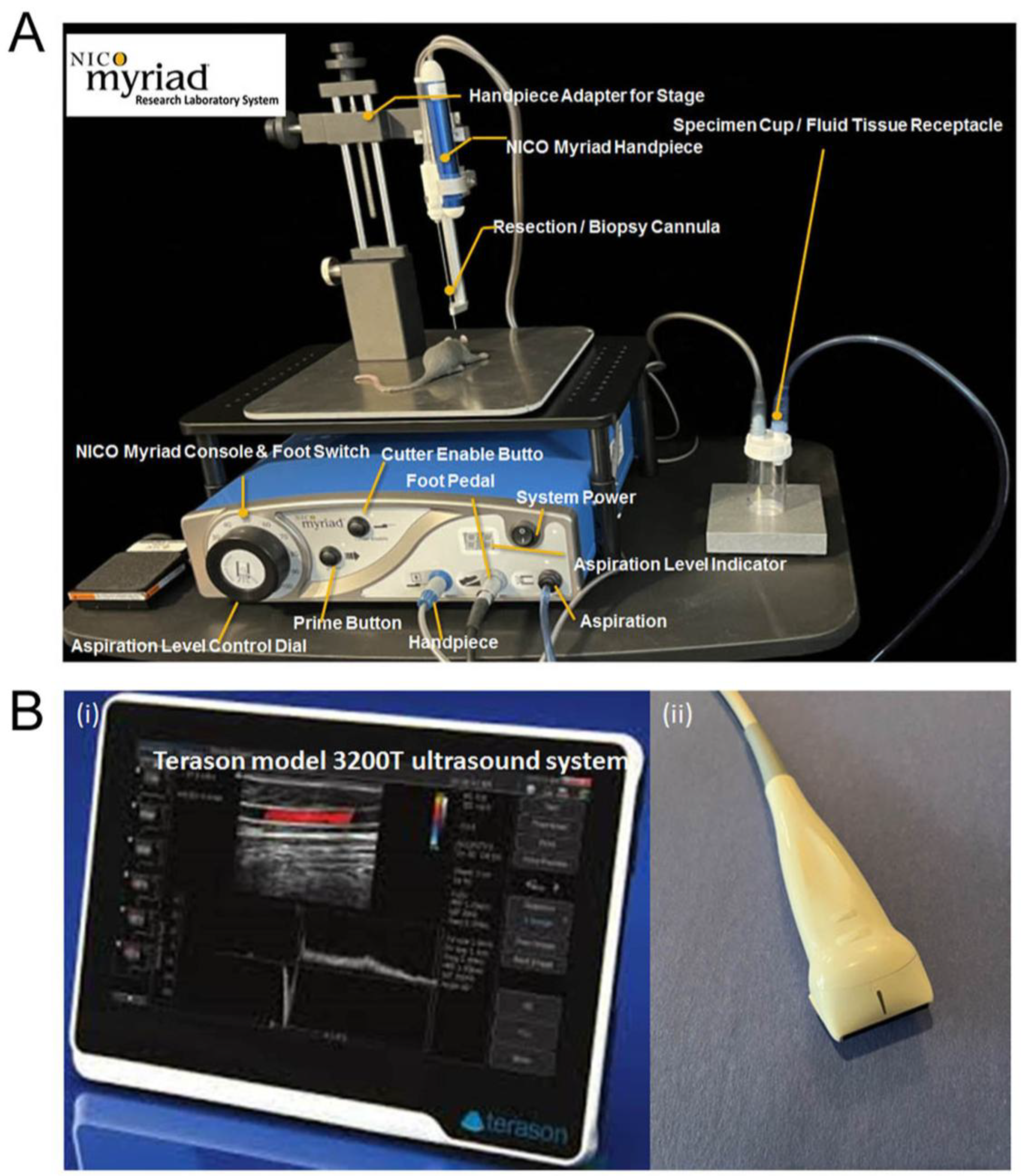
Figure 1. (A) NICO Myriad Research Laboratory System (MRLS) comprising a Myriad console and 23 g handpiece used to create a cavity for tumor cell implantation and for subsequent tumor resection. (B) (i) Terason model 3200T ultrasound system, used for monitoring tumor growth and tumor resection. (ii) Terason transducer for imaging of deep anatomical brain regions with a sweeping field of view.
Onco 06 00024 g001
Figure 2. (Phase 1) NSS was measured daily for 14 days after cavity creation to compare the effects of cavity size and method of cavity creation. The control (no-cavity) group, with injected tumor cells, was compared with groups with either 9.4 mm3 or 23.2 mm3 cavities created by MRLS or by microdissection, with and without subsequent implantation of tumor cells, to assess neurologic changes due to cavity creation or tumor progression. * p < 0.05 and ** p < 0.01 indicate a significant difference (n = 7).
Onco 06 00024 g002
Figure 3. Comparison of surgical resection models with and without cavities for tumor implantation. Cavities were created with the MRLS. Two full rotations of the MRLS cannula (Group 2) create a cavity of 23.2 mm3 at Day 0. GL261 cells were orthotopically implanted on Day 0, and tumor growth was measured on Day 14. The tumor was then resected using a single full rotation of the MRLS cannula, opening a cavity of 9.4 mm3 in both groups. Tumor growth in the cavity group (top row) and no-cavity group (bottom row) was monitored by ultrasound imaging (A) and H&E staining (B) to assess the effects of cavity creation in the development of the Sur-rGBM model (n = 5). White arrows in ultrasound images and blue arrows in H&E-stained images show sites of tumor resection and tumor reoccurrence.
Onco 06 00024 g003
Figure 4. (Phase 2) Survival (days) was compared between groups with no cavity, tumor cell injection, no resection; no cavity, standard tumor injection, resection; MRLS-created cavity, tumor cell implantation, no resection, and MRLS-created cavity, tumor cell implantation, resection—Surgical murine GBM resection model (Sur-rGBM). * p < 0.05 and ** p < 0.01 indicate a significant difference (n = 7).
Onco 06 00024 g004
Figure 5. The expression levels of a panel of commonly used markers of astrocytes, microglia/macrophages, NK regulatory cells, tumor proliferation, stem cells, and GBM tissue were determined by immunohistochemistry in the Sur-rGBM model and unresected groups. Our results demonstrated a significant increase in VEGF in Sur-rGBM, as well as increased Ki-67 and upregulation of CD133, Nestin, and Olig2 ((A) Immunostaining images and (B)) (n ≥ 3). Our results also suggested significant increases in GFAP+, CD11b+, IBA-+1 infiltration into surrounding tissue, TGM2+ and CCL22+, and CDC 26+ in Sur-rGBM compared to unresected tumor ((C) Immunostaining images and (D) Quantitative bar graphs). * p < 0.05 and ** p < 0.01 indicate a significant difference (n = 5).
Onco 06 00024 g005
Figure 6. (Phase 3) Survival of mice (days) after three therapeutic options: No therapeutics, TMZ alone, or TMZ + VEGF mAb in groups with (A) standard stereotactic cell injection methods with no cavity creation and no tumor resection or (B) Sur-rGBM model MRLS cavity creation (23.2 mm3), stereotactic cell implant, and subsequent resection prior to therapeutic treatment). * p < 0.05 and ** p < 0.01 indicate a significant difference (n = 7).
Onco 06 00024 g006
Table 1. Postoperative neurological injury severity assessment using a nine-point neurological severity scale (NSS).
Task/TestParametersPoints
Balance on a round stickInability to balance on 0.5 cm diameter round stick1
Exit circleFailure to exit 30 cm circle within 2 min1
Response to placing on floorWalks normally0
Inability to walk1
Circles1
Falls down1
Response to raising by tailFailed forelimb flexion test1
Failed hindlimb flexion test1
Failed to move head > 100 to vertical axis within 30 s1
Proprioceptive
sensory
Failed deep sensation tests
(pushing paw against table edge to stimulate limb muscles)
1
Maximum Score9
Disclaimer/Publisher’s Note: The statements, opinions and data contained in all publications are solely those of the individual author(s) and contributor(s) and not of MDPI and/or the editor(s). MDPI and/or the editor(s) disclaim responsibility for any injury to people or property resulting from any ideas, methods, instructions or products referred to in the content.