Review

Established and Emerging Less Invasive Biomarkers and Technologies for Lung Cancer Screening: Puerto Rican Context

by
Keisy Rodriguez-Villafañe
1,
Clara Santiago
2,
Juan E. Figueroa
3,
Edwin Figueroa
3 and
Yamixa Delgado
4,*
1
Pharmaceutical Sciences Department, School of Pharmacy, University of Puerto Rico, Medical Sciences Campus, San Juan, PR 00936, USA
2
School of Naturopathic Medicine, Universidad Ana G. Mendez Gurabo Campus, Gurabo, PR 00778, USA
3
Biology Department, University of Puerto Rico, Cayey Campus, Cayey, PR 00736, USA
4
Biochemistry & Pharmacology Department, San Juan Bautista School of Medicine, Caguas, PR 00726, USA
*
Author to whom correspondence should be addressed.
Submission received: 5 February 2026 / Revised: 10 March 2026 / Accepted: 19 March 2026 / Published: 1 April 2026

Simple Summary

Lung cancer is the leading cause of cancer deaths in Puerto Rico (PR), largely because most cases are found too late. Although annual low-dose computed tomography (LDCT) is the global screening standard, it is difficult to deploy at scale on the island due to high costs, limited radiology workforce and equipment, insurance and logistical barriers, and travel constraints for rural communities. This review outlines emerging biomarkers (serum protein, tumor-associated autoantibodies, and breath sensors) for early detection and a practical, island-wide biomarker-first triage model to identify who most needs LDCT. Because these tests can be performed in primary-care settings across PR, the pathway is easier to access and prioritizes imaging for those at highest risk. By aligning science with local realities, this approach offers a feasible roadmap to detect lung cancer earlier and reduce disparities for all Puerto Ricans.

Abstract

Background/Objectives: In Puerto Rico (PR), lung cancer mortality remains high because diagnoses frequently occur at advanced stages. Although low-dose computed tomography (LDCT) lowers lung cancer–specific mortality, this screening is difficult to operationalize locally due to high false-positive rates, radiology capacity constraints, payer limitations, and geographic barriers affecting rural populations. Methods: We performed a narrative review on the literature from 2001–2026 of established and emerging detection strategies—LDCT; serum biomarkers (CEA, CYFRA-21-1, NSE, ProGRP, SCC-Ag, HE4, Hp, TAAb); breath analysis (FeNO and VOCs); and liquid biopsy (ctDNAs/CTCs/miRNAs). We assessed technical performance, feasibility, and health-system fit in PR and then synthesized these findings into an implementable biomarker-first triage workflow for are. Results: Multiplex serum panels analyzed with machine learning outperform single markers and TAAb provide high specificity with biological lead time, supporting their use as a triage gateway before LDCT. Breathomics is also feasible at the point of care. Liquid biopsy has modest sensitivity in very-early disease yet provides molecular adjudication for indeterminate nodules. A stepwise pathway—expanded risk assessment, integrated multi-panel testing in primary care, LDCT reserved for biomarker-positive individuals, and liquid biopsy when imaging is inconclusive—can enrich pre-test probability, reduce unnecessary scans, align with capitation, and protect limited radiology capacity. Conclusions: An integrated, non-invasive, biomarker-first triage model offers a pragmatic, equitable route to earlier lung cancer detection in PR and resource stewardship, while reducing disparities.

1. Introduction

Lung cancer is a leading cause of cancer mortality worldwide, and Puerto Rico (PR) is no exception [1]. A primary contributor to its high mortality rate is that the majority of cases are diagnosed in advanced stages, when therapeutic options are limited, and the prognosis is unfavorable [2,3]. PR experiences persistently late-stage lung cancer diagnoses and markedly low screening uptake, despite guideline availability. Recent analyses show that the island-wide adherence to low-dose computed tomography (LDCT) screening remains very low (e.g., eligibility–uptake–adherence data from 2022 indicate that <10% of eligible adults are up-to-date), with structural barriers, such as lack of reimbursement, logistics, and patient transportation/fear, frequently cited [4,5,6]. These data reinforce the need for alternative, easier-to-deploy screening gateways that can be offered at the point of primary care. While various lung cancer detection strategies are implemented globally, their effectiveness is often constrained by a duality of challenges: the inherent technical limitations of the tests themselves, and the specific socioeconomic and infrastructural contexts in which they are deployed. It is critical to note that while screening tests are essential for early detection, a tissue biopsy remains the only definitive method for a lung cancer diagnosis. This review addresses the challenges of early detection by providing a comprehensive overview of screening strategies, from the traditional standard of LDCT to emergent non-invasive technologies. We first examine the universal challenges of these techniques and then analyze the significant implementation barriers—including access, cost, and logistics—that are particularly pronounced within PR’s unique healthcare infrastructure. Ultimately, this review argues that an integrated, multi-marker, non-invasive panel offers the most viable and effective path to overcome existing healthcare disparities and reduce lung cancer mortality across the Puerto Rican population.
Definitions and Intended Use. In this study, we distinguish three clinical terms: screening, triage, and diagnostic. Screening aims to identify individuals at increased likelihood of disease in an ostensibly asymptomatic population; screening tests prioritize high sensitivity to minimize missed cases and acceptable specificity to limit unnecessary follow-up. Triage helps to prioritize who should undergo screening and definitive testing to optimize resource use and reduce unnecessary procedures. Diagnostic use seeks to confirm or exclude disease in a clinical context with higher pretest probability; diagnostic tests emphasize high specificity (and likelihood ratios) to support clinical decision-making.

2. Methods for Structured Search

We conducted a structured, narrative review to synthesize established and emerging approaches for earlier lung cancer detection, with an emphasis on feasibility in PR. Literature searches were run in MEDLINE/PubMed, Embase, Scopus and Web of Science. Searches included studies from 2001 to 2026. To capture local implementation and equity considerations, we screened PR’s relevant reports and journals cited by the included articles. We combined controlled vocabulary and free-text terms for lung cancer and screening/diagnostics with modality-specific terms. For example: “lung neoplasms” OR “lung cancer” AND (“screening” OR “early detection” OR “risk stratification”); LDCT terms; serum biomarkers (CEA, CYFRA 21-1, SCC-Ag, NSE, ProGRP, HE4, haptoglobin, “tumor-associated autoantibodies”); breathomics (“fractional exhaled nitric oxide”, FeNO, “volatile organic compounds”, VOCs, “electronic nose”); and liquid biopsy (ctDNA, CTCs, microRNA, methylation). We used citation chaining (backward/forward) from key trials, meta-analyses, and validation cohorts to identify additional records. We included human, adult studies that reported clinical outcomes (e.g., mortality, stage shift), diagnostic performance (e.g., sensitivity/specificity/area under the curve (AUC)), health–economic findings, or implementation data relevant to screening/triage/diagnosis. We excluded editorials, narrative opinions without data, single-case reports, animal/in vitro studies, and studies not specific to lung cancer. We prioritized: (1) randomized controlled trials and systematic reviews/meta-analyses (e.g., LDCT trials and TAAb-triage RCTs); (2) prospective and/or externally validated diagnostic-accuracy studies and large cohort studies; (3) health–economic evaluations and implementation studies pertinent to constrained-resource settings; and (4) emerging technologies validation cohorts were favored over small case–control series to limit spectrum bias.

3. Screening Modalities

3.1. Established Global Standard: Low-Dose Computed Tomography (LDCT)

LDCT is the established global standard for lung cancer screening in high-risk individuals. Its worldwide adoption was prompted by robust evidence from large, randomized trials such as the National Lung Screening Trial (NLST) in the United States (US) and the NELSON trial in Europe.

3.1.1. Efficacy and Mortality Reduction

LDCT screening results in a significant reduction in lung cancer mortality. The NLST showed a 20% reduction in lung cancer-specific deaths compared to chest radiography [7,8,9], while the reduction in all-cause mortality was more modest at 6.7% [10]. The primary benefit lies in detecting cancers at an early, more treatable stage, with some studies reporting over 75% five-year survival for stage 1A disease [11]. However, LDCT screening accuracy should not be interpreted as a single fixed value across all screening settings. In a systematic review and meta-analysis focused primarily on baseline population-based LDCT screening studies, Guo et al. reported a pooled AUC of 0.98, a sensitivity of 0.97, and a specificity of 0.87. These pooled values should, therefore, be interpreted in the context of baseline screening and may vary according to the definition of a positive screen, analytic approach, and follow-up framework [12].

3.1.2. Clinical Implementation and Guidelines

International guidelines from bodies, like the US Preventive Services Task Force (USPSTF) and the American Cancer Society, recommend annual LDCT screening for high-risk individuals (typically aged 55–80 with a significant smoking history) [7,13]. However, adherence is suboptimal globally, with screening uptake rates often below 20%, even in countries with established programs [14,15,16]. Surveys of physicians, including pulmonologists and radiologists, reveal varying opinions and practices regarding screening, which can also impact implementation [15,17,18].

3.1.3. Universal Technical Limitations and Harms

LDCT has several inherent limitations. Although its high sensitivity supports early detection, it also contributes to a substantial false-positive burden, with many initially positive screens ultimately proving to be non-malignant [8,13]. In the NLST, false-positive rates were 26.3% at baseline, 27.2% at year 1, and 15.9% at year 2 [19]. This may cause patient anxiety and lead to unnecessary, and sometimes invasive, follow-up procedures. Across studies reviewed for the USPSTF, false-positive rates ranged from 7.9% to 49.3% at baseline and from 0.6% to 28.6% in incidence rounds, while needle biopsy for false-positive results occurred in 0.09% to 0.56% of all screened participants and surgical procedures in 0.5% to 1.3% [19]. Importantly, the magnitude of false-positive findings depends on the screening protocol, nodule threshold, and definition of a positive examination used across studies and programs. For example, retrospective application of Lung-RADS® to NLST data reduced the false-positive rate from 26.6% to 12.8% at baseline and from 21.8% to 5.3% after baseline, although sensitivity was also lower [20]. Lung-RADS® (Lung CT Screening Reporting and Data System) is a standard of the American College of Radiology for interpreting and reporting findings on LDCT. Overdiagnosis, defined as the detection of slow-growing tumors that may never have become clinically significant, remains another important concern, although reported estimates vary across analyses [8,13]. Additional harms include cumulative radiation exposure from annual scans and the potential for incidental findings that may require further workup [11,13].

3.1.4. The Puerto Rican Context: Lung Cancer Statistics and Amplified Barriers to Implementation

Surveillance data of the Puerto Rico Central Cancer Registry from 2018–2022 indicate that lung and bronchus cancer comprised 5.2% of all newly diagnosed cancers in men and 4.1% in women (ranking third in men and fifth in women), while accounted for 11.6% of cancer deaths in men and 9.3% in women (ranking third) over the same period. The median age for incidence was 72.5 and for death was 75. The survival rate for 5 years in these patients was 21.3%, underscoring the importance of early detection [21]. Although lung cancer is more commonly diagnosed and is the leading cause of cancer death in the US mainland, PR has a higher mortality-to-incidence ratio (~60% vs. 69%), indicating a greater fraction of diagnosed patients who die [22].
Taken together, these surveillance metrics highlight a key contrast of PR and mainland US that motivates a context-specific screening strategy. Importantly, screening uptake remains suboptimal even in the continental US (about 18% of eligible individuals up-to-date in 2022), which underscores that PR’s implementation barriers exist within a broader national challenge but are amplified by island-specific capacity, access, and payer constraints [23].
These disparities are compounded by PR’s status as a US territory (colony), which results in unequal benefits and healthcare funding [5]. The high cost of equipment, lack of specialized personnel, and fragmented healthcare infrastructure make the widespread implementation of LDCT screening unfeasible for most of the island’s high-risk population.
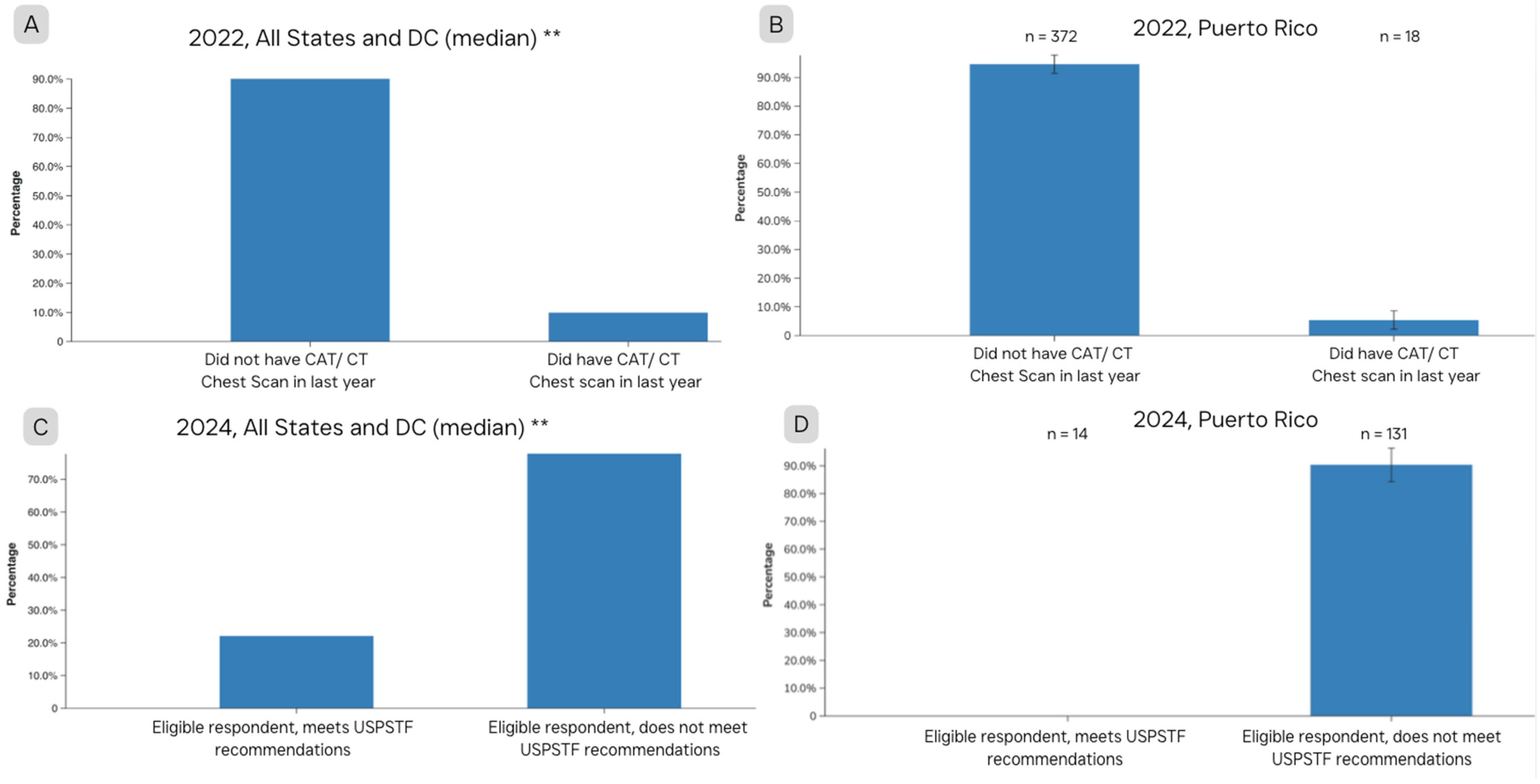
Recent data on screening eligibility and uptake on the island confirm these challenges, highlighting the persistent low adherence rates [6]. Also, population-based surveys reinforce the low screening uptake. In the 2022 BRFSS Prevalence & Trends Data, only 5.4% of PR respondents reported having a CAT/CT chest scan in the prior year for lung cancer screening (vs. 9.9% in the continental US), while 94.6% reported none (vs. 90.1% in the continental US). This disparity persists in 2024 (90.4% PR vs. 77.9% continental US), and the BRFSS eligibility module suggests that screening-eligible respondents in PR are exceedingly few (unweighted n = 14), indicating that BRFSS captured very few screening-eligible respondents in that survey cycle and reinforcing how uncommon screening exposure appears in population surveys (Figure 1) [24]. In another study by Castañeda-Avila (2025), island-specific estimates suggest that about 94,955 PR residents in 2022 met the criteria for screening with LDCT, representing about 7.9% of the population. However, only 9.8% were up to date with screening, which is lower than mainland US Hispanic (17.3%) and non-Hispanic residents (18.1%) [6]. This pattern suggests persistent implementation and adherence gaps, despite eligibility. These findings are particularly striking given that the Puerto Rico Comprehensive Cancer Control Plan explicitly targets increased utilization of LDCT/CAT/CT screening from 5.4% in 2022 to 7.5% by 2030 [25]. However, the BRFSS trend suggests that implementation has not yet translated into measurable population-level uptake. Moreover, a PR non-small cell lung carcinoma (NSCLC) real-world cohort reported that 71.1% of patients were diagnosed at stage III–IV, consistent with delayed detection and reinforcing the need for earlier detection pathways that are feasible under local constraints [26]. A plausible contributor to this gap is that policy adoption alone is insufficient: screening requires operational implementation by payers and health systems, including coverage determination, prior authorization rules, reimbursement pathways, and network capacity—steps that can substantially delay or limit real-world delivery, even when screening is recommended. Consistent with these patterns, no comprehensive lung cancer screening program currently operates island-wide in PR. In a recent survey, 77% of pulmonologists reported obstacles to performing LDCT screening, citing medical insurance plan requirements and logistical challenges as the most frequent impediments [27].
Geographic limitations likely contribute to constraints in LDCT uptake. PR is predominantly urban (91.9%); however, a meaningful minority of residents live in rural areas (8.1%), where distances to imaging sites, transportation, and specialist availability can increase friction across the multistep screening pathway [28]. These factors can also interact with participation barriers, along with fear, low health literacy, lack of insurance coverage, and limited referral continuity influencing screening implementation [4,5]. Likewise, Rodríguez-Cintrón et al. 2022, reported that many pulmonologists did not have access to chest CT services within 1 h of their practices, indicating limited local availability of imaging capacity for screening [27]. In this context, geographic dispersion and travel burdens can disproportionately reduce screening completion, especially among older adults and lower-resource patients, thereby worsening inequities.
In addition, PR faces a sustained radiologist workforce constraint and imaging bottlenecks, which make population-scale LDCT difficult to operationalize without triage. Large-scale clinician emigration has made the physician workforce decline from 14,500 in 2009 to 9000 in 2020 [29]. PR-specific radiologist-per-population estimates and LDCT-capable scanner inventories are not consistently reported in publicly available sources; therefore, we contextualize radiology capacity using the available workforce migration trends. Reports describing the radiologist shortage and global capacity gaps, combined with local physician migration after economic crises and natural disasters, suggest that any island-wide program must first enrich the pre-test probability before imaging [30,31,32].
A further constraint is that PR’s Government Healthcare Plan (Vital) operates largely on capitation; therefore, programs with high false-positive downstream costs (like LDCT without triage) are difficult to sustain. Documentation from “Administración de Seguros de Salud” (ASES) or (Health Insurance Administration), an agency that manages the government healthcare plan in PR, describes capitation as a core financing mechanism that supports the payer-incentive argument for reducing downstream utilization [33]. Thus, a high-specificity biomarker gateway that reduces negative scans and unnecessary procedures better aligns with payer incentives [34,35].
Importantly, PR’s low uptake likely reflects multiple binding constraints—participation/behavioral factors (transportation, fear, health literacy), payer implementation barriers (coverage/authorization), and imaging workforce/capacity limitations.

3.2. Emerging Non-Invasive and Minimally Invasive Technologies

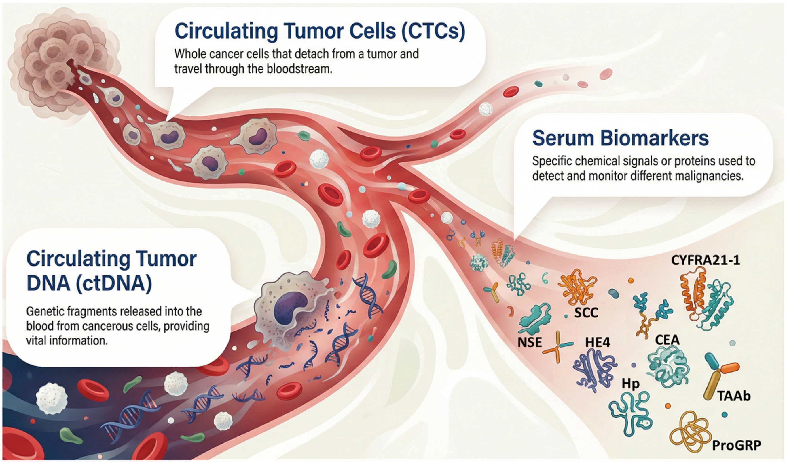
The search for alternatives that can overcome the limitations of LDCT has led to the development of numerous non-invasive and minimally invasive technologies. These globally researched strategies focus on detecting biomarkers in accessible biological samples. Figure 2 shows the circulating components (serum biomarkers, circulating tumor DNA (ctDNA) and circulating tumor cells (CTCs)) in the bloodstream relevant for cancer detection and monitoring.

3.2.1. Serum Biomarkers

A biomarker is a defined characteristic—molecular, histologic, radiographic, or physiologic—that is measured as an indicator of normal biological processes, disease processes (such as early, preclinical cancer), or responses to an exposure or intervention. In screening, biomarkers are used to identify individuals at elevated risk in order to enable earlier detection and more effective triage to diagnostic evaluation [36].
Carcinoembryonic Antigen (CEA): CEA is a glycoprotein involved in cell adhesion. While its levels are often elevated in lung cancer, particularly adenocarcinoma, it is not specific to this disease. As a general tumor marker, CEA can also be elevated in colorectal, pancreatic, gastric, and breast cancers, as well as in non-malignant conditions like smoking-related inflammation. Its sensitivity is moderate, with higher levels correlating to advanced stages and metastasis [37]. For example, Li et al., 2024 (case–control study) found that serum CEA levels were significantly higher in patients with NSCLC (n = 184) than in those with benign lung disease (n = 60) or healthy controls (n = 90) (p < 0.01) [38]. From the same study, as a single marker, CEA showed modest diagnostic discrimination for NSCLC (AUC = 0.638); however, when integrated with TuM2-PK and CYFRA21-1, accuracy was improved (AUC = 0.918). Furthermore, a retrospective analysis of newly diagnosed lung cancer focusing on metastasis assessment in 213 patients found that serum CEA levels were significantly higher in patients with metastasis (p < 0.001, AUC = 0.724), suggesting its utility in identifying advanced disease stages [37]. Based on PR-specific demographics, CEA levels could be explored as a low-cost triage adjunct. Smoking rates are lower in PR (8.5%) than in the mainland US (11.6%) making adenocarcinoma one of the most common types of lung cancer in this population [24,39,40]. Adenocarcinoma can present with elevated CEA levels; thus, this histologic distribution may increase the overall prevalence of this marker. Since smoking may cause false elevations in CEA, the lower smoking prevalence may improve CEA’s specificity in PR patients [39,41].
Cytokeratin 19 Fragment (CYFRA21-1): CYFRA21-1 detects fragments of cytokeratin 19 released from epithelial tumor cells. It is particularly useful in NSCLC, with sensitivity up to 71% and specificity up to 94%, according to a case–control study (newly diagnosed NSCLC vs. similar demographic) [42]. While more specific to NSCLC than CEA, it can also be elevated in other epithelial cancers like bladder cancer. It is especially helpful in detecting squamous cell carcinoma and shows a strong correlation with disease stage [43]. Given PR’s adenocarcinoma-predominant histology and CYFRA21-1’s stronger performance in squamous disease, CYFRA21-1 is best framed as a multi-marker panel component for risk enrichment/triage rather than a stand-alone screening marker, with value for enriching detection of the squamous NSCLC subset [26,44]. A comparative study (three group case–control study of lung cancer vs. benign pulmonary disease vs. healthy) shows that levels of CEA and CYFRA21-1 were significantly higher in lung cancer patients; the combination of these tumor markers along with NSE showed the highest sensitivity, reaching 90% for early detection, outperforming other diagnostic methods [45]. Another study comparing multiple markers (hospital cohort/case–control of malignant vs. benign/healthy) confirmed these findings, where CYFRA21-1 showed the highest sensitivity (70%) for detecting lung cancer [46]. In summary, its performance is best within multi-marker panels [42,47,48].
Squamous Cell Carcinoma Antigen (SCC-Ag): As its name suggests, SCC-Ag is most specific for squamous cell carcinoma. However, this is a cell type, not a cancer type, meaning that the marker can be elevated in squamous cell carcinomas of the lung, cervix, head, and neck. Its sensitivity for lung cancer is lower than other markers, limiting its use as a stand-alone diagnostic tool [49]. In a study by Li (2021) (three-group case–control of malignant vs. benign vs. healthy) involving 120 participants with lung cancer, serum SCC-Ag levels were significantly higher in squamous cell carcinoma compared to adenocarcinoma or small cell lung cancer, reinforcing its specific utility for this histological subtype. However, the study emphasized that a combined panel (with CEA, CYFRA 21-1, and ProGRP) yielded a superior diagnostic sensitivity of 93.29% and a specificity of 84.32%, significantly outperforming the sensitivity of markers used in isolation [50]. In adenocarcinoma-predominant settings, such as PR, SCC-Ag is unlikely to be a strong stand-alone marker; however, it may add incremental value and disease differentiation within multi-marker panels [26].
Neuron-Specific Enolase (NSE): NSE is an enzyme released from neuroendocrine cells and is strongly associated with tumors of this origin, most notably small cell lung cancer (SCLC). Its high specificity for neuroendocrine tumors makes it a valuable tool to help distinguish between SCLC and NSCLC, although its sensitivity is lower for NSCLC. In PR, NSE is best positioned as a subtype-oriented differential adjunct rather than a population-level screening marker. NSE is most effective when combined with ProGRP for SCLC screening [48,51,52]. In a case–control study, a group of researchers found that NSE showed higher sensitivity for small-cell carcinoma (AUC = 0.775) than for squamous carcinoma (AUC = 0.719) [53]. Leveraging real-world data, a retrospective descriptive utilization study showed that within 3 months before first diagnosis, NSE was obtained in ~65% of NSCLC and ~80% of SCLC cases, underscoring its screening role [54].
Progastrin-Releasing Peptide (ProGRP): ProGRP is currently regarded as one of the most accurate and specific biomarkers for SCLC, where it can yield an AUC as high as 0.973 [47,52,55,56]. Its utility for NSCLC is limited. As with NSE, ProGRP is best used to discriminate between the main classifications of lung cancer and to guide physician workups when elevated. In a study of 452 participants (diagnostic cohort/case–control of SCLC vs. NSCLC/benign), Oh et al. (2016) reported that plasma ProGRP reached a sensitivity of 85.7% and a specificity of 90.2% for SCLC, proving also to be a valuable tool for tumor staging and monitoring responses to chemotherapy [57]. In a retrospective cohort of 290 patients with lung neuroendocrine neoplasms, Rosiek et al. (2023) demonstrated that ProGRP is an exceptionally effective diagnostic marker, achieving a sensitivity of 94.8%, a specificity of 100%, and an AUC of 0.995 when distinguishing lung neuroendocrine neoplasms against healthy controls [58].
Human Epididymis Protein 4 (HE4): HE4 is an emerging marker showing promise for lung cancer but its primary clinical use is as a marker for ovarian cancer. In recent studies, HE4 has shown a high AUC for overall lung cancer detection when combined with traditional markers for both subtyping and diagnosis [47,59]. This finding suggests that it can be proposed as an investigational adjunct/panel candidate for screening in PR patients, although validation is needed for this cohort. The combination of HE4 with ProGRP and NSE was seen as a promising and optimal strategy for SCLC [47]. A case–control study by Iwahori et al. in 2012, using a novel ELISA system, evaluated 49 lung cancer patients and found that serum HE4 levels were significantly elevated in both NSCLC and SCLC, with an exceptional AUC = 0.988, a sensitivity of 89.8% and a specificity of 100% [60]. Another case–control study focusing on male lung cancer patients (n = 98) found that HE4 levels were significantly elevated compared to healthy controls (p < 0.001), with an AUC of 0.848 and a sensitivity of 64.3% at 95.9% specificity [61].
Haptoglobin (Hp): Hp is a type of acidoglycoprotein that has shown high sensitivity to lung cancer, especially when combined with CEA, NSE, and CYFRA21-1. Its positive detection rate is highest in the squamous cell carcinoma subtype [62]. For the PR population, this marker serves a complementary role in distinguishing between the most prevalent types of NSCLC on the island, improving sensitivity for squamous cell carcinoma. In a case–control study utilizing proteomic analysis followed by validation in lung cancer patients, Hp showed an AUC of 0.768 for distinguishing lung adenocarcinoma from healthy controls, with a sensitivity of 64% [63]. Furthermore, a study (three-group case–control of lung cancer vs. benign vs. healthy) involving 193 subjects with lung cancer and 87 with benign disease reported that while serum Hp alone had a moderate sensitivity of 43.5% and a specificity of 90.7%, its combination with CEA, NSE, and CYFRA 21-1 significantly increased the overall positive detection rate to 85.0%, particularly aiding in pathological typing [62].
Tumor-Associated Autoantibodies (TAAb): TAAb arise when the host immune system mounts a humoral response against tumor antigens (e.g., p53, NY-ESO-1, CAGE, GBU4-5, HuD, MAGE-A4, SOX2), providing biological signal amplification that can be detectable months to years before radiologic visibility [64]. For example, a large, pragmatic, randomized, controlled trial (12,208 participants) called the Early Detection of Cancer of the Lung Scotland (ECLS), used the EarlyCDT-Lung autoantibody blood test, offering LDCT only to test-positive participants (every 6 months for up to 2 years), while test-negative and control participants received the usual care [65]. Notably, they observed a stage shift with fewer stage III/IV diagnoses versus usual care (hazard ratio for stage III/IV = 0.64; 95% CI 0.41–0.99), supporting the role of autoantibodies as a high-specificity triage rather than a stand-alone screen. At the same time, independent evaluations reported modest sensitivity for EarlyCDT-Lung (≈30–40% overall and lower for stage I–II) [66]. These findings are relevant for PR, where contemporary cohorts indicate that a high proportion of NSCLC patients present with advanced disease, consistent with documented screening implementation barriers [26]. This proposed biomarker could help to make a more promptly diagnosis in the Puerto Rican population.

3.2.2. Exhaled Breath Analysis

Nitric Oxide (NO): Aberrant NO signaling is a central feature of cancer [67]. The measurement of fractional exhaled nitric oxide (FeNO) is a non-invasive technology that reflects changes in airway inflammation. FeNO is generally elevated in lung cancer patients when compared to healthy controls; its levels correspond to tumor burden and stage, especially in NSCLC [68]. However, FeNO can also be elevated in other inflammatory lung conditions such as asthma. It shows good sensitivity for distinguishing lung cancer patients from controls but may lack adequate clinical specificity if used alone [69]. In PR, asthma and other respiratory diseases are very prevalent due to air pollution; thus, this marker may be insufficient on its own but can serve as an adjunct in a multi-modal approach to lung cancer screening [70,71]. Furthermore, alterations in exhaled NO may also correlate with changes in cytokine profiles, such as IL-17 and VEGF, linking NO production to ongoing tumor-mediated inflammation and angiogenesis [72].
Exhaled Breath Analysis (VOCs, Sensor Arrays): Sensor-based technologies enable the multiplexed detection of volatile organic compounds (VOCs) and other exhaled gases (e.g., NO, acetone, isoprene), providing a rapid and non-invasive strategy for lung cancer screening. Recent advances in electronic nose (“e-nose”) systems with metal oxide sensors have improved both the sensitivity and selectivity of breath-based diagnostics. These platforms hold promise for point-of-care application, especially when incorporated with ensemble learning models for higher diagnostic accuracy [69,73,74]. For patients in PR, a breath-based triage may provide a low-burden primary-care gateway that helps prioritize limited imaging capacity amid workforce constraints, capitation, and payer authorization requirements.

3.2.3. Liquid Biopsy (ctDNA, CTCs, miRNA)

Liquid biopsies detect tumor-derived materials such as ctDNA and CTCs from the blood [75,76,77]. Recent tumor-informed ctDNA assays with ultrasensitive, phased-variant sequencing achieve subparts-per-million limits of detection and have doubled clinical sensitivity for post-operative minimal residual disease compared with first-generation assays, enabling recurrence prediction before imaging [78,79]. In the indeterminate pulmonary nodule setting, integrating plasma ctDNA methylation with clinical and CT features improved malignant/benign classification, especially for 5–10 mm nodules [80]. CTCs complement ctDNA by offering cellular phenotyping; epithelial/mesenchymal hybrid CTCs correlate with metastatic potential in NSCLC and can aid in subtype discrimination and resistance monitoring when combined with ctDNA in multimodal workflows [81].
As components of liquid biopsy, miRNAs are part of tumor-derived materials that can be detected without a tissue biopsy; alongside ctDNA and CTCs, a liquid biopsy includes circulating RNA, either free in plasma or packaged in extracellular vesicles such as exosomes [82]. In a large multicenter cohort of 3046 individuals, a blood-borne miRNA signature achieved 91.4% accuracy (82.8% sensitivity, 93.5% specificity) for lung cancer detection across symptomatic populations and clinically relevant controls [83], supporting utility for triage and enrichment prior to LDCT and demonstrating disease-focused specificity rather than generic inflammation signals alone. Beyond single analytes, exosomal miRNA panels measured in serum improved analytical robustness and biological specificity (vesicular protection; tumor-derived cargo), with optimized multi-miRNA combinations reporting ROC AUC > 0.93 for early-stage NSCLC in discovery/validation workflows—an attractive feature for pre-imaging screening or parallel testing with serum proteins [84]. Importantly, integrated approaches that combine plasma miRNAs (e.g., miR-21-5p) with signals from other matrices (e.g., sputum miR-31-5p/miR-210-3p) have demonstrated 85.5% sensitivity and 91.7% specificity, independent of stage and histology, reinforcing that miRNA can raise pre-test probability and increase PPV when embedded in multiple, machine learning-assisted panels alongside established serum biomarkers [85]. The aggregate evidence indicates that miRNAs—individually, in vesicles, or in integrated panels—offer lung-relevant specificity and screening performance that can be operationalized within a biomarker-first workflow to streamline LDCT utilization.
While ctDNA and methylation-based assays represent major advances in liquid biopsy, their performance for very-early, asymptomatic disease remains limited. Much of the evidence reporting high sensitivity originates from diagnostic or minimal residual disease (MRD)-oriented cohorts rather than true screening populations, as highlighted in reviews of liquid-biopsy implementation and limitations [75,79]. Moreover, the detection of low-variant-allele-frequency ctDNA can be confounded by clonal hematopoiesis, which produces non-tumor mutations that reduce specificity, particularly in older individuals [79]. This is particularly relevant in PR, given its older age structure (≈24.6% of residents are ≥65 years), which may increase the prevalence of clonal hematopoiesis and necessitates careful assay calibration and external validation in local populations [86,87]. Assay-specific differences in sequencing depth, target panels, bioinformatic pipelines, and analytical sensitivity introduce additional variability across platforms [75]. The high cost of advanced NGS-based ctDNA assays currently limits their feasibility for broad population screening. For these reasons, ctDNA and methylation assays should be interpreted as adjunct tools, with their most validated roles in: (1) the adjudication of indeterminate pulmonary nodules, where integrating plasma methylation with LDCT and protein markers improves malignancy discrimination; and (2) MRD detection and recurrence monitoring, where ultrasensitive tumor-informed approaches have demonstrated strong predictive value [78,80]. At present, no prospective population-screening trials support ctDNA or methylation assays as first-line screening tests; therefore, we position them as complementary technologies rather than primary screening modalities.
Implementation of liquid-biopsy procedures in PR should account for variability in clinician knowledge and the utilization of lung cancer biomarker testing, reinforcing the need for standardized workflows and decision support if deployed [88].

3.2.4. Universal Limitations of Emerging Biomarkers

These emergent technologies share universal technical limitations. A primary challenge is often insufficient sensitivity and specificity for early-stage disease. Many serum markers are also elevated in non-malignant inflammatory conditions, leading to false positives. It is important to highlight that reported diagnostic accuracies for serum biomarkers are often from case–control or symptomatic cohort studies, which could inflate their sensitivity, specificity, and AUC results. This is because the information is from symptomatic cohorts, clinically indicated diagnostic workups, or case–control designs, rather than true asymptomatic screening populations. This means that these settings often include a higher proportion of advanced-stage cancers and fewer benign comparators. This may create spectrum bias, meaning that performance does not generalize to asymptomatic individuals undergoing screening. In addition, in true screening settings with low disease prevalence, even the highly specific biomarkers yield lower positive predictive values (PPV), which increases false-positive referrals. Therefore, all accuracy estimates in this section should be interpreted within the context of each study setting, population, and stage distribution, and not assumed to reflect screening-level performance. Moreover, multi-biomarker panels with modest sample sizes are prone to overfitting when developed by machine learning models. The performance can be inflated by batch effects such as systematic differences in sample handling between cases and controls. Ultimately, many panels lack external validation with independent screening-like cohorts. This makes data preliminary and it may not transport to real-world screening settings.
Exhaled breath analysis is susceptible to confounding variables like diet and co-existing respiratory conditions. Liquid biopsies can struggle to detect the low levels of ctDNA present in early-stage cancer. Finally, many of these technologies are still experimental, costly, and lack the standardization needed for widespread clinical use as screening tools.
Another limitation is that PR-specific validation data for these emerging biomarkers in population-based cohorts is limited. As a result, reported trends are extrapolated from other groups. Differences in population characteristics and workflows may affect calibration and generalizability, underscoring the need for external validation. A study is currently being designed to evaluate these biomarkers directly in PR lung cancer patients and to generate local performance data.

3.3. Additional Emerging Technologies

  • Genetic/Epigenetic Markers: Assays, such as DNA methylation tests of SHOX2/RASSF1A in bronchoalveolar lavage fluid (BALF), have been shown to provide higher sensitivity than serum CEA in early lung cancer detection and can serve as a diagnostic adjunct when cytology is inconclusive [89].
  • Advanced Imaging: Near-infrared fluorescence tumor-targeted imaging offers enhanced intraoperative detection of cancerous nodules, supporting surgical navigation [90].
  • Tumor Markers in Other Fluids: Traditional and novel markers can also be assayed on pleural fluid for diagnosing malignancy [91]. Pleural fluid, defined as liquid around the lungs, has emerged as a valuable biological matrix for evaluating lung cancer–associated malignant pleural effusion. Several tumor biomarkers, including CEA, CYFRA-21-1, NSE, SCC-Ag, and ProGRP, have been rigorously studied for their diagnostic performance in this compartment. Among these, CEA consistently demonstrates the highest diagnostic accuracy: a major 2020 study analyzing 348 patients with pleural effusions identified pleural-fluid CEA at a cutoff of 5.23 ng/mL as the most effective marker, achieving 99% sensitivity and 91.6% accuracy in distinguishing malignant from benign effusions, outperforming CYFRA-21-1, SCC-Ag, and NSE [92]. NSE and ProGRP are detectable in pleural fluid but generally yield lower diagnostic performance compared with CEA and CYFRA-21-1; however, they remain clinically useful for differential diagnosis and the histological typing of malignant pleural effusion [93]. SCC-Ag is also measurable in pleural fluid but consistently performs weaker than CEA and CYFRA-21-1 in differentiating malignant from benign effusions [92]. Collectively, these studies show that pleural fluid biomarkers—especially CEA, followed by CYFRA-21-1—provide meaningful diagnostic support in lung cancer evaluation. However, it is important to note that obtaining pleural fluid requires thoracentesis, an invasive medical procedure. For this reason, it is not considered the best matrix for early screening.
Beyond pleural fluid, saliva and sputum offer non-invasive matrices that can capture the same molecular signals discussed for plasma/serum. In saliva, case–control modeling with miRNAs (e.g., let-7a-2, miR-221, miR-20a) achieved up to 90% accuracy (95% sensitivity, 85% specificity) for distinguishing lung cancer from controls, underscoring the feasibility for point-of-care triage in high-risk populations [94]. Complementarily, a salivary metabolomics study that profiled paired tumor/adjacent tissues, plasma, and saliva (n ≈ 109 pairs) identified 12 metabolites shared across matrices (polyamine/amino-acid pathways) and produced a 12-metabolite salivary panel with validation AUCs~0.74–0.79, reinforcing saliva as a proxy for early tumor biology [95]. Despite the absence of studies examining the early screening of lung cancer through the detection of the serum protein biomarkers discussed in this review, certain serum markers, including CEA, CYFRA-21-1, NSE, and Hp, have been identified in saliva [94,96,97,98]. This finding underscores the direct translational potential of serum markers in salivary testing for community-based triage. However, salivary marker studies require strict methodological control, as concentration levels can differ significantly between individuals and are affected by oral health. Therefore, creating standard criteria for normal and pathological ranges is essential for accurate diagnosis.
In sputum, an integrated panel that combines sputum miRNAs (miR-31-5p, miR-210-3p) with plasma miRNAs (miR-21-5p) improved discrimination of NSCLC, with 85.5% sensitivity and 91.7% specificity, independent of stage and histology [85]. Regarding protein biomarkers, Kalomenidis (2004) discovered that CEA, CYFRA21, and NSA attained sensitivities in sputum that were analogous to those observed in the serum of lung cancer patients [99]. In principle, sputum is produced from the respiratory tract, which could improve specificity for early lung cancer screening. Collectively, these saliva/sputum data demonstrate that other fluids can detect biomarkers already used in serum together with miRNA and metabolite signatures, enabling earlier, lung-focused case findings when deployed as machine learning–assisted multiplex panels alongside imaging. For PR, saliva/sputum triage and methylation-based adjudication are plausible for near-term applications. These non-invasive methods may be useful to increase screening capacity before LCDT referral, lowering late-stage detection. Nevertheless, PR-specific validation in local cohorts is necessary before broader adoption.

3.4. Future Developments and Ongoing Research

Future directions for LDCT screening aim to optimize eligibility criteria using individualized risk models, minimize harms by refining follow-up protocols, and enhance cost-effectiveness. There is also a push to expand screening to certain non-high-risk populations, such as never-smokers and younger adults, as evidence accumulates [7,69,100].
Table 1 consolidates the core characteristics and performance of the gold standard LDCT, serum biomarkers, breath analytes and liquid biopsy—linking each test/marker to its sample type, invasiveness, targeted lung cancer subtype(s), clinical role/utility and diagnostic value—to enable rapid, evidence-based comparison across options. This harmonized view highlights complementarities among biomarkers and technologies, clarifies where each test adds incremental value, and supports reproducible panel design tailored to histology and care settings.
Table 2 highlights a growing shift toward non-blood, lung-proximal biofluids to improve early lung cancer detection by capturing biomarkers closer to their site of origin. In pleural fluid, traditional tumor markers show strong discriminatory performance in clinically indicated effusions. However, because pleural fluid collection is inherently procedure-dependent and often reflects advanced disease contexts (e.g., malignant pleural effusion), Table 2 motivates future work focused on less invasive respiratory matrices, such as sputum and saliva, for population-relevant screening.

4. Discussion

The dual challenge of lung cancer screening—universal technical limitations combined with specific contextual barriers—is starkly evident in PR. While LDCT is the global standard, is not a good fit for an island with limited resources and a fragmented healthcare system. Similarly, while emerging biomarkers are promising, their own technical limitations (e.g., variable sensitivity/specificity) prevent any single one from being a standalone solution.
This is where the promise of a multi-faceted approach becomes clear. An integrated, multi-marker, non-invasive panel offers the most viable path forward for Puerto Rican patients. Single serum markers rarely achieve acceptable screening performance when used alone; combining markers that capture orthogonal biology (e.g., tumor load, neuroendocrine features, epithelial injury) improves accuracy.
The recommended biomarkers for our proposed first-triage panel are CEA + CYFRA-21-1 + NSE + ProGRP + SCC-Ag + HE4. These biomarkers can be analyzed with machine learning classifiers rather than fixed cut-offs, demonstrating substantially higher discrimination for lung cancer, and for small-cell histology, in particular [47,48,56,62,102]. They were selected based on their complementary histology coverage—adenocarcinoma (CEA, HE4), squamous (CYFRA 21-1, SCC-Ag), and SCLC (ProGRP, NSE)—and superior performance in combination versus any single marker, which is critical for a triage-first approach that seeks to raise specificity and contain the imaging burden. In parallel, TAAb offers a high specificity and biological lead time, making TAAb well-suited as a triage gateway before LDCT to limit downstream false-positive imaging [65]. Additionally, integrating a low-cost breath sensor array—FeNO plus VOC profiling via e-nose—into the panel is feasible; e-nose systems have distinguished lung cancer from other pulmonary diseases and are amenable to point-of-care use, especially when locally calibrated for ambient humidity and particulate conditions [69,73,74]. Finally, for indeterminate nodules or borderline-risk cases, a blood-based liquid biopsy (ctDNA/CTCs) can serve as a molecular adjudicator alongside imaging—useful to refine malignancy probability and, in postoperative settings, to detect minimal residual disease—although sensitivity in very early-stage disease remains a limiting factor [35]. In very early-stage settings, a multimodal biomarker panel (serum proteins + tumor autoantibodies ± e-nose) typically achieves higher practical sensitivity than the ctDNA assay, whereas ctDNA contributes molecular specificity and is best positioned as an adjudicator for indeterminate nodules and for recurrence monitoring rather than as a first-line screening replacement [103]. However, consistent with expert guidance emphasizing the importance of study design in biomarker evaluation, we deliberately avoid overstating early-disease sensitivity for ctDNA or methylation assays and instead highlight their current, evidence-supported roles in nodule adjudication and MRD assessment [66].
Hispanic/Latino cohorts—including Puerto Ricans—exhibit higher EGFR mutation prevalence (approaching ~24–40%) and relatively lower KRAS rates compared with non-Hispanic Whites [40,104]. Screening and triage panels for PR should, therefore, ensure the sensitive detection of EGFR (Ex19del/L858R) and consider ALK/ROS1 as part of a liquid-biopsy adjudication step for indeterminate LDCT findings. Building in-island rapid-turnaround workflows (e.g., automated NGS platforms) can shorten the time to result and reduce send-out costs. This type of research also moves medicine toward a more personalized approach, allowing for the development of diagnostic algorithms tailored to the unique genetic and environmental profile of the Puerto Rican population.
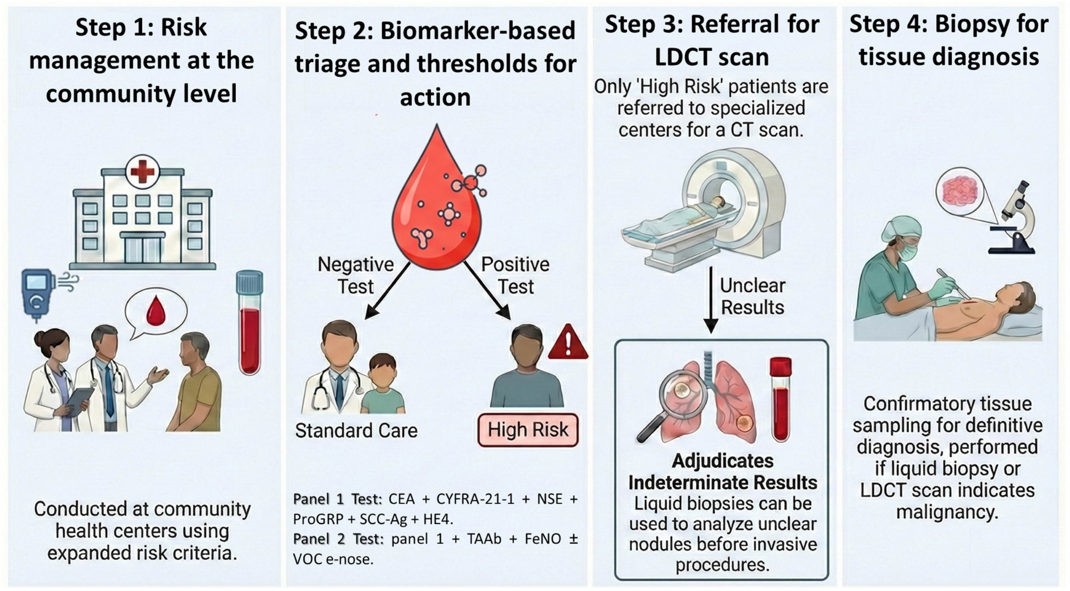
Figure 3 outlines a four-step process designed to identify and evaluate at-risk patients, likely for lung cancer.
Step 1—Risk Management at the community level. Primary-care teams in community health centers apply expanded, locally relevant risk criteria (age, tobacco exposure, environmental/occupational factors, and comorbidities) and collect a single blood sample for the core serum biomarker panel (CYFRA 21-1, CEA, SCC-Ag, ProGRP, NSE, HE4), with an optional autoantibody module and point-of-care breath testing when phlebotomy or transport is challenging. The workflow is designed for PR’s distributed care network, as samples can be drawn close to home, transported to a centralized laboratory under standardized procedures, and reported back within a turn-around time that supports timely referral. This step anchors the model in primary care, lowers access barriers for rural municipalities, and aligns testing with payer documentation and authorization needs.
Step 2—Biomarker-based triage and clear thresholds for action. Results from the multi-analyte panel are combined with clinical risk to produce a simple, operational decision: “triage-negative” individuals return to routine care and prevention (including smoking cessation and symptom vigilance), while “triage-positive” individuals are flagged as high-risk and prioritized for imaging. The thresholds are calibrated to local prevalence and the capacity to maximize specificity (thereby reducing unnecessary imaging) while maintaining acceptable sensitivity; autoantibody positivity is treated as a rule-in signal. This step is intentionally framed to conserve limited LDCT slots, contain out-of-pocket exposure under capitation models, and make the referral process predictable for primary-care clinicians. For this biomarker-based triage, we propose two types of panels: Panel 1 (minimum viable) is a serum-only panel (CEA + CYFRA-21-1 + NSE + ProGRP + SCC-Ag + HE4) from a single blood draw; Panel 2 (enhanced) adds the TAAb rule-in module and, where feasible, breathomics (FeNO ± VOC “e-nose”) with machine learning integration introduced only after local calibration/validation to reduce overfitting and batch effects. TAAb triage prior to CT has randomized trial support [65].
Step 3—Referral for LDCT scan and adjudication of indeterminate results. Only patients categorized as high-risk are referred to specialized LDCT centers, concentrating imaging resources on those with a higher post-test probability of disease. LDCT reports follow standardized screening nomenclature; when the scan is indeterminate, the pathway introduces a noninvasive liquid-biopsy adjudication (e.g., ctDNA-based or multi-omic signatures) before any invasive procedures are considered. This staged approach reduces avoidable downstream testing, supports patient navigation across island regions, and embeds explicit checks against overuse, which is particularly important where radiology capacity and appointment availability can be constrained.
Step 4—Biopsy for tissue diagnosis. Patients proceed to biopsy when LDCT or the combined imaging-plus-molecular assessment indicates malignancy or high suspicion. Tissue sampling confirms diagnosis and guides definitive management, with a multidisciplinary review to determine the safest, most efficient route (e.g., percutaneous vs. endoscopic). By deferring invasive sampling until after a structured triage and imaging-adjudication sequence, the model minimizes false-positive cascades, reduces patient travel and financial burden, and preserves specialty capacity—fulfilling the manuscript’s goal of a context-adapted pathway for PR that is pragmatic, equitable, and resource-aware.
In PR, this triage-first approach helps to enrich pre-test probability, reduce unnecessary scans/biopsies, and preserve scarce resources. Table 3 shows the advantages of this multi-marker panel approach compared with LDCT.
  • Steps for Implementation and Considerations
Effective adoption in PR depends on matching a biomarker-first triage model to day-to-day realities in primary care, radiology, and payment. Below, we outline concise but practical steps.
(a)
Laboratory readiness and quality. Integrated multi-panel biomarker testing should be conducted in primary care (multiplex serum ± TAAb; breathomics where feasible) using pre-specified thresholds tuned to improve specificity while preserving sensitivity. Assays with clear instructions, standardized procedures, and routine quality checks should be used. Test failure/invalid rates should be tracked and decision cutoffs in the local population should be checked before scaling.
Baseline risk assessment: age, smoking, clinical factors in primary care
Six core serum biomarker panel: CEA + CYFRA-21-1 + NSE + ProGRP + SCC-Ag + HE4 ± TAAb in the same blood draw;
(b)
Logistics and turnaround time. Prioritize sample types and workflows that fit primary care (blood draw or breath analysis). Ensure reliable transport where needed and aim for a turnaround time that supports timely referrals (e.g., within 3–5 business days). Monitor delays and address bottlenecks early;
(c)
Clinical workflow and thresholds. Provide a one-page protocol that states: who is eligible, which biomarker panels are ordered, the exact threshold that triggers LDCT, and when to use liquid biopsy for indeterminate imaging. Include a simple reflex pathway and criteria for multidisciplinary review. All triage-positive cases proceed to LDCT with management governed by Lung-RADS v2022 (American College of Radiology’s structured system for management of findings on LDCT lung cancer screening) to reduce unnecessary procedures and false-positive cascades;
(d)
Fit with capacity and payment. Align test volumes with radiology capacity to avoid overload. Plan the budget across the full pathway (biomarker tests, confirmatory imaging, follow-up visits, patient navigation). Engage payers early to clarify coverage, documentation, and coding;
(e)
Equity and access. Expand access beyond large centers through community clinics, mobile blood collection, or point-of-care breath testing. Provide language-concordant materials and patient navigation to reduce loss to follow-up, especially in rural areas;
(f)
Data and monitoring. Build a simple dashboard to track the time from order to result, invalid rate, proportion of biomarker-positive patients receiving LDCT, number of scans avoided, positive predictive value in practice, and stage at diagnosis. Review these metrics regularly and adjust thresholds if needed;
(g)
Training and communication. Offer brief training for primary care teams on intended use, results interpretation, and next steps. Provide standard referral letters and clear contact channels among primary care, radiology, and oncology;
(h)
Governance and ethics. Ensure appropriate consent, protect privacy, and set a clear plan for communicating incidental or indeterminate findings. Consider community input to maintain trust;
(i)
Phased rollout and pilot. Start with a small pilot in a few clinics and a centralized laboratory. Define success targets (for example, turnaround time ~ 5 days, invalid tests < 5%, fewer unnecessary scans, earlier stage at diagnosis). Use “go/no-go” criteria to decide on expansion and refine the pathway based on real-world results. We will also look for a shift toward earlier stage at diagnosis, higher positive predictive value of the pathway, and shorter time from first presentation to treatment start, with results reported by region to ensure equity.
  • Example of Clinical Implementation Scenarios:
The following scenarios illustrate how a biomarker-first triage pathway could operate within PR’s primary-care network and constrained imaging capacity. We propose the following two candidate panels for pragmatic deployment:
Case 1—Primary care-based screening in a screening-eligible smoker. A 62-year-old male residing in a rural municipality presents for routine follow-up in a community primary-care clinic. He has a 35 pack-year smoking history and meets USPSTF eligibility criteria for annual LDCT screening [7]. However, local access to LDCT is limited and requires travel to a tertiary imaging center, which may reduce screening completion. Under a biomarker-first triage workflow, the primary-care team orders the six-marker core serum panel (CEA + CYFRA-21-1 + NSE + ProGRP + SCC-Ag + HE4) proposed in Figure 3 Step 2 (Panel 1)—from a single blood-draw during the visit, with Panel 2 (enhanced) options (TAAb rule-in and/or breath testing), where feasible [65]. Samples are transported to a regional or centralized laboratory under standardized handling procedures with routine quality checks (calibrators/controls, predefined repeat/invalid criteria, and tracking invalid test rates); results are returned within a timeframe that supports timely referrals (e.g., within 3–5 business days, or faster where local capacity allows). Patients classified as triage-negative continue risk reduction and reassessment at defined intervals. Patients classified as triage-positive are prioritized for LDCT referral through a coordinated navigation pathway, with subsequent management governed by Lung-RADS v2022 to reduce unnecessary procedures and false-positive cascades [105]. This approach aims to enrich LDCT referrals, conserve limited imaging slots, and reduce drop-off related to travel and scheduling barriers.
Case 2—Incidental pulmonary nodule risk stratification. A 58-year-old female former smoker undergoes abdominal CT at a regional hospital for unrelated symptoms. Imaging identifies an incidental 6-mm subpleural pulmonary nodule. Standard management often involves serial CT follow-up, contributing to imaging burden and anxiety when pretest malignancy probability is low. In a biomarker-assisted triage model (Figure 3 Step 2), serum biomarker testing (Panel 1) is incorporated as an adjunct to clinical and radiographic risk stratification. A low-risk biomarker profile could support longer surveillance intervals consistent with Fleischner Society guidance for incidental pulmonary nodules [106]. Conversely, a high-risk biomarker signal would prompt earlier LDCT reassessment or advanced imaging evaluation and expedited referral to pulmonology or a multidisciplinary tumor board. When imaging findings remain indeterminate or management uncertainty persists, a methylation-based ctDNA assay can be reserved as an adjudicator (where cost/logistics permit) before invasive procedures are pursued, recognizing that ctDNA/methylation assays are less suited as first-line screening tests in low-prevalence settings [107,108,109,110]. This staged approach can reduce unnecessary serial imaging while accelerating the evaluation of higher-risk lesions in settings with constrained radiology capacity. The assay platforms assume that serum proteins and TAAb are measured using standard clinical immunoassay platforms (e.g., automated chemiluminescent immunoassays/CLIA or validated ELISA methods) in a regional or centralized laboratory and that breath testing uses a FeNO device and/or a VOC sensor array (“e-nose”) at the point of care (requiring device calibration and quality checks) [73,111,112].
An operational burden/cost comparison shows that: serum-only testing has the lowest complexity; adding TAAb increases lab complexity modestly; breathomics shifts requirements to point-of-care device calibration and environmental control; and ctDNA/methylation assays are higher cost and more logistically challenging, and are, therefore, best positioned as adjudicators for indeterminate imaging rather than for first-line population screening [108,109,111].

5. Conclusions

The global strategies for lung cancer screening face both universal technical hurdles and context-specific implementation barriers. In PR, where socioeconomic and healthcare system challenges are pronounced, relying on the current standard of LDCT is insufficient to address the public health crisis of late-stage lung cancer. Substantially, PR’s low screening uptake reflects multiple restrictions, such as payer and authorization barriers, workforce and imaging-capacity limitations, and participation-related factors such as transportation, health literacy, and fear. A promising and potentially more equitable path forward lies in the validation and implementation of an integrated panel of non-invasive biomarkers (biomarker-first triage strategy). This approach has the potential to transform lung cancer detection on the island, reducing mortality, improving quality of life, and ensuring that access to early diagnosis is a reality for all, not just a privilege for the few.

Author Contributions

Conceptualization, Y.D.; funding acquisition, Y.D.; investigation, K.R.-V., J.E.F., E.F., C.S. and Y.D.; methodology, K.R.-V., J.E.F., E.F., C.S. and Y.D.; project administration, Y.D.; resources, Y.D.; supervision, Y.D.; validation, Y.D.; visualization, K.R.-V. and Y.D.; writing—original draft, K.R.-V. and Y.D.; writing—review and editing, K.R.-V., C.S. and Y.D. All authors have read and agreed to the published version of the manuscript.

Funding

This project was supported by Puerto Rico IDeA Network Biomedical Research Excellence (PR-INBRE) (NIH-NIGMS Award Number P20 GM10347519); Hispanic Clinical and Translational Research Education and Career Development (HCTRECD) Program (NIH-NIMHD Award Number R25 MD007607); COBRE Puerto Rico Center for Microbiome Science (NIH-NIGMS Award Number P20 GM156713), Cancer Prevention and Control (CAPAC) Research Training Program (NIH-NCI Award Grant Number R25CA240120), and Fundación Intellectus. The contents are solely the responsibility of the authors and do not necessarily represent the official views of the sponsoring agencies. These agencies had no role in the design of the study, nor in the collection, analysis, or interpretation of data.

Institutional Review Board Statement

Not applicable.

Data Availability Statement

No new data were created or analyzed in this study. Data sharing is not applicable to this article.

Acknowledgments

The authors acknowledge the San Juan Bautista School of Medicine (SJBSM) for its sponsorship and support on the completion of this manuscript. The author utilized DeepL API v2, Gemini (Google), and NotebookLM to assist with the drafting, editing, and organization of this manuscript. These AI tools were used to refine language, synthesize literature, in the construction of figures, and to aid in structuring the arguments presented herein. The authors have reviewed and edited the output and take full responsibility for the contents of this publication.

Conflicts of Interest

The authors declare no conflicts of interest.

Abbreviations

The following abbreviations are used in this manuscript:
AIArtificial Intelligence
ALKAnaplastic Lymphoma Kinase
AUCArea Under the Curve
ATSAmerican Thoracic Society
BALFBronchoalveolar Lavage Fluid
CEACarcinoembryonic Antigen
CTComputed Tomography
CTCsCirculating Tumor Cells
ctDNACirculating Tumor DNA
CYFRA 21-1Cytokeratin 19 Fragment
EGFREpidermal Growth Factor Receptor
e-noseElectronic Nose
FeNOFractional Exhaled Nitric Oxide
HE4Human Epididymis Protein 4
HpHaptoglobin
IL-17Interleukin 17
KRASKirsten Rat Sarcoma Viral Oncogene Homolog
LDCTLow-Dose Computed Tomography
NLSTNational Lung Screening Trial
NONitric Oxide
NSENeuron-Specific Enolase
NSCLCNon-Small Cell Lung Cancer
ProGRPPro-Gastrin-Releasing Peptide
PPVPositive Predictive Value
PRPuerto Rico
ROS1c-ros Oncogene 1 (Receptor Tyrosine Kinase)
SCC-AgSquamous Cell Carcinoma Antigen
SCLCSmall Cell Lung Cancer
TAAbTumor-Associated Autoantibody
USPSTFU.S. Preventive Services Task Force
VOCVolatile Organic Compound
ECLSEarly Detection of Cancer of the Lung Scotland
Lung-RADSLung CT Screening Reporting and Data System
BRFSSBehavioral Risk Factor Surveillance System

References

  1. Sung, H.; Ferlay, J.; Siegel, R.L.; Laversanne, M.; Soerjomataram, I.; Jemal, A.; Bray, F. Global Cancer Statistics 2020: GLOBOCAN Estimates of Incidence and Mortality Worldwide for 36 Cancers in 185 Countries. CA. Cancer J. Clin. 2021, 71, 209–249. [Google Scholar] [CrossRef]
  2. American Cancer Society. Lung Cancer Survival Rates. Available online: https://www.cancer.org/cancer/types/lung-cancer/detection-diagnosis-staging/survival-rates.html (accessed on 16 June 2025).
  3. Cancer Stat Facts: Lung and Bronchus Cancer. SEER Cancer Stat Facts: Lung and Bronchus Cancer. Available online: https://seer.cancer.gov/statfacts/html/lungb.html (accessed on 20 March 2025).
  4. Rodríguez-Rabassa, M.S.; Simmons, V.N.; Vega, A.; Moreno, D.; Irizarry-Ramos, J.; Quinn, G.P. Perceptions of and Barriers to Lung Cancer Screening Among Physicians in Puerto Rico: A Qualitative Study. J. Health Care Poor Underserved 2020, 31, 973–991. [Google Scholar] [CrossRef]
  5. Olazagasti, C.; Seetharamu, N. Disparities in Lung Cancer Screening in Puerto Rico: A United States Colony with Unequal Benefits. Cancer Control 2021, 28, 10732748211051924. [Google Scholar] [CrossRef]
  6. Castañeda-Avila, M.A.; Santiago-Rodríguez, E.J.; Rodríguez-Cintrón, W.; Olazagasti, C.; Flores, E.J.; Rodríguez, E.; Velázquez Mañana, A.I.; Otero-Domínguez, Y.; Núñez, E.R. Lung Cancer Screening Eligibility, Uptake, and Adherence in Puerto Rico, 2022. JTO Clin. Res. Rep. 2025, 6, 100852. [Google Scholar] [CrossRef]
  7. US Preventive Services Task Force; Krist, A.H.; Davidson, K.W.; Mangione, C.M.; Barry, M.J.; Cabana, M.; Caughey, A.B.; Davis, E.M.; Donahue, K.E.; Doubeni, C.A.; et al. Screening for Lung Cancer: US Preventive Services Task Force Recommendation Statement. JAMA 2021, 325, 962. [Google Scholar] [CrossRef]
  8. Passiglia, F.; Cinquini, M.; Bertolaccini, L.; Del Re, M.; Facchinetti, F.; Ferrara, R.; Franchina, T.; Larici, A.R.; Malapelle, U.; Menis, J.; et al. Benefits and Harms of Lung Cancer Screening by Chest Computed Tomography: A Systematic Review and Meta-Analysis. J. Clin. Oncol. 2021, 39, 2574–2585. [Google Scholar] [CrossRef]
  9. Wender, R.; Fontham, E.T.H.; Barrera, E.; Colditz, G.A.; Church, T.R.; Ettinger, D.S.; Etzioni, R.; Flowers, C.R.; Scott Gazelle, G.; Kelsey, D.K.; et al. American Cancer Society lung cancer screening guidelines. CA. Cancer J. Clin. 2013, 63, 106–117. [Google Scholar] [CrossRef]
  10. Burzic, A.; O’Dowd, E.L.; Baldwin, D.R. The Future of Lung Cancer Screening: Current Challenges and Research Priorities. Cancer Manag. Res. 2022, 14, 637–645. [Google Scholar] [CrossRef]
  11. Lancaster, H.L.; Heuvelmans, M.A.; Oudkerk, M. Low-dose computed tomography lung cancer screening: Clinical evidence and implementation research. J. Intern. Med. 2022, 292, 68–80. [Google Scholar] [CrossRef]
  12. Guo, L.; Yu, Y.; Yang, F.; Gao, W.; Wang, Y.; Xiao, Y.; Du, J.; Tian, J.; Yang, H. Accuracy of baseline low-dose computed tomography lung cancer screening: A systematic review and meta-analysis. Chin. Med. J. 2023, 136, 1047–1056. [Google Scholar] [CrossRef]
  13. Pacheco, P.; Melo, V.; Martins, C.; Ribeiro, H. Lung Cancer Screening With Low-Dose CT: A Systematic Review. Cureus 2024, 16, e75515. [Google Scholar] [CrossRef]
  14. Shin, D.W.; Chun, S.; Kim, Y.I.; Kim, S.J.; Kim, J.S.; Chong, S.; Park, Y.S.; Song, S.-Y.; Lee, J.H.; Ahn, H.K.; et al. A national survey of lung cancer specialists’ views on low-dose CT screening for lung cancer in Korea. PLoS ONE 2018, 13, e0192626. [Google Scholar] [CrossRef]
  15. Barton, M.K. Integration of lung cancer screening into practice is lacking. CA. Cancer J. Clin. 2015, 65, 255–256. [Google Scholar] [CrossRef]
  16. Sahar, L.; Douangchai Wills, V.L.; Liu, K.K.; Kazerooni, E.A.; Dyer, D.S.; Smith, R.A. Using Geospatial Analysis to Evaluate Access to Lung Cancer Screening in the United States. Chest 2021, 159, 833–844. [Google Scholar] [CrossRef]
  17. Iaccarino, J.M.; Clark, J.; Bolton, R.; Kinsinger, L.; Kelley, M.; Slatore, C.G.; Au, D.H.; Wiener, R.S. A National Survey of Pulmonologists’ Views on Low-Dose CT Screening for Lung Cancer. Ann. Am. Thorac. Soc. 2015, 12, 1667–1675. [Google Scholar] [CrossRef]
  18. Henderson, L.M.; Marsh, M.W.; Benefield, T.S.; Jones, L.M.; Reuland, D.S.; Brenner, A.T.; Goldstein, A.O.; Molina, P.L.; Maygarden, S.J.; Rivera, M.P. Opinions and Practices of Lung Cancer Screening by Physician Specialty. N. C. Med. J. 2019, 80, 19–26. [Google Scholar] [CrossRef]
  19. Jonas, D.E.; Reuland, D.S.; Reddy, S.M.; Nagle, M.; Clark, S.D.; Weber, R.P.; Enyioha, C.; Malo, T.L.; Brenner, A.T.; Armstrong, C.; et al. Screening for Lung Cancer With Low-Dose Computed Tomography: Updated Evidence Report and Systematic Review for the US Preventive Services Task Force. JAMA 2021, 325, 971. [Google Scholar] [CrossRef]
  20. Pinsky, P.F.; Gierada, D.S.; Black, W.; Munden, R.; Nath, H.; Aberle, D.; Kazerooni, E. Performance of Lung-RADS in the National Lung Screening Trial: A Retrospective Assessment. Ann. Intern. Med. 2015, 162, 485–491. [Google Scholar] [CrossRef]
  21. Torres-Cintrón, C.R.; Suárez-Ramos, T.; Pagán-Santana, Y.; Román-Ruiz, Y.; Gierbolini-Bermúdez, A.; Ortiz-Ortiz, K.J. 2018–2022 Cancer in Puerto Rico: Incidence, Mortality and Survival; Puerto Rico Central Cancer Registry: San Juan, Puerto Rico, 2025; Available online: https://rcpr.org/Portals/0/informe%202018-2022%20-%20Ingles.pdf?ver=Itl1zSSVgNpMIzvxFfG1zw%3D%3D&utm (accessed on 4 March 2026).
  22. State Cancer Profiles: Puerto Rico. Available online: https://statecancerprofiles.cancer.gov/quick-profiles/index.php?statename=puertorico (accessed on 4 March 2026).
  23. Bandi, P.; Star, J.; Ashad-Bishop, K.; Kratzer, T.; Smith, R.; Jemal, A. Lung Cancer Screening in the US, 2022. JAMA Intern. Med. 2024, 184, 882. [Google Scholar] [CrossRef]
  24. BRFSS Prevalence & Trends Data. US Centers for Disease Control and Prevention. Available online: https://www.cdc.gov/brfss/brfssprevalence/index.html (accessed on 4 March 2026).
  25. 2025–2030 Puerto Rico Comprehensive Cancer Control Plan; Coalición para el Control de Cáncer en Puerto Rico: San Juan, Puerto Rico, 2024; Available online: https://www.iccp-portal.org/sites/default/files/2025-04/PRCCC-Plan-2025-2030.pdf (accessed on 4 March 2026).
  26. Miranda, E.I.; Gierbolini-Bermúdez, A.; Quintana, R.; Torres-Cintrón, C.R.; Ortiz-Ortiz, K.J. Treatment Patterns and Health Care Resource Utilization of Patients With Non–Small Cell Lung Cancer in Puerto Rico: The TREATLINES-ONCOLUNG Study. JCO Glob. Oncol. 2024, 10, e2400089. [Google Scholar] [CrossRef]
  27. Rodríguez-Cintrón, W.; Morales-Colón, S.; Martínez-González, J.; Albors-Sanchez, J. Lung Cancer Screening in Community-Based Practice in Puerto Rico: A Survey of Puerto Rico Pulmonologists. P. R. Health Sci. J. 2022, 41, 161–164. [Google Scholar] [PubMed]
  28. 2020 Census Urban Areas Facts. 2023. Available online: https://www.census.gov/programs-surveys/geography/guidance/geo-areas/urban-rural/2020-ua-facts.html (accessed on 4 March 2026).
  29. Varas-Díaz, N.; Rodríguez-Madera, S.; Padilla, M.; Rivera-Bustelo, K.; Mercado-Ríos, C.; Rivera-Custodio, J.; Matiz-Reyes, A.; Santiago-Santiago, A.; González-Font, Y.; Vertovec, J.; et al. On leaving: Coloniality and physician migration in Puerto Rico. Soc. Sci. Med. 2023, 325, 115888. [Google Scholar] [CrossRef] [PubMed]
  30. Goodley, P.; Evison, M. Addressing Global Disparities in Lung Cancer Screening: Lessons From Puerto Rico and Beyond. JTO Clin. Res. Rep. 2025, 6, 100919. [Google Scholar] [CrossRef]
  31. Santiago-Santiago, A.J.; Rivera-Custodio, J.; Mercado-Ríos, C.A.; González-Font, Y.; Madera, S.R.; Varas-Díaz, N.; Padilla, M.; Ramos-Pibernus, A.; Rivera-Bustelo, K.; Vertovec, J.; et al. Puerto Rican physician’s recommendations to mitigate medical migration from Puerto Rico to the mainland United States. Health Policy OPEN 2024, 7, 100124. [Google Scholar] [CrossRef] [PubMed]
  32. Hall, J. Where Things Stand with the Radiologist Shortage. 2025. Available online: https://www.diagnosticimaging.com/view/where-things-stand-with-the-radiologist-shortage (accessed on 15 January 2026).
  33. FitzPatrick, T.; Wander, S. Puerto Rico Medicaid—Vital Program: Risk Adjustment and Mitigation Assessment; Puerto Rico Department of Health: San Juan, Puerto Rico, 2022. Available online: https://docs.pr.gov/files/ASES/Comunicaciones/Avisos%20P%C3%BAblicos/Puerto-Rico-Risk-Adjustment-and-Mitigation-Assessment.pdf (accessed on 4 March 2026).
  34. How Cost-Effective is a Biomarker and Low-Dose CT Scan Compared to Standard Care in the Early Diagnosis of Lung Cancer? Society for Academic Primary Care. Available online: https://sapc.ac.uk/conference/sapc-asm-2023-brighton/abstract/how-cost-effective-biomarker-and-low-dose-ct-scan (accessed on 4 March 2026).
  35. Robles-Zurita, J.A.; McMeekin, N.; Sullivan, F.; Mair, F.S.; Briggs, A. Health Economic Evaluation of Lung Cancer Screening Using a Diagnostic Blood Test: The Early Detection of Cancer of the Lung Scotland (ECLS). Curr. Oncol. 2024, 31, 3546–3562. [Google Scholar] [CrossRef]
  36. FDA-NIH Biomarker Working Group. BEST (Biomarkers, EndpointS, and Other Tools) Resource; Food and Drug Administration (US): Silver Spring, MD, USA, 2016. Available online: http://www.ncbi.nlm.nih.gov/books/NBK326791/ (accessed on 9 March 2026).
  37. Wang, J.; Chu, Y.; Li, J.; Wang, T.; Sun, L.; Wang, P.; Fang, X.; Zeng, F.; Wang, J.; Zeng, F. The clinical value of carcinoembryonic antigen for tumor metastasis assessment in lung cancer. PeerJ 2019, 7, e7433. [Google Scholar] [CrossRef]
  38. Li, L.; Xu, Y.; Wang, Y.; Zhang, Q.; Wang, Y.; Xu, C. The Diagnostic and Prognostic Value of the Combination of Tumor M2-Pyruvate Kinase, Carcinoembryonic Antigen, and Cytokeratin 19 Fragment in Non-Small Cell Lung Cancer. Technol. Cancer Res. Treat. 2024, 23, 15330338241265983. [Google Scholar] [CrossRef]
  39. Miller, K.D.; Ortiz, A.P.; Pinheiro, P.S.; Bandi, P.; Minihan, A.; Fuchs, H.E.; Martinez Tyson, D.; Tortolero-Luna, G.; Fedewa, S.A.; Jemal, A.M.; et al. Cancer statistics for the US Hispanic/Latino population, 2021. CA. Cancer J. Clin. 2021, 71, 466–487. [Google Scholar] [CrossRef]
  40. Zheng, R.; Yin, Z.; Alhatem, A.; Lyle, D.; You, B.; Jiang, A.S.; Liu, D.; Jobbagy, Z.; Wang, Q.; Aisner, S.; et al. Epidemiologic Features of NSCLC Gene Alterations in Hispanic Patients from Puerto Rico. Cancers 2020, 12, 3492. [Google Scholar] [CrossRef]
  41. Yang, Y.; Xu, M.; Huang, H.; Jiang, X.; Gong, K.; Liu, Y.; Kuang, X.; Yang, X. Serum carcinoembryonic antigen elevation in benign lung diseases. Sci. Rep. 2021, 11, 19044. [Google Scholar] [CrossRef]
  42. Paydas Hataysal, E. Assessment of serum vascular endothelial growth factor, nitric oxide and asymmetric dimethyl arginine levels in non-small cell lung cancer. North. Clin. Istanb. 2025, 12, 45–54. [Google Scholar] [CrossRef] [PubMed]
  43. Xu, Y.; Xu, L.; Qiu, M.; Wang, J.; Zhou, Q.; Xu, L.; Wang, J.; Yin, R. Prognostic value of serum cytokeratin 19 fragments (Cyfra 21-1) in patients with non-small cell lung cancer. Sci. Rep. 2015, 5, 9444. [Google Scholar] [CrossRef] [PubMed]
  44. Fu, L.; Wang, R.; Yin, L.; Shang, X.; Zhang, R.; Zhang, P. CYFRA21-1 tests in the diagnosis of non-small cell lung cancer: A meta-analysis. Int. J. Biol. Markers 2019, 34, 251–261. [Google Scholar] [CrossRef] [PubMed]
  45. Wei, S.J.; Wang, L.P.; Wang, J.Y.; Ma, J.X.; Chuan, F.B.; Zhang, Y.D. Diagnostic Value of Imaging Combined With Tumor Markers in Early Detection of Lung Cancer. Front. Surg. 2021, 8, 694210. [Google Scholar] [CrossRef]
  46. Xu, X.; Zha, Q.; Kang, Y.; Zou, J.; Jiang, H.; Qin, H. Diagnostic value of tumor markers CEA, CYFRA21-1, SCC, and proGRP in detecting lung cancer. Indian J. Cancer 2024, 61, 509–515. [Google Scholar] [CrossRef]
  47. Wu, L.; Chen, X.; Peng, T.; Tang, E.; Bai, W.; Chen, L. Human epididymal protein 4 and its combined detection show good diagnostic value in lung cancer: A retrospective study. Int. J. Biol. Markers 2024, 39, 141–148. [Google Scholar] [CrossRef]
  48. Sua, L.F.; Serrano-Gomez, S.J.; Nuñez, M.; Amezquita-Dussan, M.A.; Fernández-Trujillo, L. Diagnostic potential of protein serum biomarkers for distinguishing small and non-small cell lung cancer in patients with suspicious lung lesions. Biomarkers 2024, 29, 315–323. [Google Scholar] [CrossRef]
  49. Vassilakopoulos, T.; Troupis, T.; Sotiropoulou, C.; Zacharatos, P.; Katsaounou, P.; Parthenis, D.; Noussia, O.; Troupis, G.; Papiris, S.; Kittas, C.; et al. Diagnostic and prognostic significance of squamous cell carcinoma antigen in non-small cell lung cancer. Lung Cancer 2001, 32, 137–144. [Google Scholar] [CrossRef]
  50. Li, J.; Chen, Y.; Wang, X.; Wang, C.; Xiao, M. The value of combined detection of CEA, CYFRA21-1, SCC-Ag, and pro-GRP in the differential diagnosis of lung cancer. Transl. Cancer Res. 2021, 10, 1900–1906. [Google Scholar] [CrossRef]
  51. Wu, Y.; Tang, Y.; Huang, W.; Zhu, C.; Ju, H.; Wu, J.; Zhang, Q.; Zhao, Y.; Kong, H. Improving the screening ability of neuron-specific enolase on small cell lung cancer. Lung Cancer 2025, 199, 108078. [Google Scholar] [CrossRef]
  52. Bi, H.; Yin, L.; Fang, W.; Song, S.; Wu, S.; Shen, J. Association of CEA, NSE, CYFRA 21-1, SCC-Ag, and ProGRP with Clinicopathological Characteristics and Chemotherapeutic Outcomes of Lung Cancer. Lab. Med. 2023, 54, 372–379. [Google Scholar] [CrossRef] [PubMed]
  53. Yang, Y.; Chang, S.; Wang, N.; Song, P.; Wei, H.; Liu, J. Clinical utility of six serum tumor markers for the diagnosis of lung cancer. iLABMED 2023, 1, 132–141. [Google Scholar] [CrossRef]
  54. Wang, H.; Zhang, J.; Li, X.; Zhang, C.; Zheng, S.; Chi, Y.; Sheng, X.; Zhang, Y. The utilization pattern of serum tumor markers in lung cancer patients: A population-based retrospective descriptive study. J. Clin. Lab. Anal. 2020, 34, e23465. [Google Scholar] [CrossRef] [PubMed]
  55. Lyubimova, N.V.; Kuz’minov, A.E.; Markovich, A.A.; Lebedeva, A.V.; Timofeev, Y.S.; Stilidi, I.S.; Kushlinskii, N.E. Pro-Gastrin-Releasing Peptide as a Marker of Small Cell Lung Cancer. Bull. Exp. Biol. Med. 2022, 173, 257–260. [Google Scholar] [CrossRef]
  56. Chen, Z.; Liu, X.; Shang, X.; Qi, K.; Zhang, S. The diagnostic value of the combination of carcinoembryonic antigen, squamous cell carcinoma-related antigen, CYFRA 21-1, neuron-specific enolase, tissue polypeptide antigen, and progastrin-releasing peptide in small cell lung cancer discrimination. Int. J. Biol. Markers 2021, 36, 36–44. [Google Scholar] [CrossRef]
  57. Oh, H.J.; Park, H.Y.; Kim, K.H.; Park, C.K.; Shin, H.J.; Lim, J.H.; Kwon, Y.S.; Oh, I.J.; Kim, Y.I.; Lim, S.C.; et al. Progastrin-releasing peptide as a diagnostic and therapeutic biomarker of small cell lung cancer. J. Thorac. Dis. 2016, 8, 2530–2537. [Google Scholar] [CrossRef][Green Version]
  58. Rosiek, V.; Kogut, A.; Kos-Kudła, B. Pro-Gastrin-Releasing Peptide as a Biomarker in Lung Neuroendocrine Neoplasm. Cancers 2023, 15, 3282. [Google Scholar] [CrossRef]
  59. He, Y.P.; Li, L.X.; Tang, J.X.; Yi, L.; Zhao, Y.; Zhang, H.W.; Wu, Z.J.; Lei, H.K.; Yu, H.Q.; Nian, W.Q.; et al. HE4 as a biomarker for diagnosis of lung cancer: A meta-analysis. Medicine 2019, 98, e17198. [Google Scholar] [CrossRef]
  60. Iwahori, K.; Suzuki, H.; Kishi, Y.; Fujii, Y.; Uehara, R.; Okamoto, N.; Kobayashi, M.; Hirashima, T.; Kawase, I.; Naka, T. Serum HE4 as a diagnostic and prognostic marker for lung cancer. Tumor Biol. 2012, 33, 1141–1149. [Google Scholar] [CrossRef]
  61. Nagy, B.; Bhattoa, H.P.; Steiber, Z.; Csobán, M.; Szilasi, M.; Méhes, G.; Müller, M.; Lázár, J.; Kappelmayer, J.; Antal-Szalmás, P. Serum human epididymis protein 4 (HE4) as a tumor marker in men with lung cancer. Clin. Chem. Lab. Med. CCLM 2014, 52, 1639–1648. [Google Scholar] [CrossRef]
  62. Wang, B.; He, Y.J.; Tian, Y.X.; Yang, R.N.; Zhu, Y.R.; Qiu, H. Clinical Utility of Haptoglobin in Combination with CEA, NSE and CYFRA21-1 for Diagnosis of Lung Cancer. Asian Pac. J. Cancer Prev. 2014, 15, 9611–9614. [Google Scholar] [CrossRef] [PubMed]
  63. Chang, Y.K.; Lai, Y.H.; Chu, Y.; Lee, M.C.; Huang, C.Y.; Wu, S. Haptoglobin is a serological biomarker for adenocarcinoma lung cancer by using the ProteomeLab PF2D combined with mass spectrometry. Am. J. Cancer Res. 2016, 6, 1828–1836. [Google Scholar] [PubMed]
  64. Allen, J.; Healey, G.; Macdonald, I. Lung cancer associated autoantibody responses are detectable years before clinical presentation. PLoS ONE 2025, 20, e0315220. [Google Scholar] [CrossRef] [PubMed]
  65. Sullivan, F.M.; Mair, F.S.; Anderson, W.; Armory, P.; Briggs, A.; Chew, C.; Dorward, A.; Haughney, J.; Hogarth, F.; Kendrick, D.; et al. Earlier diagnosis of lung cancer in a randomised trial of an autoantibody blood test followed by imaging. Eur. Respir. J. 2020, 57, 2000670. [Google Scholar] [CrossRef]
  66. Baldwin, D.R.; Callister, M.E.; Crosbie, P.A.; O’Dowd, E.L.; Rintoul, R.C.; Robbins, H.A.; Steele, R.J.C. Biomarkers in lung cancer screening: The importance of study design. Eur. Respir. J. 2021, 57, 2004367. [Google Scholar] [CrossRef]
  67. Lundberg, J.O.; Weitzberg, E. Nitric oxide signaling in health and disease. Cell 2022, 185, 2853–2878. [Google Scholar] [CrossRef]
  68. Zhou, H.; Li, J.; Chen, Z.; Chen, Y.; Ye, S. Nitric oxide in occurrence, progress and therapy of lung Cancer: A systemic review and meta-analysis. BMC Cancer 2021, 21, 678. [Google Scholar] [CrossRef]
  69. Li, J.; Li, Q.; Wei, X.; Chen, Q.; Sun, M.; Li, Y. Measurement of Exhaled Nitric Oxide in 456 Lung Cancer Patients Using a Ringdown FENO Analyzer. Metabolites 2021, 11, 352. [Google Scholar] [CrossRef]
  70. Kaya, D.; Santiago, C.; Pernas, E.; Truong, S.; Martinez, G.; Méndez, L.B.; Delgado, Y. Air Pollutants in Puerto Rico: Key Pollutants and Carcinogenic Properties. Int. J. Environ. Res. Public Health 2025, 22, 1549. [Google Scholar] [CrossRef]
  71. Lewis, L.M.; Mirabelli, M.C.; Beavers, S.F.; Kennedy, C.M.; Shriber, J.; Stearns, D.; Morales González, J.J.; Santiago, M.S.; Félix, I.M.; Ruiz-Serrano, K.; et al. Characterizing environmental asthma triggers and healthcare use patterns in Puerto Rico. J. Asthma 2020, 57, 886–897. [Google Scholar] [CrossRef]
  72. Nicola, S.; Ridolfi, I.; Rolla, G.; Filosso, P.; Giobbe, R.; Boita, M.; Culla, B.; Bucca, C.; Solidoro, P.; Brussino, L. IL-17 Promotes Nitric Oxide Production in Non-Small-Cell Lung Cancer. J. Clin. Med. 2021, 10, 4572. [Google Scholar] [CrossRef] [PubMed]
  73. Dhanush Gowda, A.M.; Dessai, A.D.; Nayak, U.Y. Electronic-Nose Technology for Lung Cancer Detection: A Non-Invasive Diagnostic Revolution. Lung 2025, 203, 76. [Google Scholar] [CrossRef] [PubMed]
  74. Dent, A.G.; Sutedja, T.G.; Zimmerman, P.V. Exhaled breath analysis for lung cancer. J. Thorac. Dis. 2013, 5, S540–S550. [Google Scholar] [CrossRef] [PubMed]
  75. Manicone, M.; Poggiana, C.; Facchinetti, A.; Zamarchi, R. Critical issues in the clinical application of liquid biopsy in non-small cell lung cancer. J. Thorac. Dis. 2017, 9, S1346–S1358. [Google Scholar] [CrossRef]
  76. Deng, Z.; Ma, X.; Zou, S.; Tan, L.; Miao, T. Innovative technologies and their clinical prospects for early lung cancer screening. Clin. Exp. Med. 2025, 25, 212. [Google Scholar] [CrossRef]
  77. Jahani, M.M.; Mashayekhi, P.; Omrani, M.D.; Meibody, A.A. Efficacy of liquid biopsy for genetic mutations determination in non-small cell lung cancer: A systematic review on literatures. BMC Cancer 2025, 25, 433. [Google Scholar] [CrossRef]
  78. Isbell, J.M.; Goldstein, J.S.; Hamilton, E.G.; Liu, S.-Y.; Eichholz, J.; Buonocore, D.J.; Rusch, V.W.; Bott, M.; Molena, D.; Rocco, G.; et al. Ultrasensitive circulating tumor DNA (ctDNA) minimal residual disease (MRD) detection in early stage non-small cell lung cancer (NSCLC). J. Clin. Oncol. 2024, 42, 8078. [Google Scholar] [CrossRef]
  79. Semenkovich, N.P.; Szymanski, J.J.; Earland, N.; Chauhan, P.S.; Pellini, B.; Chaudhuri, A.A. Genomic approaches to cancer and minimal residual disease detection using circulating tumor DNA. J. Immunother. Cancer 2023, 11, e006284. [Google Scholar] [CrossRef]
  80. Yang, M.; Yu, H.; Feng, H.; Duan, J.; Wang, K.; Tong, B.; Zhang, Y.; Li, W.; Wang, Y.; Liang, C.; et al. Enhancing the differential diagnosis of small pulmonary nodules: A comprehensive model integrating plasma methylation, protein biomarkers, and LDCT imaging features. J. Transl. Med. 2024, 22, 984. [Google Scholar] [CrossRef]
  81. Linkner, T.R.; Nagy, Z.B.; Kalmár, A.; Farkas, E.; Bányai, F.; Szakállas, N.; Takács, I.; Molnár, B. Circulating tumor cells: Indicators of cancer progression, plasticity and utility for therapies. Pathol. Oncol. Res. 2025, 31, 1612181. [Google Scholar] [CrossRef]
  82. Kim, Y.J.; Kang, D.H.; Cho, H.; Chung, C.; Lee, J.E.; Shin, Y.-B. Exosomal microRNA Panels for Detecting Early-Stage Non-Small Cell Lung Cancer. Diagnostics 2025, 15, 2735. [Google Scholar] [CrossRef]
  83. Fehlmann, T.; Kahraman, M.; Ludwig, N.; Backes, C.; Galata, V.; Keller, V.; Geffers, L.; Mercaldo, N.; Hornung, D.; Weis, T.; et al. Evaluating the Use of Circulating MicroRNA Profiles for Lung Cancer Detection in Symptomatic Patients. JAMA Oncol. 2020, 6, 714. [Google Scholar] [CrossRef] [PubMed]
  84. Jiang, H.G.; Dai, C.H.; Xu, Y.P.; Jiang, Q.; Xia, X.B.; Shu, Y.; Li, J. Four plasma miRNAs act as biomarkers for diagnosis and prognosis of non-small cell lung cancer. Oncol. Lett. 2021, 22, 792. [Google Scholar] [CrossRef] [PubMed]
  85. Liao, J.; Shen, J.; Leng, Q.; Qin, M.; Zhan, M.; Jiang, F. MicroRNA-based biomarkers for diagnosis of non-small cell lung cancer (NSCLC). Thorac. Cancer 2020, 11, 762–768. [Google Scholar] [CrossRef]
  86. Jaiswal, S.; Ebert, B.L. Clonal hematopoiesis in human aging and disease. Science 2019, 366, eaan4673. [Google Scholar] [CrossRef]
  87. Puerto Rico Census. United States Census Bureau. Available online: https://www.census.gov/quickfacts/fact/table/PR/AGE775224#AGE775224 (accessed on 4 March 2026).
  88. Muñoz-Antonia, T.; Simmons, V.N.; Sutton, S.K.; Schabath, M.B.; Alam, I.; Chiappori, A.; Quinn, G.P. Use of Biomarker Testing in Lung Cancer Among Puerto Rico and Florida Physicians: Results of a Comparative Study. J. Clincal Pathw. 2019, 5, 33–40. [Google Scholar] [CrossRef]
  89. Zhang, C.; Yu, W.; Wang, L.; Zhao, M.; Guo, Q.; Lv, S.; Hu, X.; Lou, J. DNA Methylation Analysis of the SHOX2 and RASSF1A Panel in Bronchoalveolar Lavage Fluid for Lung Cancer Diagnosis. J. Cancer 2017, 8, 3585–3591. [Google Scholar] [CrossRef]
  90. Neijenhuis, L.K.A.; De Myunck, L.D.A.N.; Bijlstra, O.D.; Kuppen, P.J.K.; Hilling, D.E.; Borm, F.J.; Cohen, D.; Mieog, J.S.D.; Steup, W.H.; Braun, J.; et al. Near-Infrared Fluorescence Tumor-Targeted Imaging in Lung Cancer: A Systematic Review. Life 2022, 12, 446. [Google Scholar] [CrossRef]
  91. Porcel, J.M. Biomarkers in the diagnosis of pleural diseases: A 2018 update. Ther. Adv. Respir. Dis. 2018, 12, 1753466618808660. [Google Scholar] [CrossRef]
  92. Zhang, H.; Li, C.; Hu, F.; Zhang, X.; Shen, Y.; Chen, Y.; Li, F. Auxiliary diagnostic value of tumor biomarkers in pleural fluid for lung cancer-associated malignant pleural effusion. Respir. Res. 2020, 21, 284. [Google Scholar] [CrossRef]
  93. Liu, Y.; Yu, L.; Lin, J. Study on the value of tumor markers ProGRP, CYFRA21-1, NSE and CEA in the differential diagnosis of pleural effusion. Zhongguo Fei Ai Za Zhi Chin. J. Lung Cancer 2006, 9, 273–276. [Google Scholar] [CrossRef]
  94. Alizadeh, N.; Zahedi, H.; Koopaie, M.; Fatahzadeh, M.; Mousavi, R.; Kolahdooz, S. Diagnosis of lung cancer using salivary miRNAs expression and clinical characteristics. BMC Pulm. Med. 2025, 25, 41. [Google Scholar] [CrossRef]
  95. Kajiwara, N.; Kakihana, M.; Maeda, J.; Kaneko, M.; Ota, S.; Enomoto, A.; Ikeda, N.; Sugimoto, M. Salivary metabolomic biomarkers for non-invasive lung cancer detection. Cancer Sci. 2024, 115, 1695–1705. [Google Scholar] [CrossRef]
  96. Joshi, S.; Kallappa, S.; Kumar, P.; Shukla, S.; Ghosh, R. Simple diagnosis of cancer by detecting CEA and CYFRA 21-1 in saliva using electronic sensors. Sci. Rep. 2022, 12, 15315. [Google Scholar] [CrossRef] [PubMed]
  97. Bel’skaya, L.V.; Sarf, E.A.; Loginova, A.I.; Vyushkov, D.M.; Choi, E.D. Potential Diagnostic Value of Salivary Tumor Markers in Breast, Lung and Ovarian Cancer: A Preliminary Study. Curr. Issues Mol. Biol. 2023, 45, 5084–5098. [Google Scholar] [CrossRef] [PubMed]
  98. Mateo, S.V.; Contreras-Aguilar, M.D.; López-Jornet, P.; Jimenez-Reyes, P.; Ceron, J.J.; Tvarijonaviciute, A.; Martinez-Subiela, S. Development and evaluation of a rapid and sensitive homogeneous assay for haptoglobin measurements in saliva. Microchem. J. 2019, 150, 104159. [Google Scholar] [CrossRef]
  99. Kalomenidis, I.; Dimakou, K.; Kolintza, A.; Vlami, K.; Papadakis, M.; Sotiropoulou, C.; Orphanidou, D.; Roussos, C.; Papiris, S. Sputum carcinoembryonic antigen, neuron-specific enolase and cytokeratin fragment 19 levels in lung cancer diagnosis. Respirology 2004, 9, 54–59. [Google Scholar] [CrossRef]
  100. Baldwin, D.R.; Callister, M.E. The British Thoracic Society guidelines on the investigation and management of pulmonary nodules. Thorax 2015, 70, 794–798. [Google Scholar] [CrossRef]
  101. Eberth, J.M.; Qiu, R.; Linder, S.K.; Gallant, N.R.; Munden, R.F. Computed Tomography Screening for Lung Cancer: A Survey of Society of Thoracic Radiology Members. J. Thorac. Imaging 2014, 29, 289–292. [Google Scholar] [CrossRef]
  102. Yao, L.; Li, Y.; Wang, Q.; Chen, T.; Li, J.; Wang, Y.; Zhang, L.; Su, L.; Li, L.; Lou, Q.; et al. Multi-Biomarkers Panel in Identifying Benign and Malignant Lung Diseases and Pathological Types of Lung Cancer. J. Cancer 2023, 14, 1904–1912. [Google Scholar] [CrossRef]
  103. Zhao, L.; Li, M.; Qi, J.; Wan, L. LungPanelNet: A machine learning-based approach for the early prediction and differentiation of non-small cell lung cancer. Front. Oncol. 2026, 15, 1702589. [Google Scholar] [CrossRef]
  104. Poei, D.; Ali, S.; Thomas, J.S.; Nieva, J.J.; Hsu, R.C. Real-World Incidence of Anaplastic Lymphoma Kinase Alterations in Hispanics with Non–Small Cell Lung Cancer at a Large Academic Institution in Los Angeles. Cancer Res. Commun. 2025, 5, 277–286. [Google Scholar] [CrossRef] [PubMed]
  105. Lung RADS V2022. American College of Radiology. Available online: https://cs.acr.org/-/media/ACR/Files/RADS/Lung-RADS/Lung-RADS-2022-Summary-_Final.pdf?utm (accessed on 6 March 2026).
  106. MacMahon, H.; Naidich, D.P.; Goo, J.M.; Lee, K.S.; Leung, A.N.C.; Mayo, J.R.; Mehta, A.C.; Ohno, Y.; Powell, C.A.; Prokop, M.; et al. Guidelines for Management of Incidental Pulmonary Nodules Detected on CT Images: From the Fleischner Society 2017. Radiology 2017, 284, 228–243. [Google Scholar] [CrossRef] [PubMed]
  107. Ma, L.; Guo, H.; Zhao, Y.; Liu, Z.; Wang, C.; Bu, J.; Sun, T.; Wei, J. Liquid biopsy in cancer: Current status, challenges and future prospects. Signal Transduct. Target. Ther. 2024, 9, 336. [Google Scholar] [CrossRef]
  108. Chan, H.T.; Chin, Y.M.; Nakamura, Y.; Low, S.K. Clonal Hematopoiesis in Liquid Biopsy: From Biological Noise to Valuable Clinical Implications. Cancers 2020, 12, 2277. [Google Scholar] [CrossRef]
  109. Liang, W.; Chen, Z.; Li, C.; Liu, J.; Tao, J.; Liu, X.; Zhao, D.; Yin, W.; Chen, H.; Cheng, C.; et al. Accurate diagnosis of pulmonary nodules using a noninvasive DNA methylation test. J. Clin. Investig. 2021, 131, e145973. [Google Scholar] [CrossRef]
  110. Liang, W.; Tao, J.; Cheng, C.; Sun, H.; Ye, Z.; Wu, S.; Guo, Y.; Zhang, J.; Chen, Q.; Liu, D.; et al. clinically effective model based on cell-free DNA methylation and low-dose CT for risk stratification of pulmonary nodules. Cell Rep. Med. 2024, 5, 101750. [Google Scholar] [CrossRef]
  111. Dragonieri, S.; Quaranta, V.N.; Portacci, A.; Ranieri, T.; Carpagnano, G.E. High Concordance of E-Nose-Derived Breathprints in a Healthy Population: A Cross-Sectional Observational Study. Sensors 2025, 25, 2610. [Google Scholar] [CrossRef]
  112. Lu, B.; Fu, L.; Nie, B.; Peng, Z.; Liu, H. A Novel Framework with High Diagnostic Sensitivity for Lung Cancer Detection by Electronic Nose. Sensors 2019, 19, 5333. [Google Scholar] [CrossRef]
Figure 1. BRFSS Prevalence and Trends Data (2022 and 2024) comparing PR vs. All States and DC (median) for lung cancer screening–related indicators. (A,B) show self-reported CAT/CT chest scans in the last year for lung cancer screening (2022) for All States/DC (median) and PR; (C,D) show USPSTF screening eligibility status (2024) for All States/DC (median) and PR. Unweighted sample sizes (n) are shown above each bar; estimates with very small denominators (e.g., PR eligible n = 14 in 2024) should be interpreted cautiously. Data source: Behavioral Risk Factor Surveillance System (BRFSS). The CT/CAT chest scan serves as a proxy for LDCT screening. ** Median value reported with no confidence intervals [24].
Onco 06 00018 g001
Figure 2. Bloodstream as a reservoir of ctDNA, CTCs and serum biomarkers for the early detection of lung cancer.
Onco 06 00018 g002
Figure 3. Schematic flowchart of a stepwise biomarker-first triage strategy for lung cancer detection.
Onco 06 00018 g003
Table 1. Established and emerging technologies for screening of lung cancer.
Test/MarkerSample TypeInvasivenessLung Cancer Subtype(s)Detection Value
LDCTImagingLowNSCLC/SCLC (high-risk screening)Reduces mortality by ~20% vs. chest radiography in randomized trials/meta-analyses. 1 Sensitivity ~97%, specificity ~87% in screening settings [8,12,17,18,101].
CEABloodLowNSCLC, SCLCUseful in combination with CYFRA21-1;
improved diagnostic accuracy in case–control/diagnostic cohorts. 2 Sensitivity ~60–70%, specificity ~70–90% [38,45].
CYFRA21-1BloodLowNSCLC (especially squamous)Elevated in NSCLC; prognostic and diagnostic utility; used in marker panels. 2 Sensitivity ~70%, specificity ~90–94%, in diagnostic/three-group case–control [43,45,46].
SCC-AgBloodLowSquamous NSCLCElevated in squamous subtype; best within multi-marker panel with sensitivity >90% per diagnostic case–control 2 [48,49,50].
NSEBloodLowSCLCElevated in neuroendocrine tumors; aids
SCLC differentiation. 2 Sensitivity ~60–75% per diagnostic cohorts [48,51,52,53].
ProGRPBloodLowSCLCHigh sensitivity/specificity for SCLC; higher efficacy with NSE, CEA and CYFRA21-1; best-performing SCLC marker per diagnostic cohort. 2 Sensitivity 85.7–94.8%, specificity >90% [48,55,56,57,58].
HE4BloodLowSCLCHigh AUC in case–control diagnostic studies and high diagnostic accuracy combined with CEA, SCC-Ag, ProGRP and NSE. 2,3 AUC 0.85–0.99; sensitivity ~64–90%, specificity > 96% [47,59,60,61].
HpBloodLowNSCLCHighest detection rate for the squamous NSCLC subtype when combined with CEA, NSE, CYFRA21-1. 2 Sensitivity ~43–64% but improves substantially in multi-marker panels [62,63].
TAAbBloodLowNSCLC and SCLCAutoantibodies detectable years prior to diagnosis; population-based RCT evidence shows earlier stage at diagnosis when test-positive individuals receive CT 4 with high specificity (~90–93%) and modest sensitivity (~30–40%) [64,65].
FeNOExhaled BreathNonNSCLCElevated in lung cancer vs. controls; sensitivity moderate, specificity variable due to inflammatory confounders per diagnostic cohorts [68,69,72].
VOCs/MetalsExhaled BreathNonNSCLC, SCLCCan differentiate LC from other pulmonary diseases per breath analyses in clinical case–control studies [73,74].
ctDNABloodLowNSCLC, SCLCScreening enrichment; indeterminate nodule adjudication when combined with LDCT 5 in diagnostic and MRD-oriented cohorts [75,77,78,79,80].
CTCBloodLowNSCLC, SCLCComplementary to ctDNA, provides prognostic information and tumor-cell phenotype profiles per diagnostic/monitoring cohort [81].
miRNABloodLowNSCLCEarly-stage enrichment; screening signal that complements other biomarkers per symptomatic/diagnostic cohorts [83,84,85].
DNA
Methylation (Lavage)
BALFLowNSCLCOutperforms CEA for early-stage detection 5 per diagnostic cohorts; not suitable for population screening [89].
Near-Infrared Fluorescence ImagingTissueHigh (Surgical)NSCLCUsed for intraoperative localization of tumors; not a screening modality [90].
1 Performance values originate from diagnostic or case–control cohorts, except for LDCT, which has randomized trial evidence. 2 Reported sensitivities, specificities, or AUC values correspond to the cited single study unless otherwise noted in the text. 3 HE4 units vary (ng/mL vs. pmol/L) depending on assay manufacturer [59,60,61]. 4 TAAb/EarlyCDT-Lung [64,65]: autoantibody positivity precedes clinical diagnosis; the RCT evaluated triage with CT, not stand-alone screening sensitivity. 5 Performance values for ctDNA and methylation assays are derived from diagnostic or MRD-oriented cohorts, not prospective screening populations. Sensitivity for stage I disease remains limited and reported values may be influenced by clonal hematopoiesis and assay-specific thresholds.
Table 2. Detection of different biomarkers in fluids different than plasma for screening of lung cancer.
Fluid TypeBiomarker NameDetection MetricPerformance LevelSource
Pleural fluidCEA89.8% sensitivity, 98.6% specificity (cutoff 5.23 ng/mL) 1High[92]
CYFRA 21-167.9% sensitivity, 90.5% specificity 1Moderate
SCC-Ag69.3% sensitivity, 54.1% specificity 1Low
NSE69.0% sensitivity, 45.9% specificity 1Low
ProGRP91.7% sensitivity, 97.3% specificity for SCLC 2High[93]
SputumCEA57% sensitivity, 95% specificity 3Moderate[99]
CYFRA2136% sensitivity, 95% specificity 3Moderate
NSE19% sensitivity, 95% specificity 3Low
Sputum + PlasmamiR-31-5p, miR-210-3p (sputum) and miR-21-5p (plasma)85.5% sensitivity, 91.7% specificityHigh[85]
Saliva12-metabolite salivary panel (polyamine/amino-acid pathways)AUC 0.74–0.79Moderate[95]
let-7a-2, miR-221, miR-20a95% sensitivity, 85% specificityHigh[94]
CEA, CYFRA-21-1, NSE, HE4, HpN/A 4Translational potential/early feasibility[94,96,97,98]
1 Pleural fluid performance metrics are from a single cohort and sensitivity/specificity values for each biomarker correspond to that same cohort [92]. 2 ProGRP pleural fluid values use the study-specific cutoff from [93]. 3 Sputum biomarker metrics are from a single case–control study [99]. 4 Metric not reported. This was taken from feasibility studies only. Therefore, no validated cutoffs or diagnostic accuracy are available in the cited papers.
Table 3. Comparative analysis: LDCT vs. multi-marker panel for lung cancer detection.
Global Standard (LDCT)Integrated Panel
InfrastructureHigh (Specialized Centers)Low (Standard Clinic)
CostHigh (Equipment/Personnel)Low to Moderate
AccessibilityUrban/Metro OnlyRural Deployable
InvasivenessRadiation ExposureNon-Invasive
PerformanceEstablished HistoryOptimized for Feasibility
Disclaimer/Publisher’s Note: The statements, opinions and data contained in all publications are solely those of the individual author(s) and contributor(s) and not of MDPI and/or the editor(s). MDPI and/or the editor(s) disclaim responsibility for any injury to people or property resulting from any ideas, methods, instructions or products referred to in the content.