Article

CRISPR-Mediated Analysis of p27 and PAK1 Phosphorylation Reveals Complex Regulation of Osteosarcoma Metastasis

1
Section of Hematology and Oncology, Department of Pediatrics, Baylor College of Medicine, Houston, TX 77030, USA
2
Texas Children’s Cancer and Hematology Center, Texas Children’s Hospital, Houston, TX 77030, USA
3
Department of Molecular and Cellular Biology, Baylor College of Medicine, Houston, TX 77030, USA
4
Advanced Cell Engineering and 3D Models Core, Baylor College of Medicine, Houston, TX 77030, USA
5
Section of Histological Research and Development, Department of Pathology, Texas Children’s Hospital, Houston, TX 77030, USA
6
Section of Pathology and Immunology, Department of Pathology, Baylor College of Medicine, Houston, TX 77030, USA
7
Dan L. Duncan Comprehensive Cancer Center, Baylor College of Medicine, Houston, TX 77030, USA
*
Author to whom correspondence should be addressed.
These authors contributed equally to this work.
Submission received: 9 July 2025 / Revised: 25 August 2025 / Accepted: 25 August 2025 / Published: 27 August 2025

Simple Summary

Osteosarcoma is the leading bone cancer diagnosed in pediatric and adolescent patients. It becomes deadly when the cancer spreads, especially to the lungs. Two proteins, called p27 and PAK1, play a role in how this cancer spreads, but the process of how changes in these proteins affect the process is still not fully understood. Therefore, we made specific changes to these proteins to see how they influence the behavior of osteosarcoma cells. Changing the p27 protein did not affect whether the cancer spread to the lungs. But when we turned off the function of PAK1, the cancer unexpectedly spread more in mice. We discovered that a specific change in PAK1, called T423 phosphorylation, was important in this process. Our results suggest that simply shutting down PAK1’s kinase activity does not stop the cancer from spreading because other factors are likely involved in how p27 and PAK1 function. We need to explore other ways to target these proteins for more effective treatments.

Abstract

Background: Osteosarcoma (OS) is a fast-growing malignant bone tumor that occurs most often in children and teenagers. Development of pulmonary metastasis is the primary cause of treatment failure and mortality. Our previous studies demonstrated that cytoplasmic p27 interacts with PAK1, enhancing PAK1 phosphorylation and promoting OS pulmonary metastasis. However, the cellular functions of p27 and PAK1 are primarily regulated by phosphorylation, and the roles of specific phosphorylation residues in modulating OS metastatic potential remain unclear. Methods: To study tumor invasiveness and lung metastasis, we employed a CRISPR-based knock-in method to introduce specific mutations—p27-T157A, p27-T157D, PAK1-T423E, and PAK1-K299R—into the 143B OS cell line, followed by in vitro invasion and orthotopic xenograft mouse experiments. These residues were selected for their therapeutic potential, as T157 regulates p27 nuclear–cytoplasmic shuttling, while T423 and K299 modulate PAK1 kinase activity. Results: No significant differences in pulmonary metastasis were observed across p27 mutants compared to parental controls. However, the p27-T157D mutant exhibited increased cytoplasmic mislocalization, elevated PAK1-S144 phosphorylation, and enhanced in vitro invasiveness compared to the p27-T157A mutant and parental 143B cells. The PAK1-K299R mutant, designed to be kinase-dead, showed negligible S144 phosphorylation, consistent with loss of kinase activity. Unexpectedly, this mutant displayed increased T423 phosphorylation and in vitro invasiveness, and significantly enhanced pulmonary metastasis in vivo compared to the PAK1-T423E mutant and parental controls. Conclusions: These findings highlight the complexity of targeting specific p27 and PAK1 phosphorylation sites as an anti-metastatic strategy for OS. While p27-T157 phosphorylation influences cytoplasmic localization and invasiveness, it does not significantly alter metastatic outcomes. Conversely, PAK1-T423 phosphorylation is critical in driving OS metastatic potential, and the kinase-dead K299R mutant’s unexpected pro-metastatic effect suggests that kinase-independent mechanisms or compensatory pathways may contribute to metastasis. Our findings suggest the necessity for a more comprehensive understanding of the phosphorylation dynamics of p27 and PAK1 in metastatic OS. They also indicate that conventional kinase inhibition may be insufficient and underscore the potential benefits of alternative or combinatorial therapeutic strategies, such as targeting kinase-independent functions or other upstream kinases involved in these regulatory pathways.

1. Introduction

Osteosarcoma (OS) is an aggressive primary malignancy of bone tissue, most common among young people, particularly those under 20 years of age. Despite advancements in local tumor control, the development of distant metastases remains the leading cause of treatment failure and death in OS patients [1,2]. Currently, there are no effective treatment options for patients with metastatic disease. The cyclin-dependent kinase inhibitor p27 (CDKN1B) and the p21-activated kinase PAK1 are two key proteins with established roles in cancer aggressiveness [3,4,5]. Although recurrent mutations in p27 or PAK1 are rare in OS patients, alterations in their expression, phosphorylation, or subcellular localization are commonly observed and associated with disease progression [3,6,7,8]. For example, cytoplasmic mislocalization of p27 is frequently reported in OS and other cancers, while dysregulated PAK1 activity is well-documented across multiple malignancies, including OS. Our previous findings revealed a critical interaction between p27 mislocalization and PAK1, leading to enhanced PAK1 phosphorylation and increased cellular motility in OS cells [3,6]. Silencing PAK1 in OS cells containing aberrantly localized p27 led to a marked loss of migration and adhesion in vitro, and a significant reduction in lung metastases in mouse xenograft models [3].
Since the cellular functions of both p27 and PAK1 are primarily regulated by post-translational phosphorylation events, a critical unresolved question has been how specific phosphorylations on these proteins modulate their roles in OS invasiveness and metastasis. Phosphorylation of p27 at Threonine 157 (T157) is one of the mechanisms for its cytoplasmic mislocalization, which can promote cell motility by sequestering p27 from the nucleus and inhibiting its classical cell cycle arrest function [7,9]. Moreover, phosphorylation of p27 at other sites such as Serine 10 (S10) and Threonine 198 (T198) has also been reported to promote migratory and invasive phenotypes in OS cells [6]. Similarly, PAK1 has several regulatory sites that depend on phosphorylation. Threonine 423 (T423) is a critical autophosphorylation site essential for its kinase activation [10]. Other important autophosphorylation sites such as Serine 21 (S21) and Serine 144 (S144) also contribute to its activation and substrate interactions [11,12]. Lysine 299 (K299) is a conserved residue within the ATP-binding pocket, and its mutation is widely considered to produce a kinase-dead phenotype [13,14]. Despite the known importance of these and other phosphorylation sites, the precise impact of their phosphorylation statuses on OS metastatic progression remains poorly understood.
To address this gap, our current study systematically investigates the impact of key phospho-mimicking and phospho-deficient mutations in p27 and PAK1 on OS invasiveness and pulmonary metastasis. We created p27-T157A (phospho-deficient) and p27-T157D (phospho-mimetic) mutants to investigate the role of T157 phosphorylation, which is known to regulate p27 nuclear–cytoplasmic localization. We hypothesized that the T157D mutant would mimic constitutive phosphorylation, thereby enhancing OS cell invasiveness and metastatic potential relative to the T157A mutant and parental controls. In parallel, we generated PAK1-T423E (phospho-mimetic, constitutively active) and PAK1-K299R (kinase-dead) mutants to examine whether modulation of PAK1 kinase activity directly influences OS metastasis. Through in vitro phenotypic analyses and orthotopic xenograft mouse models, we investigated the roles of these specific phosphorylation sites in OS metastatic progression. Our findings revealed an increased p27 cytoplasmic mislocalization, PAK1-S144 phosphorylation, and cell invasiveness in the p27-T157D mutant compared to the T157A mutant and the parental control. Despite these increments, there were no significant differences in the development of pulmonary metastases. Intriguingly, while the PAK1-K299R mutant showed reduced PAK1-S144 phosphorylation consistent with a kinase-dead phenotype, it demonstrated increased PAK1-T423 phosphorylation, enhanced cell invasiveness, and significantly promoted pulmonary metastasis compared to the PAK1-T423E mutant and the parental control. These results underscore the complexity of p27 and PAK1 signaling in OS metastasis and its implications in utilizing p27 and PAK1 as therapeutic targets in OS metastasis.

2. Materials and Methods

2.1. Mice and Human Cell Line

NOD.CB17 Prkdc SCID/J mice were acquired from The Jackson Laboratory (Bar Harbor, ME, USA). All housing and experimental work at Baylor College of Medicine complied with Institutional Animal Care and Use Committee (IACUC) regulations. The 143B cell line was obtained from the American Type Culture Collection (ATCC, Manassas, VA, USA), cryopreserved in liquid nitrogen, and grown in Dulbecco’s Modified Eagle Medium (DMEM) with 10% fetal bovine serum and 1% penicillin/streptomycin (Thermo Fisher Scientific, Waltham, MA, USA) under standard culture conditions (humidified, 5% CO2, 37 °C). A luciferase-expressing 143B variant (143B Luc), previously described [15], was used for in vivo assays.

2.2. CRISPR-Mediated Generation of p27-T157D, p27-T157A, PAK1-T423E and PAK1-K299R Mutants

143B-Luc cells were cultured for 3 days post-thaw or until they reached approximately 80% confluence. Cells in the log growth phase were trypsinized and passaged 24 h prior to electroporation. Electroporation procedure and conditions for mutant generation were similar as previously described [16]. sgRNAs used for generating the site-specific mutations are shown in Table 1. Individual clones were screened for the desired mutation via genomic PCR. In total, the desired mutations were successfully confirmed in 10 independent p27-T157A clones, 4 p27-T157D clones, 1 PAK1-K299R clone, and 2 PAK1-T423E clones. Sanger sequencing was used to validate the specific mutations in the p27 and PAK1 genes within the selected mutant clones. Potential off-target effects at other genomic loci were not assessed.

2.3. Polymerase Chain Reaction

Isolation of genomic DNA, PCR amplification conditions, and confirmation of desired mutation by Sanger sequencing were as previously described [16]. The PCR primers used are detailed in Table 2. The resulting PCR products for the PAK1-K299R, PAK1-T423E, and p27-T157D and p27–T157A mutants were subjected to restriction enzyme digestion using HpalI, HhaI, and PvuII (all from New England Biolabs, Ipswich, MA, USA), respectively, to generate fragments of the expected sizes.

2.4. Immunoblotting

143B cells from storage were grown until log phase. At about 70–80% confluence, cells were trypsinized and washed twice with ice-cold PBS. Cells were lysed and fractionated into cytoplasmic and nuclear components using the NE-PER Nuclear and Cytoplasmic Extraction Kit from Thermo Fisher Scientific, following the manufacturer’s protocol. To obtain total cellular protein, cells were lysed for 30 min on ice in ice-cold RIPA buffer supplemented with phosphatase and protease inhibitors (Cell Signaling, Danvers, MA, USA). Quantification of protein concentration, SDS-PAGE, and immunoblotting were conducted as previously described [16]. The following antibodies were used: p27 (#3656), total PAK-1 (#2602), P-PAK1-S144 (#2606), P-PAK1-T423 (#2601), HRP-linked anti-rabbit IgG (#7074), and HRP-linked anti-mouse IgG (#7076) (Cell Signaling). GAPDH (#sc-32233) and HDAC1 (#sc-81598) were purchased from Santa Cruz (Dallas, TX, USA). Protein bands were visualized using chemiluminescent substrates from Thermo Fisher Scientific using either autoradiography films or the ChemiDoc Imaging System (Bio-Rad, Hercules, CA, USA).

2.5. Invasion Assay

Invasion assays were conducted using Invasion Chambers (ECM550, EMD Millipore, Burlington, MA, USA) according to the manufacturer’s instructions. 10% FBS-supplemented DMEM in the lower chamber served as the chemoattractant. A total of 1.8 × 10⁵ cells/mL in DMEM containing 0.1% FBS per insert was used. The incubation periods were set at 4 h for the p27 experiment and 18 h for the PAK1 experiment. Quantification of invaded cells and chemotactic index (CI) were performed as previously described [16]. The experiments were performed in duplicate.

2.6. Cell Proliferation Assay

A total of 10,000 cells of the p27 and PAK1 mutants, as well as the 143B parental cells, were suspended in DMEM supplemented with 0.1% FBS and seeded in 96-well plates. Cell viability was evaluated as previously reported [16].

2.7. Mouse Studies

A total of 1 × 10⁶ 143B-Luc cells in 20 μL PBS was injected into the proximal tibia of 8- to 16-week-old NOD/SCID mice. Ten mice (weighing 18–25 mg) were randomly assigned to 5 groups, containing 50 animals: a control group and 2 groups for each of the p27 and PAK1 mutants. Due to injection-related complications, some mice were euthanized or died, resulting in final group sizes of 6 (3 males, 3 females) and 7 (3 males, 4 females) for the p27-T157A and p27-T157D mutant groups, respectively. The PAK1-K299R and PAK1-T423E groups had 9 (5 males, 4 females) and 10 (5 males, 5 females) mice, respectively, while the control group had 7 mice (4 males, 3 females). Exclusion criteria included surgery-related complications, death, or failure to develop a tumor. Tumor volume and activity were monitored weekly using an in vivo imaging system (IVIS), beginning two weeks after injection when tumors were palpable. Mice were euthanized 5 weeks post-injection or when at least half of the mice of any group had developed tumor size ≥1.5 cm. Following euthanasia, lungs were harvested, and histopathological examination of H&E-stained sections was performed to quantify pulmonary metastases using a light microscope. To mitigate bias, the identities of the mice were known only to the lab technician responsible for their care, and all tumor burden measurements were conducted by an independent assessor.

2.8. Statistical Analyses

Chemotactic indices between mutant and control cells were compared using two tailed, unpaired t tests. For animal data, we used repeated measures one way ANOVA with Tukey’s post hoc test to determine differences in tumor growth and metastatic burden. All analyses were performed in GraphPad Prism v10.1.0, and p-values below 0.05 were considered statistically significant.

3. Results

Earlier studies from our group established that forcing p27 into the cytoplasm boosts OS cell motility and promotes lung metastases in mice. Additionally, reducing PAK1 expression in OS cells with mislocalized p27 suppressed both motility and metastatic spread [3]. Since both p27 and PAK1 are mainly regulated via phosphorylation and not at the expression level, the current study further investigates the impact of key mutations that affect the phosphorylation of p27 and PAK1 on OS invasiveness and pulmonary metastasis. Utilizing the 143B OS cell line as parental control (CTL), two distinct p27 mutants, T157D and T157A, and two PAK1 mutants, K299R and T423E, were generated using CRISPR-Cas9 genome editing technology. The desired mutations of p27 or PAK1 in the mutants were confirmed by Sanger Sequencing (Supplementary Figures S1 and S2).

3.1. Effects of p27 T157 Mutations on Cellular Phenotypes and PAK1 Phosphorylation

The characterization of p27 mutants (T157D and T157A) revealed subtle phenotypic patterns. Western blot analysis demonstrated altered subcellular localization of p27 in both T157D and T157A mutants compared to parental control. Specifically, both mutants showed an increase in p27 cytoplasmic-to-nuclear ratio, with the T157D mutant revealing a more pronounced mislocalization to the cytoplasm (Figure 1A). By normalizing cytoplasmic and nuclear p27 levels to the total p27 level, our data indicate that the observed changes in subcellular localization stem from altered distribution rather than differences in overall protein expression (Figure 1A, lower; Supplementary Figure S3A). Assessment of PAK1 phosphorylation at S144, a key activity-regulatory residue, showed differential effects among the p27 mutants. The T157D mutant exhibited elevated phosphorylation levels compared to the control, whereas the T157A mutant showed a slight reduction (Figure 1B). After accounting for total PAK1 protein levels, we further confirm that the observed changes in S144 phosphorylation are due to variations in phosphorylation levels, rather than solely differences in total PAK1 expression (Figure 1B, lower; Supplementary Figure S3B). Original, uncropped Western blot membranes are provided in Supplementary Figure S4.

3.2. Elevated PAK1-S144 Phosphorylation Alone Is Insufficient in Driving OS Pulmonary Metastasis

Next, we examined whether the differential phosphorylation of PAK1 at S144 in the p27 mutants affected tumor cell invasiveness. Boyden chamber assays revealed that the p27-T157D mutant had a markedly higher chemotactic index than the T157A mutant, indicating a greater potential for invasion (Figure 1C). However, in orthotopic xenograft mouse models, intratibial injection of cells harboring the two p27 mutations resulted in no significant difference in tumor growth or metastatic burden in the lungs compared to the control, suggesting that PAK1-S144 phosphorylation alone is insufficient or is dispensable in PAK1-mediated OS pulmonary metastasis promotion (Figure 1D,E).

3.3. Increased T423-PAK1 Phosphorylation Promotes OS Metastasis

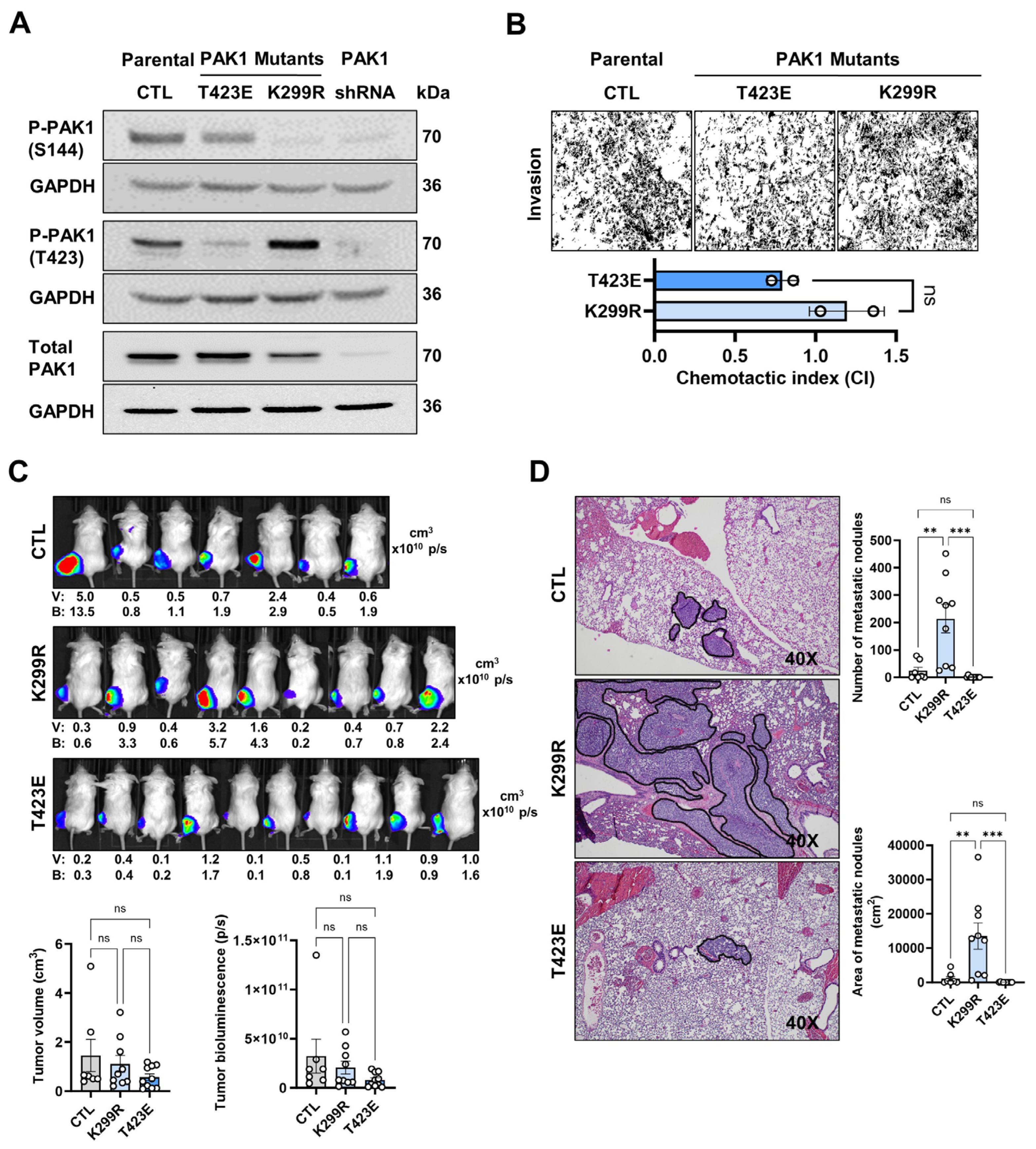
To understand the effects of the PAK1 mutations in OS cells, we first analyzed their effects on PAK1 phosphorylations at S144 and T423. While both mutants showed relatively low levels of phospho-PAK1 at S144 compared to the control, the K299R mutant displayed very low to undetectable levels (Figure 2A). Moreover, the K299R mutant exhibited higher T423 phosphorylation relative to the control (Figure 2A). The low phosphorylation of T423 in the T423E mutant is due to the mutation itself, which is a phospho-memetic mutation for constitutive activation. After considering total PAK1 expression, we confirm that the observed phosphorylation changes are due to specific post-translational modifications rather than variations in overall protein expression (Figure 2A, Supplementary Figure S3C). Original, uncropped Western blot membranes are provided in Supplementary Figure S5. Transwell invasion and proliferation assays demonstrated a non-significant, enhanced invasive propensity (Figure 2B) and a marked increase in growth rate of the K299R mutant compared to the T423E mutant and the control (Supplementary Figure S6), respectively. Despite the effect on cell proliferation in vitro, the K299R mutant had no significant effect on tumor volume and luciferase activity in mouse xenograft models (Figure 2C). The same result was observed for the T423E mutant. However, the K299R mutant markedly increased the size and number of pulmonary metastatic nodules relative to the T423E mutant (Figure 2D), suggesting that the high level of T423-PAK1 phosphorylation in K299R mutant is critical in driving the development of pulmonary metastases.

4. Discussion

Signaling networks regulating cell growth and motility are frequently dysregulated in cancer, contributing to tumor progression and metastasis [17]. Our previous studies have highlighted the vital roles of the cell cycle inhibitor p27, and p21-activated kinase PAK1 in OS aggressiveness [3,6]. While p27 is generally known as a tumor suppressor, which affects cell cycle arrest, cytoplasmic mislocalization of the protein has been implicated in promoting cell motility and metastasis, thus correlating with poor prognoses in OS and other cancers [3,4,18]. Conversely, PAK1 is widely recognized as a pro-metastatic kinase, driving aggressive cancer phenotypes through actin dynamics and upregulation of survival pathways [19,20,21]. We have shown that cytoplasmic p27 interacts with PAK1 leading to increased PAK1 phosphorylation and cell motility, and silencing PAK1 expression abrogated the cell migratory and adhesion propensities of p27-mislocalized OS cells in vitro and the development of pulmonary metastasis in an orthotopic xenograft mouse model. Since the functions of both p27 and PAK1 are mainly regulated at the level of protein phosphorylation [13,22], an unanswered question has been how specific phosphorylations of these proteins influence their roles in OS metastasis.
Our current work details the analysis of phenotypic behaviors of p27-T157A and p27-T157D mutants, and PAK1-T423E and PAK1-K299R mutants in vitro and in mouse xenograft models. We selected these mutations because the phosphorylation of the T157 residue of p27 affects the nuclear–cytoplasmic trafficking [7], while PAK1-T423E mutation produces a constitutively active kinase and the PAK1-K229R mutation abolishes ATP binding and acts as inactivating mutation [10,23]. Despite increased p27 cytoplasmic mislocalization, PAK1-S144 phosphorylation and OS invasiveness in the p27-T157D mutant compared to the T157A and the parental control, no significant differences in the development of pulmonary metastases were observed. Consistent with the kinase-dead nature of the K299R mutant, we observed a low to negligible S144 phosphorylation compared to the control and the T423E mutant. However, the K299R mutant showed increased T423 phosphorylation, elevated cell invasiveness and significantly promoted pulmonary metastasis compared to the T423E and the parental background. Our current findings extend our previous studies on p27 and PAK1 on OS metastasis by demonstrating the complexity of targeting specific p27 and PAK1 phosphorylations as an anti-metastatic strategy for OS.
Phosphorylation of p27 at Threonine 157 (T157) is a well-established mechanism for its cytoplasmic mislocalization, which can promote cell motility by sequestering p27 from the nucleus and inhibiting its classical cell cycle arrest function. The T157D mutant of p27, which mimics constitutive phosphorylation at this site, has been shown to enhance metastatic progression due to its cytoplasmic retention activity [7,18,24]. Contrarily, our results revealed that the T157D mutant did not lead to increased metastatic aggression compared to either the control or the phosphodeficient T157A mutant. Although increased S144 phosphorylation was observed in the T157D mutant, it is evidently not sufficient to promote OS metastasis. This suggests that while T157 phosphorylation and subsequent cytoplasmic p27 mislocalization may promote initial cellular motility, the T157D-mediated event alone, or in combination with induced PAK1-S144 phosphorylation, may not be the sole or dominant drivers of overt metastatic lung colonization, or that other driver mechanisms are at play. The pro-invasive effects of cytoplasmic p27, as more pronounced in the T157D, are highly dependent on the cellular microenvironment. In the simplified in vitro Boyden chamber assay, these factors are sufficient to manifest as increased chemotaxis. However, in vivo, the tumor cells may encounter numerous additional barriers and regulatory factors such as stromal interactions and distant organ niche that the T157D-induced pathway cannot overcome. For instance, while cytoplasmic p27 promotes invasion in breast cancer, its metastatic potential can be counteracted by the immune microenvironment or specific organ tropism factors [9,25,26]. Similarly, higher in vitro invasiveness does not always translate to higher in vivo metastasis in pancreatic cancer due to the complex desmoplastic stroma [27,28,29]. We have previously shown that in nuclear export signal (NES)-p27 mutants where overexpressed p27 is constitutively exported out from the nucleus, significant pulmonary metastasis via increased PAK1 activation was observed in mice, suggesting that these barriers may be overcome under sustained high level of cytoplasmic p27. Notably, cells carrying the T157A mutation, which lowers cytoplasmic p27 levels, showed no meaningful difference in PAK1-S144 phosphorylation or metastatic spread compared with controls—suggesting that inhibiting T157 phosphorylation alone is unlikely to block OS metastasis effectively.
PAK1-T423 is a vital autophosphorylation site essential for the kinase activation [11,30]. Our result showed that the T423E mutant, designed to mimic constitutive phosphorylation for protein activation [10,23], demonstrated similar phenotypes compared to parental control. Since the level of PAK1-T423 phosphorylation is quite high in the parental 143B, the T423E mutation may not further influence the metastatic phenotype. Alternatively, the phosphomimetic mutation may not fully replicate the multi-site phosphorylation pattern or precise conformational shifts required for optimal PAK1 activation [31]. In contrast, our observations regarding the PAK1-K299R mutant present an intriguing paradox.
The K299R mutation, targeting a highly conserved lysine residue within the ATP-binding pocket, is widely considered to produce a kinase-dead phenotype, effectively abolishing its catalytic activity [23]. This is evidenced by the negligible phosphorylation of S144 by Western blot analysis. Consequently, a number of studies often employed K299R as a dominant-negative mutation in research [23,32,33]. Notwithstanding, our functional assay results suggest that the K299R mutant retains or gains properties that contribute to increased metastatic activity in our OS model. One possible explanation is that the Kirsten murine sarcoma virus oncogene (K-RAS) is hyperactive in the 143B OS cell line. K-RAS is a potent activator of the RAF/MEK/ERK and PI3K/PDK1/AKT pathway, both of which contain kinases capable of phosphorylating serine/threonine residues on various substrates [20,34,35,36]. Given that K299R impairs PAK1’s autophosphorylation capabilities, any phosphorylation at T423 is likely to be catalyzed by external kinase(s) [37,38]. We reason that elevated K-RAS activity may activate specific downstream kinases, such as phosphoinositide-dependent kinase-1 (PDK1) or a member of the ERK pathway, which then preferentially phosphorylates T423 of the K299R PAK1 mutant. PDK1, a recognized upstream activator of PAK1 in cancers, phosphorylates PAK1 at critical sites like T423, essential for its activation [39]. Moreover, kinases in the ERK pathway have been implicated in PAK1 phosphorylation [40]. In COS-7 and neuroblastoma cell lines, EGF-induced T423 phosphorylation activated PAK1 through the RAS/PI3K signaling pathway, highlighting the upstream roles of RAS and PI3K in PAK1-T423 phosphorylation [34]. Moreover, it is also plausible that the unique conformation of the K299R mutant, or the specific cellular environment fostered by hyperactive K-RAS signaling in 143B cells, further creates a favorable environment for the T423 phosphorylation. This non-autophosphorylation of T423, despite the inherent kinase-dead nature of K299R, could potentially unlock a non-catalytic scaffolding function of PAK1 that is pro-metastatic [41,42].
PAK1 also serves as a scaffolding protein, facilitating protein–protein interactions independently of its kinase activity. For instance, co-transfection of cultured or in vivo-derived macrophages with either wild-type or kinase-deficient PAK1 constructs similarly suppressed the scavenger receptor class B, type I promoter, underscoring a kinase-independent role [41]. In colon cancer cell lines, overexpression of a kinase-dead PAK1 mutant enhanced cell proliferation and tumorigenesis by elevating ERK and MEK1/2 phosphorylation and promoting interactions between membrane-bound MEK and RAF [43]. Additionally, in COS-1 fibroblast-like cells, RAC1 stimulation induced kinase-independent PAK1-mediated recruitment of AKT and its interaction with PDK1, a critical component of the RAC1/PDK1/AKT pathway involved in cell growth, metabolism, and motility [44]. These findings highlight PAK1’s scaffolding role in promoting tumorigenesis, independent of its kinase function. The K299R mutation, potentially coupled with T423 phosphorylation by external kinases, may amplify or unmask these kinase-independent functions, establishing a compensatory mechanism for metastatic progression.
An alternative explanation for the observed phenotype of the K299R mutant is that PAK1 may phosphorylate downstream targets with tumor-suppressive or anti-metastatic functions, which are deactivated in the kinase-dead K299R mutant, potentially enhancing tumor growth and/or metastasis. Although PAK1 is primarily recognized as a pro-oncogenic kinase, and such anti-metastatic targets are not well-established, the phenomenon of phosphorylation-mediated inhibition is documented for other kinases. For example, AKT-mediated phosphorylation of TSC2, a tuberous sclerosis tumor suppressor, inhibits its interaction with TSC1, disrupting a critical complex for mTOR suppression [45]. Similarly, PAK1 phosphorylation of pyruvate dehydrogenase E1α (PDHA1) inhibits its activity, altering pyruvate metabolism in pancreatic ductal adenocarcinoma (PDAC) [46]. The PAK family has six members (PAK1–6). PAK1/2/4 have been shown to overlap roles in cell proliferation, motility and metastasis [47,48,49]. It is also possible that the K299R mutation in PAK1 might cause the cell to upregulate or hyperactivate other PAK family members, such as PAK2 or PAK4, to compensate for the kinase-dead PAK1 phenotype. For example, as PAK4 is a known driver of cell migration and invasion in many cancers [50,51], upstream signals that normally activate PAK1 could be redirected to activate PAK4 instead, providing a sustained compensatory pathway for increased metastasis. Consequently, targeting only PAK1’s catalytic activity may be insufficient to prevent metastasis if its scaffolding functions or other compensatory mechanisms are activated. Therefore, therapeutic strategies for PAK1 should address both its kinase activity, scaffolding roles, and potential compensatory mechanisms.

5. Conclusions

Despite the novel findings, our current study suffers from several limitations. Only limited phosphorylation sites were analyzed. Analysis of other important phosphorylation sites in p27 and PAK1 will help us to understand their roles in OS metastasis. This study was conducted using the 143B cell line, a widely utilized model for investigating metastatic OS. However, potential clonal variation and off-target effects among the mutant cell lines were not assessed. Incorporating additional OS cell lines with diverse genetic backgrounds, examining multiple independent clones per mutation, and assessing potential off-target effects would help minimize selection bias, address clonal and off-target impacts, and enhance the generalizability of our findings. Further experimental investigation into the kinase-independent functions of PAK1, especially for the K299R mutant, will be critical for fully elucidating its pro-metastatic role.
With the potential limitations in mind, the implications of these findings are substantial, particularly for therapeutic development in OS. If the pro-metastatic activity of the PAK1-K299R mutant is indeed driven by the phosphorylation of T423 by an external kinase or kinases under the influence of K-RAS, it suggests that merely inhibiting PAK1 autophosphorylation might not be sufficient to abrogate metastasis. Instead, a more effective therapeutic approach needs to inhibit both the autophosphorylation and the specific external kinase responsible for T423 phosphorylation on PAK1. For instance, combination therapies targeting RAS signaling and its downstream effectors have demonstrated therapeutic potential in lung cancer [52,53]. Furthermore, if the K299R mutant exerts its pro-metastatic effects through a non-catalytic scaffolding mechanism, future PAK1 inhibitors could be designed to interfere with this specific scaffolding function, rather than solely targeting the ATP-binding site. Future studies to identify the precise kinase(s) responsible for T423 phosphorylation in the PAK1-K299R mutant will be warranted for developing novel, highly effective therapeutic strategies to combat metastatic OS, particularly in tumors with elevated K-RAS activity.

Supplementary Materials

The following supporting information can be downloaded at: https://www.mdpi.com/article/10.3390/onco5030040/s1, Figure S1: Sequence alignment verification of the CRISPR-mediated p27 inactivating (T157A) and activating (T157D) mutations; Figure S2: Sequence alignment verification of the CRISPR-mediated knock in (KI) PAK1 mutations; Figure S3: Protein densitometry of immunoblots analyzed using ImageJ (v1.53e; Java 1.8.0_172); Figure S4: Original, uncropped Western blot membranes used to generate Figure 1A,B; Figure S5: Original, uncropped Western blot membranes used to generate Figure 2A; Figure S6: CCK8 proliferation assay of PAK1-K299R mutant compared to PAK1-T423E mutant and the parental control.

Author Contributions

J.W.: Data Curation, Formal analysis, Investigation, Methodology, Validation, Visualization, Writing—Original Draft, Writing—Review and Editing. B.B.G.: Data Curation, Formal analysis, Investigation, Methodology, Validation, Visualization, Writing—Original Draft, Writing—Review and Editing. J.X.: Methodology, Writing—Original Draft. A.M.M.: Formal analysis. J.H.: Conceptualization, Methodology, Supervision. T.-K.M.: Conceptualization, Data curation, Formal analysis, Funding acquisition, Investigation, Methodology, Project administration, Resources, Supervision, Validation, Writing—Original Draft, Writing—Review and Editing. All authors have read and agreed to the published version of the manuscript.

Funding

The study was generously funded by the Cancer Prevention and Research Institute of Texas (RP200135) to TKM, and by the NIH (2P30CA125123-1) to the Advanced Cell Engineering and 3D Models Core Facility at Baylor College of Medicine.

Institutional Review Board Statement

The animal study protocol was approved by the Institutional Animal Care and Use Committee (IACUC) at the Baylor College of Medicine, Houston, TX, USA (Protocol Code: AN-5460; Approval Date: 24 February 2023–23 February 2026).

Data Availability Statement

Supporting data are available from the corresponding author upon request.

Acknowledgments

We are grateful to Xiang Chen, Margaret Clement, Weidong Jin, Sonal Somvanshi and Matthew Weiser for their skillful technical help in this study.

Conflicts of Interest

Authors report no potential conflicts of interest. Funder had no role in the study design, collection, analyses and interpretation of data, and the writing and publication of the manuscript.

Abbreviations

The following abbreviations are used in this manuscript:
OSOsteosarcoma
P27Cyclin-dependent kinase inhibitor 1B
PAK1P21-activated kinase 1
CRISPRClustered Regularly Interspaced Short Palindromic Repeats

References

  1. Kager, L.; Zoubek, A.; Pötschger, U.; Kastner, U.; Flege, S.; Kempf-Bielack, B.; Branscheid, D.; Kotz, R.; Salzer-Kuntschik, M.; Winkelmann, W.; et al. Primary metastatic osteosarcoma: Presentation and outcome of patients treated on neoadjuvant Cooperative Osteosarcoma Study Group protocols. J. Clin. Oncol. 2003, 21, 2011–2018. [Google Scholar] [CrossRef]
  2. Smeland, S.; Bielack, S.S.; Whelan, J.; Bernstein, M.; Hogendoorn, P.; Krailo, M.D.; Gorlick, R.; Janeway, K.A.; Ingleby, F.C.; Anninga, J.; et al. Survival and prognosis with osteosarcoma: Outcomes in more than 2000 patients in the EURAMOS-1 (European and American Osteosarcoma Study) cohort. Eur. J. Cancer 2019, 109, 36–50. [Google Scholar] [CrossRef]
  3. Chen, X.; Cates, J.M.M.; Du, Y.C.; Jain, A.; Jung, S.Y.; Li, X.N.; Hicks, J.M.; Man, T.K. Mislocalized cytoplasmic p27 activates PAK1-mediated metastasis and is a prognostic factor in osteosarcoma. Mol. Oncol. 2020, 14, 846–864. [Google Scholar] [CrossRef] [PubMed]
  4. Razavipour, S.F.; Harikumar, K.B.; Slingerland, J.M. P27 as a transcriptional regulator: New roles in development and cancer. Cancer Res. 2020, 80, 3451–3458. [Google Scholar] [CrossRef] [PubMed]
  5. Ong, C.C.; Jubb, A.M.; Haverty, P.M.; Zhou, W.; Tran, V.; Truong, T.; Turley, H.; O’Brien, T.; Vucic, D.; Harris, A.L.; et al. Targeting p21-activated kinase 1 (PAK1) to induce apoptosis of tumor cells. Proc. Natl. Acad. Sci. USA 2011, 108, 7177–7182. [Google Scholar] [CrossRef] [PubMed]
  6. Li, Y.; Nakka, M.; Kelly, A.J.; Lau, C.C.; Krailo, M.; Barkauskas, D.A.; Hicks, J.M.; Man, T.K. P27 is a candidate prognostic biomarker and metastatic promoter in osteosarcoma. Cancer Res. 2016, 76, 4002–4011. [Google Scholar] [CrossRef]
  7. Shin, I.; Yakes, F.M.; Rojo, F.; Shin, N.-y.; Bakin, A.V.; Baselga, J.; Arteaga, C.L. PKB/Akt mediates cell-cycle progression by phosphorylation of p27Kip1 at threonine 157 and modulation of its cellular localization. Nat. Med. 2002, 8, 1145–1152. [Google Scholar] [CrossRef]
  8. Ye, D.Z.; Field, J. PAK signaling in cancer. Cell. Logist. 2012, 2, 105. [Google Scholar] [CrossRef]
  9. Liang, J.; Zubovitz, J.; Petrocelli, T.; Kotchetkov, R.; Connor, M.K.; Han, K.; Lee, J.-H.; Ciarallo, S.; Catzavelos, C.; Beniston, R.; et al. PKB/Akt phosphorylates p27, impairs nuclear import of p27 and opposes p27-mediated G1 arrest. Nat. Med. 2002, 8, 1153–1160. [Google Scholar] [CrossRef]
  10. Zenke, F.T.; King, C.C.; Bohl, B.P.; Bokoch, G.M. Identification of a central phosphorylation site in p21-activated kinase regulating autoinhibition and kinase activity. J. Biol. Chem. 1999, 274, 32565–32573. [Google Scholar] [CrossRef]
  11. Chong, C.; Tan, L.; Lim, L.; Manser, E. The mechanism of PAK activation. Autophosphorylation events in both regulatory and kinase domains control activity. J. Biol. Chem. 2001, 276, 17347–17353. [Google Scholar] [CrossRef]
  12. Zhou, G.-L.; Zhuo, Y.; King, C.C.; Fryer, B.H.; Bokoch, G.M.; Field, J. Akt Phosphorylation of Serine 21 on Pak1 Modulates Nck Binding and Cell Migration. Mol. Cell. Biol. 2003, 23, 8058–8069. [Google Scholar] [CrossRef]
  13. Lei, M.; Lu, W.; Meng, W.; Parrini, M.C.; Eck, M.J.; Mayer, B.J.; Harrison, S.C. Structure of PAK1 in an autoinhibited conformation reveals a multistage activation switch. Cell 2000, 102, 387–397. [Google Scholar] [CrossRef]
  14. Wang, J.; Wu, J.W.; Wang, Z.X. Structural insights into the autoactivation mechanism of p21-activated protein kinase. Structure 2011, 19, 1753–1761. [Google Scholar] [CrossRef] [PubMed]
  15. Ingvarsen, S.Z.; Gårdsvoll, H.; van Putten, S.; Nørregaard, K.S.; Krigslund, O.; Meilstrup, J.A.; Tran, C.; Jürgensen, H.J.; Melander, M.C.; Nielsen, C.H.; et al. Tumor cell MT1-MMP is dispensable for osteosarcoma tumor growth, bone degradation and lung metastasis. Sci. Rep. 2020, 10, 19138. [Google Scholar] [CrossRef] [PubMed]
  16. Gyau, B.B.; Wang, J.; Chen, X.; Clement, M.A.; Man, Z.D.; Major, A.M.; Weiser, M.C.; Xu, J.; Hicks, J.; Man, T.K. The metastatic role of the CXCL10-CXCR3 axis and its therapeutic potential in osteosarcoma. J. Bone Oncol. 2025, 52, 100690. [Google Scholar] [CrossRef] [PubMed]
  17. Kolch, W.; Halasz, M.; Granovskaya, M.; Kholodenko, B.N. The dynamic control of signal transduction networks in cancer cells. Nat. Rev. Cancer 2015, 15, 525–527. [Google Scholar] [CrossRef]
  18. Kim, J.; Jonasch, E.; Alexander, A.; Short, J.D.; Cai, S.; Wen, S.; Tsavachidou, D.; Tamboli, P.; Czerniak, B.A.; Do, K.A.; et al. Cytoplasmic sequestration of p27 via AKT phosphorylation in renal cell carcinoma. Clin. Cancer Res. 2009, 15, 81–90. [Google Scholar] [CrossRef]
  19. Radu, M.; Semenova, G.; Kosoff, R.; Chernoff, J. PAK signalling during the development and progression of cancer. Nat. Rev. Cancer 2014, 14, 13–25. [Google Scholar] [CrossRef]
  20. Papakonstanti, E.A.; Stournaras, C. Association of PI-3 kinase with PAK1 leads to actin phosphorylation and cytoskeletal reorganization. Mol. Biol. Cell 2002, 13, 2946–2962. [Google Scholar] [CrossRef]
  21. Sells, M.A.; Knaus, U.G.; Bagrodia, S.; Ambrose, D.M.; Bokoch, G.M.; Chernoff, J. Human p21-activated kinase (Pak1) regulates actin organization in mammalian cells. Curr. Biol. 1997, 7, 202–210. [Google Scholar] [CrossRef]
  22. Jäkel, H.; Peschel, I.; Kunze, C.; Weinl, C.; Hengst, L. Regulation of p27Kip1 by mitogen-induced tyrosine phosphorylation. Cell Cycle 2012, 11, 1910–1917. [Google Scholar] [CrossRef]
  23. Lei, M.; Robinson, M.A.; Harrison, S.C. The active conformation of the PAK1 kinase domain. Structure 2005, 13, 769–778. [Google Scholar] [CrossRef]
  24. Zhao, D.; Besser, A.H.; Wander, S.A.; Sun, J.; Zhou, W.; Wang, B.; Ince, T.; Durante, M.A.; Guo, W.; Mills, G.; et al. Cytoplasmic p27 promotes epithelial-mesenchymal transition and tumor metastasis via STAT3-mediated Twist1 upregulation. Oncogene 2015, 34, 5447–5459. [Google Scholar] [CrossRef] [PubMed]
  25. Xie, H.Y.; Shao, Z.M.; Li, D.Q. Tumor microenvironment: Driving forces and potential therapeutic targets for breast cancer metastasis. Chin. J. Cancer 2017, 36, 36. [Google Scholar] [CrossRef] [PubMed]
  26. Friedl, P.; Alexander, S. Cancer invasion and the microenvironment: Plasticity and reciprocity. Cell 2011, 147, 992–1009. [Google Scholar] [CrossRef]
  27. Merika, E.E.; Syrigos, K.N.; Saif, M.W. Desmoplasia in pancreatic cancer. Can we fight it? Gastroenterol. Res. Pract. 2012, 2012, 781765. [Google Scholar] [CrossRef]
  28. Ren, B.; Cui, M.; Yang, G.; Wang, H.; Feng, M.; You, L.; Zhao, Y. Tumor microenvironment participates in metastasis of pancreatic cancer. Mol. Cancer 2018, 17, 108. [Google Scholar] [CrossRef]
  29. Bulle, A.; Lim, K.H. Beyond just a tight fortress: Contribution of stroma to epithelial-mesenchymal transition in pancreatic cancer. Signal Transduct. Target. Ther. 2020, 5, 249. [Google Scholar] [CrossRef]
  30. Yu, J.S.; Chen, W.J.; Ni, M.H.; Chan, W.H.; Yang, S. Der. Identification of the regulatory autophosphorylation site of autophosphorylation-dependent protein kinase (auto-kinase): Evidence that auto-kinase belongs to a member of the p21-activated kinase family. Biochem. J. 1998, 334, 121–131. [Google Scholar] [CrossRef]
  31. Ng, Y.W.; Raghunathan, D.; Chan, P.M.; Baskaran, Y.; Smith, D.J.; Lee, C.H.; Verma, C.; Manser, E. Why an A-Loop Phospho-Mimetic Fails to Activate PAK1: Understanding an Inaccessible Kinase State by Molecular Dynamics Simulations. Structure 2010, 18, 879–890. [Google Scholar] [CrossRef]
  32. Zegers, M.M.P.; Forget, M.A.; Chernoff, J.; Mostov, K.E.; Ter Beest, M.B.A.; Hansen, S.H. Pak1 and PIX regulate contact inhibition during epithelial wound healing. EMBO J. 2003, 22, 4155–4165. [Google Scholar] [CrossRef]
  33. Zhu, Y.; Liu, H.; Xu, L.; An, H.; Liu, W.; Liu, Y.; Lin, Z.; Xu, J. P21-activated kinase 1 determines stem-like phenotype and sunitinib resistance via NF-κB/IL-6 activation in renal cell carcinoma. Cell Death Dis. 2015, 6, e1637. [Google Scholar] [CrossRef]
  34. Menard, R.E.; Mattingly, R.R. Cell surface receptors activate p21-activated kinase 1 via multiple Ras and PI3-kinase-dependent pathways. Cell. Signal. 2003, 15, 1099–1109. [Google Scholar] [CrossRef] [PubMed]
  35. Yan, J.; Roy, S.; Apolloni, A.; Lane, A.; Hancock, J.F. Ras isoforms vary in their ability to activate Raf-1 and phosphoinositide 3-kinase. J. Biol. Chem. 1998, 273, 24052–24056. [Google Scholar] [CrossRef] [PubMed]
  36. Peyssonnaux, C.; Provot, S.; Felder-Schmittbuhl, M.P.; Calothy, G.; Eychène, A. Induction of Postmitotic Neuroretina Cell Proliferation by Distinct Ras Downstream Signaling Pathways. Mol. Cell. Biol. 2000, 20, 7068–7079. [Google Scholar] [CrossRef] [PubMed]
  37. Bagheri-Yarmand, R.; Mandal, M.; Taludker, A.H.; Wang, R.A.; Vadlamudi, R.K.; Kung, H.J.; Kumar, R. Etk/Bmx Tyrosine Kinase Activates Pak1 and Regulates Tumorigenicity of Breast Cancer Cells. J. Biol. Chem. 2001, 276, 29403–29409. [Google Scholar] [CrossRef]
  38. Rider, L.; Shatrova, A.; Feener, E.P.; Webb, L.; Diakonova, M. JAK2 tyrosine kinase phosphorylates PAK1 and regulates PAK1 activity and functions. J. Biol. Chem. 2007, 282, 30985–30996. [Google Scholar] [CrossRef]
  39. King, C.C.; Gardiner, E.M.M.; Zenke, F.T.; Bohl, B.P.; Newton, A.C.; Hemmings, B.A.; Bokoch, G.M. p21-activated kinase (PAK1) is phosphorylated and activated by 3-phosphoinositide-dependent kinase-1 (PDK1). J. Biol. Chem. 2000, 275, 41201–41209. [Google Scholar] [CrossRef]
  40. Chang, T.W.; Lin, M.C.; Yu, C.J.; Lee, F.J.S. The phosphorylation of Pak1 by Erk1/2 to drive cell migration requires Arl4D acting as a scaffolding protein. J. Cell Sci. 2025, 138, jcs263812. [Google Scholar] [CrossRef]
  41. Hullinger, T.G.; Panek, R.L.; Xu, X.; Karathanasis, S.K. p21-activated Kinase-1 (PAK1) Inhibition of the Human Scavenger Receptor Class B, Type I Promoter in Macrophages Is Independent of PAK1 Kinase Activity, but Requires the GTPase-binding Domain. J. Biol. Chem. 2001, 276, 46807–46814. [Google Scholar] [CrossRef]
  42. Bokoch, G.M.; Reilly, A.M.; Daniels, R.H.; King, C.C.; Olivera, A.; Spiegel, S.; Knaus, U.G. A GTPase-independent mechanism of p21-activated kinase activation. Regulation by sphingosine and other biologically active lipids. J. Biol. Chem. 1998, 273, 8137–8144. [Google Scholar] [CrossRef]
  43. Wang, Z.; Fu, M.; Wang, L.; Liu, J.; Li, Y.; Brakebusch, C.; Mei, Q. P21-activated kinase 1 (PAK1) can promote ERK activation in a kinase-independent manner. J. Biol. Chem. 2013, 288, 20093–20099. [Google Scholar] [CrossRef]
  44. Higuchi, M.; Onishi, K.; Kikuchi, C.; Gotoh, Y. Scaffolding function of PAK in the PDK1-Akt pathway. Nat. Cell Biol. 2008, 10, 1356–1364. [Google Scholar] [CrossRef]
  45. Inoki, K.; Li, Y.; Zhu, T.; Wu, J.; Guan, K.L. TSC2 is phosphorylated and inhibited by Akt and suppresses mTOR signalling. Nat. Cell Biol. 2002, 4, 648–657. [Google Scholar] [CrossRef] [PubMed]
  46. Murugan, S.; B, S.S.S.; Gopinath, P.; Saravanan, R.; Sundaram, S.; Shanmugasundaram, G.; Venkatraman, G.; Rayala, S.K. Pak1 dysregulates pyruvate metabolism in PDAC cells by exerting a phosphorylation-mediated regulatory effect on PDHA1. J. Biol. Chem. 2025, 301, 108409. [Google Scholar] [CrossRef] [PubMed]
  47. Wang, C.; Wang, J.; Xu, R.; Li, Q.; Huang, X.; Zhang, C.; Yuan, B. PAK2 promotes proliferation, migration, and invasion of lung squamous cell carcinoma through the LIMK1/cofilin signaling pathway. J. Biomed. Res. 2025, 39, 184–197. [Google Scholar] [CrossRef] [PubMed]
  48. Dart, A.E.; Box, G.M.; Court, W.; Gale, M.E.; Brown, J.P.; Pinder, S.E.; Eccles, S.A.; Wells, C.M. PAK4 promotes kinase-independent stabilization of RhoU to modulate cell adhesion. J. Cell Biol. 2015, 211, 863–879. [Google Scholar] [CrossRef]
  49. Liu, H.; Liu, K.; Dong, Z. The Role of p21-Activated Kinases in Cancer and Beyond: Where Are We Heading? Front. Cell Dev. Biol. 2021, 9, 641381. [Google Scholar] [CrossRef]
  50. He, L.F.; Xu, H.W.; Chen, M.; Xian, Z.R.; Wen, X.F.; Chen, M.N.; Du, C.W.; Huang, W.H.; Wu, J.D.; Zhang, G.J. Activated-PAK4 predicts worse prognosis in breast cancer and promotes tumorigenesis through activation of PI3K/AKT signaling. Oncotarget 2017, 8, 17573–17585. [Google Scholar] [CrossRef]
  51. Blankenstein, L.J.; Cordes, N.; Kunz-Schughart, L.A.; Vehlow, A. Targeting of p21-Activated Kinase 4 Radiosensitizes Glioblastoma Cells via Impaired DNA Repair. Cells 2022, 11, 2133. [Google Scholar] [CrossRef] [PubMed]
  52. Jansen, R.A.; Mainardi, S.; Dias, M.H.; Bosma, A.; van Dijk, E.; Selig, R.; Albrecht, W.; Laufer, S.A.; Zender, L.; Bernards, R. Small-molecule inhibition of MAP2K4 is synergistic with RAS inhibitors in KRAS-mutant cancers. Proc. Natl. Acad. Sci. USA 2024, 121, e2319492121. [Google Scholar] [CrossRef] [PubMed]
  53. Zhao, X.; Zheng, Y.; Wang, Y.; Zhang, M.; Dong, Z.; Liu, Y.; Sun, M. The Potential Treatment Options and Combination Strategies of KRAS-Mutated Lung Cancer. OncoTargets Ther. 2024, 17, 1041–1057. [Google Scholar] [CrossRef] [PubMed]
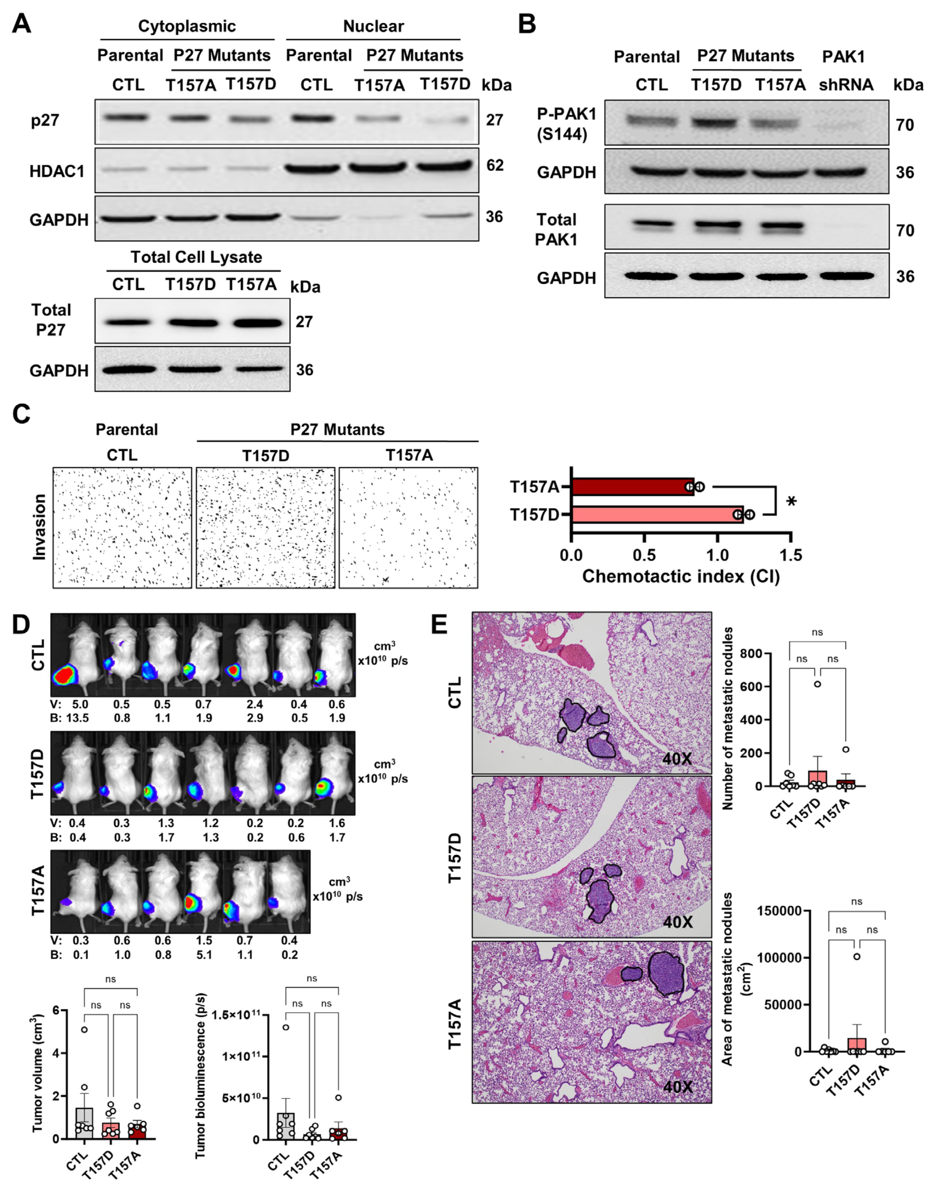
Figure 1. Phenotypic analyses of p27-T157A and p27-T157D mutants. (A) Fractionated (cytoplasmic or nuclear) and total p27 immunoblotting of the T157A and T157D mutants. 143B cells served as parental control (CTL). Cells were cultured in 10% FBS-supplemented DMEM until 80% confluence before analysis. Cell lysates were fractionated into nuclear and cytoplasmic fractions for the fractionated p27 immunoblotting while whole cell lysates were used for the total p27. Loading controls for cytoplasmic/total and nuclear proteins were GAPDH and HDAC1, respectively. (B) Phospho-PAK1-S144 (P-PAK1-S144) and total PAK1 immunoblotting of 143B (parental CTL), the 2 p27 mutants and the PAK1-shRNA mutant. Cells were cultured in 10% FBS-supplemented DMEM until 80% confluence before analysis. GAPDH served as the loading control. (C) Shown are example images (left) and quantification (right) from transwell invasion assays comparing control cells with 2 p27 mutants in low-serum (0.1% FBS) DMEM (n = 2). All cells were maintained in 10% FBS medium prior to testing. (D) Top: Bioluminescence scans of xenograft-bearing mice injected intratibially with control or mutant cells, taken five weeks after implantation. Different colors denote different bioluminescence intensity levels with red being high and blue being low. Bottom: Graphs of tumor volume (calculated as 0.5 × height × width2) and luminescence intensity. The inserted numbers correspond to individual tumor volume (V) and bioluminescence intensity (B) measurements taken from each mouse. (E) Representative H&E-stained lung sections (left) alongside quantification (right) of metastatic nodule counts and total area. Each dot denotes one animal. Experiments were repeated with consistent results. Error bars show standard deviations; “*” marks significant differences (p < 0.05), while “ns” denotes no significance.
Onco 05 00040 g001
Figure 2. Phenotypic analyses of the PAK1-T423E and PAK1-K299R mutants. (A) Phospho-PAK1 (P-PAK1) immunoblotting at S144 (upper) and T423 (middle) phosphorylation sites, and total PAK1 (lower) immunoblotting of 143B (parental CTL), the 2 PAK1 mutants and the PAK1-shRNA mutant. Cells were cultured in 10% FBS-supplemented DMEM until 80% confluence before analysis. GAPDH served as the loading control. (B) Representative photographs (top) and analysis (bottom) of transwell invasion data comparing parental control to 2 PAK1 mutants in 0.1% FBS DMEM (n = 2). (C) Top: Five-week post-injection bioluminescence images from mice injected intratibially with control or mutant cells. Different colors denote different bioluminescence intensity levels with red being high and blue being low. Bottom: Tumor volume and luminescence comparisons. The inserted numbers correspond to individual tumor volume (V) and bioluminescence intensity (B) measurements taken from each mouse. (D) Representative lung histology (left) with metastasis quantification (right). Each symbol represents one mouse. Findings were consistent across repeated trials. Standard deviations are displayed with error bars; ** p < 0.01, *** p < 0.001, “ns” = not significant.
Onco 05 00040 g002
Table 1. sgRNA for the site-specific CRISPR mutation generation.
GeneKI LocussgRNAssODN
PAK1K299RcugaagauucaucugcuuaaataatcagctctttcttgggctgctgctgaagattcatctgCCGGatCgccacctgaaatcaagagtatattcaatgtgcaaccatatg
PAK1T423EgcagagcaaacggagcaccaggattctgtgcacagataaccccagagcagagcaaGcgCagcGAAatggtaggaaccccatactggatggcaccagaggttgtgac
P27T157DaggaagcgaccugcaaccgaaagcggggccccaaacacattctatggttgggaaagggtcattaccgtcgTCAgcTggtcgcttccttattcctgcgcattgctccgctaaccccgtctgg
P27T157AaggaagcgaccugcaaccgaaagcggggccccaaacacattctatggttgggaaagggtcattaccgtcggCAgcTggtcgcttccttattcctgcgcattgctccgctaaccccgtctgg
Table 2. PCR amplification primers and restriction enzymes.
GeneKI LocusPCR Primer Fw’PCR Primer Rev’Restriction Enzyme
PAK1K299RgcattcttggcttttgccgtatttgactcaggcagatgggttgHpalI
PAK1T423EccaaaatgggcagcttggacaccacagagaacaccctggaHhaI
P27T157DtgtgtcttttggctccgaggtgagagggaccgcgatgtatPvuII
P27T157AtgtgtcttttggctccgaggtgagagggaccgcgatgtatPvuII
Disclaimer/Publisher’s Note: The statements, opinions and data contained in all publications are solely those of the individual author(s) and contributor(s) and not of MDPI and/or the editor(s). MDPI and/or the editor(s) disclaim responsibility for any injury to people or property resulting from any ideas, methods, instructions or products referred to in the content.